Comparative analysis of the
electroencephalogram in patients
with Alzheimer’s disease, diffuse axonal
injury patients and healthy controls using
LORETA analysis
Jéssica Natuline Ianof¹, Francisco José Fraga2, Leonardo Alves Ferreira2, Renato Teodoro Ramos3, José Luiz Carlos Demario4, Regina Baratho4,
Luís Fernando Hindi Basile1,5, Ricardo Nitrini¹, Renato Anghinah¹
ABSTRACT. Alzheimer’s disease (AD) is a dementia that affects a large contingent of the elderly population characterized by the presence of neurofibrillary tangles and senile plaques. Traumatic brain injury (TBI) is a non-degenerative injury caused by an external mechanical force. One of the main causes of TBI is diffuse axonal injury (DAI), promoted by acceleration-deceleration mechanisms. Objective: To understand the electroencephalographic differences in functional mechanisms between AD and DAI groups. Methods: The study included 20 subjects with AD, 19 with DAI and 17 healthy adults submitted to high resolution EEG with 128 channels. Cortical sources of EEG rhythms were estimated by exact low-resolution electromagnetic tomography (eLORETA) analysis. Results: The eLORETA analysis showed that, in comparison to the control (CTL) group, the AD group had increased theta activity in the parietal and frontal lobes and decreased alpha 2 activity in the parietal, frontal, limbic and occipital lobes. In comparison to the CTL group, the DAI group had increased theta activity in the limbic, occipital sublobar and temporal areas. Conclusion: The results suggest that individuals with AD and DAI have impairment of electrical activity in areas important for memory and learning.
Key words: dementia, Alzheimer disease, electroencephalography, brain waves.
ANÁLISE COMPARATIVA DO ELETROENCEFALOGRAMA EM PACIENTES COM DOENÇA DE ALZHEIMER, LESÃO AXONAL DIFUSA E CONTROLES SAUDÁVEIS: E ANÁLISE LORETA
RESUMO. A dooença de Alzheimer (DA) é uma demência que acomete uma grande parcela da população idosa e caracteriza-se pela presença de emaranhados neurofibrilares e placas senis. O traumatismo cranioencefálico (TCE) é uma lesão não degenerativa causada por uma força mecânica externa. Uma das principais causas de TCE é a lesão axonal difusa (LAD), causada por mecanismos de aceleração-desaceleração. Objetivo: Entender as diferenças dos mecanismos funcionais entre os grupos – DA e LAD do ponto de vista eletroencefalográfico. Métodos: Participaram deste estudo 56 indivíduos adultos. Destes, 20 com DA, 19 com LAD e 17 adultos saudáveis submetidos ao EEG de alta resolução com 128 canais. As fontes corticais dos ritmos do EEG foram estimadas pela análise por tomografia eletromagnética exata de baixa resolução (eLORETA). Resultados: A análise por eLORETA mostrou que, em comparação ao grupo controle (CTL), o grupo DA apresentou aumento da atividade teta nos lobos parietal e frontal e diminuição da atividade alfa 2 nos lobos parietal, frontal, límbico e occipital. Em comparação ao grupo CTL, o grupo LAD apresentou aumento da atividade teta nas áreas límbica, occipital sub-lobar e temporal. Conclusão: Os resultados sugerem que os indivíduos com DA e com LAD apresentam comprometimento da atividade elétrica em áreas importantes para a memória e aprendizagem.
Palavras-chave: demência, doença de Alzheimer, traumatismos encefálicos, lesão axonal difusa, eletroencefalografia, ondas encefálicas
This study was conducted at the Neurology Department, University of São Paulo Medical School Hospital (FMUSP-HC), São Paulo, SP, Brazil.
1Neurology Department, University of São Paulo Medical School Hospital (FMUSP-HC), São Paulo, SP, Brazil. 2Engineering, Modeling and Applied Social Sciences
Center (CECS) – Federal University of ABC (UFABC), São Paulo, SP, Brazil. 3Psychiatry Institute – University of São Paulo, São Paulo, SP, Brazil. 4Department of
Actuarial and Quantitative Methods – Pontifical Catholic of São Paulo, São Paulo, SP, Brazil. 5Laboratory of Psychophysiology – Methodist University of São Paulo,
São Paulo, SP, Brazil.
Jéssica Natuline Ianof. Neurology Department, University of São Paulo Medical School Hospital (FMUSP-HC) – Av. Dr. Enéas de Carvalho Aguiar, 255 – 05403-000 São Paulo SP – Brazil. E-mail: [email protected]
Disclosure: The authors report no conflicts of interest.
Received March 03, 2017. Accepted in final form May 24, 2017.
INTRODUCTION
A
lzheimer’s disease (AD) is the most common form of dementia among the elderly,1 accounting for 35to 80% of cases of dementia in these individuals.2 It is
also the most frequent cause of dementia in the Brazil-ian elderly population.3 Characterized by progressive
dementia,4 AD is a neurodegenerative disease whose
deinitive diagnosis cannot yet be established without histological analysis of the brain (biopsy or necropsy). he disease is associated with speciic degeneration in brain tissue, especially in pyramidal neurons, with marked intracellular presence of neuroibrillary tangles (hyperphosphorylation of β-amyloid) – resulting from the abnormal metabolism of amyloid precursor protein (APP) – in the extracellular compartment, accompanied by other structural alterations such as granulovacu-olar degeneration, dendritic atrophy and loss of neural synapses.5 Patients with AD may have limited access
to memories that shape their awareness and self-image, resulting in a compromised sense of identity.6
Over the course of the disease, other symptoms may emerge, such as disorientation, mood swings, confu-sion, more severe memory deicits, behavioral changes, as well as diiculties speaking, swallowing, and walking.7
Traumatic brain injury (TBI) is a nondegenerative and noncongenital insult to the brain from an external mechanical force. It is associated with diminished or altered state of consciousness and it can lead to perma-nent or temporary impairment of cognitive, physical, and psychosocial functions.8 he most common causes
of TBI are car crashes, falls, assaults and thefts, and accidents during recreational activity.9 It is considered
a “silent epidemic”,10 being the major cause of morbidity
and mortality11 and leading cause of death and sequelae
in children and young adults in western industrialized countries12. Annually, TBI afects around 10 million
people, leading to death or hospitalization.13
he acceleration-deceleration mechanism, respon-sible for difuse axonal injury (DAI), causes the fast rotational forces attributed to shear strain, damaging the axons.14 his mechanism often damages the lateral
and ventral regions of the frontal and temporal lobes. Deicits in attention and memory, diiculty learning new information, solving problems and planning, and problems associated with impulsivity and self-control are common sequelae.2 Long-term memory is
usu-ally restored, but some individuals ind it diicult to learn and retain new information. Working memory is often afected at diferent stages - coding, storage and retrieval. hese changes have a signiicant impact on the social and professional reintegration of the individual.15
Although AD and DAI have diferent mechanisms of injury, complaints such as memory deicits and diiculty learning new information are common to both groups of patients. For the investigation of both, electroencepha-lographic (EEG) recording in awake-resting state condi-tion is an ideal low-cost and non-invasive methodology. Indeed, EEG recording has a high temporal resolution (milliseconds) that provides an optimal investigational tool for the emerging features of brain physiology.9 EEG
procedures are well-tolerated by patients, unafected by task diiculty and are widely available in any country. hey can be repeated over time without habituation efects.17
When compared to groups of normal elderly sub-jects, AD groups are characterized by high power wide-spread delta and theta rhythms, as well as by low power posterior alpha and/or beta rhythms.16-19 Moreover, EEG
amplitude modulation analysis has been shown to be useful for characterizing AD progression from mild to moderate stages.20
he EEG immediately after TBI initially shows epi-leptiform activity,21 followed by suppressed cortical
activity - which may last from seconds to about a min-ute.22 Many patients return to normal within an hour,
while others continue to present focal or generalized slowing - which can last from weeks to a few months.23
he theta/alpha ratio increases after mild TBI and tends to return to normal within weeks to months.24
Quanti-tative EEG also shows an immediate reduction in the mean frequency of alpha and an increase in theta slow activity. hese changes usually take weeks to months to resolve. Improvement is associated with a reduction in symptoms.23
Low-resolution brain electromagnetic tomography (LORETA) is a mathematical algorithm that estimates the sources of EEG recorded on the scalp25 and is widely
used in EEG studies. New improved versions of LORETA have been developed such as Standardized low-resolu-tion brain electromagnetic tomography (sLORETA) and Exact low resolution brain electromagnetic tomography (eLORETA). sLORETA26 and eLORETA27 have the same
low spatial resolution, with zero localization error, but the eLORETA provides better localization of the signal source in the presence of noise.28
To establish the electroencephalographic diferences in functional mechanisms between Alzheimer’s disease and difuse axonal injury patients, both with memory complaints, among other shared symptoms.
METHODS
Hospital School of Medicine, University of São Paulo. All recruited participants provided written consent.
Inclusion criteria. Elderly patients diagnosed with prob-able AD, as determined by the National Institute of Neurological and Communicative Disorders and Stroke and the Alzheimer’s disease and Related Disorders Association29 with CDR=1 or 2 and Mini-Mental State
Examination (MMSE) scores from 13 to 29 – mild or moderate phase – were included. Subjects were recruited by the Cognitive Neurology and Behavior Group of the Neurology Department, University of São Paulo Medical School Hospital (FMUSP-HC). Patients who participated in this study had the diagnosis of AD for at least 6 months. he patients were not taking anti-cholinergic drugs.
Subjects aged 18 years or older diagnosed with mild/ moderate (Rancho Los Amigos ≥5 and MMSE scores from 13 to 28) DAI in the chronic phase and that pre-sented memory complaints were included. hey were recruited by the Cognitive Rehabilitation Group after TBI in the Division of Neurology, Clinicas Hospital, School of Medicine, University of São Paulo. he subjects with DAI who participated in this study were examined in the chronic stage (≥6 months after TBI). Patients with DAI were diagnosed based on the following criteria:30
1) A loss of consciousness from the time of injury that persisted beyond 6 h;
2) No apparent hemorrhagic contusion on computed tomography (CT);
3) he presence of white matter injury on MRI. In addition, healthy adults with no memory com-plaints were recruited to serve as controls. Individuals of both sexes were included in the study. he instruments used for cognitive assessment in patients and adult con-trols were the MMSE, verbal luency test (VF, animals category) and clock-drawing test (CDT). Demographic data and performance on screening tests are reported in the next section.
Exclusion criteria. Patients using medications that can modify the EEG record (such as antidepressant drugs, tricyclic compounds, nefazodone, benzodiazepines, lithium, neuroleptics) were excluded. Subjects with other neurological and/or psychiatric disorders, or that had sufered more than one TBI were also excluded from this study.
EEG recordings and data acquisition. EEG signals were recorded with a digital high-resolution 128-channel device (Brain Vision) using the International 10–10
system.17 Sampling frequency was 10000 Hz and
impedance of all electrodes was maintained below 10 kΩ. he recordings were performed at resting state, with participants comfortably seated in a reclined chair for approximately 25 min. During this period, the subjects kept their eyes closed most of the time (20 minutes). When drowsiness was noticed, they were asked to open their eyes and this event was duly noted in the EEG recording. Two neurophysiologists, who are board certiied, selected the EEG tracings for further analysis.
EEG pre-processing. he EEGLAB software package,31
which runs in the MATLAB (MathWorks®) software environment, was used to perform all pre-processing steps. All iltering was done in the zero-phase mode. First of all, the EEG was downsampled from 10000 Hz to 1000 Hz and once again to 400 Hz after lowpass iltering to 115 Hz using a 5th-order Chebyshev II ilter.
Subsequently, two Butterworth 4th-order ilters were
applied, one to eliminate power grid interference (60 Hz, notch) and the other to remove very slow luc-tuations (0.4 Hz, highpass). Eyes-opening and closing events were identiied, since analysis focused on the second eyes-closed moment onwards while every-thing before that was discarded. Average referencing was performed and the 128-channel EEG signal was divided into 4-second epochs. Epochs with strong arti-facts (outside the ±450 μV range or with slope above 250 μV/50 ms) were eliminated. Finally, ocular and muscular artifacts were removed with the EEGLAB Independent Component Analysis (ICA) tool.31 After all
these pre-processing steps, seventy 4-second epochs for each participant were stored in .txt format to serve as input EEG iles to the LORETA-KEY software (http:// www.uzh.ch/keyinst/loreta).
EEG source localization. eLORETA was used to analyze the cortical distribution of current source density. he head model of eLORETA and the electrode coor-dinates are based on the Montreal Neurological Insti-tute average MRI brain map (MNI152).32 he solution
space was limited to the cortical gray matter, including 6239 voxels with spatial resolution of 5 cubic mm. he eLORETA tomography has been previously used in several studies.18,33,34 Selected artifact-free EEG
Figure 1. Representation of brain areas with statistically significant differences in comparison CTL × AD for theta band on 32-channel analysis (CTL<AD) [A], for alpha 2 band on 32-channel analysis (CTL>AD) [B] and for alpha 2 band on 64-channel analysis (CTL>AD) [C].
C
A
alpha 2 (10.5-12 Hz), beta 1 (12.5-18 Hz), beta 2 (18.5-21 Hz), beta 3 (18.5-30 Hz) and omega – full-band (1.5-30 Hz).
Statistical analysis. Frequency tables and descriptive statistics were used to describe the proile of the sample. he Mann-Whitney U and Kruskal-Wallis tests were used to compare the continuous variables between two or three groups, respectively. Pearson’s Chi-square test was used to compare the categorical variables between the diagnostic groups. For statistical analysis, the Social Package for Social Science (SPSS) version 20 by International Business Machines (IBM) was used. he signiicance level adopted for the statistical tests was 5%, i.e. a p-value <0.05.
For the statistical analysis of current source den-sity, the statistical nonparametric mapping method (SnPM)35 was used, available as a software tool in the
LORETA-KEY package. he diference in cortical source localization between groups was assessed for each
fre-quency band with voxel-by-voxel independent F-ratio-tests, based upon eLORETA log-transformed current source density power. Cortical voxels with signiicant diferences were identiied by means of a nonparamet-ric randomization procedure, in the three-dimensional statistical mapping. he mean source power in each voxel and the distribution in the permutated values was compared, with threshold set at a 5% signiicance level. A total of 5000 data randomizations were used to deter-mine the critical probability threshold values for the actually observed log F-ratio values, with correction for multiple comparisons across all voxels and frequencies.
RESULTS
he inal sample consisted of 17 control individuals, 20 patients with Alzheimer’s disease and 19 patients with DAI. Table 1 summarizes the relevant demographic data for the CTL, AD and DAI participants. Table 2 summa-rizes the relevant clinical data and screening test results for the participants.
Table 1. Demographic data in the subgroup of Alzheimer’s disease, diffuse axonal injury and healthy control participants.
Variables CTL AD DAI Statistical analyses
N 17 20 19
Gender – – – p=0.765a, 0.149b, 0.224c
(Chi-square test)
Male 8 11 14 –
Female 9 9 5 –
Age 47.94 (±20.82 SE) 77.35 (±6.19 SE) 50 (±11.28 SE) P<0.001a,c, 0.832b
(Mann-Whitney U test)
Education (years) 13.94 (±3.96 SE) 7.35 (±4.68 SE) 7.47 (±5.21 SE) p<0.001a,b, 0.806c
(Mann-Whitney U test)
CTL: control. AD: Alzheimer’s disease. DAI: diffuse axonal injury. aCTL × AD; bCTL × DAI; cAD × DAI.
Table 2. Performance on screening tests in the subgroup Alzheimer’s disease, diffuse axonal injury and healthy control participants.
Variables CTL AD DAI Statistical analyses
CDR – 1.25 (±0.55 SE) –
Rancho los Amigos Scale – – 7 (±1.05 SE) –
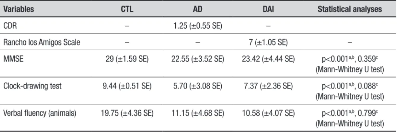
MMSE 29 (±1.59 SE) 22.55 (±3.52 SE) 23.42 (±4.44 SE) p<0.001a,b, 0.359c
(Mann-Whitney U test)
Clock-drawing test 9.44 (±0.51 SE) 5.70 (±3.08 SE) 7.37 (±2.36 SE) p<0.001a,b, 0.088c
(Mann-Whitney U test)
Verbal fluency (animals) 19.75 (±4.36 SE) 11.15 (±4.68 SE) 10.58 (±4.07 SE) p<0.001a,b, 0.799c
(Mann-Whitney U test)
Table 3. Brain structures with statistically significant difference on eLORETA in comparison – CTL × AD (32-channel analysis).
Theta band
Lobe Brodmann areas
Frontal 4
Parietal 1, 2, 3, 40
Alpha 2 band
Lobe Brodmann areas
Parietal 5, 7, 19, 31, 40
Frontal 5, 31
Limbic 23, 31
Occipital 7, 18, 19, 31
Table 4. Brain structures with statistically significant difference for alpha 2 band on eLORETA (64-channel analysis) – CTL × AD.
Lobe Brodmann areas
Limbic 23, 31
Table 5. Brain structures with statistically significant difference for theta band on eLORETA in comparison – CTL × DAI.
32 channels
Lobe Brodmann areas
Limbic lobe 19, 23, 27, 28, 29, 30, 31, 34, 35, 36
Occipital 17, 18, 19, 30, 31
Sub-lobar 13
Temporal 41
64 channels
Lobe Brodmann areas
Limbic lobe 23, 27, 29, 30
Occipital 17, 18, 19, 30
Sub-lobar 13
EEG findings. he mean alpha frequency peak was 10.23 Hz (±0.90 SE) for the CTL participants, 9.30 Hz (±0.72 SE) for the AD group and 9.73 Hz (±1.02 SE) for the DAI group. Statistical analysis (Mann-Whitney U test) was performed to test possible diferences in the alpha frequency peak between the groups. A statisti-cally signiicant diference was found between the CTL and AD groups (p<0.05). No statistically signiicant diference was found between the CTL and DAI groups (p>0.125) or between the AD and DAI groups (p>0.136).
eLORETA analysis. he eLORETA analysis was performed with 32, 64 and 128 channels (10-20 and 10-10 system). On the comparison between CTL and AD groups, the analysis with 32 channels revealed difer-ences in the theta and alpha2 bands. Brain structures containing voxels with statistically signiicant difer-ences are shown in Table 3 and in Figure 1a and 1b. he analysis with 64 channels also revealed diferences, but only in the alpha2 band. Brain structures in which voxels with statistically signiicant diferences were found are shown in Table 4 and in Figure 1c.
On the comparison between CTL and DAI groups, the analysis with 32 channels showed diferences only in the theta band. he brain structures in which voxels with statistically signiicant diferences were found are given in Table 5 and Figure 2a. he 64-channel analy-sis also showed signiicant diferences only in the theta band (Table 5, Figure 2b).
No diferences were found between the CTL and AD groups or the CTL and DAI groups for the 128-chan-nel analysis. Also, there were no statistical diferences between AD and DAI groups at any of the settings (32, 64 or 128 channels).
DISCUSSION
Although AD and DAI patients difered in age and schooling, they had the same cognitive impairment proile, since they had many common symptoms. For this reason, we sought to compare them and determine electroencephalographic diferences.
Regarding gender, no signiicant statistical difer-ences were found between the CTL and AD groups or CTL and DAI groups. he DAI group had a higher num-ber of males than the AD group, with a statistically sig-niicant diference. he literature shows that 2-3 times more men are afected by TBI than women.
he mean age of the DAI group was lower than that of the CTL group and the AD group. his was expected since TBI tends to afect younger adults whereas AD occurs mostly in elderly individuals.
he diagnosis of most cognitive disorders is clinical, but the EEG plays a role in the evaluation, classiication and follow-up of these disorders. It is an important method for evaluation of cortical processing and physi-ological changes.36 A decrease in alpha and beta rhythms
and increase in delta and theta frequencies are related to brain lesions and cognitive decline.37 Babiloni et al.
A
B
Figure 2. Representation of brain areas with statistically significant differences on comparison CTL × DAI for theta band on 32-channel analysis (CTL<DAI) [A], and for theta band on 64-channel analysis (CTL<DAI).
compared to healthy elderly subjects in all areas, par-ticularly in the central, parietal, temporal and limbic areas.38 Gianotti et al. (2006) failed to ind diferences
between mild/moderate AD groups and controls for the alpha 1 band. However, diferences were observed for the alpha 2 band, which decreased in the occipital area more prominently in the right hemisphere.39 Babiloni et
al. (2009) studied a group of individuals with mild cogni-tive impairment (MCI) and AD and found a reduction in alpha 1 activity in the occipital, temporal and parietal areas.40 Canuet et al. (2012) found diferences between
groups, using the eLORETA method, only for the alpha 1 band in the parieto-occipital region, mainly in the right pre-cuneus region.28 Caso et al. (2012), based on spectral
analysis and sLORETA, observed that patients with AD,
in comparison with controls, had lower activity for fast frequencies (alpha 1, alpha 2, beta 1 and beta 2) in the central and posterior regions.41 Babiloni et al. (2016)
studied controls, patients with Parkinson’s disease and with AD. Using LORETA, they observed that, relative to controls, patients with AD had lower alpha 1 activity in the central, parietal, occipital and temporal areas and less alpha 2 activity in the parietal and occipital areas.42
Our literature search found no papers using LORETA analysis in patients with DAI. Indeed, there is scant liter-ature involving TBI and LORETA. Tomkins et al. (2011) found, using sLORETA, that TBI patients had slower delta waves than controls.43 he study of Corradini and
adjacent to the temporal lobe in individuals with mild TBI.44
In our study, no diferences were found between the AD and DAI groups for any EEG rhythm in any conigu-ration (32, 64 or 128 channels). One possible explana-tion for this is that, although the mechanisms of injury difer in the two diseases, there are many similarities in their neuroanatomy and physiology, which leads the patients to present similar symptoms. At the begin-ning of AD, the main characteristic of clinical features is episodic memory impairment, i.e., the patient has dif-iculty remembering recent events and conversations, repeats questions and stories, and has trouble inding personal objects.45 Decreased autobiographical memory,
decreased ability to learn new information as quickly as before the TBI, less proiciency to remember faces of new acquaintances, and delayed task execution after rapid or new visual presentations, are common com-plaints among post-TBI patients,44 especially in DAI9.
he irst pathological link between a single TBI and AD was the observation that β-amyloid plaques are pres-ent in up to 30% of patipres-ents dying in the acute phase of TBI.46 APP can be found accumulated in damaged axons
within 2 hours after injury.47 Several studies have
identi-ied that the history of a single TBI is an epigenetic risk factor for the later development of clinical syndromes of cognitive impairment, such as AD.48 he activation of
posterior and medial portions of the parietal-occipital cortex (covering the cuneus and precuneus) has been reported with considerable consistency in PET and func-tional MRI studies during memory tasks,49 speciically
for success in memory recall. he onset of amnestic syn-dromes is related to damage in portions of the medial temporal cortex, including the hippocampus and para-hippocampal gyrus.50
he onset of amnestic syndromes is related to dam-age in portions of the medial temporal cortex, including the hippocampus and para-hippocampal gyrus (Zola-Morgan et al., 1986). At the beginning of AD, the main characteristic of the clinical feature is episodic memory impairment.
here is a pathological link between a single TBI and AD, and it is well established that TBI patients have a higher risk for developing dementia in the future. hus, the objective of the present study was to ascertain whether these groups had similarities in EEG.
Our analyzes have shown that with fewer channels, the resolution of LORETA is improved, since a channel is responsible for a larger portion of the scalp. For this reason, the analysis using 32 channels revealed more voxels with statistical diferences between the groups.
Limitations of the study include the sample size and cross-sectional methodology employed, since patients were not followed during disease progression. It should be emphasized that this study provides contributions to the national literature, since the line of research involv-ing cognitive electroencephalography is scarce. We found no articles comparing the group of Alzheimer’s disease and difuse axonal injury in the literature with regard to electroencephalographic proile. In fact, papers investi-gating difuse axonal injury are very scarce. he use of the LORETA methodology is restricted to a few research groups and existing studies generally compare demen-tia x controls, or two types of demendemen-tia. We found no articles involving patients with traumatic brain injury and comparing them with dementia patients.
In conclusion, our indings showed a neurofunctional similarity between AD and DAI, and that the two groups difered in relation to the controls, which was expected, since it is a comparison between pathology and normal-ity. his neurofunctional similarity helps to understand the functionality of these diseases – our results show that the areas involved in memory and learning are com-promised in both pathologies and this knowledge can be extrapolated to studies aimed at the treatment of these conditions. Drugs known to prevent memory decline in Alzheimer’s disease could also be used in difuse axonal injury patients. Also, these indings could pave the way for the use of drugs that are commonly used in the treat-ment of AD in patients with DAI.
Further studies on the physiology of these dis-eases, from an electroencephalographic perspective in a greater number of individuals are necessary, and it is also important to correlate the electroencephalographic indings and cognitive performance of patients.
Acknowledgments. We would like to thank the São Paulo Research Foundation (FAPESP, grant #2015/09510-7) for the partial funding of this research
Hindi Basile: writing and reviewing of the data. Ricardo Nitrini: responsible for evaluation protocols and contributed by referring patients with the diagnosis of
Alzheimer’s disease. Renato Anghinah: project design, EEG analysis, clinical correlations and discussion of the methodology and results.
REFERENCES
1. Fratiglioni L, Launer LJ, Andersen K, Breteler MM, Copeland JR, Dartigues JF, et al. Incidence of dementia and major subtypes in Europe: a collaborative study of population-based cohorts. Neurology 2000;54 (11 Suppl 5):S10-5.
2. Grinberg LT, Nitrini R, Suemoto CK, Lucena Ferretti-Rebustini RE, Leite RE, Farfel JM, et al. Prevalence of dementia subtypes in a developing country: a clinicopathological study. Clinics (Sao Paulo). 2013;68(8):1140-5.
3. Nitrini R, Caramelli P, Herrera-Jr E, Bahia VS, Caixeta LF, Radanovic M, et al. Incidence of dementia in a community-dwelling Brazilian popula-tion.Alzheimer Dis Assoc Disord. 2004;18(4):241-6.
4. Nizzari M, Thellug S, Corsaro A, Villa V, Pagano A, Porcile C et al. Neuro-degeneration in Alzheimer disease: role of the amyloid precursor protein and presenilin 1 intracellular signaling. J Toxicol. 2012;187-297. 5. James OG. Doraiswamy PM, Borges-Neto S. PET Imaging of Tau
Pathology in Alzheimer’s Disease and Tauopathies. Front Neurol. 2015; 6:38.
6. Haj EL, Antoine P. Describe yourself to improve your autobiographical memory: A study in Alzheimer’s disease. Cortex. 2017;88:165-72. 7. Winblad B, Amouyel P, Andrieu S, Ballard C, Brayne C, Brodaty H, et al.
Defeating Alzheimer’s disease and other dementias:a priority for Euro-pean science and society. Lancet Neurol. 2016;15(5):455-532. 8. Jang SH. Review of motor recovery in patients with traumatic brain injury.
Neurorehabilitation. 2009;24:349-53.
9. Freire FR; Coelho F; Lacerda JR; Silva MF; Gonçalves VT; Machado S et al. Cognitive rehabilitation following traumatic brain injury. Dement. Neuropsychol. 2011;5(1):17-25.
10. Langlois JA, Sattin RW. Traumatic brain injury in the United States: research and programs of the Centers for Disease Control and Preven-tion (CDC). J Head Trauma Rehabil. 2005;20(3):187-8.
11. Hay J, Johnson VE, Smith DH, Stewart W. Chronic Traumatic Encepha-lopathy: The Neuropathological Legacy of Traumatic Brain Injury. Annu Rev Pathol. 2016;11:21-45.
12. McArthur DL, Chute DJ, Villablanca JP. Moderate and severe traumatic brain injury: epidemiologic, imaging and neuropathologic perspectives. Brain Pathol. 2004;14:185-94.
13. Hyder AA, Wunderlich CA, Puvanachandra P, Gururaj G, Kobusingye OC. The impact of traumatic brain injuries: a global perspective. Neuro-Rehabilitation. 2007;22(5):341-53.
14. Adams JH, Graham DI, Murray LS, Scott . Diffuse axonal injury due to nonmissile head injury in humans: an analysis of 45 cases. Ann Neurol.1982; 12(6):557-63.
15. Vallat-Azouvi C, Weber T, Legrand L, Azouvi P. Working memory after severe traumatic brain injury. J Int Neuropsy chol Soc. 2007;13:770-80. 16. Babiloni C, Lizio R, Del Percio C, Marzano N, Soricelli A, Salvatore E,
et al. Cortical sources of resting state EEG rhythms are sensitive to the progression of early stage Alzheimer’s disease. J Alzheimers Dis. 2013;34(4):1015-35.
17. Cassani R, Falk TH, Fraga FJ, Cecchi M, Moore DK, Anghinah R. Towards automated electroencephalography-based Alzheimer’s disease diagnosis using portable low-density devices. Biomed Signal Processing and Control. 2017;33:261-71.
18. Dierks T, Jelic V, Pascual-Marqui RD, Wahlund LO, Julin P, Linden DEJ, et al. Spatial pattern of cerebral glucose metabolism (PET) correlates with localization of intracerebral EEG-generators in Alzheimer’s disease. Clin Neurophysiol. 2000;111:1817-24.
19. Jeong J. EEG dynamics in patients with Alzheimer’s disease. Clin Neuro-physiol. 2004;115(7):1490-505.
20. Fraga FJ, Falk TH, Kanda PAM, Anghinah R. Characterizing Alzheimer’s disease severity via resting-awake EEG amplitude modulation analysis. PLoS One. 2013;8(8):e72240.
21. Walker AE, Kollros JJ, Case TJ. The physiological basis of concussion. J Neurosurg. 1944;1:103-16.
22. Shaw NA. The neurophysiology of concussion. Prog Neurobiol. 2002;67: 281-344.
23. Nuwer MR, Hovda DA, Schrader LM, Vespa PM. Routine and quan-titative EEG in mild traumatic brain injury. Clin Neurophysiol. 2005; 116:2001-25.
24. McCrea M, Prichep L, Powell MR, Chabot R, Barr WB. Acute effects and recovery after sport-related concussion:a neurocognitive and quantitative brain electrical activity study. J Head Trauma Rehabil. 2010;25(4):283-92. 25. Babiloni C, Frisoni G, Pievani M, Vecchio F, Lizio R, Buttiglione M et
al. Hippocampal volume and cortical sources of EEG alpha rhythms in mild cognitive impairment and Alzheimer disease. Neuroimage. 2009a; 44(1):123-35.
26. Pascual-Marqui RD. Standardized low-resolution brain electromagnetic tomography (sloreta): technical details. Methods Find Exp Clin Phar-macol. 2002a;24 Suppl D:5-12.
27. Pascual-Marqui R. Discrete, 3D distributed, linear imaging methods of electric neuronal activity. Part 1: exact, zero error localization. 2007. Available online at: http://arxiv.org/pdf/0710.3341.
28. Canuet L, Tellado I, Couceiro V, Fraile C, Fernandez-Novoa L, Ishii R, et al. Resting-state network disruption and APOE genotype in Alzheim-er’s disease: a lagged functional connectivity study. PLoS One 2012; 7:e46289.
29. NINCDS-ADRDA, National Institute of Neurological and Communicative Disorders and Stroke and the Alzheimer’s Disease and Related Disorders Association.
30. Ezaki Y, Tsutsumi K, Morikawa M, Nagata I. Role of diffusion-weighted magnetic resonance imaging in diffuse axonal injury. Acta Radiol. 2006;47(7):733-40.
31. Delorme A and Makeig,S. EEGLAB: an open source toolbox for analysis of single-trial eegdynamics including independent component analysis. J Neurosci Methods. 2004;134:9-21.
32. Mazziotta J, Toga A, Evans A, Fox P, Lancaster J, Zilles K, et al. A probabilistic atlas and reference system for the human brain: International Consortium for Brain Mapping (ICBM). Philos Trans R Soc Lond B Biol Sci 2001;356:129-322.
33. Worrell GA, Lagerlund TD, Sharbrough FW, Brinkmann BH, Busacker NE, Cicora KM, O’Brien TJ. Localization of the epileptic focus by low-resolution electromagnetic tomography in patients with a lesion demon-strated by MRI. Brain Topography 2000;12:273-82.
34. Zumsteg D, Friedman A, Wieser HG, Wennberg RA. Propagation of interictal discharges in temporal lobe epilepsy: correlation of spatiotem-poral mapping with intracranial foramen ovale electrode recordings. Clin Neurophysiol. 2006;117:2615-26
35. Holmes AP, Blair RC, Watson JDG, Ford I. Nonparametric Analysis of Statistic Images from Functional Mapping Experiments. J Cerebral Blood Flow Metabol. 1996;16(1):7-22.
36. Kanda PAM, Anghinah R, Schmidt MT, Jorge MS. The clinical use of quantitative EEG in cognitive disorders. Dement Neuropsychol. 2009; 3(3):195-203.
37. Anghinah R, Kanda PA, Lopes HF, Basile LF, Machado S, Ribeiro P, et al. Alzheimer’s disease qEEG: spectral analysis versus coherence. Which is the best measurement? Arq Neuropsiquiatr. 2011;69(6):871-4. 38. Babiloni C, Binetti G, Cassetta E, Cerboneschi D, Dal Forno G, Del
Percio C, et al. Mapping distributed sources of cortical rhythms in mild Alzheimer’s disease. A multicentric EEG study. Neuroimage. 2004; 22(1): 57-67.
39. Gianotti LR, Künig G, Lehmann D, Faber PL, Pascual-Marqui RD, Kochi K, Schreiter-Gasser U. Correlation between disease severity and brain electromagnetic LORETA tomography in Alzheimer’s Disease. Clin Neurophysiol. 2006;118(1):186-96.
40. Babiloni C, Frisoni GB, Pievani M, Vecchio F, Lizio R, Buttiglione M, et al.. Hippocampal volume and cortical sources of EEG alpha rhythms in mild cognitive impairment and Alzheimer disease. Neuroimage. 2009b;44(1):123-35.
42. Babiloni C, Del Percio C, Caroli A, Salvatore E, Nicolai E, Marzano N, et al. A. Cortical sources of resting state EEG rhythms are related to brain hypometabolism in subjects with Alzheimer’s disease: an EEG-PET study.Neurobiol Aging. 2016;48:122-34.
43. Tomkins O, Feintuch A, Benifla M, Cohen A, Friedman A, Shelef I. Blood-brain barrier breakdown following traumatic brain injury: a possible role in posttraumatic epilepsy. Cardiovasc Psychiatry Neurol. 2011;2011:765923.
44. Corradini PL, Persinger MA. Standardized Low Resolution Electro-magnetic Tomography (sLORETA) is a Sensitive Indicator of Protracted Neuropsychological Impairments Following “Mild” (Concussive) Traumatic Brain Injury. J Neurol Neurophysiol. 2013;4:176.
45. Parmera JB, Nitrini R. Da investigação ao diagnóstico. Rev Med (São Paulo). 2015;94(3):179-84.
46. DeKosky ST, Abrahamson EE, Ciallella JR, Paljug WR, Wisniewski SR,
Clark RS, Ikonomovic MD. Association of increased cortical soluble abeta42 levels with diffuse plaques after severe brain injury in humans. Arch Neurol. 2007;64:541-4.
47. Johnson VE, Stewart W, Smith DH. Axonal pathology in traumatic brain injury. Exp Neurol. 2013;246:35-43.
48. Plassman BL, Havlik RJ, Steffens DC, Helms MJ, Newman TN, Dros-dick D, et al. Documented head injury in early adulthood and risk of Alzheimer’s disease and other dementias. Neurology. 2000;55:1158-66. 49. Busatto G, Howard RJ, Ha Y, Brammer M, Wright I, Woodruff PW,
Simmons A, Williams SC, David AS, Bullmore ET. A functional magnetic resonance imaging study of episodic memory. Neuroreport. 1997;8(12):2671-5.
![Figure 1. Representation of brain areas with statistically significant differences in comparison CTL × AD for theta band on 32-channel analysis (CTL<AD) [A], for alpha 2 band on 32-channel analysis (CTL>AD) [B] and for alpha 2 band on 64-channel ana](https://thumb-eu.123doks.com/thumbv2/123dok_br/15189636.527268/4.955.140.866.108.1122/figure-representation-statistically-significant-differences-comparison-analysis-analysis.webp)
![Figure 2. Representation of brain areas with statistically significant differences on comparison CTL × DAI for theta band on 32-channel analysis (CTL<DAI) [A], and for theta band on 64-channel analysis (CTL<DAI).](https://thumb-eu.123doks.com/thumbv2/123dok_br/15189636.527268/7.955.102.813.473.1129/figure-representation-statistically-significant-differences-comparison-analysis-analysis.webp)
