Systematic Review
Revisão Sistemática ISSN 2317-1782 (Online version)
This is an Open Access article distributed under the terms of the Creative Commons Attribution License, which permits unrestricted use, distribution, and reproduction in any medium, provided the original work is properly cited.
Characteristics of auditory evaluation in
Williams syndrome: a systematic review
Características da avaliação auditiva na
síndrome de Williams: revisão sistemática
Liliane Aparecida Fagundes Silva1
Chong Ae Kim2
Carla Gentile Matas1
Keywords
Williams Syndrome Hearing Loss Cochlea Audiology Hearing
Descritores
Síndrome de Williams Perda Auditiva Cóclea Audiologia Audição
Correspondence address:
Liliane Aparecida Fagundes Silva R. Cipotânea, 51, Cidade
Universitária, São Paulo (SP), Brasil, CEP: 05360-160.
E-mail: [email protected]
Received: December, 13, 2017
Accepted: March 27, 2018
Study conducted at Departamento de Fisioterapia, Fonoaudiologia e Terapia Ocupacional, Faculdade de Medicina – FMUSP, Universidade de São Paulo – USP - São Paulo (SP), Brasil.
1 Departamento de Fisioterapia, Fonoaudiologia e Terapia Ocupacional, Faculdade de Medicina, Universidade
de São Paulo – USP - São Paulo (SP), Brasil.
2 Unidade de Genética, Instituto da Criança, Hospital das Clinicas, Faculdade de Medicina, Universidade de
São Paulo – USP - São Paulo (SP), Brasil.
Financial support: nothing to declare.
Conflict of interests: nothing to declare.
ABSTRACT
Purpose: Identify the characteristics of the clinical audiological evaluation of individuals with Williams syndrome by means of a systematic literature review. Research strategies: The following research question was initially determined: “What are the characteristics of clinical auditory assessment in individuals with Williams syndrome?”. From this, a bibliographic search was conducted in four databases using the descriptors: Williams syndrome, Hearing loss, and Audiology. Selection criteria: Only full articles with evidence levels 1 or 2, published in Brazilian Portuguese or English, were selected. Data analysis: Results obtained in the auditory tests used in the clinical routine, namely: immittance test, pure-tone audiometry, otoacoustic emissions, and brainstem auditory evoked potential were analyzed. Results: Two hundred nine studies were found, but only 12 met the inclusion criteria for the study. It was possible to observe prevalence of type A tympanometry curve, which may occur with absence of acoustic reflexes, mild to moderate sensorineural hearing loss, affecting mainly the high frequencies, absent or less amplified otoacoustic emissions, and brainstem auditory evoked potential without retrocochlear alteration. Conclusion: Cochlear impairment is common in individuals with Williams syndrome and the main disorders found in the hearing assessment in this population are absence of otoacoustic emissions and acoustic reflexes, as well as presence of mild to moderate sensorineural hearing loss, mainly in the high-frequency range, observed by audiometry.
RESUMO
Objetivo: Identificar por meio de uma revisão sistemática da literatura quais são as características da avaliação
INTRODUCTION
Williams Syndrome (WS) is a neurodevelopmental disorder
resulting from a hemizygous microdeletion of approximately
20 to 28 genes belonging to the long arm of chromosome 7q11.23(1-4).
The WS phenotype is characterized by several physical(4-6) and neurological(7) disabilities that manifest concomitantly in
a very peculiar behavioral and cognitive framework. On the one hand, individuals with WS have a high level of sociability, with preserved linguistic and face recognition skills; on the
other hand, they present a global cognitive deficit, including extreme visuospatial processing impairment(8-12). In addition,
the characteristic of the auditory phenotype has also been a
subject of significant scientific investigation: hypersensitivity
to sounds, manifested as phonophobia and hyperacusis, is a very common feature in WS(13-15), which contrasts with the
fascination with sounds and musical interest also frequently observed in this population(16-18).
Recent studies have investigated the action of some genes localized in some positions of chromosome 7q11.23, namely, Elastin (ELN), General Transcription Factor 21 (GTF21), and Lim Domain Kinase 1 (LIMK1), which may cause anatomical and physiological changes when absent and thus compromise the structural functioning of the auditory system as a whole(14,15).
It is believed that deletion of the ELN gene may be responsible for compromising the hearing function, because it is capable of altering the perfusion of the cochlea by vascular stenosis, stiffening the basilar membrane, and deregulating cell proliferation, and it is also able to impair the signal transduction of the hair cells(19). In addition, a deficiency in this gene may be able to
hinder the synchrony of the stereocilia, delaying activation of the cochlear nerve(20).
Moreover, studies conducted with mice with deletion of genes of the GTF21 family have reported that they were hypersensitive to sounds, as individuals with WS(21,22). Research has describe
that this gene is highly expressed in the neurosensory tissues of
the cochlea, serving as a receptor for hair cells in neurons of the spiral ganglion, which are responsible for triggering the action potential to conduct the auditory stimulus to the central auditory pathways, in the Reissner’s membrane, and several other cell types within the organ of Corti. Therefore, it is possible that dysfunction of these cells contribute to impairment in cochlear
amplification by means of a disturbance in the ionic gradient,
thus resulting in hypoacusis(23).
Furthermore, the LIMK1 gene has been reported as important for synaptic transmission, functioning of the central nervous system(24-27), and regulation of hair cell mobility in the cochlea. Thus deletion of this gene may also be associated with the auditory phenotype observed in WS(28).
In addition to the aforementioned genes, other genes localized in regions typically deleted in individuals with WS may also contribute to the hearing impairments observed in this
syndrome. Studies have described expression of the FZD9 and
STX1A genes in spiral ganglion neurons in the cochlea(29,30).
Moreover, the STX1A gene seems to be associated with the synaptic activity of the organ of Corti(29) and with serotonin
level, which can lead to enhancement of specific cognitive
functions, such as musical abilities(31).
In addition, there is the hypothesis of an exclusive genetic
model for the processing of sounds in individuals with WS
that extends beyond the peripheral system(32). Studies assessing
anatomopathological changes in the brain of individuals with WS, using magnetic resonance imaging, have reported increased
volume of the auditory cortex in the left hemisphere(32-34), or in
both hemispheres(35).
Considering that changes in the auditory system due to genetic alterations may compromise the functionality of hearing, it is important to learn more about the most common impairments found in patients with WS with the objective of guiding the choice of the main tests to be used in the clinical routine of auditory assessment of these individuals.
PURPOSE
The present study aimed to identify the characteristics of the clinical audiological evaluation of individuals with WS by
means of a systematic review of the specific scientific literature.
SEARCH STRATEGY
The following study question was prepared to begin the literature search: “What are the characteristics of clinical auditory assessment in individuals with WS?”
After that, a search in the Descriptors in Health Sciences
(DeCS) system was conducted to define the keywords to
start the bibliographic survey. Three keywords were selected in English and Brazilian Portuguese: Williams syndrome
(síndrome de Williams), hearing loss (perda auditiva), and
audiology (audiologia). Next, four searches were performed in
each database between May and July 2017 with the following keyword combinations:
• “Williams syndrome” and “hearing loss”;
• “Williams syndrome” and “audiology”;
• “síndrome de Williams” and “perda auditiva”;
• “síndrome de Williams” and “audiologia”.
The following databases were selected for the search: SciELO, ScienceDirect, Biblioteca Virtual em Saúde (BVS), and PubMed.
SELECTION CRITERIA
Considering the small number of articles found with the topic of interest, articles published in any year were included for analysis and those with levels of evidence 1 or 2 were accepted,
according to the criteria of the Oxford Centre for Evidence-based
Medicine(36). Clinical-case studies, book chapters, conference
summaries, letters to the editor, and expert opinions were excluded from the study.
DATA ANALYSIS
A table was filled with the reference of each study searched to
enable calculation of the total number of articles found. At the end of the bibliographic survey with each combination of keywords
in each database, a search was conducted to identify and exclude
repeated titles. Sequentially, two independent reviewers read the
titles of all studies found. When it was not possible to exclude
the article only by reading the title, its abstract was also read. When the reading of the abstract placed the article within the inclusion criteria, it was selected to be read in full.
After completing this stage, the studies selected for the present review were analyzed with regards to the important aspects to answer the research question within the scope of objective, methodology, results obtained, and conclusion. Divergences in the analysis of the studies were resolved through discussion between the reviewers.
RESULTS
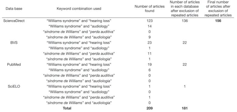
Results of the electronic databases
A total of 209 articles were found in each search, with the largest number of articles found in the ScienceDirect database. A larger number of articles were also found using keywords in
English. After exclusion of repeated titles, a total of 156 published
articles were obtained (Table 1).
Considering the inclusion criteria, as well as the research question, the titles of the 156 articles found were read.
From this stage, 119 articles were excluded and the abstracts
of the remaining 37 articles were read. After reading of the abstracts, 18 articles were selected to be read in full, and a total of 12 articles covered all the inclusion criteria and were considered for analysis in the present review (Figure 1).
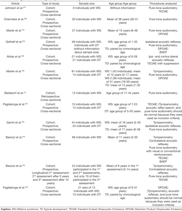
Considering that each study used different types of procedures, for greater clarity of the selected studies, a choice was made for an initial description of the main methodological criteria of each study (Table 2). The results of each study are described ahead, with analysis of each procedure of interest of the present study (Immittance test, Pure-tone audiometry, OAE, and BAEP.
Table 1. Results from the literature data collection
Data base Keyword combination used Number of articles
found
Number of articles in each database after exclusion of repeated articles
Final number of articles after
exclusion of repeated articles
ScienceDirect “Williams syndrome” and “hearing loss” 123 136 156
“Williams syndrome” and “audiology” 14
“síndrome de Williams” and “perda auditiva” 0 “síndrome de Williams” and “audiologia” 9
BVS “Williams syndrome” and “hearing loss” 23 22
“Williams syndrome” and “audiology” 1
“síndrome de Williams” and “perda auditiva” 11 “síndrome de Williams” and “audiologia” 1
PubMed “Williams syndrome” and “hearing loss” 19 22
“Williams syndrome” and “audiology” 6
“síndrome de Williams” and “perda auditiva” 0 “síndrome de Williams” and “audiologia” 0
SciELO “Williams syndrome” and “hearing loss” 1 1
“Williams syndrome” and “audiology” 0
“síndrome de Williams” and “perda auditiva” 1 “síndrome de Williams” and “audiologia” 0
Total 209 181
Analysis of the selected studies
Results of this literature review demonstrated that, although there was no restriction with respect to year of publication, studies on hearing assessment conducted with patients with
WS are recent (published in the past 20 years), suggesting that this area of study is relatively new. It was possible to observe
that most of the studies used samples composed of an extensive
age group, and that some of them presented a large sample size. However, not all participants underwent all procedures, that
Table 2. Detailing of the methodological aspects of each study
Article Type of study Sample size Age group Age group Procedures analyzed
Johnson et al.(37) Cohort;
Prospective; Cross-sectional
9 individuals with WS Without information Pure-tone audiometry; TEOAE
Cherniske et al.(38) Cohort;
Prospective; Cross-sectional
20 individuals with WS Mean of 38 years (30-51 years)
Pure-tone audiometry
Marler et al.(19) Cohort;
Prospective; Cross-sectional
27 individuals with WS Mean of 15 years (6-48 years)
Pure-tone audiometry; DPOAE
Gothelf et al.(20) Cohort;
Prospective; Cross-sectional
49 individuals with WS; Individuals with DT (without information about sample size)
WS: age group of 1-35 years;
TD: paired by chronological age
Ipsilateral acoustic reflexes; Pure-tone audiometry;
DPOAE; BAEP
Attias et al.(39) Cohort;
Prospective; Cross-sectional
21 individuals with WS; 21 individuals with DT
WS: age group of 6-26 years;
TD: paired by chronological age
Ipsi- and contra-lateral acoustic reflexes; TEOAE with suppression
Marler et al.(40) Cohort;
Prospective; Cross-sectional
81 individuals with WS; 14 individuals with DT
WS 1 (43 individuals): mean of 12 years (5-17 years); WS 2 (38 individuals): mean
of 31 years (18-59 years); TD: mean of 13 years (7-32
years)
Tympanometry; Pure-tone audiometry;
DPOAE
Bedeschi et al.(41) Cohort;
Retrospective; Cross-sectional
13 individuals with WS Age group of 17-44 years Pure-tone audiometry;
Paglialonga et al.(42) Cohort;
Prospective; Cross-sectional
13 individuals with WS; 14 individuals with DT
WS: age group of 7-23 years;
DT: age group of 5-20 years
TEOAE; (Tympanometry, acoustic reflex search, and pure-tone audiometry should be normal because they were
used as inclusion criteria)
Zarchi et al.(43) Cohort;
Prospective; Cross-sectional
44 individuals with WS; 23 individuals with DT
WS: mean of 16 years (5-35 years);
TD: mean of 17 years (6-38 years)
Tympanometry; Ipsilateral acoustic reflexes;
Pure-tone audiometry
Barozzi et al.(44) Cohort;
Prospective; Cross-sectional
69 individuals with WS Mean of 11 years (2-30 years)
Tympanometry; Contralateral acoustic
reflexes; Pure-tone audiometry with visual or conventional
reinforcement; TEOAE;
BAEP
Barozzi et al.(45) Cohort;
Prospective; Longitudinal (1st assessment;
2nd assessment after 5 years
and 3rd assessment after 10
years)
24 individuals with WS participated in the 1st
and 2nd assessments,
but only 10 of them participated in the 3rd
assessment
Mean of 8 years in the 1st
assessment (5-14 years)
Tympanometry; Contralateral acoustic
reflexes; Pure-tone audiometry;
TEOAE
Paglialonga et al.(46) Cohort;
Prospective; Cross-sectional
21 ears of 13 individuals with WS; 13 individuals with DT
WS: age group of 9-31 years;
TD: paired by chronological age
DPOAE; (Tympanometry, acoustic
reflexes, and pure-tone audiometry should be normal
is, the sample size for each procedure was different within the same study, which requires caution in the analysis of the results.
Immittance test
Among the 12 selected articles, six presented immittance measurements, four described findings of the tympanometry
curve(37-40), and two presented only findings of acoustic reflexes(20,41). Of the four studies that analyzed the tympanometry curve, the type A curve was predominant(37-40), and presence of the type B tympanometry curve was not observed in two of them(37,40).
In the first study, normal results were observed in 100% of the children evaluated (34 cases), and seven of the 32 adults
assessed presented other types of tympanometry curve other than the type A curve: type Ad and type C curves were observed in
five and two cases, respectively(37). In the second survey, type
A tympanometry curve was found in more than 65% of all
evaluations. Type C tympanometry curve was recorded only
in the first two assessments, and was observed in 20% of the cases in the first assessment and in 8-12% of the cases in the
second assessment. In the aforementioned study, contralateral
acoustic reflexes were present in all individuals with type A
tympanometry curve(40).
In contrast, two of these four studies reported type B
tympanometry curve in 23% of the cases(38,39). Type C curve was
also observed in 8.1% of the cases and acoustic reflexes were absent in more than 50% of the cases that presented normal
middle ear conditions (type A tympanometry curve)(38). In the
other study, type A tympanometry curve was observed in 76.5%
of the patients and, contrary to the previous study, contralateral
acoustic reflexes were present in all individuals who presented
type A tympanometry curve(39).
Both studies that analyzed only the characteristic of acoustic
reflexes obtained a higher percentage of absence of reflexes in individuals with WS. In the first study, a higher percentage of absence of acoustic reflexes was found in individuals with WS
compared with that of individuals with typical development (TD)(20). In the second study, absence of reflexes was observed
in 62-86% of the individuals evaluated, and in the patients who presented reflexes, the threshold was higher than that in
individuals with TD(41). In both studies, the authors associated this
finding with the complaint of hyperacusis frequently observed
in this population, considering that one of the functions of the
stapedial muscle reflex is to protect the auditory system from
intense sounds.
In general, it has been reported that type A curve is predominant in this population, suggesting that middle ear changes do not
seem to be a specific feature in this syndrome.
Concerning acoustic reflexes, although two studies did not confirm this observation(39,40), three other surveys reported
absence of acoustic reflexes as a feature commonly found even
in patients with no middle ear involvement(20,38,41). It is worth
noting that in two of these studies the authors agreed that this possible impairment in the stapedius muscle function seems to justify the hyperacusis reported by these patients(20,41).
Pure-tone audiometry
With respect to auditory threshold data in individuals with
WS, nine studies reporting this finding were identified.
In a preliminary study, 16 out of the 20 assessed patients were diagnosed with hearing loss, eight of them presented bilateral mild-to-moderate sensorineural hearing loss, one had unilateral mild sensorineural hearing loss at the high-frequency range, and three individuals presented sensorineural hearing loss at the high- and low-frequency ranges or conductive hearing loss(42). In contrast, another survey described a much lower
hearing loss prevalence, presenting six patients with normal
auditory thresholds and three patients with hearing loss at high frequencies(43)
One research observed higher auditory thresholds in individuals with WS compared with those of individuals with TD for frequencies as of 3 kHz, with predominantly mild-to-moderate sensorineural hearing loss. Conductive hearing loss at frequencies
below 2 kHz was found in 10% of the individuals, whereas 60%
of them presented cochlear hearing loss at higher frequencies
(3-8 kHz), with 75% of the cases showing bilateral hearing
loss. The degree of hearing loss at the high-frequency range varied from 25 to 55 dB in the right ear and from 25 to 110 dB in the left ear(20).
Another survey also reported presence of mild hearing loss
in most cases, with only 11.3% of the cases showing moderate to profound hearing loss (threshold >40 dB), with high frequencies
(6-8 kHz) as the most affected. Regarding the type of hearing
loss, 26.1% of the cases were sensorineural, 21.6% were mixed, and 9.1% were conductive(38).
In another study, mild to moderately severe hearing loss was
also observed in 63% of the schoolchildren and 92% of the adults
evaluated, and sensorineural hearing loss was detected in at least
50% of cases. Another study conducted with 13 patients with
WS that used a very similar methodology observed bilateral mild
hearing loss in eight individuals (61.6%), with one conductive, two mixed, and five sensorineural cases(44).
A survey that performed hearing screening in 19 individuals observed that 16 of them failed the test. In addition, the authors
reported that six out of the eight individuals who underwent
conventional audiometry presented sensorineural hearing loss(9).
Based on these findings, prevalence of hearing loss was remarkable, found in over 60% of the cases of individuals with
WS assessed; only one article described hearing loss in only
approximately 33% of the cases, but its small sample size may have influenced the results(43). In addition to this article, another
survey that evaluated young children also found a lower percentage of hearing loss(39). Among the 16 children assessed by means
of pure tone audiometry with visual reinforcement, only two presented hearing loss, conductive in both cases, of mild and moderate degrees. Among the patients assessed by conventional audiometry (53 cases), hearing loss was observed at frequencies
13.2% sensorineural) and at higher frequencies (mean of the thresholds obtained at 4, 6, and 8 kHz) in 30% of the cases(39).
Some studies have associated hearing loss with age, and this seems to be progressive in individuals with WS(39,40,43,44), beginning in early adolescence(39) or early adulthood(44). A research found
higher incidence of hearing loss in individuals aged >15 years
(46%) than in younger individuals (23%)(39), whereas another
study showed appearance of hearing loss in these individuals
at the age of approximately 25 years(44).
Considering also the progressive profile of hearing loss, a
study performed a longitudinal auditory assessment of patients (three evaluations over a 10-year follow-up). The authors observed hearing loss at the lower frequencies (mean of the thresholds
obtained at 0.5, 1, and 2 kHz) in 12.5%, 12.5%, and 30% of the cases, respectively, for the first, second, and third assessments, with predominance of sensorineural hearing loss (only 4% of conductive hearing loss observed in the first two evaluations).
For the high-frequency range (mean of the thresholds obtained
at 4, 6, and 8 kHz), hearing loss was predominant in 25%, 50%, and 80% of the cases, respectively, in the three assessments(40).
Sensorineural was the most commonly observed type of
hearing loss. Six studies observed presence of conductive
hearing loss(20,38-40,42,44); however, the survey that found the highest percentage of this type of hearing loss reported a value
of 10%(20). With respect to hearing losses of the mixed type,
they were described in only two articles, in which they were
observed in approximately 15%(44) and 21%(38) of the cases.
Therefore, it is possible to observe that middle ear impairment is not a dominant feature in individuals with WS.
Data concerning the degree and configuration of hearing loss
are also worth noting. Prevalence of mild-to-moderate hearing loss was observed in all articles that described this variable(20,38,39,42,44).
The description of hearing loss configuration was also very similar between the articles; descending configuration was predominant
in all studies that described this variable, with greater impact on the high-frequency range(20,38-40,42,43): as of 3 kHz for some authors(20,39,40) and between 6 and 8 kHz for others(38).
Otoacoustic Emissions (OAE)
Nine articles performed OAE: four assessed Distortion Product Otoacoustic Emissions (DPOAE)(19,20,45) and five analyzed Transient Evoked Otoacoustic Emissions (TEOAE)(39-41,43,46).
In the studies that conducted DPOAE, the results were convergent when showing smaller amplitude responses in individuals with WS compared with those of individuals with TD(19,20,37,45), which could differ between frequencies from 2 to 11 dB(45). In addition, one of the articles observed absence
of DPOAE in 23% of the cases(20), whereas another survey
reported higher involvement in the high-frequency range(37).
In these four studies, the authors agreed when they reported cochlear impairment in this population, especially at the medium and high frequencies, and indicated the DPOAE evaluation as
an important method to detect subclinical findings in cochlear
hair cell damage.
Regarding assessment using TEOAE, one of the surveys observed four patients who presented normal hearing thresholds,
but absent TEOAE(43). Moreover, another study found absent
TEOAE in 39-48% of the patients with normal hearing and no
middle ear impairment(39). These results demonstrate loss of
the cochlear function of the hair cells and the importance of auditory monitoring by TEOAE, considering that this measure seems to be useful to diagnose auditory impairment, even when auditory thresholds are not yet altered.
Another research verified the TEOAE measurements by means of three analyses: energy extracted from the broad-band TEOAE recordings, energy extracted from each of the narrow-band frequency components of the TEOAE, and latency extracted only
from the frequency components. The authors observed lower energy, both in the broad-band TEOAE responses in individuals with WS (23.5 dB NPS) compared with that of individuals with
TD (30.8 dB NPS) and for energy extracted from the frequency
components (with mean difference between the two groups of 5-9 dB NPS, distributed almost evenly across the frequency range). In the latency analysis in the frequency components, higher latency was observed in individuals with WS compared with that in individuals with TD, with the difference between the two groups varying from 0.6 to 1.5 ms(46).
Furthermore, one of the studies evaluated TEOAE with suppression. The results showed higher suppression of the effect of TEOAE in patients with WS than in individuals with TD, which, according to the authors, suggests a higher activity of the medial olivocochlear efferent system and that this functional alteration may contribute to the presence of hyperacusis in these patients(41).
One last study that monitored TEOAE responses over time
also observed a progressive profile of TEOAE loss. Absence of TEOAE was found in approximately 50, 60 and 70% of
the patients that presented means of the threshold values in normal low frequencies, respectively, in the three assessments. According to the authors, these data suggest a cochlear fragility in individuals with WS(40).
Analysis of the TEOAE responses described in these nine
articles showed very similar findings for both acoustic stimuli
employed. The results have demonstrated absence or decrease in the amplitude of TEOAE in the WS population, even in individuals with normal hearing thresholds. Overall, the authors
reported that this finding demonstrates a cochlear fragility or
impairment in these individuals, and suggested that TEOAE is a measure of paramount importance for this population.
Brainstem Auditory Evoked Potential (BAEP)
Regarding analysis of the BAEP, only two studies considered
and described these findings. In the first study, despite the
observation an increased latency values (in individuals with WS compared with those in individuals with TD) of wave I in
61.9% of the cases (means of 1.78 and 1.63 ms for both groups, respectively), of wave III in 42.9% of the cases (means of
3.98 and 3.7 ms for both groups, respectively), and of wave
V in 23.8% of the evaluated cases (means of 5.9 and 5.52 ms
waves III and V were due to a delay in the latency of wave I, and discarded neural conduction impairment(20).
In the second study, BAEP was performed in 14 patients.
The latency values observed in individuals with WS were as
follows: between 1.38 and 1.94 ms for wave I, 3.44 and 4.16 ms
for wave III, and 5.08 and 6.02 ms for wave V. As for the interpeak intervals, the following values were observed: between 1.92 and 2.58 ms for interpeak I-III, 1.52 and 1.88 ms for interpeak III-V,
and 3.6 and 4.32 ms for interpeak I-V. The authors considered
these results as normal and suggested no retrocochlear involvement in this population(39).
Although both studies have discarded retrocochlear involvement in these individuals, one of them(20) observed increased latency
values for all waves, which is an important finding to be
considered in the clinical routine. Thus, the results of only two
articles seem to be insufficient to determine a conclusion about expected latency values in individuals with WS.
Therefore, further studies addressing BAEP in larger samples
of this population would be useful to confirm the results regarding
the functionality and integrity of the central auditory brainstem pathways in individuals with WS. Although the WS phenotype is not so thought-provoking, a general convergence in the results of the studies was observed, evidencing a remarkable cochlear impairment in this population. Data from all of these assessments jointly analyzed reinforce the need for routine otolaryngology follow-up with complete auditory monitoring, including multiple auditory tests, in the WS population. Such
monitoring should begin on the first days of life, seeking early
diagnosis and, consequently, intervention and improvement of the quality of life of these individuals.
CONCLUSION
Based on the articles analyzed in the present literature review, we conclude that the main alterations in auditory assessment in individuals with WS are due to absence of OAE and acoustic
reflex and presence of mild-to-moderate sensorineural hearing
loss, mainly in the high-frequency range in pure-tone audiometry, and these results show a cochlear impairment in this population.
Regarding the immittance test, it was possible to observe a type A tympanometry curve, demonstrating absence of middle ear impairment. As for the brainstem auditory-evoked potentials, no retrocochlear alteration was observed in individuals with WS.
REFERENCES
1. Korenberg JR, Chen XN, Hirota H, Lai Z, Bellugi U, Burian D, et al. Genome structure and cognitive map of Williams syndrome. J Cogn Neurosci. 2000;12(Suppl. 1):89-107. http://dx.doi.org/10.1162/089892900562002. PMid:10953236.
2. Rossi NF, Moretti-Ferreira D, Giacheti CM. Genética e linguagem na Síndrome de Williams-Beuren: uma condição neuro-cognitiva peculiar. Pro Fono. 2006;18(3):331-8. http://dx.doi.org/10.1590/S0104-56872006000300013. PMid:17180802.
3. Deutsch SI, Rosse RB, Schwartz BL. Williams Syndrome: a genetic deletion disorder presenting clues to the biology of sociability and clinical challenges of hypersociability. CNS Spectr. 2007;12(12):903-7. http:// dx.doi.org/10.1017/S1092852900015686. PMid:18163035.
4. Sugayama SM, Leone C, Chauffaille ML, Okay TS, Kim CA. Síndrome de Williams: proposta de sistema de pontuação para diagnóstico clínico. Clinics. 2007;62(2):159-66. http://dx.doi.org/10.1590/S1807-59322007000200011. PMid:17505701.
5. von Beust G, Laccone FA, del Pilar Andrino M, Wessel A. Clinical aspects and genetics of Williams-Beuren syndrome- Clinical and molecular genetic study of 44 patients with suspected Williams-Beuren syndrome. Klin Padiatr. 2000;212(2):299-307. PMid:11190824.
6. Meyer-Lindenberg A, Mervis CB, Faith Berman K. Neural mechanisms in Williams syndrome: a unique window to genetic influences on cognition and behaviour. Nat Rev Neurosci. 2006;7(5):380-93. http://dx.doi.org/10.1038/ nrn1906. PMid:16760918.
7. Gagliardi C, Martelli S, Burt MD, Borgatti R. Evolution of neurologic features in Williams syndrome. Pediatr Neurol. 2007;36(5):301-6. http:// dx.doi.org/10.1016/j.pediatrneurol.2007.01.001. PMid:17509461. 8. Bellugi U, Lichtenberger L, Jones W, Lai I, St George M. The neurocognitive
profile of Williams syndrome: a complex pattern of strengths and weaknesses. J Cogn Neurosci. 2000(12, Suppl 1):7-29. http://dx.doi. org/10.1162/089892900561959. PMid:10953231.
9. Bellugi U, Korenberg JE, Klima ES. Williams syndrome: an exploration of neurocognitive and genetic features. Clin Neur Res. 2001;1(3):217-29. http://dx.doi.org/10.1016/S1566-2772(01)00008-1.
10. Volterra V, Caselli MC, Capirci O, Tonucci F, Vicari S. Early linguistic abilities of italian children with Williams syndrome. Dev Neuropsychol. 2003;23(1-2):33-58. http://dx.doi.org/10.1080/87565641.2003.9651886. PMid:12730019.
11. Järvinen-Pasley A, Bellugi U, Reilly J, Mills DL, Galaburda A, Reiss AL, et al. Defining the social phenotype in Williams syndrome: a model for linking gene, the brain, and behavior. Dev Psychopathol. 2008;20(1):1-35. http://dx.doi.org/10.1017/S0954579408000011. PMid:18211726. 12. Järvinen-Pasley A, Vines BW, Hill KJ, Yam A, Grichanik M, Mills D, et al.
Cross-modal influences of affect across social and non-social domains in individuals with Williams syndrome. Neuropsychologia. 2010;48(2):456-66. http://dx.doi.org/10.1016/j.neuropsychologia.2009.10.003. PMid:19822162. 13. Levitin DJ, Cole K, Lincoln A, Bellugi U. Aversion, awareness, and
attraction: investigating claims of hyperacusis in the Williams syndrome phenotype. J Child Psychol Psychiatry. 2005;46(5):514-23. http://dx.doi. org/10.1111/j.1469-7610.2004.00376.x. PMid:15845131.
14. Zarchi O, Attias J, Gothelf D. Auditory and visual processing in Williams syndrome. Isr J Psychiatry Relat Sci. 2010;47(2):125-31. PMid:20733255. 15. Attias J. New findings on hyperacusis in Williams syndrome. ENT and
Audiology News. 2013;21-6:76-8.
16. Lenhoff HM, Perales O, Hickok G. Absolute pitch in Williams syndrome. Music Percept. 2001;18(4):491-503. http://dx.doi.org/10.1525/mp.2001.18.4.491. 17. Levitin DJ, Cole K, Chiles M, Lai Z, Lincoln A, Bellugi U. Characterizing the musical phenotype in individuals with Williams syndrome. Child Neuropsychol. 2004;10(4):223-47. http://dx.doi.org/10.1080/09297040490909288. PMid:15621847.
18. Lense MD, Dykens EM. Musical learning in children and adults with Williams syndrom. J Intellect Disabil Res. 2013;57(9):850-60. http:// dx.doi.org/10.1111/j.1365-2788.2012.01611.x. PMid:22974236. 19. Marler JA, Elfenbein JL, Ryals BM, Urban Z, Netzloff ML. Sensorineural
hearing loss in children and adults with Williams syndrome. Am J Med Genet. 2005;138(4):318-27. http://dx.doi.org/10.1002/ajmg.a.30970. PMid:16222677.
20. Gothelf D, Farber N, Raveh E, Apter A, Attias J. Hyperacusis in Williams syndrome: Characteristics and associated neuroaudiologic abnormalities. Neurology. 2006;66(3):390-5. http://dx.doi.org/10.1212/01. wnl.0000196643.35395.5f. PMid:16476938.
21. Li HH, Roy M, Kuscuoglu U, Spencer CM, Halm B, Harrison KC, et al. Induced chromosome deletions cause hypersociability and other features of Williams-Beuren syndrome in mice. EMBO Mol Med. 2009;1(1):50-65. http://dx.doi.org/10.1002/emmm.200900003. PMid:20049703. 22. Lucena J, Pezzi S, Aso E, Valero MC, Carreiro C, Dubus P, et al. Essential
Genet. 2010;11(1):61. http://dx.doi.org/10.1186/1471-2350-11-61. PMid:20403157.
23. Canales CP, Wong ACY, Gunning PW, Housley GD, Hardeman EC, Palmer SJ. The role of GTF2IRD1 in the auditory pathology of Williams–Beuren Syndrome. Eur J Hum Genet. 2015;23(6):774-80. http://dx.doi.org/10.1038/ ejhg.2014.188. PMid:25248400.
24. Meng Y, Zhang Y, Tregoubov V, Falls DL, Jia Z. Regulation of spine morphology and synapticfunction by LIMK1 and the action cytoskeleton. Rev Neurosci. 2003;14(3):233-40. http://dx.doi.org/10.1515/REVNEURO.2003.14.3.233. PMid:14513866.
25. Meng Y, Zhang Y, Tregoubov V, Janus C, Cruz L, Jackson M, et al. Abnormal spine morphology and enhanced LTP in LIMK-1 knockout mice. Neuron. 2002;35(1):121-33. http://dx.doi.org/10.1016/S0896-6273(02)00758-4. PMid:12123613.
26. Hoogenraad CC, Akhmanova A, Galjart N, De Zeeuw CI. LIMK1 and CLIP-115: linking cytoskeletal defects to Williams syndrome. BioEssays. 2004;26(2):141-50. http://dx.doi.org/10.1002/bies.10402. PMid:14745832. 27. Scott RW, Olson MF. LIM kinases: function, regulation and association
with human disease. J Mol Med (Berl). 2007;85(6):555-68. http://dx.doi. org/10.1007/s00109-007-0165-6. PMid:17294230.
28. Matsumoto N, Kitani R, Kalinec F. Linking LIMK1 deficiency to hyperacusis and progressive hearing loss in individuals with Williams syndrome. Commun Integr Biol. 2011;4(2):208-10. http://dx.doi.org/10.4161/cib.4.2.14491. PMid:21655442.
29. Safieddine S, Wenthold RJ. SNARE complex at the ribbon synapses of cochlear hair cells: analysis of synaptic vesicle- and synaptic membrane-associated proteins. Eur J Neurosci. 1999;11(3):803-12. http://dx.doi. org/10.1046/j.1460-9568.1999.00487.x. PMid:10103074.
30. Shah SM, Kang YJ, Christensen BL, Feng AS, Kollmar R. Expression of Wnt receptors in adult spiral ganglion neurons: frizzled 9 localization at growth cones of regenerating neurites. Neuroscience. 2009;164(2):478-87. http://dx.doi.org/10.1016/j.neuroscience.2009.08.049. PMid:19716861. 31. Di Rosa C, Cieri F, Antonucci I, Stuppia L, Gatta V. Music in DNA: from
Williams syndrome to music genes. Open J Genet. 2015;5(1):12-26. http:// dx.doi.org/10.4236/ojgen.2015.51002.
32. Wengenroth M, Blatow M, Bendszus M, Schneider P. Leftward lateralization of auditory cortex underlies holistic sound perception in Williams syndrome. PLoS One. 2010;5(8):e12326. http://dx.doi.org/10.1371/journal.pone.0012326. PMid:20808792.
33. Lenhoff HM, Wang PP, Greenberg F, Bellugi U. Williams syndrome and the brain. Sci Am. 1997;277(6):68-73. http://dx.doi.org/10.1038/ scientificamerican1297-68. PMid:9388834.
34. Holinger DP, Bellugi U, Mills DL, Korenberg JR, Reiss AL, Sherman GF, et al. Relative sparing of primary auditory cortex in Williams syndrome. Brain Res. 2005;1037(1-2):35-42. http://dx.doi.org/10.1016/j. brainres.2004.11.038. PMid:15777750.
35. Martens MA, Reutens DC, Wilson SJ. Auditory cortical volumes and musical ability in Williams syndrome. Neuropsychol. 2010;48(9):2602-9. http://dx.doi.org/10.1016/j.neuropsychologia.2010.05.007. PMid:20457168. 36. Centre for Evidence-based Medicine. Oxford Centre for Evidence-based
Medicine – Levels of Evidence. Oxford: CEBM; 2009 [citado em 2017
Jun 2]. Disponível em: http://www.cebm.net/oxford-centre-evidence-based-medicine-levels-evidence-march-2009/
37. Marler JA, Sitcovsky JL, Mervis CB, Kistler DJ, Wightman FL. Auditory function and hearing loss in children and adults with Williams syndrome: Cochlear impairment in individuals with otherwise normal hearing. Am J Med Genet C Semin Med Genet. 2010;154C(2):249-65. http://dx.doi. org/10.1002/ajmg.c.30262. PMid:20425785.
38. Zarchi O, Attias J, Raveh E, Basel-Vanagaite L, Saporta L, Gothelf D. A comparative study of hearing loss in two microdeletion syndromes: velocardiofacial (22q11.2 Deletion) and Williams (7q11.23 Deletion) Syndromes. J Pediatr. 2011;158(2):301-6. http://dx.doi.org/10.1016/j. jpeds.2010.07.056. PMid:20846670.
39. Barozzi S, Soi D, Comiotto E, Borghi A, Gavioli C, Spreafico E, et al. Audiological findings in Williams syndrome: a study of 69 patients. Am J Med Genet. 2012;158A(4):759-71. http://dx.doi.org/10.1002/ajmg.a.35241. PMid:22411878.
40. Barozzi S, Soi D, Spreafico E, Borghi A, Comiotto E, Gagliardi C, et al. Audiological follow-up of 24 patients affected by Williams syndrome. Eur J Med Genet. 2013;56(9):490-6. http://dx.doi.org/10.1016/j.ejmg.2013.07.001. PMid:23886711.
41. Attias J, Raveh E, Ben-Naftali NF, Zarchi O, Gothelf D. Hyperactive auditory efferent system and lack of acoustic reflexes in Williams syndrome. J Basic Clin Physiol Pharmacol. 2008;19(3-4):193-207. http://dx.doi.org/10.1515/ JBCPP.2008.19.3-4.193. PMid:19025031.
42. Cherniske EM, Carpenter TO, Klaiman C, Young E, Bregman J, Insogna K, et al. Multisystem study of 20 older adults with Williams syndrome. Am J Med Genet. 2004;131(3):255-64. http://dx.doi.org/10.1002/ajmg.a.30400. PMid:15534874.
43. Johnson LB, Comeau M, Clarke KD. Hyperacusis in Williams syndrome. J Otolaryngol. 2001;30(2):90-2. http://dx.doi.org/10.2310/7070.2001.20811. PMid:11770962.
44. Bedeschi MF, Bianchi V, Colli AM, Natacci F, Cereda A, Milani D, et al. Clinical follow-up of young adults affected by Williams syndrome: experience of 45 Italian patients. Am J Med Genet. 2011;155(2):353-9. http://dx.doi.org/10.1002/ajmg.a.33819. PMid:21271653.
45. Paglialonga A, Barozzi S, Brambilla D, Soi D, Cesarani A, Spreafico E, et al. Analysis of subtle auditory dysfunctions in young normal-hearing subjects affected by Williams syndrome. Int J Pediatr Otorhinolaryngol. 2014;78(11):1861-5. http://dx.doi.org/10.1016/j.ijporl.2014.08.010. PMid:25193583.
46. Paglialonga A, Barozzi S, Brambilla D, Soi D, Cesarani A, Gagliardi C, et al. Cochlear active mechanisms in young normal-hearing subjects affected by Williams syndrome: Time-frequency analysis of otoacoustic emissions. Hear Res. 2011;272(1-2):157-67. http://dx.doi.org/10.1016/j. heares.2010.10.004. PMid:20969939.
Author contributions
LAFS was responsible for collection, classification and analysis of the data and