of Cyphomyiactia costai
Artigas, Papavero & Serra, 1991
(Asilidae, Laphriinae, Atomosiini):
description of the male, and illustration
of the holotype and structures of
male and female terminalia
Rodrigo Vieira¹²⁵ & Lucas de Araújo Cezar³⁴⁶
¹ Secretaria de Estado de Educação e Qualidade de Ensino do Amazonas (SEDUC/Amazonas). Manaus, AM, Brasil. ² Instituto Nacional de Pesquisas da Amazônia (INPA), Coordenação de Biodiversidade (CBIO),
Programa de Pós-Graduação em Entomologia (PPG-ENT). Manaus, AM, Brasil. ³ Universidade de São Paulo (USP), Museu de Zoologia (MZUSP). São Paulo, SP, Brasil.
⁴ Universidade de São Paulo (USP), Faculdade de Filosofia, Ciências e Letras de Ribeirão Preto (FFCLRP), Programa de Pós-Graduação em Entomologia. Ribeirão Preto, SP, Brasil.
⁵ ORCID: 0000-0002-3382-9638. E-mail: [email protected] ⁶ E-mail: [email protected]
Abstract. The male of Cyphomyiactia costai Artigas, Papavero & Serra, 1991 is described and illustrated for the first time. New records are provided from the states of Bahia, Maranhão and Mato Grosso, Brazil. The holotype is illustrated, as well as struc-tures of male and female terminalia.
Key-Words. Asiloidea; Neotropical; Cyphomyia.
INTRODUCTION
Artigas, Papavero & Serra (1991) proposed the genus Cyphomyiactia to include an undescribed Brazilian species, Cyphomyiactia costai Artigas, Papavero & Serra, 1991, because of its singular morphology. The generic description was based on a single female specimen collected in Goiás state.
Cyphomyiactia costai possesses a relatively
wide face and an evenly convex, first flagellomere prolonged into a filiform process; the abdomen is very wide; short, and cup-shaped. Due to the overall coloration, the body being blackish with blue reflections and with a golden tomentose occiput, these robber-flies resemble stratiomy-ids of the genus Cyphomyia Wiedemann (Artigas, Papavero & Serra, 1991).
In this work, the male of Cyphomyiactia costai
Artigas, Papavero & Serra, 1991 is described and illustrated for the first time. New records are pro-vided from Bahia and Maranhão states, Brazil. The holotype is illustrated, as well as structures of male and female terminalia.
MATERIALS AND METHODS
In the present study, we examined specimens housed at INPA – Instituto Nacional de Pesquisas da Amazônia, CEIOC – Coleção Entomológica do Instituto Oswaldo Cruz, and CZMA – Coleção Zoológica do Maranhão, Universidade Estadual do Maranhão, Caxias, Maranhão, Brasil, as well as the holotype of C. costai housed in the Museu de Zoologia da Universidade de São Paulo (MZUSP) collection.
General morphological terminology follows Cumming & Wood (2009).
Dissected terminalia were treated in hot 10% KOH for varying periods of time, washed in wa-ter, subsequently treated in acetic acid and fi-nally examined in excavated slides in glycerin. Illustrations of the terminalia were prepared us-ing Adobe Illustrator CS5 software. After exam-ination and illustration, the detached parts were placed in microvials containing glycerin and the vials were pinned with their respective speci-mens. Images were taken with a ZEISS AxioCam
ISSN On-Line: 1807-0205 ISSN Printed: 0031-1049 ISNI: 0000-0004-0384-1825 Pap. Avulsos Zool., 2018; v.58: e20185846
http://doi.org/10.11606/1807-0205/2018.58.46
Mrc5 digital camera attached to a Zeiss Discovery V20 Stereomicroscope, using the lighting methods described by Kerr et al. (2008). Focus stacks of photos of asilid spec-imens were combined using CombineZP (Hadley, 2010).
Label data is cited in full, with the original spellings, punctuations, and dates. Information presented within square brackets – “[ ]” – is complementary data not in-cluded on the labels. Data for the same specimen but from different labels are separated by slashes ().
The map was generated on the website SimpleMappr (www.simplemappr.net) (Fig. 1).
Cyphomyiactia costai Artigas, Papavero & Serra, 1991
(Figs. 2‑3)
Cyphomyiactia costai Artigas, Papavero & Serra, 1991: 62;
Papavero, 2009: 92 (catalogue).
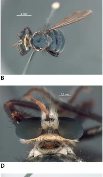
Original description is transcribed below: “Face densely silvery-white tomentose. Hair of mystax white [Fig. 2C]. Occiput [Fig. 2D] gold-en tomgold-entose at margins, brownish-black to-mentose around foramen, with long, sparse, fine white pile bellow; dorsal occipital bristles yellowish. Scape brown, pedicel brownish-yel-low, flagellum red-brown and white pollinose [Figs. 2C, 2D]. Frons golden tomentose with a slender black stripe running from anterior ocellus to base of antennae. Vertex black. Thorax blackish in ground color, with blu-ish reflections [Fig. 2B]. Pleura with mixed silvery-white and golden-brown tomentum in some areas. Antepronotum predominant-ly golden tomentose with a brownish-black stripe on the anterior border and yellow hairs. Lateral margins of antepronotum sil-very-white tomentose. Postpronotum
brown-ish-golden tomentose, hairs brown [Fig. 2C]. Proepisternum silvery-white tomentose, as well as the anepisternum, which shows a polished brown spot and brown hairs. Anepimeron brown, with white pollinosity. Anatergite goldish-silvery-white pollinose with a fringe of yellowish hairs behind pos-terior callus. Meron brown, white pollinose, with a few brown hairs. Katatergite brown, silvery-white tomentose, with brown bristles. Posterior callus brown.
Wings [Fig. 2F] hyaline, slightly fumose along veins. Halteres yellow.
Abdomen [Figs. 2A, 2B, 2E] blackish in ground color, with blue shine. Hairs of abdomen white. Tergites 3-4 silvery-white tomentose spots dorsolaterally [Fig. 2E].
Holotype female. BRAZIL, Goiás, Goiânia (Campinas), 1935 (Borgmeier & Souza Lopes), in the MZUSP.”
Addenda to the holotype female description: Total length, excluding antenna, 10.6 mm; length of wing, 8.8 mm; greatest width of abdomen, 3.4 mm. Head: Eyes greenish; occiput silvery-golden-pollinose on ventral 1/4. Thorax: Postpronotum with mixed brownish-gold-en and silvery-white tombrownish-gold-entum, setae brown and white (Fig. 2C); anepisternum with mixed silvery-white and golden-brown tomentum; 3 long and strong anepister-nal macrosetae, among others; anepimeron dark-brown; 3 postalar setae (not 1 postalar + 3 on “posterior callus”).
Wing (Fig. 2F): Cell c wide; r₅ closed, with short stalk; m₃ widening posteriorly; cup with short stalk; anal lobe wide. Legs (Figs. 2A-2C, 2E): Entirely dark-brown, with dark-brown setae; tarsi with golden vestiture.
Label transcript: “Campinas, Goyaz [Goiás], Borgmeier
et. S. Lopes, XII.[1]935 / [handwritten] Cyphomyiactia
cy-phomyioides, gen. n. sp. n. Artigas, Papavero & Serra / [red
label] HOLOTYPE Cyphomyiactia costai, det. L.A. Cezar, 2014.”
Holotype condition: Pinned; well preserved; lacking right antenna, anepisternal and postalar setae on the left side, and left wing.
Female, additional specimen: Total length, excluding antenna, 9.2 mm; length of wing, 7.1 mm; greatest width of abdomen, 3.2 mm.
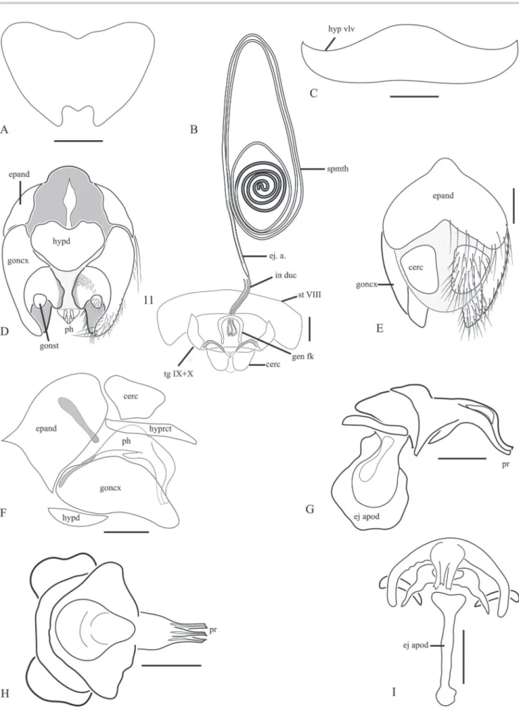
Female terminalia, additional specimen (Figs. 3A‑3C):
Anterior margin of sternite VII sinuous (Fig. 3A); hypog-ynium almost hemispherical, with hypogynial valves continuous with lateral margins of the sclerite (Fig. 3C); smooth median protrusion between hypogynial valves (Fig. 3C); smooth retraction on anterior margin (Fig. 3C); hypogynium softly setate posteriorly. Three
sperma-Figure 1. Distribution of Cyphomyiactia costai.
Vieira, R. & Cezar, L.A.: Addition to the knowledge on Cyphomyiactia costai Artigas, Papavero & Serra, 1991 Pap. Avulsos Zool., 2018; v.58: e20185846
thecae (Fig. 3B), occupying abdominal segments 4-8; reservoirs cylindrical, disposed from a wider elliptical spiral going to an inner round and more sclerotized spiral distally (Fig. 3B); spermathecal ducts opening in-dependently at the bursa; genital fork U-shaped, arms
anteriorly thick, posteriorly truncate, divergent (Fig. 3B). Eggs brown and oval.
Male (Figs. 3D‑3I): Total length, excluding antenna, 10.6 mm; length of wing, 7.6 mm; greatest width of
Figure 3.Cyphomyiactia costai. Addtional specimen. (A) Female, sternite VII; (B) Female genitalia; (C) Female, sternite VIII; (D) Male terminalia, ventral view; (E) Male terminalia, dorsal view; (F) Male terminalia, lateral view; (G) Phallus, lateral view; (H) Phallus, ventral view; (I) Phallus, dorsal view. Abbreviations: cerc: cercus; ej a: ejection apparatus; ej apod: ejaculatory apodeme; epand: epandrium; gen fk: genital fork; goncx: gonocoxite; gonst: gonostylus; hyprct: hypoproct; hypd: hypandrium; hyp hlv: hypoginial valves; in duc: individual duct; ph: phallus; pr: phallic prongs; st VIII: sternite VIII; tg IX + X: tergite IX + X. Scale bar = 1 mm.
Vieira, R. & Cezar, L.A.: Addition to the knowledge on Cyphomyiactia costai Artigas, Papavero & Serra, 1991 Pap. Avulsos Zool., 2018; v.58: e20185846
men, 3.2 mm. Similar to female except for: postalar callus with 4 macrosetae; antepronotum predominantly with dark-brown setae; anepimeron with white and brown tomentum. Male Terminalia: Hypopygium inconspic-uous in both dorsal and lateral views, hidden under a strongly-convex cup-shaped abdomen. Hypandrium subtriangular, wider than long, with blunt posterior mar-gin, and a median concavity on anterior margin (Fig. 3D). Gonocoxites free, with setae near median margin basal-ly (Fig. 3D); gonocoxal prolongations blunt, smoothbasal-ly curved inwards, with strong medially-directed setae near apex; gonostyli reduced, round, attached to the dor-sal base of gonocoxites (Fig. 3D). Lateral lobes of hypo-proct subquadratic (abruptly blunt and parallel-sided), in dorsal view; lateral lobes long, extending beyond the apex of gonocoxal prolongation, and diverging posteri-orly. Cerci free, subtriangular, slightly longer than wide, distant from each other (Figs. 3E, 3F). Phallus strongly arched laterally (towards gonocoxal apodemes), pro-truded mediodorsally, in lateral view (Fig. 3G); moderate dilation proximal to the prongs (Fig. 3G); apex of phallus with three short prongs (Fig. 3H); lateral ejaculatory pro-cesses wide (Fig. 3I).
Remarks: The specimen deposited in MZUSP did not bear an explicit label clarifying its name-bearing status. Apart from the original specimen label, there was only a handwritten identification label that read “Cyphomyiactia
cyphomyioides gen. n. sp. n. Artigas, Papavero & Serra”.
Given that the morphology and sampling data of this specimen agree with those cited by the authors in the original description, and that there was no other speci-men speci-mentioned for the species in the original work, it is sensible to assume that “C. cyphomyioides” is an unavail-able manuscript name and that this specimen is indeed the holotype of C. costai (N. Papavero, pers. comm.). Although Cyphomyiactia’s singular compactness cannot be identified in any other group within the Atomosiini, its spermathecal disposal greatly resembles that of
Hybozelodes lucidus (Hermann, 1912), as well as the shape
of antennae, the bluish reflections of thorax and abdo-men, and the length of the lateral lobes of hypoproct of the male. These characters could indicate a closer rela-tionship between the monotypic genus Cyphomyiactia
and Hybozelodes Hermann, 1912.
Additional material examined: BRASIL, MA[ranhão],
Mirador, Parque Est.[adual] Mirador, Base da Geraldina [06°43’S 44°58’W] / Armadilha Malaise, 15-26.x.2006,
F. Limeira-de-Oliveira (1 male INPA); same data, ex-cept 17.xii.2006 (1 female CZMA); BR[ASIL], BA[HIA],
Igrapiuna, Res.[erva] Michelin Vilas [13°49’S 39°08’W], Malaise / 20.i-24.ii.2013, M. Menezes & E. Aragão col.
(1 male INPA); Campinas [Goiânia] [16°40’S 49°15’W] – Goyas [Goiás], [BRASIL] Borgmeier et S. Lopes, xii. [1]925
/ Atractia / Det. S.H. Lopes (1 male CEIOC); Chapada
dos Guimarães [15°27’39”S 55°45’00”W], Mato Grosso,
BRASIL, xi. 1963, Alvarenga e Werner (1 male CEIOC). Distribution (Fig. 1): BRAZIL (Maranhão, Bahia, Goiás, Mato Grosso).
ACKNOWLEDGMENTS
To the curators of the collections housing the exam-ined material, Dr. Francisco Limeira-de-Oliveira (CZMA), Dr. Carlos Lamas (MZUSP), Dra. Jane Costa (CEIOC) and Dr. Marcio Oliveira (INPA). To Alexssandro Camargo for the records of specimens deposited at CEIOC. To CNPq: Biodiversidade e Monitoramento do processo de des-matamento do cerrado maranhense no Parque Estadual do Mirador, 457440/2012-0 – CNPq PPBio Rede Cerrado; Riqueza, Diversidade e Composição dos táxons de inse-tos do Parque Estadual do Mirador, estado do Maranhão, Brasil, FAPEMA, Processos: APP-00852/10 – FAPEMA; APP-00498/12 – FAPEMA; CBIOMA-03001/12 – FAPEMA. To CNPq for the fellowships that have allowed us to com-plete this work. Thanks to anonymous referees for valu-able comments that greatly improved the manuscript.
REFERENCES
Artigas, J.N.; Papavero, N. & Serra, A.L. 1991. The American genera of Asilidae (Diptera): Keys for identification with an atlas of female spermathecae and other morphological details. VI. Tribe Atomosiini Hermann (Laphriinae), with descriptions of two new genera and three new species, and a catalogue. Gayana Zoologia, 55(1): 53-85.
Cumming, J.M. & Wood, D.M. 2009. Adult morphology and terminology [Chapter] 2. In: Brown, B.V.; Borkent, A.; Cumming, J.M.; Wood, D.M.; Woodley, N.E. & Zumbado, M.A. (Eds.). Manual of Central American Diptera. Ottawa, National Research Council Research Press. v. 1, p. 9-50. Hadley, A. 2010. CombineZP Image Stacking Software. Disponível em: https://
combinezp.software.informer.com.
Kerr, P.H.; Fisher E.M. & Buffington M.L. 2008. Dome lighting for insect imaging under a microscope. American Entomologist, 54(4): 198-200. Papavero, N. 2009. Catalogue of Neotropical Diptera. Asilidae. Neotropical
Diptera, 17: 1-179.
Published with the financial suppor
t of the "Pr
ograma de Apoio às P
ublic
ações C
ientífic
as P
eriódic
as da USP"
Seç
ão de P
ublic
ações – Museu de Z
oologia da Univ
ersidade de S
ão P
aulo