Absence of mutagenicity in somatic and germ cells of mice submitted
to subchronic treatment with an extract of
Croton cajucara
Benth.
(Euphorbiaceae)
Fabio V. Santos
1, Suzana F.P. Mesquita
1, Maria José S.S. Faria
1, Aline Poersh
1,
Maria Aparecida M. Maciel
2, Angelo C. Pinto
2, Helena K. Morimoto
3and Ilce Mara S. Cólus
11
Universidade Estadual de Londrina, Centro de Ciências Biológicas, Departamento de Biologia Geral,
Londrina, Paraná, Brazil.
2Universidade Federal do Rio de Janeiro, Centro de Tecnologia, Instituto de Química, Rio de Janeiro,
RJ, Brazil.
3
Universidade Estadual de Londrina, Centro de Ciências da Saúde, Departamento de Patologia
Aplicada à Legislação e Deontologia, Londrina, Paraná, Brazil.
Abstract
The plantCroton cajucaraBenth. (Euphorbiaceae) is a medicinal plant from the Brazilian Amazon where it is com-monly known assacaca.The principal compound isolated fromC. cajucarastem-bark extracts is the clerodane-type diterpene trans-dehydrocrotonin (DCTN) which presents several biological activities, including antiulcerogenic, anti-inflammatory, hypoglycemic, antimutagenic and antitumoral activity. However, few studies have been carried out to evaluate the therapeutic potential of rawC. cajucaraextracts. We studied mutagenicity and antimutagenicity effects ofC. cajucaramethanol extract using the micronucleus assay in bone marrow cells and the dominant lethal assay in mice submitted to subchronic treatments. The blood testosterone levels of the mice were also measured to assess the effects of the methanol extract on testes function. Statistical analysis of the data obtained in this study showed no statistically significant mutagenicity attributable toC. cajucarastem-bark extracts, nor did such extracts show antimutagenic activity at the concentrations assessed. The testosterone concentration was normal in all the mice studied.
Key words:antimutagenicity, mutagenicity, dominant lethal, micronucleus,Croton cajucara, medicinal plant. Received: January 10, 2005; Accepted: July 5, 2005.
Introduction
The medicinal use of plants has been important in medical history and contributed to the development of modern pharmacotherapy. The secondary metabolism of higher plants has been shown to be an almost inexhaustible source of compounds with possible biological activity. However, even widely used plants that apparently present no risk to health should only be used in short treatment peri-ods since their use in disease prevention may create new in-firmities caused by active secondary metabolites that do not have a known function but accumulate in the plants. Brazil-ian medicinal plants are being commercialized on an ever increasing scale to treat and cure various diseases and it is therefore important to assess the safety of these plants.
The plantCroton cajucaraBenth. (Euphorbiaceae) is an arboreal or bushy medicinal plant from the south of the state of Pará in the Brazilian Amazon where it is commonly known in Portuguese assacacaand is commonly used in traditional medicine to treat diabetes, diarrhea and fever as well as stomach, hepatic and kidney problems and is also used to control high cholesterol levels (Maciel et al., 1998a). The clerodane diterpene trans-dehydrocrotonin (DCTN) is present in high concentrations in stem-bark ex-tracts of adultC. cajucaraplants more than three years old (Macielet al., 1998a, b). This compound has shown direct correlation with the therapeutic effects popularly attributed toC. cajucara, such as antiulcerogenic (Britoet al., 1998), anti-inflammatory (Carvalho et al., 1996) and hypo-glycemic effects (Fariaset al., 1997). In addition, DCTN has been shown to also have antimutagenic (Agneret al., 2001) and antitumoral (Grynberget al., 1999) activities.
www.sbg.org.br
Send correspondence to Fabio Vieira dos Santos. Universidade Estadual de Londrina, Centro de Ciências Biológicas, Departa-mento de Biologia Geral, 86051-990 Londrina, PR, Brazil. E-mail: santos_fv@yahoo.com.br.
Although there is much published work on DCTN there are few pharmacological studies on the biological ef-fects of raw extracts obtained from the bark or leaves ofC. cajucara, this plant being widely commercialized on the Brazilian phytotherapeutic market where excessive con-sumption of concentratedC. cajucaraleaf or bark infusions have been implicated in cases of toxic hepatitis (Macielet al., 1998a), because of which a broad study is needed in-volving simple extracts of this plant.
The study described in this paper used the bone mar-row micronucleus assay and the lethal dominant assay to assess mutagenic and antimutagenic activity in mice sub-mitted to subchronic treatment withC. cajucaramethanol stem-bark extracts.
Material and Methods
Plant extract
The methanol plant extract obtained from the bark of a nativeCroton cajucaratree, collected in Jacunda, Pará state (Amazon region) was used. The plant was identified by Nelson A. Rosa, and a voucher specimen (n. 247) was deposited in the Emilio Goeldi Paraense Museum (Belém, Brazil).
The methanol extraction was carried out on ground bark (in powder) via Soxhlet, for 48 h. Six kilograms of powder and 46 liters of methanol were used in the extrac-tion, resulting in 202 g of the methanol extract. The plant extract was diluted in a solution of DMSO (Dimethyl sulfo-xide) + water at a ratio of 2:1 (v/v) and administered to the animals via gavage in doses of 312.5, 625 or 1,250 mg kg-1 body weight (bw). These doses were chosen with base in the LD50of the DCTN via gavage (555 mg kg-1bw),
deter-mined by Carvalhoet al.(1996).
Cyclophosphamide (CP)
Cyclophosphamide (Sigma - CAS: 50-18-0) was di-luted in distilled water and used as positive control and as damage inducing agent in the antimutagenicity tests at a dose of 150 mg kg-1 bw and was administered intra-peritoneally. The choice of this dose was based on the works of Velez de la Calleet al.(1989) and Glodeet al.
(1981).
Animals
Five to six-week old albino Swiss mice (Mus musculus), weighing approximately 30 g, from the Central Animal Facility of the State University of Londrina (Parana, Brazil) were kept individually in polypropylene cages following the conditions for animal care recom-mended by the Canadian Council on Animal Care (Olfertet al., 1993).
Animal treatment
The animals were distributed into ten treatment groups to assess the mutagenic and antimutagenic potential of theCroton cajucarabark methanol extract. Each group was composed of ten animals, five males and five females
Mutagenicity test:to assess the mutagenic potential of theCroton cajucaraextract, the animals of the groups 1, 2 and 3 were treated with doses of 312.5, 625 and 1250 mg kg-1 bw via gavage, once a week for 28 days. In addition to these groups, three other groups were set up: a positive control group (group 4 - cyclophosphamide 150 mg kg-1 bw), a negative control group (group 5 - distilled water) and a sol-vent control group (group 6 - DMSO + water).
Antimutagenicity test:simultaneous treatment of the extract with the damage-inducing agent was performed to assess the antimutagenic activity in Croton cajucara. Cyclophosphamide was administrated in a single intra-peritoneal dose, one hour after the plant extract was admin-istered via gavage at doses of 312.5, 625 and 1250 mg kg-1 bw for groups 7, 8 and 9, respectively, every seven days for 28 days.
The time difference of one hour between the two treatments allowed the simultaneous entry of cyclophos-phamide and the plant extract components in the blood-stream, as the latter were subject to digestive transport. In the solvent-control group 10 cyclophosphamide was ad-ministered one hour after animals had received DMSO + water (2:1, v/v) via gavage. To carry out the lethal domi-nant test male treated animals were mated with untreated virgin females on the 21stday. All treated animals were sac-rificed 24 h after the last dose administration; bone marrow was removed and micronucleus test was performed. About 18 days after mating with treated males, nontreated females were sacrificed to assess intra-uterine contents.
Micronucleus assay
All treated mice were humanely sacrificed by cervical dislocation (females) or by decaptation (males) 24 h after the last treatment (on day 29) and bone marrow collected for the micronucleus assay (modified from Schmid, 1975) by washing the femurs with 1 mL of fetal calf serum (Cultilab-Brazil) in a centrifuge tube containing an addi-tional 1 mL of serum, homogenizing the cell suspension and centrifuging it at 800 rpm for 10 min, after which the supernatant was partially discarded to leave about 0.3 mL of fetal calf serum in which the cell pellet was resuspended and then smeared on clean and dry slides which were dried at ambient temperature for 24 h, fixed with absolute metha-nol for 10 min and stained for 8 min with 5% (v/v) Giemsa stain diluted in phosphate buffer (Na2HPO4 0.06 M and
KH2PO4 0.06 M, pH 6.8). One thousand polychromatic
1983; Titenko-Hollandet al., 1997) using a Nikon binocu-lar optical microscope fitted with a 100x objective lens.
Dominant lethal assay
On the 21stday after starting treatment each of the five male mice in each test group was mated with two un-treated virgin females who had not received any treatment, mating being confirmed by the presence of a vaginal plug. The mated females were sacrificed in mid-pregnancy (18 days) and their intra-uterine content examined to establish the number of pregnant females and embryos (implanted, live and dead) from which male fertility could be calcu-lated.
Testosterone dosage and histologycal analyses
Serum was separated from blood and frozen (-20 °C) until assayed quantitatively for testoterone using a chemi-luminescent immunoassay system (IMMULITE, Diagnos-tic Products Corporation). Fifty untreated control male mice were also tested to determine the normal range of tes-tosterone for mice. The testes of all the male mice were re-moved, weighed, fixed with Bouin’s fixative, embedded in paraffin and sectioned to produce 6m sections which were stained with hematoxylin and eosin (HE) and examined by optical microscopy.
Data analysis
The micronucleus and Dominant Lethal assay raw data were transformed according to the equation y = (x+1/2)1/2 and testosterone data using the equation y = log10y. For the micronucleus assay the mean
frequen-cies of micronucleated cells and the standard deviations were calculated for one thousand cells for each treatment
group and the Student t-test (p < 0.05) used to test for sig-nificance.
The Student t-test (p < 0.05) was also used to test the dominant lethal data for significance between the different groups regarding the proportion of pregnant and non-pregnant females, the proportion of dead implanted em-bryos, and the mean embryo implantation per pregnant fe-male. The percentage dominant lethal frequency (%LDF) was calculated from the mean number of live implants (LI) per pregnant female in the experimental (exp) and negative
control (con) groups using the formula
%LDF = [1 - (LIexp x 1-LIcon-1)] x 100 (Haseman and
Soares, 1976), which takes into consideration the rates of spontaneous lethal dominants present in the negative con-trol group.
Analysis of variance (ANOVA) and the Tukey test were used to test for significance in the testosterone data.
Results
The frequency of micronucleated cells in mice treated with a methanol extract ofCroton cajucarabark at doses of 312.5, 625 and 1250 mg kg-1did not differ significantly (student t-test, p = 0.05) from negative control mice admin-istered water in place of the methanol extract (Table 1).
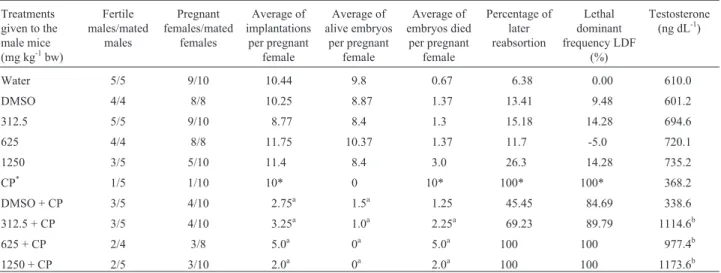
The mouse dominant lethal assay which used male mice treated with the different doses of the methanol ex-tract mated with female mice which did not receive any treatment also showed no statistically significant (student t-test, p = 0.05) mutagenic activity for the sacacaat the doses tested, there being no significant difference between the mean number of live implants and late embryo reabsorptions per untreated pregnant female mated with males that received doses of the vegetal extract or water (Table 2). Of the five males treated with 150 mg kg-1bw
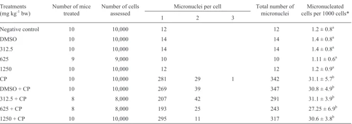
Table 1- Micronucleus per cell, micronucleus total number and micronucleated cells frequency for a thousand polynucleated erythrocytes (PCEs) after the sub-chronic treatment with methanol extract ofCroton cajucaraBenth.
Treatments (mg kg-1bw)
Number of mice treated
Number of cells assessed
Micronuclei per cell Total number of micronuclei
Micronucleated cells per 1000 cells*
1 2 3
Negative control 10 10,000 12 12 1.2 ± 0.8a
DMSO 10 10,000 14 14 1.4 ± 0.8a
312.5 10 10,000 14 14 1.4 ± 0.8a
625 9 9,000 10 10 1.11 ± 0.6a
1250 10 10,000 12 12 1.2 ± 0.9a
CP 10 10,000 281 29 1 342 31.1 ± 5.7b
DMSO + CP 10 10,000 269 39 347 30.8 ± 4.9b
312.5 + CP 8 8,000 207 42 291 31.1 ± 3.9b
625 + CP 8 8,000 193 25 243 27.25 ± 6.9b
1250 + CP 10 10,000 295 11 317 30.6 ± 3.8b
*Values following by the same letter do not differ statistically (Student t-test, p < 0.05). CP = Cyclophosphamide (150 mg kg-1bw).
cyclophosphamide (positive control) only one succeeded in producing a pregnant female but no live embryos were pro-duced, this result being statistically significant compared to males who had received extract or water only. Mice treated simultaneously with methanol extract ofC. cajucaraand cyclophosphamide showed no significant reduction in cyclophosphamide mutagenicity (Tables 1 and 2).
In the 50 untreated male mice tested, testosterone concentrations were between 20 to 1200 ng dL-1, and there was no significant difference between these levels and those found in mice administered water only (negative con-trol) or treated with the methanol extract, cyclophos-phamide or both (Table 2). This indicates that testosterone levels were high enough to permit normal spermatogenesis.
Discussion
According to Agneret al. (2001) and Macielet al.
(2000) the population in the north of Brazil (Amazon re-gion) uses extracts fromCroton cajucaraleaves and bark on a large scale to treat various diseases, and the medicinal use of this plant spread to the southeast of Brazil at the end of the 1990s. Similarly toCroton cajucara, other medicinal plants are intensely used by the world population without the proper knowledge of the risk that these treatments can incur. Cases of toxic hepatitis have been reported in several hospitals in Belém (state of Pará, Brazil) because of exces-sive consumption of extremely strongCroton cajucaratea (Macielet al., 1998a,b; Macielet al., 2000). According to the “Institute of Chinese Materia Medica” (apudChan and Critchley, 1996) only about 230 of the thousands of species of medicinal plants used in China have been studied
phar-macologically and clinically, and only ten were considered toxic. This low incidence of side effects is a factor encour-aging the low-income population to ingest medicinal plant.
In this study, the animals were submitted to sub-chronic treatment every seven days for 28 days via gavage of the methanol extract solutions. This treatment regimen and the administration method were considered the most suitable because they were closer to the form that treat-ments are conducted andCroton cajucaraproducts (teas and tablets) are ingested by the population.
Statistical analysis of our micronucleus assay results (Table 1) revealed no significant difference between the frequency of micronucleated cells in mice treated with 312.5, 625 or 1250 mg kg-1bw of the extract and those that received distilled water, indicating that subchronic admin-istration of the methanol extract of C. cajucara is not mutagenic in mice. Similarly, the DMSO solvent also showed no statistically significant mutagenicity by the micronucleus assay. Our results agree with those of Agner
et al. (1999) who subjected mice to acute treatment with DCTN (the major ingredient ofC. cajucara) and found no significant genotoxic activity for this compound. In our ex-periments, the absence of genotoxicity may have been due to the absence of forbol diterpene esters which are present in many toxicCrotonspecies and are known to cause vari-ous toxic effects and to be carcinogenic (Hecker and Schmidt, 1974; Weber and Hecker, 1978).
The stem bark of C. cajucara is a rich source of clerodane-type diterpenes such as DCTN (Maciel et al., 2000) that present many therapeutic properties (Fariaset al., 1997; Macielet al., 2002). Agneret al. (2001) showed that acute treatment of mice with DCTN administered by
Table 2- Lethal Dominant Assay and testosterone concentration in Swiss albino mice submitted to sub-chronic treatment with the methanol extract of
Croton cajucarafor the assessment of mutagenicity and antimutagenicity.
Treatments given to the male mice (mg kg-1bw)
Fertile males/mated
males
Pregnant females/mated
females
Average of implantations
per pregnant female
Average of alive embryos
per pregnant female
Average of embryos died
per pregnant female
Percentage of later reabsortion
Lethal dominant frequency LDF
(%)
Testosterone (ng dL-1)
Water 5/5 9/10 10.44 9.8 0.67 6.38 0.00 610.0
DMSO 4/4 8/8 10.25 8.87 1.37 13.41 9.48 601.2
312.5 5/5 9/10 8.77 8.4 1.3 15.18 14.28 694.6
625 4/4 8/8 11.75 10.37 1.37 11.7 -5.0 720.1
1250 3/5 5/10 11.4 8.4 3.0 26.3 14.28 735.2
CP* 1/5 1/10 10* 0 10* 100* 100* 368.2
DMSO + CP 3/5 4/10 2.75a 1.5a 1.25 45.45 84.69 338.6
312.5 + CP 3/5 4/10 3.25a 1.0a 2.25a 69.23 89.79 1114.6b
625 + CP 2/4 3/8 5.0a 0a 5.0a 100 100 977.4b
1250 + CP 2/5 3/10 2.0a 0a 2.0a 100 100 1173.6b
CP - cylophosphamide (150 mg kg-1bw).
DMSO group received DMSO + water 2:1 (v/v); DMSO + CP group received DMSO + water 2:1 (v/v) and CP at 150 mg kg-1bw. *In this group only one female was pregnant and all of the embryos died.
a
gavage protects their bone marrow cells against cyclo-phosphamide-induced damage. Our studies with 312.5, 625 and 1250 mg kg-1bw of the methanol extract administered subchronically to mice did not demonstrate any protective activity in the bone marrow cells against cyclophospha-mide-induced damage. There was no significant reduction at any of the plant extract doses tested in the frequency of micronucleated cells in the treated groups that suffered cyclophosphamide-induced damage, possibly because of the low concentration of DCTN (1.4%) inC. cajucara ex-tracts (Macielet al., 1998a) as compared with the doses (138.75, 277.50 and 416.25 mg kg-1bw) of pure DCTN used by Agneret al. (2001). Another explanation for the difference between the results published by Agner et al. (2001) and our results is that Agner’s study used single doses of both pure DCTN and cyclophosphamide while our study involved subchronic treatment. Another possible rea-son why extract failed to show a protector effect in our study is that we used a higher concentration of cyclophos-phamide (150 mg kg-1bw) than the 10 mg kg-1bw used by Agner et al. (2001). We used a high cyclophosphamide concentration to enable this mutagenic chemotherapeutic alkylating agent to cross the testicular barrier and induce damage in testicular tissue which resulted in lethal do-minants. However, the increased toxicity resulting from the high dose of cyclophosphamide may have masked any pro-tector effect of the methanol extract. In spite of the high cyclophosphamide dose used in our experiments the mice showed no toxic effects (e.g.mortality, diarrhea, prostra-tion or behavioral alteraprostra-tions), supporting the work of Ve-lez de la Calleet al. (1989) who found no toxic effects in adult rats injected with 100 mg kg-1bw in a treatment re-gime similar to that used in our study. In summary, our micronucleus assay indicate that the methanol extract of
Croton cajucaradid not cause genetic alterations in mice bone marrow erythrocytes but was not capable of reducing the genotoxic effects of cyclophosphamide administered to the mice intraperitoneally at a dose of 150 mg kg-1bw. Our results for the dominant lethal assay (Table 2) showed no statistically significant evidence for the extract analyzed exerting mutagenic activity on male Swiss albino mice germ cells. The frequencies of live implantations and late reabsorptions per pregnant female were not altered in groups 1, 2 and 3 treated with the vegetal extract as com-pared to males in the negative control (distilled water). There was no statistically significant difference in fertility between the male mice in groups 1, 2 and 3 which had been treated with extract and the negative control group.
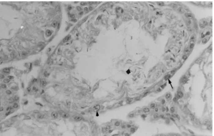
Cyclophosphamide was fairly effective in inducing lethal dominants as shown by the fact that 80% of males treated with doses of 150 mg kg-1cyclophosphamide were infertile after treatment with this alkylating agent and only one of the ten females mated presented implanted embryos, which were all dead (Table 2). The Dominant Lethal Assay protocol used (Generoso, 1978,apudLeber, 1988) was ex-pected to induce mutational events in testicular spermatid
or sperm cells but our assay results suggest that cyclo-phosphamide-induced mutations occurred in sperm located in the epididimus, with histological analysis of the testes showing changes in the seminiferous tissue and a few sperm cells in the seminiferous tubules (Figure 1).
Our results show that cyclophosphamide causes testi-cular dysfunction in male mice without reducing blood testosterone levels (Table 2), indicating that cyclophos-phamide did not alter Leydig cell function. Our results sup-port those of Velez de la Calleet al. (1989), who obtained similar results in adult rats treated for five consecutive weeks with cyclophosphamide. According to Vigil and Bustos-Obregon (1991) cyclophosphamide interferes with late spermatogenesis, possibly by damaging germ cell DNA or its products. Our observation that cyclophospha-mide reduced fertility but did not affect serum testosterone levels agrees with the suggestion by Howell and Shalet (2002) that germinal epithelium is more sensitive than Leydig cells to the effects of cytotoxic drugs. In our study, although the testosterone levels of the cyclophosphamide-treated male mice were within the normal range they were lower than those seen for male mice in groups 1 to 5 which received no cyclophosphamide.
The increase in testosterone production after co-administration of methanol extract and cyclophosphamide to male mice in groups 7, 8 and 9 suggests that the extract protected against cyclophosphamide-induced testicular gametogenic and androgenic dysfunction, possibly because of the restoration of testicular androgenesis given that an-drogen is a prime regulator of gametogenesis (Huanget al., 1987).
The simultaneous administration of extract with cyclophosphamide to males in groups 7, 8 and 9 did not al-leviate the effects of cyclophosphamide because in females mated to these males the frequency of live implantations and the total number of implantations were very close to the values observed for females mated with males from the
positive control group and much less than those observed for females mated with males belonging to the negative control group. The percentage of late reabsorption was also very high in females mated to group 7, 8 and 9 males and was, in fact, similar to the values observed for females mated with males from the positive control. Table 2 shows that the late reabsorption frequency in females mated to group 7, 8 and 9 males was much lower than in females mated with cyclophosphamide-treated males from the posi-tive control group. However, the results for the posiposi-tive control group males should be treated with caution because only one male in this group succeeded in making a female pregnant and all the embryos in this female were dead, cre-ating a controversial value for the implantation means. Shukla and Taneja (2001) showed that the dominant lethal assay could be useful for detecting the antimutagenic po-tential of substances, but our study showed no reduction in cyclophosphamide-induced dominant lethal frequency group 7, 8 and 9 mice.
To clarify the situation involving the interaction be-tween cyclophosphamide and theC. cajucaramethanol ex-tract more work needs to be carried out involving the use of higher doses of extract association with cyclophosphamide or the use of a new protocol with continuous administration of extract and a single weekly dose of cyclophosphamide.
Overall, our results show no statistically significant mutagenicity caused by application of different doses of
Croton cajucarastem-bark methanol extract to Swiss al-bino mice under the conditions used. These results support the data from studies concerning the biological activities of DCTN, the active principal ofC. cajucara bark extract, which indicate that consumption of this phytotherapeutic agent by the population may be safe, and hence may serve as a stimulus to the pharmaceutical industry for develop-ment ofCroton cajucara-derived products.
However, in spite of the positive data for antimuta-genic activity of DCTN (Agneret al., 2001) our results in-dicate that it is still early to recommend the use of alcoholic extracts of stem-bark in the prevention of diseases such as cancer or to protect healthy cells during chemotherapy with cyclophosphamide.
Acknowledgments
We would like to thank Patrick Garcia and Carlos Lourenço for support in maintaining the animals and carry-ing out the experiments and also to the Brazilian agency CAPES and the State University of Londrina (Brazil) for funding.
References
Agner AR, Maciel MAM, Pinto AC and Cólus IMS (2001) Anti-genotoxicity oftrans-dehydrocrotonin, a clerodane diter-pene fromCroton cajucara. Planta Med 67:815-819. Agner AR, Maciel MAM, Pinto AC, Pamplona SGRS and Cólus
IMS (1999) Investigation of genotoxic activity of
trans-dehydrocrotonin, a clerodane diterpene from Croton cajucara. Teratog Carcinog Mutagen 19:377-384.
Brito AR, Rodriguez JA, Hiruma-Lima CA, Haun M and Nunes DS (1998) Antiulcerogenic activity of trans-dehidrocrotonin fromCroton cajucara.Planta Med 64:126-129.
Carvalho JC, Silva MF, Maciel MA, Pinto AC, Nunes DS, Lima RM and Sarti SJ (1996) Investigation of anti-inflammatory and antinociceptive activities of trans-dehydrocrotonin, a 19-nor-clerodane diterpene fromCroton cajucara.Part 1. Planta Med 62:402-404.
Chan TYK and Critchley JAJH (1996) Usage and adverse effects of Chinese herbal medicines. Hum Exp Toxicol 15:5-12. Farias RA, Rao VS, Viana GS, Silveira ER, Maciel MA and Pinto
AC (1997) Hypoglycemic effect of trans-dehydrocrotonin, a nor-clerodane diterpene fromCroton cajucara.Planta Med 63:558-560.
Grynberg NF, Echevarria A, Lima JE, Pamplona SSR, Pinto AC and Maciel MAM (1999) Anti-tumor activity of two 19-nor-clerodane diterpenes, dehydrocrotonin and trans-crotonin, fromCroton cajucara. Planta Med 65:687-689. Haseman JK and Soares ER (1976) The distribution of fetal death
in control mice and its implication on statistical tests for dominant lethals effects. Mutat Res 41:277-288.
Hecker E and Schmidt R (1974) Phorbolesters - The irritants and cocarcinogens of Croton tiglium L Fortschr Chem Org Naturst 31:377-467.
Howell SJ and Shalet SM (2002) Effect of cancer therapy on pitu-itary-testicular axis. Internat J Andrology 25:269-276. Huang HFS, Marshall GR, Rosenberg RA and Nieschalg E (1987)
Restoration of spermatogenesis by high levels of testoster-one in hypophysectomized rats after long-term regression. Acta Endocrinol 16:433-444.
Huber R, Streng S and Bauchinger M (1983) The suitability of the human lymphocyte micronucleus assay system for biologi-cal dosimetry. Mutat Res 111:185-193.
Leber AP (1988) Chemical induction of dominant lethal muta-tions in mammals. In: Ballantyne B (ed) Perspectives in Ba-sic and Applied Toxicology. John Wrigth, London, pp 199-205.
Maciel MAM, Pinto AC, Veiga Jr VF, Martins JR, Grynberg NF, Echevarria A, Lapa AJ and Vanderlinde FA (2002)Croton cajucaraas an alternative to traditional medicine in a mod-ern health system. Recent Prog in Med Plant 8:502-517. Maciel MAM, Pinto AC, Arruda AC, Pamplona SGSR,
Vander-linde FA, Lapa AJ, Cólus IMS, Echevarria A, Grynberg NF, Farias RAF, Luna AM and Rao VSN (2000) Ethno-pharmacology, phytochemistry and pharmacology: A suc-cessful combination in the study of Croton cajucara. J Ethnopharmacol 70:41-55.
Maciel MAM, Pinto AC, Brabo SN and Arruda AC (1998a) Estudo da variação dos teores de terpenóides bioativos isola-dos das cascas deCroton cajucara, nativos e cultivados no estado do Pará. Rev Univers Rural 20:17-34.
Maciel MAM, Pinto AC, Brabo SN and Da Silva MN (1998b) Terpenoids fromCroton cajucara. Phytochemistry 49:823-828.
Olfert ED, Cross BM and McWilliam AA (1993) The Guide to the Care and Use of Experimental Animals. V. 1. Canadian Council on Animal Care, Ontario, 211 pp.
Preston RJ, San Sebastian JR and Mcfee AF (1987) Thein vitro
human lymphocyte assay for assessing the clastogenicity of chemical agents. Mutat Res 189:175-183.
Schmid W (1975) The micronucleus test. Mutat Res 31:9-15. Shukla Y and Taneja P (2001) Antimutagenic effect of black tea
extract using ‘rodent dominant lethal mutation assay’. Toxi-cology 168:269-274.
Titenko-Holland N, Windham G, Kolachana P, Reinisch F, Parva-tham S, Osorio AM and Smith MT (1997) Genotoxicity of malathion in human lymphocytes assessed using the
micro-nucleus assayin vitroandin vivo: A study of malathion-exposed workers. Mutat Res 338:85-95.
Velez de la Calle JF, Queiroz F, Garnier DH, Kercret H, Folliot R and Jégou B (1989) Reproductive effects of the anticancer drug cyclophosphamide in male rats at different ages. Arch Androl 22:251-263.
Vigil P and Bustos-Obregon E (1985) Alkylating agents and mouse spermatogenesis: Effects of a single injection of cyclophosphamide. Andrology 17:276-282.
Weber J and Hecker E (1978) Cocarcinogens of the diterpene es-ter type from Croton flavens L. and esophageal cancer in Curacao. Experientia 34:679-82.