Si m u l t a n e o u s A sse ssm e n t o f Pl a sm a t i c,
Acrosom al, and M it ochondrial M em branes of
Roost er Sperm at ozoa
M ail Address
Ke yw ord s Aut hor(s)
Arrived: January / 2007 Approved: August / 2007
ABSTRACT
This experiment w as designed w ith the objective of developing a simple, practical, and high repeatability technique for the simultaneous evaluation of the integrity of the plasmatic and acrosomal membranes, as w ell as funcional mitochondria of domestic fow l spermatozoa using an association of fluorescent probes. Four ejaculates (motility ≥80% and abnormal morphology ≤10% ) from each of six Ross male broiler b r eed er (n = 2 4 ) w er e d ilu t ed in TA LP sp er m m ed iu m (2 5 x1 06 spermatozoa/mL) and split into tw o aliquots, and one of these aliquots w as flash frozen in liquid nitrogen and thaw ed to damage all cellular membranes. Three treatments w ere prepared from these aliquots, w ith the follow ing ratios of Fresh semen:Flash frozen semen: 100:0 (T100), 50:50 (T50), and 0:100 (T0). A 150-µL aliquot of diluted semen w as placed in a microcentrifuge tube w ith the addition of 2-µL PI, 2-µL M ITO, and 50-µL FITC-PSA, and incubated at 38.5o C/8 min in the dark. An 8-µL sam ple w as placed on a slide, coverslipped, and exam ined by epifluorescence microscopy. Each sample had 200 cells counted and classif ied based on t he f luorescence em it t ed by each probe. By regression analysis, plasma membrane integrity, as detected by PI, w as determined as: v=4.17+0.82X (R2=0.95). Acrosome integrity, as detected by FITC-PSA , generat ed t he equat ion: v= 4.19+ 0.84X (R2= 0.96). Functional mitochondria w as estimated by the equation v=3.20+0.83X (R2=0.96). This is an ef f icient t echnique t o simult aneously evaluat e plasmatic, acrosomal, and mitochondrial membranes in fow l sperm. It is suggested that its application in flow cytometry systems allow s this methodology to be applied in large scale.
INTRODUCTION
Fert ilit y is one of t he most import ant economic t rait s in poult ry production, together w ith egg hatchability. M ale fertility potential may be defined as the capability to produce and to ejaculate spermatozoa that are able to fertilize eggs, w hich includes accomplishment of all st eps of t he f ert ilizat ion process: sperm moving across t he f emale reproductive tract and reaching the sperm storage tubule, binding and penetration into the perivitelline layer, and fertilization (Celeghini et al., 2001).
Sire selection in most commercial poultry breeds is performed on a subjective basis, determined by secondary sexualtraits, such as the comb development (Celeghini et al., 2001). Given the importance of fertility rat e in a b reed er f lo ck, st u d ies h ave in vest ig at ed m o re p recise evaluations of male fertility capacity, since one male is responsible for f er t ilizin g sever al f em ales. Th u s, t h e an alysis o f f o w l sem en characteristics can be used as a tool for the selection of sires kept either under natural mating, or as semen donors in artificial insemination
Celeghini ECC1
Arruda RP1*
Albuquerque R2
Silva FHA3
Faria DE3
Andrade AFC1
Nascimento J1
Raphael CF1
1 Laborat ório de Biot ecnologia do Sêmen e
Andrologia, Departamento de Reprodução Animal, Faculdade de M edicina Veterinária e Zoot ecnia (FM VZ), Universidade de São Paulo (USP).
2 Dep art am en t o d e Nu t rição e Pro d u ção
Animal, FM VZ, USP.
3 Departamento de Zootecnia, Faculdade de
Zo o t ecn ia e En g en h ar ia d e A lim en t o s (FZEA), USP.
RP Arruda
Av. Duque de Caxias Norte, 225 Post-box 23
13.635-900. Pirassununga, SP, Brazil. Telephone: +55 019 3565-4221 Fax: +55 019 3565-4060 E-mail: [email protected]
Flu o rescen t p ro b es, m em b ran es, ro o st er, sperm .
systems (Wilson et al., 1979). Some researchers found significant correlations betw een semen characteristics and egg fertility (Harris Jr. et al., 1984), sustaining that male fertilizing potential is dependent on semen quality. It is know n that, in order to ensure high egg fertility rate, the semen needs to present some characteristics, w h ich evalu at io n is b ased o n p h ysical an d morphological examination. M ore objective techniques t o evaluat e sem en-f ert ilizing pot ent ial have been proposed, such as tests of sperm penetration in the blastodisc region (Bramw ell et al., 1995; Barbarato et al., 1998), and sperm membrane evaluations (Bilgili & Renden, 1984; Chalah & Brillard, 1998).
Despite being highly correlated to fertility, the sperm penetration test is labor-intensive, and difficult to apply in large breeding st ocks (Bram w ell et al., 1995; Barbarato et al., 1998). How ever, as membranes play an essential role in maintaining sperm ability to fertilize, t h ey h ave b een evalu at ed b y m o r e o b ject ive techniques, such as the use of fluorescent probes (Bilgili & Renden, 1984; Graham et al., 1990; Chalah & Brillard, 1998; Celeghini et al., 2005).
Sp er m p lasm a m em b r an e is r esp o n sib le f o r est ab lish in g a b arrier b et w een in t racellu lar an d ext racellular environm ent s, w hich is im port ant t o maintain osmotic equilibrium and cellular homeostasis. Damages in this structure lead to cellular instability caused by homeostasis loss, resulting in cellular death. Therefore, plasma membrane integrity exerts a crucial role on sperm survival in the female reproductive tract and its fertilizing ability (Parks & Graham, 1992).
Earlier studies evaluating the integrity of the sperm plasma membrane in fow l, utilizing fluorescent probes, mention the use of ethidium bromide (Bilgili & Renden, 1984). Nevertheless, due to its high toxicity, ethidium bromide application is limited. Propidium iodide (PI), a f luorescent dye w it h propert ies similar t o et hidium bromide, but less toxic, has DNA affinity, and stains damaged plasma membrane cell nucleus in red (Bayyari et al., 1990; Graham et al., 1990; Chalah & Brillard, 1998). Because it is a stable fluorescent stain, PI has been the most frequently utilized probe in research, w it h successf ul result s in several species, bot h by fluorescent microscopy (Garner et al., 1997; Sukardi et al., 1997; Thomas et al., 1997; Chalah & Brilllard, 1998) and by flow cytometry system (Bayyari et al., 1990; Graham et al., 1990; Pintado et al., 2000; Gillan et al., 2005).
In addition to plasmatic membrane integrity, it is important to consider acrosomal membrane integrity and t he maint enance of it s enzymes, as acrosomal
reaction, characterized by the release of the acrosomal enzymes, is essential for the sperm to penetrate the blast odisc region, and t o egg f ert ilizat ion (Bakst & How arth, 1977). Acrosome integrity can be checked by different fluorescent techniques. Among them, the use of marked lectins is emphasized (Graham et al., 1990), such as Pisum sativum agglutinin (PSA), Ricinus communis agglutinin (RCA), Arachis hypogea agglutinin (PNA ) (Cross & M eizel, 1989), and Concanavalia en sif o r m is (Co n A ), w h ich ar e f lu o r escein isothiocyanate-conjugates (FITC). PSA is an agglutinin from edible peas, and binds to the glycoconjugate of the acrosomal matrix (Cross & M eizel, 1989). It has af f init y f or t erminal a-D-glucosyl and a-D-mannosyl residues of glycoproteins, and binds specifically to the sugar a-m annoside f ound in acrosom al cont ent s (Sukardi et al., 1997). This agglutinin, w hen conjugated to FITC, marks the sperm acrosome in yellow -green, and identifies acrosome damage. It can be applied to spermatozoa of different species (Graham et al., 1990; Arruda et al., 2002).
a high correlat ion w as observed bet w een sperm stained by M ITO and sperm motility (r = 0.96), as w ell as sperm viability, as detected by SYBR-14 (r = 0.97), indicating that this probe reflects the functional status of mitochondria (Garner et al., 1997).
Considering that, in order for to fertilize the oocyte, sperm need to have all its membranes intact, it is then vital that sperm evaluation be simultaneous, supplying information on the number of spermatozoa w ith intact plasm at ic and acrosom al m em branes, as w ell as preserved mitochondrial function. The association of fluorescent probes allow s the simultaneous evaluation of more than one sperm cell compartment, as w ell as simultaneous evaluation of plasmatic and acrosomal m em branes, using Hoechst 33258 and FITC-PSA association (Arruda et al., 2002), PI and LYSO-G or PI + SYTO-17 and FITC-PNA (Thom as et al., 1997), phycoeryt hrin (PE)-conjugat ed PNA associat ed t o probes SYBR-14 and PI (NAGY et al., 2003), or PI and FITC-PSA association (Centola et al., 1990; Graham et al., 1990; Sukardi et al., 1997; Arruda et al., 2002).
Graham et al. (1990) demonstrated that at least t h r ee b o vin e sp er m co m p ar t m en t s can b e simultaneously evaluated by addition of three probes. These aut hors ut ilized PI f or plasm at ic m em brane int egrit y evaluat ion, PSA t o det erm ine acrosom al integrity, and R123 to verify mitochondrial function. For sim u lt an eo u s evalu at io n , sin g le f lo w cyt o m et ry apparatus w as used, and the validation w as performed separately for each probe.
The m ore sperm param et ers are evaluat ed in a semen sample, t he higher t he value in t he in vit ro fertility prognostic. How ever, in commercial poultry product ion, a large num ber of sam ples m ust be processed and evaluated. Consequently, this technique may require to be faster and cheaper to be routinely applied.
This experiment w as designed w ith the objective of developing a simple, practical, and highly repeatable t echnique f or sim ult aneous int egrit y evaluat ion of plasmat ic and acrosomal membranes, as w ell as of mitochondrial function in domestic fow l spermatozoa by the association of fluorescent probes.
M ATERIALS AND M ETHODS
Sem en Preparat ion
Four ejaculates from six Ross male broiler breeder (n=24) w ere utilized, all presenting motility ≥80% ,and ab n o rm al m o rp h o lo g y ≤1 0 % . Im m ed iat ely u p o n collect ion, t he sem en w as dilut ed in TALP sperm
medium (Bavister et al., 1983) to a final concentration of 25x106 sperm at ozoa/m L. The sam ple of dilut ed semen w as split into 2 aliquots, and 1 aliquot w as flash frozen in liquid nitrogen and thaw ed in 3 continuous cycles in order to damage cellular membranes and to disturb mitochondrial function. Three treatments w ere prepared from these aliquots, w ith the follow ing ratios of Fresh semen:Flash frozen semen: 100:0 (T100), 50:50 (T50) and 0:100 (T0).
Sperm at ozoa St ain
After the preparation, the three samples, T100, T50 and T0, w ere submitted to a stain technique adapted from Arruda & Celeghini (2003). A 150-µL aliquot of diluted semen w as placed in a microcentrifuge tube, and 2-µL PI (3 mM , Sigma, 28,707-5, in DPBS), 2-µL M ITO (500 mM , M olecular Probes, M -7514, in DM SO), and 50-µL FITC-PSA (100 µg/mL, Sigma, L-0770, in DPBS) w ere added. The sample w as incubated for 8 minutes at 38.5o C, in the dark.
Fluorim et ric Assessm ent
An 8-µL sample of stained spermatozoa suspension w as placed on a slide, a coverslip added, and t he exam in at io n w as im m ed iat ely p erf o rm ed u n d er epifluorescence microscopy (Nikon, model Eclipse 80i) in a triple filter (D/F/R, C58420), w ith the set: UV-2E/C (340-380 nm excitation and 435-485 nm emission), B-2 E/C (4 6 5 -4 9 5 n m excit at io n an d 5 1 5 -5 5 5 n m emission), and G-2E/C (excitation 540-525 and emission 605-655), at 1.000 x magnification. Each sample had 2 0 0 cells co u n t ed an d classif ied b ased o n t h e fluorescence emitted by each probe.
St at ist ical Analysis
Data obtained from T0, T50, and T100 treatment groups w ere evaluated by analysis of variance (ANOVA) (SAS, 1998). Treatment means across all samples w ere compared by Fisher’s LSD test. The data of plasmatic and acrosomal membranes integrity, and mitochondrial function (dependent variables) in the treatments T0, T50, and T100 (independent variables) w ere submitted t o sim p le lin ear reg ressio n an alysis b y St at View softw are (SAS, 1998).
RESULTS
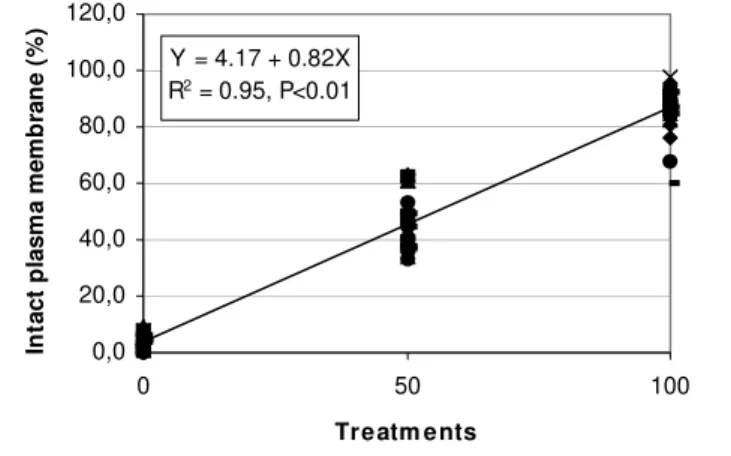
Y = 4.17 + 0.82X R2 = 0.95, P<0.01
0,0 20,0 40,0 60,0 80,0 100,0 120,0
0 50 100
Treatm ents
In
tact
p
lasm
a m
e
m
b
ran
e
(
%
)
Figure 2 - Linear regression of plasma membrane integrity, as verified by exclusion of propidium iodide in PI/FITC-PSA/M ITO association, in fow l spermatozoa submitted to treatments T0 (Fresh semen:Flash frozen semen, 0:100), T50 (Fresh semen:Flash frozen semen, 50:50) and T100 (Fresh semen:Flash frozen semen, 100:0.
Figure 1 - Photomicrography of fow l sperm stained w ith PI, FITC-PSA, and M ITO association (1,0000x magnification). A. int act plasm a m em brane, int act acrosom e and f unct ional mit ochondria sperm. B. int act plasma membrane, damaged acrosome and functional mitochondria sperm. C. intact plasma membrane, intact acrosome and non-functional mitochondria sperm. D. intact plasma membrane, damaged acrosome and non-f unct ional m it ochondria sperm . E. dam aged plasm a membrane, damaged acrosome and mit ochondrial f unct ion sperm. F. and G. damaged plasma membrane, intact acrosome and non-functional mitochondria sperm. H. damaged plasma m em b r an e, d am ag ed acr o so m e an d n o n - f u n ct io n al mitochondria sperm.
Table 1 - Classification of fow l sperm cells according to fluorescence emitted by PI, FITC-PSA, and M ITO probes.
Sperm Cells PI* FITC-PSA* * M ITO* * *
Int act plasmat ic membrane, int act acrosome, and f unct ional mit ochondria --- --- +++++ Int act plasmat ic membrane, int act acrosome and non-f unct ional mit ochondria --- --- ---Int act plasmat ic membrane, damaged acrosome and f unct ional mit ochondria --- +++++ +++++ Int act plasmat ic membrane, damaged acrosome and non-f unct ional mit ochondria --- +++++ ---Damaged plasmat ic membrane, int act acrosome and f unct ional mit ochondria +++++ --- +++++ Damaged plasmat ic membrane, int act acrosome and non-f unct ional mit ochondria +++++ --- ---Damaged plasmat ic membrane, damaged acrosome and f unct ional mit ochondria +++++ +++++ +++++ Damaged plasmat ic membrane, damaged acrosome and non-f unct ional mit ochondria +++++ +++++ ---* PI positive (+) = red-stained nucleus. ---* ---* FITC-PSA positive (+) = yellow -green acrosome region. ---* ---* ---* M ITO positive (+) = bright green in midpiece region.
by sperm stained by the fluorescent probes association is show n in Figure 1.
A NOVA an d Fish er t est s d et ect ed st at ist ical differences (p<0.0001) betw een the T100, T50, and T0 treatment groups, as show n in Table 2, validating the submission of data to linear regression analysis.
Table 2 - M ean ± standard deviation of intact plasma membrane (IPM ), intact acrosome (IA) and functional mitochondria (M F) in fow l spermatozoa submitted to treatments 100, 50 and 0.
Characteristic T100 T50 T0
IM P 86.29±8.4c 46.40±8.6b 3.71±2.8a IA 87.71±6.9c 47.89±8.8b 3.42±2.6a M F 86.06±8.1c 46.81±8.3b 2.33±2.4a Dif f erent superscript let t ers in t he sam e row indicat e st at ist ical dif f erences (p<0.0001).
Regression analysis results for plasma membrane integrity, as detected by PI, are displayed in Figure 2. Acrosome integrity, as detected by FITC-PSA probe, g en er at ed t h e eq u at io n sh o w n in Fig u r e 3 . M itochondrial function estimated by M ITO is displayed in Figure 4.
DISCUSSION
In this experiment, the association of fluorescent probes w as tested and validated for the simultaneous evaluation of plasmatic (PI) and acrosomal (FITC-PSA) m em branes int egrit y, as w ell as of m it ochondrial f u n ct io n (M ITO) in f o w l sp erm . Th ese t est s are im port ant in order t o obt ain a highly repeat able technique that presents results reflecting the real status of each structure. It is still necessary to identify w hich probes associat e bet t er, as t here may be dif f erent results w ith different associations due to changes in its characteristics and fluorescence standards.
(Centola et al., 1990; Sukardi et al., 1997; Arruda et al., 2002; Nagy et al., 2003) and poultry sperm (Chalah & Brilllard, 1998). The PI and FITC-PSA probe association to assessplasmatic and acrosome membranes integrity, respectively, as utilized in this experiment, w as reported in sheep (Sukardi et al., 1997), human (Centola et al., 1990), horse (Arruda et al., 2002), and cattle (Graham et al., 1990). PI w as also associated w ith probes to evaluate mitochondrial function as R123, M ITO and JC-1; t hese probes show ed high posit ive correlat ion (r>0.96) w it h sperm mot ilit y (Garner et al., 1997). How ever, this experiment validated the association of PI an d FITC- PSA w it h an ad d it io n al p r o b e f o r mitochondrial function evaluation in fow l sperm.
PI, FITC-PSA and M ITO association resulted in very consistent marking of spermatozoa, making it easy to identify the evaluated structures. The association of PI and M ITO probes w as mentioned by Garner et al. (1997) in bovine spermatozoa assessment. T100, T50
and T0 treatment groups w ere prepared to validate the technique, according to the methodology described by Thomas et al. (1997). The results obtained w ere submitted to ANOVA, and means w ere compared using the Fisher test. The comparison of the results and the confirmation of differences among treatments w ere important to determine the efficacy of each treatment f or t echnique validat ion bef ore submit t ing t hem t o linear regression analysis.
As conf irmed by regression analysis result s, t his t ech n iq u e p r esen t ed g o o d r esu lt s an d h ig h r ep eat ab ilit y. So m e M ITO ch ar act er ist ics w er e observed: w hen it binds t o mit ochondria t hat have membrane potential, it emits bright green fluorescence. How ever, it is important to stress that M ITO also binds to regions of the membrane of the head and tail of the sperm in a non-specific fashion, emitting less intense fluorescence, as observed by Garner et al. (1997). This unspecific binding of M ITO is, in a w ay, beneficial to the evaluation, because it makes it possible to visualize t he shape of cells w it h int act plasm a m em brane. How ever, it is important to differentiate midpiece stain nuances in functional and non-functional mitochondria. In an attempt to investigate more objectively these associations in bovine sperm, Celeghini et al. (2005) valid at ed t w o f lu o rescen t -p ro b e t ech n iq u es f o r simultaneous evaluation of plasmatic, acrosomal, and mitochondrial membranes, using PI, Hoechst 33342, FITC-PSA, and CM XRos or JC-1, and obt ained very consistent results. Nevertheless, it is must be noted that the addition of one additional probe (H342) increases the technique cost.
In t he regression analysis equat ion obt ained f or plasmatic membrane integrity (Figure 2), evaluated by the exclusion of PI from the nucleus, it is possible to verify that the value of point “ a” (w here the line crosses the Y axis, i.e., show ing the value of Y w hen X= 0) is next to zero (a=4.17), as w ould be expected from a sample that w as submitted to flash freezing w ith the objective of damaging all membranes. The value of “ b” (regression coefficient, i. e., how much X varies in relation to Y) is also near the expected (b= 0.82) for T100, represent ing a sample w it h at least 80% of motility. The regression coefficient of 95% confirms t hese observat ions. Sim ilar result s w ere f ound by Graham et al. (1990), w ho compared the efficiency of PI w ith the stain technique by eosin/nigrosin, and found a positive correlation (r = 0.78) betw een techniques, and confidence interval of 95% . Positive correlations betw een PI and eosin/nigrosin w ere also observed in dogs (r = 0.88) (Peña et al., 1998), boars (r= 0.71) and Y = 3.20 + 0.83x
R2 = 0.96, P<0.01
0,0 20,0 40,0 60,0 80,0 100,0 120,0
0 50 100
Treatments
M
it
oc
hondr
ia
l f
unc
ti
on
(%)
Figure 4 - Linear regression of mitochondrial function, verified by M itoTracker Green FM , in PI/FITC-PSA/M ITO association in fow l spermatozoa, submitted to treatments T0 (Fresh semen:Flash frozen semen, 0:100), T50 (Fresh semen:Flash frozen semen, 50:50) and T100 (Fresh semen:Flash frozen semen, 100:0).
Y = 4.19+ 0.84X R2 = 0.96, P<0.01
0,0 20,0 40,0 60,0 80,0 100,0 120,0
0 50 100
Treatments
in
ta
ct
a
c
ro
s
so
m
e
(
%
)
bulls (r =0.83) (Pintado et al., 2000). Pintado et al. (2000) also observed high positive correlations betw een PI and H258 in sw ine (r=0.96) and bovine (r = 0.94) sperm.
Similar coefficients w ere found for the same probe association by regression equations that calculated for acrosome integrity (Figure 3), as verified by the FITC-PSA probe. The value of “ a” (4.19) is close t o t he expect ed. The value of “ b” (0.84) also ref lect s t he desired value and the determination coefficient of 95% dem onst rat es t he ef f iciency of t he t echnique. The FITC-PSA efficiency w as evaluated by Graham et al. (1990), comparing it to naphthol yellow /erythrosin b, finding a confidence interval of 95% .
The equation obtained for mitochondrial function, w ith M ITO associated to PI and FITC-PSA, show ed a similar characteristics to plasmatic membrane integrity and acrosomal int egrit y (a = 3.20, and b = 0.83). Nevertheless, Arruda & Celeghini (2003) obtained a regression equation, for the same association of probes in bovine sperm, (Y= 35.0 + 0.55X) dif f erent t han expect ed, in spit e of f inding a high det erminat ion coefficient (R2=0.84). This same technique, w ith minor changes, w as validated in equine sperm (Celeghini et al., 2004), using the same methodology, w hich yielded an equation (v= 9.57 + 0.78X) w ith results similar to t hose obt ained in t he present experim ent , w it h a determination coefficient of 93% . These differences could be explained by differences among species or by the greater ability of the technique to differentiate stain nuances of M ITO w hen bound to mitochondrial membranes presenting potential or not.
Th is is an ef f icien t an d easy t ech n iq u e t o sim ult aneously evaluat e plasm at ic, acrosom al, and mitochondrial membranes of fow l sperm. It is possible to suggest that the application of this methodology in large scale can be m axim ized by t he use of f low cyt om et ry syst em s, providing higher accuracy and sw iftness, as it is to read approximately 10,000 cells in a few seconds.
REFERENCES
Arruda RP, Celeghini ECC. Validation of a technique to evaluation sim ult aneous of t he plasm at ic, acrosom al and m it ochondrial membranes in bovine spermatozoa. Acta Scientiae Veterinariae 2003; 31:230-231.
Arruda RP, Souza NL, M arques A, Celeghini ECC, Gobesso AAO, M eirelles FV, Binelli M , Blasques FJH. Evaluation of techiniques using CFDA /PI, H258/FITC-PSA and Trypan Blue/Giem sa f or assessment of the viability and acrosomal Integrity of cryopreserved equine spermatozoa. Theriogenology 2002; 57(1):477.
Bakst M R, How arth B. Hydrolysis of hen’s perivitelline layer by cock sperm. Biology of Reproduction 1977; 17:370-379.
Barbarato GF, Cramer PG, Hammestedt RH. A pratical in vitro sperm-egg binding assay t hat det ect s subf ert ile m ales. Biology of Reproduction 1998; 58:686-699.
Bavist er BD, Leib f ried M L, Lieb erm an G. Develo p m en t o f preimplantation embryos of the golden hamster in a defined culture medium. Biology of Reproduction 1983; 28(1):235-47.
Bayyari GR, Cook JR, Harris Jr GC, M acy LB, Slavick M F, Skeeles JK. Research not e: The evaluat ion of chicken sperm at ozoa using fluorescent staining in a 96-w ell format. Poultry Science 1990; 69: 1602-1605.
Bereit er-Hahn J. Behavior of m it ochondria in t he living cell. International Review of Cytology 1990; 122:1-63.
Bilgili SF, Renden JA. Fluorimetric determination of avian sperm viability and concentration. Poultry Science 1984; 63(11):2275-77.
Bramw ell RK, M arks HL, How arth B. Quantitative determination of spermatozoa penetration of the perivitelline layer of the hen’s ovum as assessed on oviposited eggs. Poultry Science 1995; 74(11):1875-1883.
Celeghini ECC, Albuquerque R, Arruda RP, Lima CG. Seminal characteristics evaluation of the male broiler breeder selected by comb development to reproduction. Brazilian Journal of Veterinary Research and Animal Science 2001; 38(4):177-183.
Celeghini ECC, Arruda RP, Andrade AFC, Raphael CF, Nascimento J. Sim ult aneous evaluat ion of t he plasm at ic, acrosom al, and mitochondrial membranes in equine spermatozoa. Proceedings of the 15th Internacional Congress Of Animal Reproduction; 2004; p. 511. Porto Seguro, BA-Brazil.
Celeghini ECC, Nascimento J, Andrade AFC, Raphael CF, Souza LW O, Arruda RP. Use of CM XRos and JC-1 on m it ochondrial function evaluation, associated to fluorescent probes to plasmatic and acrosomal membranes evaluation in bovine spermatozoa. Acta Scientiae Veterinariae 2005; 33:321.
Centola GM , M attox JH, Burde S, Leary JF. Assessment of the viability an d acr o so m e st at u s o f f r esh an d f r o zen - t h aw ed h u m an spermatozoa using single-w avelength fluorescence microscopy. M olecular of Reproduction and Development 1990; 27(2):130-135.
Chalah T, Brillard JP. Comparison of assessment of fow l sperm viabilit y by eosin-nigrosin and dual f luorescence (SYBR-14/PI). Theriogenologv 1998; 50:487-493.
Cross NL, M eizel S. M ethods for evaluating the acrosomal status of mammalian sperm. Biology of Reproduction 1989; 41:635-641.
Gillan L, Evans G, M axw ell WM C. Flow cytometric evaluation of sperm parameters in relation to fertility potential. Theriogenology 2005; 63:445-457.
Graham JK, Kunze E, Hammerstedt RH. Analysis of sperm cell viability, acrosom al int egrit y, and m it ochondrial f unct ion using f low cytometry. Biology of Reproduction 1990; 43:55-64.
Harris Junior GC, Benson JA, Sellers RS. The influence of daylength, body w eight, and age on the reproductive ability of broiler breeder cockerels. Poultry Science 1984; 63(9):1705-1710.
Haugland, RP. Introduction to fluorescence techniques. In: Spence, M TZ. The handbook: a guide to fluorescent probes and labeling technologies. 10th ed. Invitrogen Corp.; 2005. p.1-6.
Nagy S, Jansen J, Topper EK, Gadella BM . A t riple-st ain f low cytometric method to assess plasma –and acrosome membrane integrity of cryopreserved bovine sperm immediately after thaw ing in presence of egg-yolk particles. Biology of Reproduction 2003; 68:1828-1835.
Parks JE, Graham JK. Effect of cryopreservation procedures on sperm membranes. Theriogenology 1992; 38:209-222.
Pintado B, De La Fuente J, Roldan ERS. Permeability of boar and bull spermatozoa to the nucleic acid stains propidium iodide or Hoechst 33258, or the eosin: accuracy in the assessment of cell viability. Journal of Reproduction and Fertility 2000; 118:145-152.
Raw e VY, Galaverna GD, Acosta AA, Olmedo SB, Chemes HE. Incidence of tail structure distortions associated w ith dysplasia of the fibrous sheath in human spermatozoa. Human Reproduction 2001; 16(5):879-886.
SAS Institute. User’s guide: statistic. version 6.11. 2nd ed. Cary; 1998. Sukardi S, Curry M R, Watson PF. Simultaneous detection of the acrosomal status and viability of incubated ram spermatozoa using fluorescent markers. Animal Reproduction Science 1997; 46:89-96.
Thomas CA, Garner DL, Dejarnette JM , M arshall CE. Fluorometric assessments of acrosomal integrity and viability in Cryopreserved bovine spermatozoa. Biology of Reproduction 1997; 56:991-998.