RESUMO.- [A PCR em tempo real de swab nasal não é adequada para o diagnóstico in vivo de tuberculose bo-vina.]A tuberculose bovina (bTB) é uma zoonose que cau-sa perdas econômicas e riscos à cau-saúde pública em muitos países. O diagnóstico da doença em animais vivos é reali-zado pelo teste intradérmico da tuberculina, que é baseado em reações de hipersensibilidade tardia. Como a tubercu-lose tem resposta imunológica complexa, este teste tem li-mitações em termos de sensibilidade e especificidade. Este estudo procurou desenvolver uma abordagem alternativa para o diagnóstico in vivo da tuberculose bovina, com base na reação em cadeia da polimerase (PCR) em tempo real. As amostras de DNA, extraídas de suabes nasais de vacas
Nasal swab real-time PCR is not suitable for
in vivo
diagnosis
of bovine tuberculosis
1Fabiana Q. Mayer2*, Emily M. dos Reis2, André Vinícius A. Bezerra2,3, Rogério O.
Rodrigues4, Thais Michel5, Cristine Cerva2 and Angélica C. Bertagnolli3
ABSTRACT.- Mayer F.Q., Reis E.M., Bezerra A.V.A., Rodrigues R.O., Michel T., Cerva C. & Ber -tagnolli A.C. 2017. Nasal swab real-time PCR is not suitable for in vivo diagnosis of bo-vine tuberculosis. Pesquisa Veterinária Brasileira 37(6):549-554. Laboratório de Biologia Molecular, Instituto de Pesquisas Veterinárias Desidério Finamor, Fundação Estadual de Pesquisa Agropecuária, Estrada Municipal do Conde 6000, Eldorado do Sul, RS 92990-000, Brazil. E-mail: bimmayer@gmail.com
Bovine tuberculosis (bTB) is a zoonosis causing economic losses and public health risks in many countries. The disease diagnosis in live animals is performed by intradermal tuber-culin test, which is based on delayed hypersensitivity reactions. As tuberculosis has complex immune response, this test has limitations in sensitivity and specificity. This study sought to test an alternative approach for in vivo diagnosis of bovine tuberculosis, based on real-time polymerase chain reaction (PCR). DNA samples, extracted from nasal swabs of live cows, were used for SYBR® Green real-time PCR, which is able to differentiate between Mycobacterium tuberculosis and Mycobacterium avium complexes. Statistical analysis was performed to com -pare the results of tuberculin test, the in vivo gold standard bTB diagnosis method, with real-time PCR, thereby determining the specificity and sensitivity of molecular method. Cervical comparative test (CCT) was performed in 238 animals, of which 193 had suitable DNA from nasal swabs for molecular analysis, as indicated by amplification of glyceraldehyde-3-phos -phate dehydrogenase (GAPDH) gene, and were included in the study. In total, 25 (10.5%) of the animals were CCT reactive, of which none was positive in the molecular test. Of the 168 CCT negative animals, four were positive for M. tuberculosis complex at real time PCR from nasal swabs. The comparison of these results generated values of sensitivity and specificity of 0% and 97.6%, respectively; moreover, low coefficients of agreement and correlation (-0.029 and -0.049, respectively) between the results obtained with both tests were also observed. This study showed that real-time PCR from nasal swabs is not suitable for in vivo diagnosis of bovine tuberculosis; thus tuberculin skin test is still the best option for this purpose.
INDEX TERMS: Nasal swab real-time PCR, bovine tuberculosis, tuberculin test, molecular diagnosis, Mycobacterium tuberculosis complex, Mycobacterium avium complex, Mycobacterium bovis.
1 Received on June 12, 2015.
Accepted for publication on November 2016.
2 Laboratório de Biologia Molecular, Instituto de Pesquisas Veterinárias
Desidério Finamor, Fundação Estadual de Pesquisa Agropecuária, Estrada Municipal do Conde 6000, Eldorado do Sul, RS 92990-000, Brazil. *Corres -ponding author: bimmayer@gmail.com
3 Laboratório de Histopatologia, Instituto de Pesquisas Veterinárias
De-sidério Finamor, Fundação Estadual de Pesquisa Agropecuária, Estrada Municipal do Conde 6000, Eldorado do Sul, RS 92990-000, Brazil.
4 Laboratório de Leptospirose, Instituto de Pesquisas Veterinárias
De-sidério Finamor, Fundação Estadual de Pesquisa Agropecuária, Estrada Municipal do Conde 6000, Eldorado do Sul, RS 92990-000, Brazil.
5 Laboratório de Parasitologia, Instituto de Pesquisas Veterinárias
vivas, foram usadas para PCR em tempo real com SYBR®
Green, capaz de diferenciar os complexos Mycobacterium tuberculosis e Mycobacterium avium. A análise estatística foi realizada para comparar os resultados de teste de tu-berculina, padrão ouro para o diagnóstico in vivo da bTB, com PCR em tempo real, determinando-se assim a espe-cificidade e sensibilidade do método molecular. O teste cervical comparativo (TCC) foi realizado em 238 animais, dos quais 193 tiveram DNA dos suabes nasais adequados para análise molecular, como indicado pela amplificação do gene gliceraldeído-3-fosfato-desidrogenase (GAPDH), e foram incluídos no estudo. No total, 25 (10,5%) animais foram reativos no TCC, dos quais nenhum foi positivo no teste molecular. Dos 168 animais negativos no TCC, quatro foram positivos para o complexo M. tuberculosis na PCR em tempo real a partir dos suabes nasais. A comparação des-tes resultados gerou valores de sensibilidade e especifici -dade de 0% e 97,6%, respectivamente; além disso, baixos coeficientes de concordância e correlação (-0,029 e -0,049, respectivamente) entre os resultados obtidos com ambos os testes também foram observados. Este estudo mostrou que a PCR em tempo real a partir de suabes nasais não é adequada para o diagnóstico in vivo da tuberculose bovi-na; portanto, o teste da tuberculina ainda é a melhor opção para este fim.
TERMOS DE INDEXAÇÃO: PCR em tempo real, swab nasal, tu -berculose bovina, teste da tuberculina, diagnóstico molecular, complexo Mycobacterium tuberculosis, complexo Mycobacterium avium, Mycobacterium bovis.
INTRODUCTION
Bovine tuberculosis (bTB) is a globally distributed zoo-nosis caused mostly by Mycobacterium bovis (Michel et al. 2010). In addition to public health implications, bTB is res-ponsible for economic losses in agriculture, even in develo-ped countries (Karolemeas et al. 2012). In countries where bTB prevalence is high, the main way of bTB control and eradication is based on diagnostic and slaughter of infec-ted animals (Brasil 2006); thus, efficient in vivo diagnosis becomes necessary.
In vivo bTB diagnoses is made mainly by the intrader-mal tuberculin test, which involves inoculation of purified protein from Mycobacterium bovis (PPD-B) and measure-ment of local hypersensitivity reaction after 72 h. When applied in combination with slaughter surveillance and herd movement control, this test has been efficient in bTB control and eradication in some countries (Karolemeas et al. 2012). Despite this, in several situations the tuberculin test has limitations due to lack of sensitivity and specificity, even when applied as a comparative cervical test (CCT) us-ing a purified protein derivative of Mycobacterium avium (PPD-A) to avoid false positive results (Zarden et al. 2013). This limited efficiency may contribute to the incidence maintenance of bTB in some countries.
The problems associated with tuberculin test led re-searchers to seek for new methods to diagnose bTB in livestock. Among them, interferon gamma (IFN-γ) assay is performed in vitro to detect specific cell-mediated im -mune responses (Wood et al. 1990). This is an ELISA test
that detects IFN-γ released from in vitro stimulated T lym-phocytes. It also evaluates the difference between values achieved with PPD-A and PPD-B stimulation, being analo-gous to CCT (Schiller et al. 2010). IFN-γ assay has increased sensitivity, but has limitations such as reduced specificity, high logistical demands, high costs, and difficulties in the standardization of purified proteins (Schiller et al. 2010). Another emerging strategy is bTB antibody detection, which had some difficulties since immunological response to Mycobacterium bovis, especially in early stages, is mainly cellular-mediated. Preliminary studies have indicated the potential for these tests, but large-scale field trials at differ -ent sites may be required to define serological test accura -cy (Schiller et al. 2010).
Among the novel approaches to M. bovis detection, PCR-based methods are efficient to differentiate among Mycobac-terium species and are being widely studied as an alterna-tive to postmortem diagnosis. As bTB pathogenesis studies suggest that the main route of the disease transmission is via respiratory system, Mycobacterium spp. investigation in nasal swabs can be an efficient way to detect the disease (Cassidy 2006). In this sense, the aim of the present study was to evaluate the effectiveness of a molecular test, using DNA from bovine nasal swabs, for in vivo bTB diagnosis. To reach this objective, the results of a real time PCR to detect Mycobacterium tuberculosis complex bacteria were com-pared to the tuberculin test, the in vivo gold standard test.
MATERIALS AND METHODS
This study was approved by the Ethics Committee on Animal Use (CEUA-IPVDF) under protocol 01/2012. The study population was comprised by 238 bovines which were tested by intradermal CCT test between July and August 2012. These animals derived from 27 dairy and mixed herds of Rio Grande do Sul (RS), the Bra -zilian southern state (Table 1).
Nasal swabs were collected by vigorous swabbing inside the nos-trils (5 to 10 cm deep) of the animal and immediately kept on 300 µL of 1x Tris-EDTA (TE) buffer at -20°C until analysis. The sam -ples were collected immediately prior to tuberculin test.
All the animals were subjected to CCT tests conducted accor-ding to recommendations by Brazilian Department of Agriculture (Brasil 2006). The avian and bovine PPD tuberculin were inocu -lated intradermal in the middle third of the neck of each animal The avian tuberculin PPD was inoculated cranially and the bovine tuberculin PPD was inoculated caudally (12 cm apart) at a dose of 0.1ml of each PPD. The skin thickness was measured with cutime-ter before the inoculations and 72 hours lacutime-ter. Differences in the skin thickness between pre-injection and post-injection measu-rements were calculated to classify the animal as positive, suspi-cious, or negative. An animal was classified as PPD-positive if the skin thickness at the inoculation site for the PPD was at least 4mm greater than the skin thickness at the inoculation site for avian PPD. If the difference on skin thicknesses at the inoculation sites for the bovine and avian PPD was between 2.0 and 3.9mm, the animal was considered inconclusive. In the evaluated population, the farmers agreed in slaughter animals with inconclusive tests. All tuberculin tests were conducted by the same veterinarian. Animals with positive reactions to CCT were slaughtered under official sanitary inspection, also following plan’s instructions.
laboratory for a carefully inspection. This inspection consisted in a systematic evaluation, through visual and tactile examination with subsequent deep longitudinal incision in all collected organs. Nodular lesions, with purulent, caseous or calcificated areas were considered as suggestive of bTB.
All the collected tissues were submitted to decontamination by Petroff’s method and Mycobacterium spp. culture in Stone -brink and Lowenstein Jensen medium according to World Health Organization (WHO). The isolates were subjected to Zihel-Neel-sen staining to confirm the preZihel-Neel-sence of acid-fast bacilli.
For molecular analysis, DNA extraction protocol was adapted from Zumárraga et al. (2005). Briefly, the swab in TE 1X was in -cubated at 80 °C for 30 minutes for Mycobacterium spp. inactiva-tion. After removing the swab from the tube, the samples were incubated at 37°C for 30 minutes with 20 mg/mL of Lysozyme (Sigma-Aldrich, St Louis, MO, USA). Samples were incubated at 65°C for 10 minutes after addition of 100µL 10% SDS and 25mg/ mL of Proteinase K (Ambion, Foster City, CA, USA). Then, 100µL of 0.27 M N-cetyl-N,N,N,trimethlyl ammonium bromide/0.7 M NaCl and 140µL of 3.6 M NaCl were added. Samples were mixed vigorously and incubated at 65°C for 10 minutes. After this step, one volume of chloroform/isoamyl alcohol (24:1) was added, the samples were homogenized and centrifuged at 14,000rpm at 4°C for 5 minutes. DNA precipitation was performed by adding 0.6 volume of isopropanol. Samples were centrifuged at 14,000rpm at 4°C for 10 minutes and washed with 70% ethanol. DNA was eluted in 30µL of 1 x TE and quantified at Nanodrop 1000 (Ther -mo Fisher Scientific, Wilmington, DE, USA). A conventional PCR for Glyceraldehyde-3-phosphate dehydrogenase (GAPDH) gene was performed as described by Cerva et al. (2014) to confirm the absence of inhibitors in DNA; then, the suitable samples were sub -jected to a SYBR® Green (Invitrogen, Carlsbad, CA, USA) real-time
PCR able to differentiate, with the same primer pair, between Mycobacterium tuberculosis and Mycobacterium avium complexes by the melting temperature (90.1°C and 92.7°C, respectively - Su -pplementary Fig.1) (Jaime et al. 2010, Bezerra et al. 2015). The reactions were performed in duplicate at StepOne thermocycler (Life Technologies, USA) with 6.25µL of Platinum® SYBR® Green
qPCR SuperMix-UDG (Applied Biosystems, USA), 0.2µM of each primer, 0.5µL of ROX and ultra-pure water to a final volume of 12.5µL. The temperature cycles were hold at 50°C for 2 minutes, hold at 95°C for 2 minutes, followed by 40 cycles of 95°C for 15s and 60°C for 1 min. Melting curve was performed with a gradual
increase of 0.3°C from 60°C to 95°C (Mayer et al. 2012). Controls were added in each run; bacterial DNA from both complexes was used as amplification control and ultra-pure water as non-tem -plate control. Real-time PCR analytical sensitivity was 1,000 mo-lecules of Mycobacterium DNA with 90.4% efficiency (r2 = 0.996),
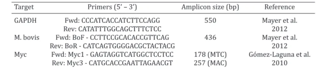
according to data previously published by our group (Mayer et al. 2012). All the primers used in this study are presented in Table 2.
Statistical analysis was performed using Stata software 10.1 (Stata Corporation, College Station, Texas, USA). The sensitivity, specificity, positive and negative predictive values between real time PCR from nasal swabs and CCT were calculated with the CCT criteria for as the gold standard for in vivo bTB. The agreement be-tween the real time PCR from nasal swabs and CCT was assessed by the Kappa coefficient (Landis & Kock 1977). The correlation between the tests was analyzed by determining Pearson coeffi -cient. Statistical significance was considered with p<0.05.
RESULTS
Out of 238 tested animals, 13 were positive and 12 were inconclusive on CCT test. Inconclusive results were con-sidered as positive, thus all 25 animals were sent to slau-ghter under official inspection. This represents 10.5% of bTB frequency of evaluated animals. Tissue samples were obtained from 23 of 25 CCT positive animals. Of collected samples, 12 (52.2%) had visible lesions consistent with tu-berculosis and 4 (17.4%) had isolated Mycobacterium spp. (Table 3). Of the isolates, 3 were retrieved from tissues with visible lesions and one of them from a tissue with no visible lesion (Table 3).
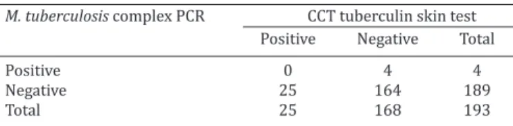
DNA was extracted from 238 swabs samples; however, only 193 were positive for GAPDH amplification, being in -cluded in the study. From these samples, 25 were positive on CCT test (Tables 2 and 3) and of these none was positive to Mycobacterium tuberculosis real-time PCR; moreover, 4 animals, which were negative at CCT, resulted positive for M. tuberculosis complex with the molecular method (with CTs of 31.9, 31.7, 32.0 and 29.0). Thus, the sensitivity (true positive within the positive results) and specificity (true negatives within the negative results) of the proposed molecular method, when compared to CCT were 0% and 97.6%, respectively. Positive predictive value (PPV) and negative predictive value (NPV) were 0% and 86.77%, respectively (Table 4). Low agreement coefficient (Kappa, -0.029, p=0.499) and correlation coefficient (Pearson, -0.049, p=0.502) were observed.
Regarding M. avium complex, 4 samples were positive in this test, of which one was inconclusive at CCT test (sample number 1, Table 3). The other three samples were negative at CCT. There was no correlation between the positivity to M. avium complex at the real-time PCR and the reactivity to PPD-A (data not shown).
Table 1. Characteristics of animals included in study
Variable Frequency n (%)
Sex Female 175 (91.14)
Male 17 (8.86)
Age (x, years) x < 1 39 (20.32)
1 ≥ x < 2 27 (4.06)
x ≥ 2 126 (65.62)
Breed Jersey 46 (23.95)
Holand 72 (37.5)
Mixed 74 (38.55)
Table 2. Primers used in the present study
Target Primers (5’ – 3’) Amplicon size (bp) Reference
GAPDH Fwd: CCCATCACCATCTTCCAGG 550 Mayer et al.
Rev: CATATTTGGCAGCTTTCTCC 2012
M. bovis Fwd: BoF - CCTTCCGCACACCGTTCAG 436 Mayer et al.
Rev: BoR - CATCAGTGGGGACGCTACTACG 2012
Myc Fwd: Myc1 - GAGTAGGTCATGGCTCCTCC 178 (MTC) Gómez-Laguna et al.
DISCUSSION
In vivo tests for bTB approved by Organization of Interna-tional Epizootics (OIE) include intradermal tuberculin skin test as prescribed test and IFN-γ ELISA assay as alternative test (OIE 2004). These tests can be applied together to in -crease sensitivity and specificity. Nevertheless, false results are still obtained, which is a problem to overcome by con-trol and eradication programs. False negative results have been reported due to early infection, immunosuppression and advanced or generalized bTB (Kehrli et al. 1989, Mo-naghan et al. 1994, Charleston et al. 2001, Pollock & Neill
2002). Factors related to the storage, manufacturing and method of administration of tuberculin might also cause false negative results (Monaghan et al. 1994). Thus, the de-velopment of an alternative in vivo test is important for a more appropriate disease control and would facilitate the study of tuberculosis in wild animals, that also can be re-servoirs of this disease.
Based on that, the present study evaluated if PCR from nasal swabs for Mycobacterium spp. detection is effective for in vivo bTB diagnosis. The advantages of using nasal swabs to bTB diagnosis would be great, since they can be easier collected from live animals, and could be applied to study Mycobacterium spp. in wildlife species, for which slaughter is not allowed and tuberculin test is not opti-mized. However, the results indicated that this technique was not effective when compared to tuberculin skin test, with low agreement values and lower sensitivity. From all animals tested positive for CCT, none was positive for M. tuberculosis complex with nasal swabs real-time PCR (Table 4). On the other hand, 4 animals that were negative for CCT, tested positive for M. tuberculosis complex with the molecular method. It is possible that these animals were in advanced stage of disease, being anergic to tuberculin skin test and with higher bacterial loads on their nasal secre-tion. If this this is case, it is possible that nasal swab real time PCR could be useful to detect anergic animals, which is one of tuberculin skin test limitations; however, as CCT was negative, these animals were not slaughtered, which prevented bTB confirmation. Thus, more studies on this subject should be carried out to confirm this hypothesis.
Despite the present study has shown inefficiency of PCR from nasal swabs to diagnose bTB in vivo, previous studies showed different results and interpretations. Figueiredo et al. (2010) identified 5.9% of positive PCR results in nasal swabs among 34 cattle positive in intradermal tuberculin test. Even with low sensitivity, the authors suggested that this method can be used as ancillary for surveillance of bo-vine tuberculosis in herds or as confirmatory for animals with inconclusive tuberculin results. Their conclusion is based on the fact that swab PCR had higher positivity fre-quency than swab culture, which was 0%. In the present study, the swab culture was not performed, since the goal was to evaluate the efficiency of real time PCR as an alter -native to in vivo bTB diagnose; then the comparison was made only to the tuberculin test, which is the gold standard for this purpose. Moreover, the swab culture would have the limitations related to Mycobacterium spp. handling as biosafety issues and time to obtaining the result.
Vitale et al. (1998) showed low sensitivity and high specificity of nasal swab PCR; however, the study compared Supplementary Fig.1. Melt curve of qPCR reaction showing the
Mycobacterium tuberculosis (purple line) and Mycobacterium avium (red line) melting temperatures.
Table 3. Tissue evaluation of animals slaughtered under
official inspection. The nasal swabs of all the animals
listed in this table were negative on real time PCR for
Mycobacterium tuberculosis and M. avium complexes
Animal CCT Tissue Visible lesion Culture
1* Inconclusive Lymph node Yes Negative
2 Inconclusive Lungs No Negative
3 Positive Lymph node and lungs Yes Negative 4 Positive Lymph node and lungs No Negative 5 Positive Lymph node and lungs No Negative
6 Positive Lymph node Yes Negative
7 Positive Lymph node Yes Positive
8 Positive Lymph node and lungs Yes Negative
9 Positive Liver Yes Negative
10 Positive Lungs Yes Negative
11 Positive Lungs No Negative
12 Positive Lymph node Yes Positive
13 Positive Lungs No Negative
14 Positive Lymph node Yes Negative
15 Inconclusive Lymph node No Negative
16 Positive Not tested Not tested Not tested
17 Inconclusive Lymph node No Negative
18 Positive Lungs Yes Negative
19 Positive Lungs Yes Positive
20 Positive Lymph node and lungs No Negative
21 Positive Lungs No Positive
22 Inconclusive Liver No Negative
23 Positive Not tested Not tested Not tested
24 Positive Lungs Yes Negative
25 Inconclusive Lungs No Negative
*Sample positive for Mycobacterium avium complex at real-time PCR.
Table 4. Comparison between the results obtained with tuberculin and real-time PCR of nasal swabs (Sensitivity:
0%; specificity: 97.6%; PPV: 0%; NPV: 86.77%) M. tuberculosis complex PCR CCT tuberculin skin test
Positive Negative Total
Positive 0 4 4
Negative 25 164 189
the nasal swab PCR with results of tissue PCR; the authors suggested the association of both techniques in samples from the same subjects. Another study found 100% of agreement between nasal swabs PCR and tuberculin re-sults, with 3 of 210 evaluated animals resulting positive in both tests (Solmaz et al. 2009). The difference on the results from that study to ours can be related to the DNA extraction method, which was made by commercial kit in the first study and in-house in the present; however, as we could detect Mycobacterium spp. DNA in a few samples, the ability of the extraction method was proven to be effective. Moreover, we used an internal control (GAPDH) to be sure of the success of DNA extraction. Thus, other possibilities to the different observed outcomes can be pointed, such as the disease stage of the tested animals and the routes of infection by which they were infected.
Several explanations can be pointed for the failure to detect Mycobacterium spp. in nasal swabs of tuberculous cattle by real-time PCR. Usually, when applied in bacteri -al cultures the efficiency of PCR is excellent; however, in samples in which there is the presence of the matrix (such as tissues, mucous, etc.), the efficiency will be dependent on the bacterial loads in the sample. This can be one pos-sible explanation to the observed results, since bacterial loads lower than 1000 (detection limit of the PCR used in the present study) may be secreted by nasal fluids. This is what seems to happen in field situations, as Palmer et al. (2002) has shown that experimental infections with aero-sols are more similar to natural infection with lower bacilli doses. Following this rationality, Neill et al. (1991), through a mathematical model, suggested that infection could be established following inhalation of a single bacillus. In this sense, the results of the present study are in accordance with other authors have been discussed, who pointed out that it is unrealistic to consider PCR as alternative to im-munological tools for tuberculosis routine diagnosis in live cattle, since it requires samples with high bacillary burdens (De la Rua-Domenech et al. 2006). In humans, sputum has been used for tuberculosis diagnosis and studies show good sensitivities values. A recent study showed sensitiv-ity of 90.3% of a real-time (RT)-PCR targeting mpt64 gene. Sputum seems to be more adequate for diagnosis purpos -es, since it is obtained from the inferior respiratory tract, and therefore it is more likely to have higher bacillary loads than nasal secretions. In cattle, the evaluation of sputum could be considered, although there would be difficulties related to the collection, and it does not solve the detection problem in cases of non-pulmonary or not active tubercu-losis.
The number of excreted bacilli may be influenced by dis -ease stage (Pollock et al. 2006). Although there are studies suggesting that animals with tuberculosis can excrete bacilli from the onset of disease, the shedding rate may be influ -enced by disease severity (Stamp 1944). Animals experi -mentally infected with higher doses of bacilli had higher severity on pathological lesions and enhanced frequency of shedding (McCorry et al. 2005). Moreover, although cattle are most likely to be infected with M. bovis through inhala-tion of aerosolized droplets, oral routes are also responsible
for infections (Neill et al. 1994, Palmer et al. 2004). Some authors state that oral route may be responsible for lesions preferentially on mesenteric lymph nodes (Biet et al. 2005, Riet-Correa & Garcia 2007). However, there is evidence that oral route lead to lesions limited to the lung, pulmonary and cranial lymph nodes (Palmer et al. 2004). The fact is that animals without lung or pulmonary lymph nodes lesions are not able to spread M. bovis through respiratory routes. Studies have suggested that only 9% to 19% of infected cattle shed M. bovis in nasal or tracheal secretions (Palmer & Waters 2006). In the present study, 12 animals (52.2%) were positive for CCT and had lesions at postmortem exam-ination, however only 4 (16%) had Mycobacterium spp. iso-lation. When considering the affected organ, only 6 animals had lung lesions compatible with tuberculosis and in just 2 of them was possible to isolate Mycobacterium spp. Never-theless, as none had positive results on nasal swab real-time PCR, we can assume that intradermal tuberculin skin test is more sensitive than nasal swabs PCR for in vivo bovine tuberculosis diagnosis. It is important to point that more studies should be carried out to evaluate the effectiveness of real time PCR to detect advanced bTB stages, in which the tuberculin results false negative and bacterial loads may be higher in animal secretions.
Acknowledgements.- Emily Marques dos Reis was recipient of Financia -dora de Estudos e Projetos (FINEP)/ Conselho Nacional de Desenvolvi -mento Científico e Tecnológico (CNPq) scholarship and André Vinícius An -drade Bezerra was recipient of Fundação de Amparo à Pesquisa do Estado do Rio Grande do Sul (FAPERGS) scholarship. This work was financially supported by CNPq with grant number 478660/2013-8. The sponsor had no role on study design; in the collection, analysis and interpretation of data; in the writing of the manuscript; and in the decision to submit the article for publication.
REFERENCES
Bezerra A.V., Dos Reis E.M., Rodrigues R.O., Cenci A., Cerva C. & Mayer F.Q. 2015. Detection of detection of Mycobacterium tuberculosis and Myco-bacterium avium Complexes by Real-Time PCR in bovine milk from Bra-zilian dairy farms. J. Food Prot. 78:1037-1042.
Biet F., Boschiroli M.L., Thorel M.F. & Guilloteau L.A. 2005. Zoonotic as -pects of Mycobacterium bovis and Mycobacterium avium-intracellulare complex (MAC). Vet. Res. 36:411-436.
Brasil 2006. Programa Nacional de Controle e Erradicação da Brucelose e da Tuberculose Animal (PNCEBT)/organizers, Vera Cecilia Ferreira de Figueiredo, José Ricardo Lôbo, Vitor Salvador Picão Gonçalves. Ministé -rio da Agricultura, Pecuária e Abastecimento, MAPA/SDA/DSA, Brasília, DF. 188p.
Cassidy J.P. 2006. The pathogenesis and pathology of bovine tuberculosis with insights from studies of tuberculosis in humans and laboratory ani-mal models. Vet. Microbiol. 112:151-161.
Cerva C., Bremm C., Dos Reis E.M., Bezerra A.V., Loiko M.R., Cruz C.E., Cenci A. & Mayer F.Q. 2014. Food safety in raw milk production: Risk factors associated to bacterial DNA contamination. Trop. Anim. Health Prod. 46:877-882.
Charleston B., Hope J.C., Carr B.V. & Howard C.J. 2001. Masking of two in vitro immunological assays for Mycobacterium bovis (BCG) in calves acutely infected with non-cytopathic bovine viral diarrhoea virus. Vet. Rec. 149:481-484.
Figueiredo E.E.S., Carvalho R.C.T., Silvestre F.G., Lilenbaum W., Fonseca L.S., Silva J.T. & Paschoalin V.M.F. 2010. Detection of Mycobacterium bovis
Gómez-Laguna J., Carrasco L., Ramis G., Quereda J.J., Gómez S. & Pallarés F.J. 2010. Use of Real-Time and Classic Polymerase Chain Reaction As -says for the diagnosis of porcine tuberculosis in formalin-fixed, paraffin --embedded tissues. J. Vet. Diagn. Investig. 22:123-127.
Karolemeas K., la Rua-Domenech R., Cooper R., Goodchild A.V., Clifton-Ha-dley R.S., Conlan A.J., Mitchell A.P., Hewinson R.G., Donnelly C.A., Wood J.L. & McKinley T.J. 2012. Estimation of the relative sensitivity of the comparative tuberculin skin test in tuberculous cattle herds subjected to depopulation. PLoS One 7:e43217.
Kehrli M.E., Nonnecke B.J. & Roth J.A. 1989. Alterations in bovine lym -phocyte function during the periparturient period. Am. J. Vet. Res. 50: 215-220.
de la Rua-Domenech R., Goodchild A.T., Vordermeier H.M., Hewinson R.G., Christiansen K.H. & Clifton-Hadley R.S. 2006. Ante mortem diagnosis of tuberculosis in cattle: a review of the tuberculin tests, γ-interferon assay and other ancillary diagnostic techniques. Res. Vet. Sci. 81:190-210.
Mayer F.Q., Cerva C., Driemeier D., Cruz C.E., Loiko M.R., Coppola M.M., Cibulski S. & Bertagnolli A.C. 2012. Mycobacterium bovis infection in a collared peccary (Tayassu tajacu): insights on tuberculosis wild reser -voirs. Vet. Microbiol. 160:549-551.
McCorry T., Whelan A.O., Welsh M.D., McNair J., Walton E., Bryson D.G., Hewinson R.G., Vordermeier H.M. & Pollock J.M. 2005. Shedding of
Mycobacterium bovis in the nasal mucus of cattle infected experimen-tally with tuberculosis by the intranasal and intratracheal routes. Vet. Rec. 157:613-618.
Michel A.L., Müller B. & Van Helden P.D. 2010. Mycobacterium bovis at the animal-human interface: a problem, or not? Vet. Microbiol. 140:371-381.
Monaghan M.L., Doherty M.L., Collins J.D., Kazda J.F. & Quinn P.J. 1994. The tuberculin test. Vet. Microbiol. 40:111-124.
Neill S.D., O’Brien J.J. & Hanna J. 1991. A mathematical model for Mycobac-terium bovis excretion from tuberculous cattle. Vet. Microbiol.
28:103-109.
Neill S.D., Pollock J.M., Bryson D.B. & Hanna J. 1994. Pathogenesis of Myco-bacterium bovis infection in cattle. Vet. Microbiol. 40:41-52.
OIE 2004. World Organization for Animal Health. Manual of Diagnostic Tests and Vaccines for Terrestrial Animals. 5th ed. Web version, Vol.1 and 2, Paris.
Palmer M.V., Waters W.R. & Whipple D.L. 2002. Aerosol delivery of virulent
Mycobacterium bovis to cattle. Tuberculosis 82:275-282.
Palmer M.V. & Waters W.R. 2006. Advances in bovine tuberculosis diagno
-sis and pathogene-sis: what policy makers need to know. Vet. Microbiol.
112:181-190.
Palmer M.V., Waters W.R., Whipple D.L., Slaughter R.E. & Jones S.L. 2004. Evaluation of an in vitro blood-based assay to detect production of interferon-gamma by Mycobacterium bovis-infected white-tailed deer (Odocoileus virginianus). J. Vet. Diagn. Invest. 16:17-21.
Pollock J.M. & Neill S.D. 2002. Mycobacterium bovis infection and tubercu-losis in cattle. Vet. J. 163:115-127.
Pollock J.M., Rodgers J.D., Welsh M.D. & McNair J. 2006. Pathogenesis of bovine tuberculosis: The role of experimental models of infection. Vet. Microbiol. 112:141-150.
Riet-Correa F. & Garcia M. 2007. Tuberculose, p.432-442. In: Riet-Correa F., Schild A., Méndez M. & Lemos R. (Eds), Doenças Ruminantes e Equi -nos. Pallotti, Santa Maria.
Schiller I., Oesch B., Vordermeier H.M., Palmer M.V., Harris B.N., Orloski K.A., Buddle B.M., Thacker T.C., Lyashchenko K.P. & Waters W.R. 2010. Bovine tuberculosis: a review of current and emerging diagnostic tech -niques in view of their relevance for disease control and eradication. Transbound. Emerg. Dis. 57:205-220.
Solmaz H., İlhan Z., Aksakal A., Gülhan T. & Ekin İ.H. 2009. Detection of bovine tuberculosis by tuberculin test and polymerase chain reaction in Van, Turkey. Turk. J. Vet. Anim. Sci 33:229-233.
Stamp J.T. 1944. A review of the pathogenesis and pathology of bovine tu -berculosis with special reference to practical problems. Vet. Rec 56:443-446.
Vitale F., Capra G., Maxia L., Reale S., Zooprofilattico I. & Della S. 1998. De -tection of Mycobacterium tuberculosis complex in cattle by PCR using milk, lymph node aspirates, and nasal swabs. J. Clin. Microbiol. 36:1050-1055.
Wood P.R., Corner L.A. & Plackett P. 1990. Development of a simple, rapid in vitro cellular assay for bovine tuberculosis based on the production of gamma interferon. Res. Vet. Sci. 49:46-49.
Zarden C.F.O., Marassi C.D., Carvalho A.C., Figueiredo E.E.S. & Lilenbaum W. 2013. Bacteriological and molecular detection of Mycobacterium bovis
in cattle with inconclusive results to intradermal tuberculin tests. Epi -demiol. Infect. 141:1390-1393.