Comparison of nine DNA extraction methods for the diagnosis
of bovine tuberculosis by real time PCR
Comparação de nove métodos de extração de DNA para diagnóstico de tuberculose bovina por PCR em tempo real
André MouraI Mikael Arrais HodonI Paulo Martins Soares FilhoI Marina de Azevedo IssaI
Ana Paula Ferreira de OliveiraI Antônio Augusto Fonseca JúniorI*
ISSN 1678-4596
AbStRACt
Bovine tuberculosis is an infectious disease with a high impact on the cattle industry, particularly in developing countries. PCR is a very sensitive method for detection of infectious agents, but the sensitivity of molecular diagnosis is largely dependent on the efficiency of the DNA extraction methods. The objective of this study was to evaluate DNA extraction methods for direct detection of Mycobacterium bovis in bovine tissue. Nine commercial kits for DNA extraction were evaluated when combined with two real time PCRs. The DNeasy Blood & Tissue Kit from QIAGEN showed better performance and sensitivity followed by the DNA Mini Kit RBC and FTA Elute Micro Card. Results suggested that, even when the analytical sensitivity of the qPCR is very high, the extraction method can influence the diagnostic sensitivity.
Key words: bovine tuberculosis, DNA extraction, real time PCR.
RESUMO
A tuberculose bovina é uma doença infecciosa com um alto impacto na pecuária, particularmente em países em desenvolvimento. A PCR é um método muito sensível para a detecção de agentes infecciosos, mas a sensibilidade do diagnóstico molecular é em grande parte dependente da eficiência dos métodos de extração de DNA. O objetivo deste estudo foi avaliar métodos de extração de DNA para detecção direta de Mycobacterium bovis em tecido bovino. Nove kits comerciais para extração de DNA foram avaliados, quando combinados com duas PCRs em tempo real. O Kit Dneasy Blood & Tissue da Qiagen apresentou melhor desempenho e sensibilidade, seguido dos kits DNA Mini RBC e FTA Elute Micro Card (protocolo modificado com digestão enzimática prévia). Os resultados sugerem que, mesmo quando a sensibilidade analítica do qPCR é muito elevada, o método de extração pode influenciar na sensibilidade de diagnóstico.
Palavras-chave: tuberculose bovina, extração de DNA, PCR
em tempo.
INtRODUCtION
Bovine tuberculosis (bTB) is an infectious disease with a high impact on the cattle industry, particularly in developing countries. It is characterized by the development of nodular granulomatous lesions, predominantly located in the respiratory tract and bronchial and mediastinal lymph nodes. The economic losses are related to the direct impact of infection due to reduced weight gain, decreased milk production and condemnation of carcasses or indirect losses as the depreciation of meat price due to sanitary barriers (BRAZIL, 2006; HEINEMANN et al., 2008).
Lesions found at post-mortem examinations
can be confirmed by bacterial isolation, the gold
standard method for detection of Mycobacterium bovis. However, this technique is laborious and time consuming and may require months to reach its conclusion, which slows the development of health programs, which aim to control and eradication of the disease (De La RUE-DOMNENECH et al., 2006). Thus, to reduce the time of diagnosis of tuberculosis in cattle, new molecular methods are proposed.
The polymerase chain reaction (PCR) is a very sensitive method for the detection of infectious agents, including the evaluation of animals in epidemiological surveys (YOON et al., 2005). The speed of the methodology can be increased by using real-time PCR (qPCR) which, besides providing
ILaboratório Nacional Agropecuário de Minas Gerais, Av. Rômulo Joviano, CP 50, 33600-000, Pedro Leopoldo, MG, Brasil. E-mail:
[email protected]. *Corresponding author.
better precision, reproducibility and quality control in the process, reduced contamination and enabled the analysis of a large number of samples in a shorter period of time (SALES et al., 2013).
The sensitivity of molecular diagnosis
is largely dependent on the efficiency of the DNA
extraction methods (NAKATANI et al., 2004). Isolation of bacterial DNA in tissues is a highly complex procedure, mainly because of the low concentration of microorganisms in the tissue sample and the presence of large amounts of contaminant
genetic material, making difficult to obtain high
quality DNA (BURGGRAF & OLGEMÖLLER, 2004). Currently, there are several commercial kits for the extraction of bacterial genetic material directly from tissues. Although some studies have
shown a significant variation in the sensitivity of
PCR according to the extraction method used, there
is no definitive view regarding the best method of
extraction of DNA from M. bovis in bovine tissue samples (YOSHIKAWA et al., 2011). The objective of this study was to evaluate nine DNA extraction methods for the direct detection of M. bovis in bovine tissue.
MAtERIAlS AND MEtHODS
Sample preparation
All samples were derived from cattle carcasses in slaughterhouses inspected by the Serviço de
Inspeção Federal (SIF) and sent to the official diagnosis
facility in Brazil. Tissues contained granulomatous lesions suggestive of bTB in retropharyngeal, mediastinal and mesenteric lymph nodes, liver and lung fragments. A small fraction of each sample was extracted in a biological safety cabinet Class II A and aliquoted into 2ml tubes containing 500µL of ATL buffer (Qiagen, Germany). Enzymatic digestion was done adding 50µL of Proteinase K to each sample and incubating overnight at a temperature of 56.5°C. Subsequently, the samples were inactivated at 87.5ºC and subjected to DNA extraction.
DNA extraction
There were five rounds of testing, in which
seventy different randomly chosen samples were assessed. The tests were performed in rounds, due to
insufficient volume of each sample being available
for simultaneous evaluation of all extraction methods. In each round, the samples were extracted with two or three different commercial kits. The kit with the best
performance in the first round was compared to other
kits in the next round and so on.
The first round of tests comparing the
extraction of 70 samples with three commercial extraction kits: Maxwell 16 (Promega, USA), DNeasy Blood & Tissue kit (Qiagen, Germany) and Cador Pathogen (Qiagen, Germany), using initial volumes for extraction of 400µL and 200µL. The second round of tests compared the extraction of 70 samples with another three commercial extraction kits: NucleoSpin TriPrep (Macherey-Nagel, Germany) DNA Blood & Tissue kit (Qiagen, Germany) and innuPREP DNA Mini Kit (Analytik Jena, Germany). The third round of tests compared the extraction of 70 samples with three further commercial extraction kits: Wizard
Genomic DNA Purification Kit (Promega, USA),
DNA Blood & Tissue kit (Qiagen, Germany) and Genomic DNA Mini Kit (Real Biotech Corporation, RBC, Taiwan). The fourth round of tests compared the extraction of 70 samples with two more commercial extraction kits: DNA Blood & Tissue kit (Qiagen, Germany) and MagNA Pure LC DNA Isolation Kit II (Roche, Germany) using equipment Magna Pure
(Roche, Germany). The fifth round of tests compared
the extraction of 70 samples with two additional commercial extraction kits: DNA Blood & Tissue kit (Qiagen, Germany) and Whatman FTA Elute cards.
All methods and extraction kits tested followed the extraction protocols recommended
by the companies without modification, except
for the extraction method used for the Whatman FTA Elute Cards, in which there was the following adaptation: cards were impregnated with 20µL of each sample after enzymatic digestion and left at room temperature atmosphere for three hours to dry. After drying, a Harris Uni-Core™ Micro-3mm punch was used to cut a disk with diameter of 3mm from on each card. Resultant disks were immersed in 500µL of sterile DEPC water. Then, each sample
was homogenized three times for five seconds by
vortexing. Subsequently, using a micropipette, all water was removed. Each sample was centrifuged for
five seconds and the resulting liquid was discarded.
50µL of sterile DEPC water was added in each sample and incubated at a temperature of 95°C for 30 minutes. After this process, the samples were quickly homogenized, followed by centrifugation for 30 seconds in order to separate the matrix from the
liquid containing the purified DNA. The microtubes containing the final DNA were placed in a refrigerator
at -20°C until use.
Due to the high initial DNA concentration obtained in the Genomic DNA extraction kits Mini Kit and Wizard® Genomic DNA Purification Kit,
sterile DEPC water. After the extractions were carried out at different concentrations, it was concluded that the best dilution for performing qPCR was 10-3 whose mean concentration was 100ng µL-1.
PCRs
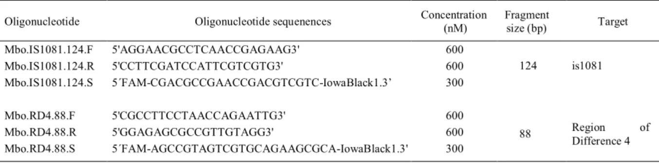
Extracted DNA was subjected to qPCR on the unit QuantStudio 7 Flex™ Real-Time PCR System (Life Technologies, USA) and used in a 25µL reaction containing the following reagents: 3µL DNA, 4.0µL RNase free water, 12.5µL of RealQ PCR 2 x Master Mix (Amplicon, Denmark), 4.0µL of MgCl2 (25nM). Primers and probes for the two PCRs used are described in table 1. The following cycling regime was used: 50°C for 2min, 95°C for 10min and 50 cycles at 95°C for 15s and 60°C for 1min. Positive
samples were those that had amplified Cq’s less than or equal to 42.0. All samples amplified with Cq’s
above this value were considered negative.
The positive control for all PCRs were the reference strain of M. bovis AN5 (CANEVARI CASTELÃO et al., 2014). In addition to the positive controls, all tests relied on negative control for DNA extraction and negative control to check contamination of PCR reagents.
Statistical analysis
McNemar test with 5% significance level
was used to determine the independence of the results and disagreement frequencies found between extraction kits in each round, comparing them individually. Finally, to get the actual correlation between them, the kappa test was applied to two kits with smaller discrepancy between themselves in each round (KRAEMER, 1992). To calculate the
Kappa coefficient, the criteria described by McGINN
et al. (2004) were followed, with values greater than 0.80 representing an “almost perfect” concordance; between 0.60 and 0.80 being “substantial”; between
0.40 and 0.60 as “moderate”; and below 0.40 representing “weak” agreement.
RESUltS AND DISCUSSION
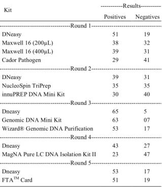
Table 2 shows the number of samples detected as positive and negative for each extraction kit evaluated. Table 3 shows the results of the McNemar test performed between all kits and the Kappa test performed on both kits with the lowest level of disagreement.
In the first round of testing results for the
McNemar test were: DNeasy x Cador x2 = 12.9, DNeasy x Maxwell 16 (200 µL) x2 = 1.39, DNeasy x Maxwell 16 (400µL) x2 = 6.05 and Maxwell 16 (200µL) x 16 Maxwell (400µL) x2 = 0.64. Extraction kits with less disagreement in this round were the Maxwell 16 (200µL) x 16 Maxwell (400µL). However, the kappa test was conducted with the results obtained with the DNeasy kit x Maxwell 16 (200µL), because the kit with the lowest mismatch did not show better sensitivity. The value obtained for the Kappa test in the round was K = 0.37.
In the second round of testing, results for the McNemar test were DNeasy NucleoSpin x = 0.75 and x2, Analytik x DNeasy x2 = 5.06,
NucleoSpin x Analytik x2= 0.94. Extraction kits with
less disagreement in this round were the DNeasy and Nucleospin. Kappa test was performed with the results obtained and gave K = 0.64.
In the third round of testing, results for the McNemar test were DNeasy x Promega x2 =
12.07, DNeasy x RBC x2 = 0.13, Promega x RBC x2
= 7.69. Extraction kits with less disagreement in this round were the DNeasy and RBC. The Kappa test was performed with the results obtained and gave K = 0.25.
In the fourth round of testing, results for the McNemar test were DNeasy x Roche x2 = 18.37.
Table 1 - Oligonucleotides used in this research.
Oligonucleotide Oligonucleotide sequenences Concentration
(nM)
Fragment
size (bp) Target
Mbo.IS1081.124.F 5'AGGAACGCCTCAACCGAGAAG3' 600
Mbo.IS1081.124.R 5'CCTTCGATCCATTCGTCGTG3' 600
Mbo.IS1081.124.S 5´FAM-CGACGCCGAACCGACGTCGTC-IowaBlack1.3’ 300
124 is1081
Mbo.RD4.88.F 5'CGCCTTCCTAACCAGAATTG3' 600
Mbo.RD4.88.R 5'GGAGAGCGCCGTTGTAGG3' 600
Mbo.RD4.88.S 5´FAM-AGCCGTAGTCGTGCAGAAGCGCA-IowaBlack1.3' 300
88 Region of
Kappa test was performed with results obtained and
met K = 0.28. In the fifth round of testing results for
the McNemar test were DNeasy x FTA Card x2 =
0.56. Kappa test was performed with results obtained and gave K = 0.41.
This study evaluated and compared the performance of nine extraction kits in clinical samples with suggestive tuberculosis lesions. The use of the extract control was important to prove the
efficiency of the procedures and the quality of the
obtained material.
The DNeasy Blood & Tissue Kit from QIAGEN showed better performance and sensitivity for the detection of M. bovis in comparison with the
other extraction kits evaluated in all five rounds. In
addition, unlike the results obtained by QUEIPO-ORTUÑO et al. (2008) and DURNEZ et al. (2009), cross-contamination was not observed in the extraction of DNA in the tests performed with this kit.Genomic DNA Mini Kit RBC and FTA Elute Micro Card extraction kits were the most similar to the QIAGEN kit, balanced by the number of positive samples detected.
The Genomic DNA Mini Kit, which uses no columns or the like in the centrifugation steps,
performed well and with good sensitivity. According
to ALDOUS et al. (2005), although DNA purification
columns tend to be less conducive to contamination by inhibiting substances, the procedure does not
guarantee greater efficiency of the extraction process,
so it is possible to extract DNA from lesions suspected of bTB even without these columns.
The FTA Elute Micro Card proved to be a promising method for DNA extraction (if tissue is submitted to enzymatic digestion previously to impregnation), due to the convenience of sample storage cards, simple method of implementation and good sensitivity displayed by the kit, agreeing with
the findings of WOLFGRAMM et al. (2009) and
GONZALEZ et al. (2012). A disadvantage of the FTA is the time required for the extraction; approximately 240 minutes for 30 samples, as compared to other kits, which take 100 to 120 minutes.
Low sensitivity displayed by the other kits is probably related to the small amount of bacterial
DNA present in the tissue, which difficult detection by qPCR even when extracted with a highly efficient
and sensitive technique (TOMASO et al., 2010). Despite the strong correlation observed between the RBC and DNeasy extraction kits in the
third round of testing, the Kappa coefficient was low,
which can be explained by the prevalence of a different distribution presented by the sum of the marginal, resulting in a relatively low Kappa, even when there is a high similarity between the tests (FEINSTEIN & CICCHETTI, 1990). Another possibility would be the detection limit of the technique, since some samples extracted with the kit Genomic DNA Mini Kit RBC (unlike the DNeasy Blood & Tissue) needed
to be confirmed with specific primers Mbo.RD4.88,
after being previously detected with the primers Mbo. is1081.124.
PCR sensitivity can still be improved. This study did not include tissue mechanical lysis by equipment like Tissue Lyzer (Qiagen, Germany) or MagNA Lyser (Roche, Germany). Mycobacterial DNA extraction from tissue is not an easy task and
the use of mechanical lysis will definitely improve
results obtained with any kit (COSTA et al., 2013).
CONClUSION
The objective of this study was to evaluate nine DNA extraction methods to detect M. bovis in bovine tissue. Results suggested that nucleic acid
extraction kit influences deeply the diagnosis of
bovine tuberculosis by qPCR in bovine tissue samples suggestive of tuberculosis lesions.
Table 2 - Number of positive and negative samples submitted for each PCR extraction kit. 70 samples were used for each round of testing.
---Results---Kit
Positives Negatives ---Round
1---DNeasy 51 19
Maxwell 16 (200µL) 38 32
Maxwell 16 (400µL) 39 31
Cador Pathogen 29 41
---Round
2---DNeasy 39 31
NucleoSpin TriPrep 35 35
innuPREP DNA Mini Kit 30 40
---Round
3---Dneasy 65 5
Genomic DNA Mini Kit 63 07
Wizard® Genomic DNA Purification 53 17
---Round
4---Dneasy 43 27
MagNA Pure LC DNA Isolation Kit II 23 47
---Round
5---Dneasy 53 17
ACKNOwlEDgEMENtS
The authors are grateful to Laboratório Nacional Agropecuário (Lanagro-MG), INCT Pecuáriaand Conselho Nacional
de Desenvolvimento Cientifico e Tecnológico (CNPq). SAGRES Project: 457417/2012-9 for financial support and fellowships.
REFERENCES
ALDOUS, W.K. Comparison of six methods of extracting
Mycobacterium tuberculosis DNA from processed sputum for testing byquantitative real time PCR. J Clin Microbiol, v.43, p.2471-2473, 2005. Available from: <http://dx.doi. org/10.4103/0974-777X.91057>. Accessed: Mar. 02, 2016. doi: 10.4103/0974-777X.91057.
BRAS IL. Ministério da Agricultura Pecuária e Abastecimento.
Programa Nacional de Controle e Erradicação da brucelose e tuberculose – PNCEbt. [National Program for Control and Eradication of Brucellosis and Tuberculosis]. Brasil: MAPA/DAS/ DAS, 2006. 184p. Available from: <http://www.agricultura.gov.br/
arq_editor/file/Aniamal/programa%20nacional%20sanidade%20
brucelose/Manual%20do%20PNCEBT%20-%20Original.pdf>. Accessed: Mar. 02, 2016.
BURGGRAF, S.; OLGEMÖLLER, B. Simple technique for
internal control of real-time amplification assays. Clin Chem,
v.50, p. 819-825, 2004. Available from: <http://www.clinchem.org/ content/50/5/819.long>. Accessed: Mar. 02, 2016. doi: 10.1373/ clinchem.2003.027961.
CANEVARI-CASTELÃO, A.B. et al. Draft genome sequence of Mycobacterium bovis strain AN5, used for production of
Purified Protein Derivative. genome Announc., v.2
p.e00277-14, 2014. Available from: <http://www.ncbi.nlm.nih.gov/pmc/ articles/PMC3974946/>. Accessed: Mar. 02, 2016. doi: 10.1128/ genomeA.00277-14.
COSTA, P. et al. Enhanced detection of tuberculous mycobacteria in animal tissues using a semi-nested probe-based real-time PCR.
PloS One, v.11, p.e81337, 2013. Available from: <http://journals. plos.org/plosone/article?id=10.1371/journal.pone.0081337>. Accessed: Mar. 02, 2016. doi: 10.1371/journal.pone.0081337. De La RUE-DOMMENEC, R. et al. Ante-mortem diagnosis in cattle: A review of the tuberculin tests, γ-interferon assay and other ancillary diagnostic techniques. Res Vet Sci, v.81, p.190-210, 2006. Available from: <http://www.sciencedirect.com/science/ article/pii/S0034528806000026>. Accessed: Mar. 02, 2016. doi: 10.1016/j.rvsc.2005.11.005.
DURNEZ, L. et al. A comparison of DNA extraction procedures for the detection of Mycobacterium ulcerans, the causative agent of Buruli ulcer, in clinical and environmental specimens. J MicrobiolMeth, v.76, p.152-158, 2009. Available from: <http:// www.sciencedirect.com/science/article/pii/S0167701208003539>. Accessed: Mar. 02, 2016. doi: 10.1016/j.mimet.2008.10.002. EINSTEIN, A.R.; CICCHETTI, D.V. High agreement but low kappa: I. The problems of two paradoxes. J ClinEpidemiol, v.43, p.543-549, 1990. Available from: <http://linkinghub.elsevier.com/ retrieve/pii/0895-4356(90)90158-L>. Accessed: Mar. 02, 2016. doi: 10.1016/0895-4356(90)90158.
GONZALEZ, P. et al. Evaluation of the FTA carrier device for human papillomavirus testing in developing countries. J Clin Microbiol, v.50, p.3870-3876, 2012. Available from: <http://jcm. asm.org/content/50/12/3870.abstract>. Accessed: Mar. 02, 2016. doi: 10.1128/JCM.01698-12.
Table 3 - Results of McNemar and Kappa tests.
Kits McNemar (ns)* Kappa
---Round
1---Dneasy x Maxwell 16 (200µL) 1.39** 0.37
Dneasy x Maxwell 16 (400µL) 6.05 0.36
Dneasy x Cador Pathogen 12.9 0.21
Maxwell 16 (200µL) x Maxwell 16 (400µL) 0.64** 0.56
---Round
2---Dneasy x NucleoSpin TriPrep 0.75** 0.64
Dneasy x innuPREP DNA Mini Kit 5.06 0.51
NucleoSpin TriPrep x innuPREP DNA Mini Kit 0.94** 0.40
---Round
3---Dneasy x Wizard® Genomic DNA Purification 12.07 0.28
Dneasy x Genomic DNA Mini Kit 0.13** 0.25
Wizard® Genomic DNA Purification x Genomic DNA Mini Kit 7.69 0.29
---Round
4---Dneasy x MagNA Pure LC DNA Isolation Kit II 18.37 0.28
---Round
5---Dneasy x FTA Card 0.56** 0.41
HEINEMANN, M.B. et al. Bovine tuberculosis: an introduction to the aetiology, epidemiological chain, pathogenesis and clinical signs [Tuberculose bovina: uma introdução à etiologia, cadeia epidemiológica, patogenia e sinais clínicos]. Cad tec Vet Zootec, v.59, p.1-12, 2008. Available from: <http://www.crmvmg.org.br/novoportal/ Institucional/detalheCadernoTecnico.aspx>. Accessed: Mar. 02, 2016. KRAEMER, H.C. Evaluating medical tests. Objective and quantitative guidelines. Newbury Park (CA): Sage Publications, 1992. 296 p. McGINN, T. et al. Tips for learners of evidence-based medicine: 3. Measures of observer variability (Kappa statistic). Can Med AssocJ, v.171, p.1369-1373, 2004. Available from: <http://www. cmaj.ca/content/171/11/1369.long>. Accessed: Mar. 02, 2016. doi: 10.1503/cmaj.1031981.
NAKATANI, S.M. et al. Efficient method for mycobacterial DNA extraction in blood cultures aids rapid PCR identification of
Mycobacterium tuberculosis and Mycobacterium avium. Eur J Clin Microbiol Infect Dis, v.23, p.851-854, 2004. Available from: <http://link.springer.com/article/10.1007/s10096-004-1236-z>. Accessed: Mar. 02, 2016. doi: 10.1007/s10096-004-1236-z. QUEIPO-ORTUÑO, M. et al. Comparison of seven commercial DNA extraction kits for the recovery of Brucella DNA from spiked human serum samples using real-time PCR. Eur J Clin Microbiol Infect Dis, v.27, p.109-114, 2008. Available from: <http://europepmc.org/abstract/med/17973130>. Accessed: Mar. 02, 2016. doi: 10.1007/s10096-007-0409-y.
SALES, M.L. et al. Validation of a real-time PCR assay for the
molecular identification of Mycobacterium tuberculosis. braz
J Microbiol, v.45, p.1362-1369, 2014. Available from: <http:// www.ncbi.nlm.nih.gov/pmc/articles/PMC4323311/>. Accessed: Mar. 02, 2016. doi: 10.1590/S1517-83822014000400029. TOMASO, H. et al. Comparison of commercial DNA preparation kits for the detection of Brucellae in tissue using quantitative real-time PCR. bMC Infect Dis, v.10, p.100, 2010. Available from: <http://link.springer.com/article/10.1 186%2F1471-2334-10-100>. Accessed: Mar. 02, 2016. doi: 10.1186/1471-2334-10-100.
WOLFGRAMM, E.V. et al. Simplified buccal DNA extraction
with FTA elute cards. J Forensic Sci International genetics, v.3, p.125-127, 2009. Available from: <http://www.sciencedirect. com/science/article/pii/S1872497308001853>. Accessed: Mar. 02, 2016. doi: 10.1016/j.fsigen.2008.11.008.
YOON, H.A. et al. Molecular survey of latent pseudorabies virus infection in nervous tissues of slaughtered pigs by nested and real-time PCR. J Microbiol, v.43, p.430-436, 2005.