Functional and Structural Studies of
Two Enzymes: Membrane Bound
Kinase and Z-DNA/Z-RNA-Binding
Protein.
Dissertação para obtenção do Grau de Doutor em
Sistemas de Bioengenharia
Orientador: Margarida Archer, Investigador Auxiliar, ITQB-UNL
Co-orientador: Li-Huei Tsai, Investigador/Director, PILM-MIT
Júri: (Font: Arial, 10 pt normal)
Presidente: Prof. Doutor Manuel Luís Magalhães Nunes da Ponte
Arguentes: Prof. Doutor Domingos Manuel Pinto Henrique
Prof. Doutor Luis Miguel Gales Pereira Pinto
Vogais: Prof. Doutora Maria Arménia Abreu Fonseca Carvalho T. Carrondo
Prof. Doutor Shabir Husein Najmudin
II
Functional and structural studies of two enzymes: membrane bound kinase and
Z-DNA/Z-RNA-binding protein.
18th November 2013, ITQB, Oeiras, Portugal First edition, November 2013
ISBN 978-989-20-4348-7
Copyright © 2013, Ana Lúcia Rebelo do Rosário, FCT-UNL e UNL.
III
This work was developed at the laboratory of Prof Li-Huei Tsai (co-supervised by Dr. Froylan Calderon de Anda), at the Picower Institute for Learning and Memory, Massachusetts Institute of Technology, Cambridge, USA. Section 2 of this chapter, describes the work developed to characterize hTAOK2α biochemical and structurally by X-ray crystallography. The work herein described was carried out at the Membrane Protein Crystallography laboratory at ITQB, under the supervision of Dr Margarida Archer and in collaboration with Dr Gonçalo Real and Dr. Marco Patrone (Animal Cell Technology Unit, IBET).
V
different fields. Dra Margarida Archer was always able to motivate me in decisive moments, to advice me in the right directions, to make me understand my possibilities, and for making me believe it was possible to go further. To her, my most sincere thanks. It was a pleasure and a privilege to do my PhD under your supervision. In second, I want to aknowledge Prof Maria Arménia Carrondo, for receiving me in the crystallography unit at ITQB, for all your support and motivation, thank you.
Professor Li-Huei Tsai, for receiving me in your laboratory, for the challenge of working and debating with you my work. It made me stronger! I want to particular thank Froylan, my mentor at MIT, my friend for life. You taught me so many things that are immeasurable. You helped me to construct a critical spirit in my work, to think outside the box, to wonder at our findings and, when looking at a neuron for the thousand time, we would see it for the first. It was a pleasure to learn with you. Konstatinos, for working with me and making me a litle easier with details on
experiments, you’re always saying “Ana, this is not rocket science”. I want to thank all the people from the Tsai lab and from the Picower Institute that helped me and worked with me during my stay. A special thanks to all my colleagues from the MIT-Portugal program that were in Boston at the time. We had great moments together.
My colleagues from the crystallography unit at ITQB, all of you have made my life easier. A special thanks to Dr Pedro Matias and Dr Carlos Frazão, always prepared to help me when collecting data at synchrotrons and in-house. Dr Colin McCvey for all your help, support and suggestions with my purification experiments. I want to thank my colleagues from the Membrane Protein Crystallography Laboratory. A special thanks to José Brito for your capacity in getting everything we need in time, your support and friendship in the lab. A very very special thanks to Tânia, for all your help and support, our discussions about the work, your tips and tricks in crystallization, for all your friendship in the hard times of thesis “depression”. For all
VI
I wish you all the best in life and thank you for all the good laughs we have.
Finalmente, mas não menos importante. Quero agradecer a toda a minha família. O meu porto de abrigo, onde a palavra casa, amizade e amor tem um significado imenso. Aos meus pais, António e Mabília, foram sempre meus amigos e companheiros. Deram-me tudo o que eu precisei e muito mais, é por vocês que sou assim, e que estou hoje a escrever estas palavras. Aos meus avós, que sempre fizeram parte da minha vida e ajudaram em tudo. À minha irmã e ao meu afilhado, Vera e Mateus, porque o simples facto de fazerem parte da minha vida, já a torna mais preenchida, cheia de significado, e muito mais rica. Sem vocês, tudo faria menos sentido, e eu seria pobre.
Um agradecimento muito especial a ti, Bruno, porque estás aqui novamente comigo, neste momento. A tua companhia e amizade, o teu amor, a tua persistência, o teu bom humor torna tudo mais colorido. Obrigada.
VII
This work represents the first approach aimed at understanding the mechanisms responsible for the delineation of basal and apical dendrites during pyramidal neuron development in the embryo, and how such mechanism may evolve to neocortical disconnection disorders. Additional work performed on humanTAOK2α focused at the determination of its three-dimensional structure by X-ray crystallography to elucidate its regulatory mechanism.
The 20th century exciting discovery of the DNA left-handed conformation, and the fact it binds to certain classes of proteins with high affinity and specificity, indicated a biological role to it. However, a full function is still to be elucidated. The human double-stranded RNA adenosine deaminase (ADAR1) is the best characterized of all Z-DNA binding proteins, where Zα domain binds and stabilizes Z-DNA/Z-RNA forms upon binding. The second part of this thesis describes the work on the ZαADAR1 domain that binds to Z-RNA/Z-DNA. When a section of a DNA or RNA molecule forms a left-handed Z-DNA/Z-RNA segment, two B-Z/A-Z junctions are formed. Herein, we describe the study carried out on the formation of Z-Z junctions from DNA and, also, the approach on trying to describe the Z-Z junction for RNA when interacting with ZαADAR1. The structure of the Z-Z-DNA junction consists of a single base pair that leads to partial or full disruption of the helical stacking. The junction region allows intercalating agents to insert themselves into the left-handed helix, which is otherwise resistant to intercalation.
IX
corticais. Este estudo representa a primeira tentativa para compreender os mecanismos responsáveis pela delienação entre dendrites basais e apicais no densenvolvimento dos neurónios piramidais, e de que forma esses mecanismos podem resultar em doenças de disconecção cortical. Apresentamos também o trabalho realizado no âmbito da determinação da estrutura tridimensional da humanaTAOK2α por cristalografia de raios-X, para esclarecer o seu mecanismo de regulação.
O ADN de rotação contrária (Z-ADN), foi uma importante descoberta do século XX. O facto do Z-ADN se ligar a determinadas classes de proteínas com alta afinidade e especificidade auspiciou uma função biológica para o mesmo. No entanto, a sua função continua por identificar. A ARN de dupla-cadeia adenosine deaminase humana (ADAR1) é a mais bem caracterizada de todas as proteínas de ligação ao Z-ADN, onde o domínio Zα se liga e
estabiliza a forma Z-ADN/Z-ARN. Sempre que uma secção de ADN/ARN forma um segmento Z-ADN/Z-ARN, dá-se a formação de duas junções, B-Z/A-Z respectivamente. Nesta tese, descrevemos o trabalho realizado no estudo da formação de junções Z-Z-ADN, de forma concomitante ao estudo realizado da junção Z-Z-ARN, aquando em interacção com o ZαADAR1. A determinação da estrutura da junção Z-Z-ADN, permitiu verificar que um único par de bases é responsável pela disrupção parcial ou total do empilhamento da dupla hélice. Esta região da junção permite a inserção de agentes de intercalação na hélice de rotação contrária que, de outra forma, é resistente à intercalação.
XI
BPs Basal progenitors CD Circular Dichroism CNS Central Nervous System
CP Cortical Plate
DNA Deoxyribonucleic acid
et al. and other people
e.g. for example
F-GFP Membrane-bound GFP
G Guanine
GFP Green Fluorescent Protein
HEPES 2-[4-(2-hydroxyethyl)piperazin-1-yl]ethanesulfonic acid
I Inosine
i.e. in other words/that is
IFN Interferon
IMAC Immobilized affinity chromatography
INM Interkinetic nuclear migration
IZ Intermediate Zone
JNK c-Jun N terminal kinase
kDa kiloDalton
Ni-NTA Nickel-nitrilotriacetic acid
MALDI Matrix Assisted Laser Desorption/Ionization
MAP2K Mitogen-activated protein kinase kinase
MAP3K Mitogen-activated protein kinase kinase kinase
MAPK Mitogen-activated protein kinase
MP(s) Membrane Protein(s)
MTs Microtubules
NMR Nuclear Magnetic Resonance
PAGE Polyacrylamide gel electrophoresis
PDB Protein Data Bank
PEGs Polyethyleneglycol(s)
PSK Prostate-derived STE20-like kinase
RGCs Radial Glial Cells
XII
shRNA Short-hairpin RNA
SVZ Subventricular Zone
T Thymine
TAOK2α Thousand-and-one amino acid 2 kinase isorform 1
TOF Time of Flight
TM(s) Transmembrane Helice(s)
VZ Ventricular Zone
ZαADAR1 ADAR1 Zalpha domain
XIII
Mammalian Brain Development ... 7
Neuronal Migration ... 9
Neuronal polarity: axon and dendrites ... 12
Axon Elongation ... 14
Dendritic arborization mechanims ... 15
Semaphorin3A signaling in cortical development ... 16
The TAOK2 protein ... 18
1.1.3. Material and Methods ... 20
1.1.4. Results and Discussion ... 25
1.1.5. Conclusion ... 55
1.1.6. References ... 59
Chapter 1 – Section 2 Deciphering human TAOK2α structure – an unfinished business 1.2.1. Abstract ... 70
1.2.2. Introduction ... 71
The biological membrane ... 71
From Gene to Protein – recombinant DNA ... 72
Studying Membrane Proteins ... 73
Overexpression of membrane proteins ... 75
Detergent-based crystallization of membrane proteins ... 77
TAOK2 secondary structure predictions ... 79
1.2.3. Material and Methods ... 82
1.2.4. Results and Discussion ... 88
1.2.5. Conclusion ... 110
XIV
2.1. Abstract ... 118
2.2. Introduction ... 119
Nucleic Acids ... 119
DNA – Deoxyribonucleic Acid ... 119
The discovery of Z-DNA, the left-handed conformation of nucleic acids ... 121
RNA - ribonucleic acid ... 123
Proteins that bind Z-DNA/Z-RNA ... 127
ADAR1 ... 127
ADAR1 Zalpha Domain ... 129
Other proteins that bind Z-DNA/Z-RNA ... 133
The biological role of Z-DNA ... 134
2.3. Materials and Methods ... 136
2.4. Results and Discussion ... 140
2.5. Conclusion ... 159
2.6. References: ... 160
Chapter 3 General Conclusions General Conclusions ... 168
XV
Figure 1.1.4.4. Taok2 shRNA’s specifically down-regulate TAOK2 expression in dissociated cortical neurons
and Ht22 cells. ... 28
Figure 1.1.4.5. TAOK2 down-regulation or overexpression affects the differentiation of isolated cortical neurons. ... 29
Figure 1.1.4.6. TAOK2 down-regulation or overexpression affects axon elongation of isolated cortical neurons. ... 31
Figure 1.1.4.7. TAOK2 down-regulation or overexpression affects basal dendrite arborization in layer V neurons in the developing cortex. ... 33
Figure 1.1.4.8. TAOK2 down-regulation or overexpression affects basal dendrite arborization in layer II-III neurons in the developing cortex. ... 34
Figure 1.1.4.9. TAOK2 down-regulation affects callosal axon projection in the developing cortex. ... 35
Figure 1.1.4.10. TAOK2 overexpression affects axonal projection at the corpus callosum in the developing cortex. ... 36
Figure 1.1.4.11. TAOK2 interacts with Nrp1. ... 36
Figure 1.1.4.12. TAOK2 interacts with Nrp1 at the intermediate zone and cortical plate in the developing cortex. ... 37
Figure 1.1.4.13. TAOK2 interacts with Nrp1 to modulate TAOK2 phosphorylation... 38
Figure 1.1.4.14. TAOK2 and Sema3A modulate the activity of JNK1: TAOK2 down-regulation descreases JNK1 signal in isolated neurons. ... 40
Figure 1.1.4.15. TAOK2 and Sema3A modulate the activity of JNK1: Sema3A treatment of isolated neurons increases JNK1 signal. ... 41
Figure 1.1.4.16. TAOK2 and Sema3A modulate the activity of JNK1: Nrp1 knock-out mouse has decreased activity of JNK1. ... 42
Figure 1.1.4.17. TAOK2 counteracts the dendritic arborization deficit in neurons expressing a deficient Nrp1 receptor. ... 43
Figure 1.1.4.18. Nrp1 shRNA specifically down-regulates Nrp1-mCherry expression in HEK293Tcells. ... 44
Figure 1.1.4.19. TAOK2 counteracts the dendritic arborization deficit in neurons with Nrp1 down-regulation. . 45
Figure 1.1.4.20. Activated JNK1 predominates in basal dendrites. ... 46
Figure 1.1.4.21. Activated JNK1 ameliorates deficient basal dendrite formation following TAOK2 down-regulation. ... 47
Figure 1.1.4.22. Activated JNK1 ameliorates axonal projection deficits following TAO2 down-regulation. ... 48
Figure 1.1.4.23. TAOK2 down-regulation affects neuronal migration in the developing cortex, while the overexpression of MKK7-JNK1 partially rescues the phenotype ... 50
Figure 1.1.4.24. TAOK2 downregulation affects cortex layering. ... 51
Figure 1.1.4.25. The overexpression of the constituve active form MKK7-JNK1 rescues the cortial layering defect. ... 52
Figure 1.1.4.26. TAOK2 down-regulation affects in vivo cell body movement speed and displacement. ... 54
Figure 1.1.4.27. TAOK2 modulates the formation of basal dendrites and axonal elongation in pyramidal neurons. ... 57
Figure 1.2.2.1. Model of fluid mosaic membrane first described by Singer and Nicolson. ... 71
Figure 1.2.2.2. Recombinant DNA technology... 73
Figure 1.2.2.3. Number of unique membrane protein structures. Currently, 427 unique membrane protein structures are present at the ‘Membrane protein of known 3D structure’ database (http://blanco.biomol.uci.edu/mpstruc/). ... 74
Figure 1.2.2.4. Schematic diagram of detergent-based crystallization of membrane proteins. ... 78
Figure 1.2.2.5. Schematic diagram of secondary structure predictions for humanTAOK2. ... 80
XVI
Figure 1.2.4.3. OPPF construct, full length hTAOK2-GFP, tested in different Escherichia coli strains. ... 93
Figure 1.2.4.4. Small scale expression and purification of hTAOK2 constructs. ... 93
Figure 1.2.4.5. Schematic diagram of full-length constructs designed in house. ... 94
Figure 1.2.4.6. HEK293T cells 48 hours after transfection with the different constructs. ... 95
Figure 1.2.4.7. His-GFP-hTAOK2 overexpression evaluated by western blot. ... 95
Figure 1.2.4.8. E. coli strains 48 hours after transfection with the different constructs. ... 96
Figure 1.2.4.9. Western blot of BL21(DE3) and C43(DE3) probed for αTAOK2 polyclonal antibody. ... 96
Figure 1.2.4.10. Affinity chromatography (Ni-NTA) for hTAOK2 overexpressed in HEK293T cells. ... 98
Figure 1.2.4.11. Time-course expression screening of His-GFP-hTAOK2 and strep-GFP-hTAOK2 in Baculovirus-infected insect cell lines ... 99
Figure 1.2.4.12. Hi5 cell cultures were infected with the indicated recombinant Baculoviruses. ... 100
Figure 1.2.4.13. Microscale Ni-NTA from Hi5 cells infected with His-GFP-hTAOK2 expressing recombinant Baculovirus. ... 101
Figure 1.2.4.14. Immunoblot with α-TAOK2 polyclonal antibody. ... 102
Figure 1.2.4.15. Initial solubilization tests performed on overexpressed His-GFP-hTAOK2 in HEK293T cells. ... 103
Figure 1.2.4.16. Western blot of soluble samples extracted from the membranes of Hi5 cells with higher yields. ... 105
Figure 1.2.4.17. Western blot of soluble samples extracted from the membranes of HEK293T cells with higher yields. ... 106
Figure 1.2.4.18. Graphical representation of detergent solubilization capacity of His-GFP-hTAOK2 construct when expressed in HEK293T cells. ... 107
Figure 1.2.4.19. Western blot analysis after manual affinity chromatography of strep-GFP-hTAOK2. ... 108
Figure 1.2.4.20. Analytical SEC of eluted fractions from affinity column Ni-NTA. ... 109
Figure 2.2.1: Diagram of DNA structure as proposed by Watson and Crick in 1953. ... 119
Figure 2.2.2. The chemical structure of DNA. ... 120
Figure 2.2.3. Representative double-helical structures of DNA. ... 121
Figure 2.2.4. B to Z-DNA transition. ... 122
Figure 2.2.5. The chemical structure of RNA. ... 123
Figure 2.2.6. First structure of Z-RNA by NMR. ... 125
Figure 2.2.7. Full-turn model of the Z-RNA helix (12.4 bp per turn) (left) compared to the 12 bp per turn models of ZI- (middle) and ZII-DNA (right). ... 126
Figure 2.2.8. The ADAR family protein. ... 128
Figure 2.2.9. Model for regulation of ADAR1 activity by Z-DNA. ... 129
Figure 2.2.10. Crystal structure of ZαADAR1:Z-DNA complex (PDB ID 1QBJ). ... 130
Figure 2.2.11. Crystal structure showing a B-Z-DNA junction (PDB ID: 2ACJ). ... 131
Figure 2.2.12. Crystal structure showing a ZαADAR1:Z-RNA complex (PDB ID 2GXB). ... 132
Figure 2.2.13. Base Stacking and Backbone Hydration of two Z-RNA Conformations one Z-DNA. ... 132
Figure 2.2.14. Proteins containing a Zα domain. ... 133
Figure 2.4.1. SDS-PAGE after His-tag cleavage and ion-exchange chromatography. ... 140
Figure 2.4.2. Crystals for ZαADAR1:Z-Z-RNA (a-f) and for ZαADAR1:A-Z-RNA (g-l). ... 141
Figure 2.4.3. CD measurements on A-Z-RNA oligonucleotide incubated with different concentrations of NaClO4. ... 142
Figure 2.4.4. A-Z-RNA oligonucleotide incubated with ZαADAR1. ... 144
Figure 2.4.5. Z-Z-RNA oligonucleotide incubated with NaClO4. ... 144
Figure 2.4.6. MS spectra of A-Z oligos (a and b), purified ZαADAR1 (c), and crystals from the A-Z-RNA complex (d). ... 147
Figure 2.4.7. Data processing of Z-Z-RNA dataset showing A-dsRNA. ... 148
Figure 2.4.8. Overall structure of a Z-Z junction. ... 150
Figure 2.4.9. Circular dichroism spectra of the Z-Z DNA duplex and its titration with Zα. ... 151
Figure 2.4.10 .Conservation of the protein–DNA interactions in the ZαADAR1:Z-Z DNA complex. ... 152
Figure 2.4.11.The Z-Z junction structure in the presence of Hepes. ... 155
XVII
Table 2.4.1. X-ray diffraction data collection and processing statistics of putative RNA oligonucleotides in complex with ZαADAR1 ... 145
Chapter 1
________________________________________
Functional and structural studies of the
Chapter 1 - Section 1
________________________________________
The role of TAOK2
α in neocortical
4
Nothing in biology, it is said, makes sense unless viewed in light of evolution. Thus the mechanisms underlying expansion of the cerebral cortex are central to understanding the potential and limits of our mental capacity.
- Pasko Rakic
This work is/will be published in the following manuscripts:
Froylan Calderon De Anda, Ana Lúcia Rosário, Omer Durak, Tracy Tran, Johannes Gräff,
Kostantinos Meletis, Damien Rei, Takahiro Soda, Ram Madabhushi, David D Ginty, Alex L Kolodkin & Li-Huei Tsai, 2012. Autism spectrum disorder susceptibility gene TAOK2 affects basal dendrite formation in the neocortex. Nature Neuroscience 15, 1022–1031.
Froylan Calderon De Anda, Ana Lúcia Rosário, Omer Durak, Konstantinos Meletis & Li-Huei
Tsai. TAOK2 acts as player on pyramidal neuron migration from the mouse neocortex. In preparation.
5
Neuronal Migration ... 9
Neuronal polarity: axon and dendrites ... 12
Axon Elongation ... 14
Dendritic arborization mechanims ... 15
Semaphorin3A signaling in cortical development ... 16
The TAOK2 protein ... 18
1.1.3. Material and Methods ... 20
1.1.4. Results and Discussion ... 25
Expression profile of TAOK2 in cultured cortical neurons and in the developing cerebral cortex. ... 25
TAOK2 impacts neuronal differentiation in cultured cortical neurons... 27
TAOK2 affects basal dendrite formation and axon elongation in vivo. ... 32
TAOK2 interacts with the Neuropilin 1 receptor to modulate neuronal differentiation. ... 36
TAOK2 and Sema3A modulate the activity of JNK in cortical neurons. ... 39
TAOK2 modulates basal dendrite formation downstream of Sema3A-Nrp1. ... 43
Activated JNK modulates basal dendrite formation downstream of TAOK2. ... 45
TAOK2 down regulation affects cortical neuronal migration, JNK1 rescues the phenotype ... 48
TAOK2 down-regulation afects in vivo leading process displacement and speed of migration ... 53
1.1.5. Conclusion ... 55
The Sema3A-Nrp1 signaling cascade is coupled to TAOK2-JNK1 to modulate differentiation of cortical pyramidal neurons. ... 55
TAOK2 and Autism Spectrum Disorders. ... 58
6
1.1.1.
Abstract
How neurons develop their morphology is an important and challenging question in neurobiology. Little is known concerning the establishment of distinct neuronal dendritic architectures. Specifically, how different molecular pathways define distinct compartments, including apical versus basal dendrites in pyramidal neurons, remains to be elucidated.
In this chapter is described a novel pathway that specifically affects the formation of basal dendrites and axonal projections in cortical pyramidal neurons, which had a simultaneous role in the migration of pyramidal cortical neurons. TAOK2α kinase (thousand-and-one amino acid 2 kinase isorform 1), the product of an Autism Spectrum Disorder susceptibility gene, plays an essential role in dendrite morphogenesis and axonal projection. TAOK2α down-regulation impairs basal dendrite formation in vivo without affecting apical dendrites. Moreover, TAOK2α
interacts with Neuropilin 1 (Nrp1), a receptor protein that binds the secreted guidance cue Semaphorin 3A (Sema3A), which has previously been associated with basal dendrite morphogenesis and axon elongation. TAOK2α overexpression restores dendrite formation in primary cortical neurons cultured from Nrp1Sema-mice, which express an Nrp1 receptor incapable
of binding Sema3A. TAOK2 overexpression also ameliorates the basal dendrite impairment resulting from Nrp1 down-regulation in vivo. Finally, Sema3A and TAOK2 modulate the formation of basal dendrites and axonal elongation through the activation of the c-Jun N-Terminal Kinase (JNK). The TAOK2 pathway through JNK is also associated with the pyramidal neuron migration during development.
7
connections with a thousand others, according to a regular and predictable wiring plan. Contrary to a man-made computer, the wiring between neurons is not so precise, simply because brains
don’t work as computers and are much more tolerant to failures in wiring between its components. Nevertheless, the brain outstrips all other biological structures in its organized complexity (Alberts et al., 2002a).
The adult brain has a diversified and elaborated architecture, a product of genes, signaling mechanism and, eventually, the external world. The first stages of brain development occur prior to synaptic activity. Those include the establishment of primordial nervous system in the embryo; the first born neurons from precursor cells, the migration of neurons from their origin towards their final position and the formation of brain regions. If any of these processes goes awry, the consequences can be disastrous, as most of the congenital brain defects result when an event – genetic mutation, disease, exposure to toxic agent – interferes with these normal mechanisms of development.
Early in the generation of the embryo, the cells that will give rise to the brain become distinct quite early. Neurulation in vertebrates, the first event in organogenesis, results in the formation of the neural tube which gives rise to the spinal cord and the brain. Neural crest cells, also born during neurulation, lie between the neural tube and the overlying epidermis, migrate away from the neural tube and will give rise to the diversity of cells present in the brain, including neurons.
Three different events take place for the formation of the central nervous system (CNS). The neural tube and its lumen bulge and constrict to form the chambers of the brain and spinal cord. At the cellular level, the neuroepithelial cells differentiate into numerous types of neurons and glia present in the brain. Simultaneously, at tissue level, there is a rearrangement of the cell populations present in the wall of the neural tube leading to a formation of different functional regions of the brain and spinal cord (Alberts et al., 2002a).
8
Figure 1.1.2.1. Different types of neurons.
A. Purkinje cell B. Granule cell C. Motor neuron D. Tripolar neuron E. Pyramidal Cell F. Chandelier cell G. Spindle neuron H. Stellate cell [Credit: Ferris Jabr; based on reconstructions and drawings by Ramon y Cajal (from Jabr, 2012)].
The mammalian neocortex is a unique structure, an evident hallmark in evolutionary biology. Thoughts, consciousness, resolution of intricate mathematical problems, writing an opera or simply reading a thesis are high cerebral cognitive and associative functions, all seating in the neocortex. The vertebrate cortex is organized in six layers and tangentially subdivided –
arealization - into functional areas deputed to the elaboration of sensory information, association between different stimuli, and selection and triggering of voluntary movements. Each area of the cortex is associated with a peculiar function, possesses unique connectivity and cytoarchitecture. In mammals, there is an extreme conservation of the neocortical areas, their conserved position and wiring within the brain suggested an identical genetic program governing the neocortical patterning (Alfano and Studer, 2013).
9
during development to the establishment of the complex network of connections in the central nervous system.
Neuronal Migration
Motility is one of the primordial features of prokaryotic and eukaryotic cells. A key feature in the developing nervous system is neuronal migration. Understanding its molecular mechanisms is a milestone for neuroscientists in part because, direct or indirectly, many human disorders are related with abnormal migration. Neuronal migration defects have been associated with multiple disorders related with development, Autism Spectrum Disorder (ASDs), schizophrenia, epilepsy, lissencephaly, between others (Liu, 2011; McManus and Golden, 2005; Murray et al., 1991).
Neuronal migration occurs during neurogenesis and consists in an accurate execution of sequentially molecular processes that confer identity and positioning to neuronal cells with subsequent maturation and connection between those cells. A neuron needs to be in the right place at the right time for differentiation and integration in a circuit that mediates behavior. However, once neurons reach a final position, cell death occurs and there is a decrease in number of neurons due to histogenetic cell death mechanisms (Finlay and Slattery, 1983). Histogenesis events are extended in time; proceed widely in all areas of the brain, comprising structural diversity with unique properties. In the neocortex, cell death is not uniform, e.g. it occurs in several of the areas from cyngulate and frontal cortex lack layer IV neurons, while the primary visual and sensory cortex have highly developed layer IV; cell loss has been suggested to determine local features of cortical differentiation (Finlay and Slattery, 1983). Yet these events are coordinated to arrive at a neuronal density that is approximately the same from region to region of the same brain and in the brains of diverse mammalian species (Rockel et al., 1980; Schüz and Palm, 1989).
10
cells), asymmetrical progenitor divisions (one daughter cell arises as a neuron, the other as a progenitor) (Chenn and McConnell, 1995) and symmetrical terminal divisions (generation of two neurons – terminally differentiated) (Takahashi et al., 1996).
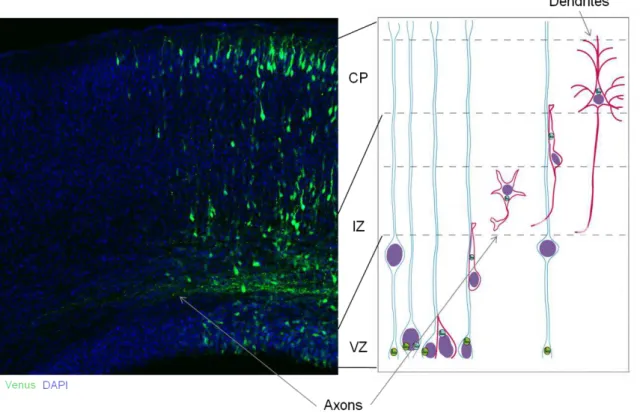
The first phenotypic change occurs at E9/E10 of the mouse telenchephalon. With the onset of neurogenesis at the telenchephalon germinal zones – the ventricular zone (VZ, also known as proliferative zone or germinal matrix, a pseudostratified columnar neuroepithelial region lining the lateral ventricles). At the VZ, neuroepithelial cells start to express glial markers, forming RGCs (Götz and Huttner, 2005; Kriegstein and Götz, 2003). RGCs are non-neuronal cells present in the developing brain which serve as molecular scaffolds for the migration of future neurons due to the particularity that these cells have their body in the VZ but extend processes to the pial (apical process) and ventricular surface (basal process), covering vertically, all length of the nascent cortex (for revision see Campbell and Götz, 2002). As neurons, RGCs also perform interkinetic nuclear migration (INM), whereas their nucleus travels within the VZ. RGCs are regarded as neural stem cells (Figure 1.1.2.2); they undergo self-renewal by symmetric division or give rise to neurons or secondary progenitors by asymmetric divisions (Noctor et al., 2001, 2004, 2008). Also, there is supporting evidence of an existing lineage relationship between neurons and the proliferative RGC, reporting that clonally related neurons migrate along radial glial fibers to form functional cortical columns, the radial organization of the neocortex (Noctor et al., 2001).
Figure 1.1.2.2. Modes of cell division in the mammalian neocortex.
Radial glial cells divide asymmetrically in the ventricular zone (VZ), and intermediate progenitor cells divide symmetrically in the subventricular zone (SVZ). A radial glial cell (green) divides asymmetrically at the ventricular surface to generate a daughter neuron (red). The radial glial cell remains mitotic to generate another daughter cell. Radial glial cells (green) also generate intermediate progenitor cells (blue) that divide symmetrically in the SVZ to generate two daughter cells, neurons (blue) that migrate toward the cortical plate (CP). Terminal symmetric divisions occur in the SVZ (from Noctor et al., 2004).
11
The distance a population of neurons migrates varies with neuronal type and brain region. In the human cerebral cortex, pyramidal neurons migrate radially about 2 cm, to those that travel tangentially the distance may be several fold increased (Tsai and Gleeson, 2005).
Figure 1.1.2.3. The model of radial glial migration in the mammalian cortex.
Microscopical picture of the mouse neocortex evidanciating the different cortical layers (left). Scheme depicting a neuron (red) undergoing the migration along a radial glial fiber (blue): initial radial migration at the VZ, SVZ arrest, establishment of neuronal polarity, second phase of migration until final destination and maturation of the neuron (right; credits of picture to Froylan Calderon de Anda).
12
followed by a sequential wave of migrating neurons that will occupy a more superficial layer (Berry and Rogers, 1965; Rakic, 1972; Sidman and Rakic, 1973; SIDMAN et al., 1959). Cortical layers are populated by arising neurons and each layer is constituted by a specific subset of approximately three quarters of pyramidal neurons (glutamatergic excitatory projection neurons) and the remaining are interneurons (GABAergic inhibitory neurons) (Jones, 1986). Deep layer and subplate neurons (L5-L6) project mainly to other areas of the nervous system, as spinal cord, thalamus and striatum, and other subcortical structures. More superficial neurons (L2-L4) are cortical projection neurons. Projection neurons are responsible for connections within the cortex: interhemispheric or contralaterally, mostly trough the corpus callosum, and intrahemispheric connections (Berry and Rogers, 1965; Rakic, 1972; Sidman and Rakic, 1973; SIDMAN et al., 1959).
During migration, neurons establish interactions between other migrating neurons and the surfaces of neighboring cells. Such interactions play a pivotal role in the appropriate selection of migratory pathways, as well in orientation, navigation and conclusion of neuronal movement in their final destination, where neuronal maturation takes place (Hatten, 2002; Pearlman et al., 1998).
The migration of neurons progresses in a way where two consecutive events are repeated, those events underlie at first an extension and retraction of the leading neurite, oriented forward, together with the centrosome, followed by nuclear displacement (Bellion et al., 2005; Gregory et al., 1988; Rakic, 1972). This migratory fashion requires major alterations in the cellular cytoskeleton, both in actin and microtubules (MTs), and action of very specialized molecules. The leading process is composed of several organelles and usually terminates in several protrusions oriented toward the cortical plate that by sensing the extracellular environment will determine the migratory pathway. Alteration to this dynamics usually is impairment for migration in cortical development studies.
The precise regulation of neuronal migration is responsible for cortical lamination and critical for proper development of the brain architecture. Therefore, its deregulation has a huge impact in the whole development of the neocortex causing a diversity of disorders (Dobyns and Truwit, 1995). The establishment of neuronal polarity with axon development and growth is crucial for the migration of neurons. Also, it is well accepted that cytoskeleton dynamics enables the movement of the growth cone, determining its direction, and regulating axon elongation during migration (Lowery and Van Vactor, 2009).
Neuronal polarity: axon and dendrites
13
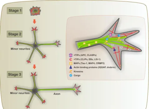
cell body and propagates down to the axon to the presynaptic terminal, and is transferred to other neurons. Hence, polarization is an indispensable event during neuronal development and a pre-requisite for the integration and transmission of information within the brain (Neukirchen and Bradke, 2011). Axon formation is the key event for the initiation of neuronal polarization. Banker and colleagues developed a culture system, of embryonic rat hippocampal neurons, for the observation in living CNS neurons. With those, they could observe the morphological events taking place during differentiation from a neuroblast to a polarized cell (Dotti et al., 1988). This in vitro model is a powerful tool to uncover the developmental mechanism of neurons and most of the knowledge comes from it, in vivo studies are then necessary for the validation of the proposed mechanism.
Figure 1.1.2.4. Schematic depiction of a developing hippocampal neuron during neuronal polarization.
14
The role of localized instability of the actin network in specifying axonal fate was examined by Bradke and Dotti in 1999 (Bradke and Dotti, 1999) with the use of rat hippocampal neurons in culture. They proved that local instability of actin, restricted to a single growth cone, present in one of the neurites, is a physiological signal specifying neuronal polarization. Disruption of the actin network in the growth cone induces axon elongation. From the observation of cultured hippocampal neurons, different developmental stages were identified during polarization (Figure 1.1.2.4.): born as an unpolar and symmetric sphere, neurons end up mature when achieving a highly polarized structure.
Defining the mechanism that regulate growth, guidance and branching of axons will tell us how the cerebral cortex becomes wired to appropriate targets during development. In the mammalian nervous system, axons establish connections through the activity of the growth cone and by extending branches towards a cell target from the collateral primary axon shaft branches (Kalil et al., 2000). Once the connectivity is incorrect or damaged; the brain’s ability to
work normally is severely incapacitated.
Axon Elongation
The growth cone responds to attractive cues by turning toward the source of a positive guidance cue, in contrary, inhibitory guidance cues repel axons away (Huber et al., 2003). It is now clear that the response to attraction versus repulsion is not due to the properties of a specific cue, but due to the intrinsic properties of the growth cone – the receptors engaged in the growth cone, and its signaling milieu. The growth cone comprises the necessary elements to determine how environmental direction lead to a given guidance response during axon
“navigation” (Chilton, 2006). Neuronal growth cones adhere to extracellular substrates: cell adhesion molecules (CAMs) or extracellular matrices, in order to properly migrate towards their final destination. Microtubules play an important role, as they are responsible for the connectivity between the growth cone and the extracellular substrates, possibly acting as guidance sensors, i.e. introducing an adhesive molecule leads to an increase in exploratory MTs (Lee and Suter, 2008).
15
T-zone, and generates actin arcs (Medeiros et al., 2006; Schaefer et al., 2008). Actin arcs orient perpendicular to the axis of filopodia and hinder the invasion of microtubules into the P-domain. So, while actin functions as a steric hindrance for microtubules, microtubules push the actin arcs cytoskeleton towards the growth direction, creating an area free of actin, enabling microtubules engorgement and delivery of proteins (Forscher and Smith, 1988). Finally, the actin cytoskeleton depolymerizes at the neck of the growth cone and microtubules bundle, enabling the proximal part of the growth cone to assume a cylindrical shape, becoming part of the axonal shaft (Bradke and Dotti, 1999; Lowery and Van Vactor, 2009; Neukirchen and Bradke, 2011).
Interstitial branching mechanism occurs from the axon shaft after the growth cone has extended past the target. Such mechanism are extremely important, those possibilitate the wiring of the cortical neurons to distinct targets, e.g. spinal cord (efferent corticospinal) and contralateral cortex (callosal axons) (Bastmeyer and O’Leary, 1996; Halloran and Kalil, 1994; O’Leary et al.,
1990).
Dendritic arborization mechanims
Dendrites are the neuronal processes specialized in the input of information, differing morphologically and functionally from the axons (Craig and Banker, 1994). The main excitatory synaptic sites, spines, are only present in dendrites. Different types of neurons meet different requirements accordingly with their physiological function. They are so readily classified based on their dendritic field dimensions and dendrite branching patterns (Ram n y a al, 1995).
16
dynamic microtubule invasion of spines and NMDA (N-methyl-D-aspartarte) receptor mediates signaling that affects dendrite organization in the somatosensory cortex during development and in the mature nervous system.
The development of dendritic branch diversity though guidance and targeting, happens in a cell-type specific way, where the first level of control arises from intrinsic programs that are linked to cell lineage and identity controlling how dendrites respond to attractive and repulsive cues in their environment (Kelsch et al., 2007; Komiyama and Luo, 2007). Local interactions between dendrites help to define and refine dendritic target boundaries. Later dendritic branching is thought to be associated with neuronal activity. As primary dendrites form first, side branches emerge later from the shaft of the primary dendrite, first as transient slender projections, or filopodia – highly dynamic actin structures. Those transient projections, if stabilized, will become a secondary dendritic branch. Stabilization occurs at the formation of a synapse, so, postsynaptic signaling may influence such process, at the same time that a dendritic spine is stabilized, likely, due to the involvement of calcium signaling (Konur and Ghosh, 2005). Side branching formation requires dynamic actin for the formation and remodeling of filipodia, followed by actin stabilization as the secondary branching pattern develops. RhoGTPases are the best studied molecules that provide regulation of actin dynamics in neuronal development (Etienne-Manneville and Hall, 2002). In vitro studies have shown that Rho inhibits dendrite growth and branching, while Rac1 play as inducer (Govek et al., 2005). Cdc42, another RhoGTPase involved in branching and cell polarity, is thought to be activated more in some layers of the cortex than in others, revealing a layer-specific environmental signaling, which is thought to interact with other players to determine the dendritic arborization at a specific cortical layer (Rosário et al., 2012; Simó and Cooper, 2012).
Recent genetic studies revealed a complex network of intrinsic and extrinsic regulators, transcription factors and ligands for cell surface receptors, respectively, that are needed to produce the distinct morphology of the four classes of larval dendritic arborization neurons in Drosophila melanogaster peripheral nervous system (revised in Corty et al., 2009).
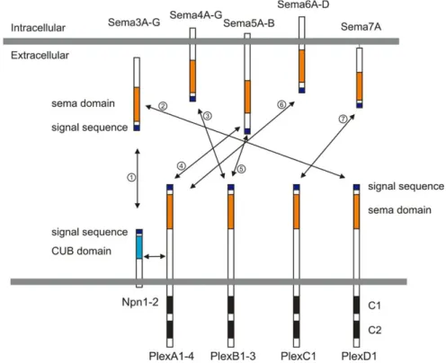
Semaphorin3A signaling in cortical development
First characterized as repulsive guidance cues (Kolodkin et al., 1992), semaphorins (Semas) are their largest family (20 members), comprising both secreted and membrane-bound proteins. In vertebrates, the major receptors of semaphorins are plexins (Plex) and neuropilins (Nrps), and all are subdivided in subfamilies (Figure 1.1.2.5).
17
Figure 1.1.2.5. Semaphorins and their receptors, plexins and neuropilins, present in vertebrates.
Semaphorins are secreted (Sema3A-G), glycosylphosphatidylinositol (GPI)-anchored (Sema7), or transmembrane (Sema4A-G, Sema5A-B and Sema6A-D) family members. Neuropilins consist of two transmembrane molecules (Nrp1–2), and plexins consist of transmembrane A (1–4), B (1–3), C1, and D1 family members. Most class 3 semaphorins require an obligate neuropilin co-receptor. Sema3E binds to PlexD1 without neuropilins. Class 4 and 5 semaphorins interact with plexinBs. Class 6 semaphorins interact with plexinAs. Sema7A interacts with PlexC1. CUB indicates complement binding. One to seven indicate the interactions between semaphorins and their receptors; 1: class 3 semaphorins and neuropilin1/2, 2: Sema3E and PlexD1, 3: Sema4D and PlexB1/B2 or Sema4C/4G and PlexB1, 4: Sema5A/5B and PlexA1/A3, 5: Sema5A and PlexB3, 6: Sema6A/6B and PlexA2/A3 or Sema6C/6D and PlexA1, 7: Sema7A and PlexC1. Other semathorins classes (e.g. Class 8, viral semaphorin) are not here respresented, because they are absent in vertebrate (from Yoshida, 2012).
18
Most class 3 semaphorins (Sema3) bind to Nrps. Plexin A family members associate to Nrps to transduce Sema3 signals across the neuronal membrane (Takahashi et al., 1999; Tamagnone et al., 1999) Sema3A and Nrp1 are known to play an important role in motor axon fasciculation and branching during embryogenesis (Behar et al., 1996; Gu et al., 2003; Kitsukawa et al., 1997; Taniguchi et al., 1997). Huber and colegueas have found that Sema3A regulates the timing motor axons ingrow to the limb (Huber et al., 2005).
Extensive studies to unravel the role of Sema3A, in the mouse neocortex, showed that Sema3A signaling is important for the developmental of axon and dendrites during development. Polleux and colleagues show that Sema3A repels axons but attracts dendrites in cortical neurons, so Sema3A promotes dendrite formation (Polleux et al., 2000). Quite recently, those findings were further proved by several studies (Nishiyama et al., 2011; Shelly et al., 2011). Sema3A signaling results in inhibition of PKA activity, and consequently to decreased activity of axon-promoting kinases. However, Sema3A knockout mice do not display defects in neuronal polarity, suggesting that alternative mechanism may also regulate axon and dendrite specification.
The TAOK2 protein
Thousand-and-one-aminoacids kinase 2 (TAOK2), a close relative of TAOK1, was first described the group of Melanie H. Cobb (Chen et al., 1999), and simultaneously by Moore and colleagues (Moore et al., 2000) under the name of PSK (Prostate-derived STE20-like kinase 1), as a member of the mitogen-activated protein kinase kinase kinase (MAP3K) family of signaling modules, ubiquitous to eukaryotic cell regulation.
The human Taok2 gene is encoded on the 16p11.2 chromosome, a genomic region associated with developmental and psychiatric disorders, where TAOK2 misregulation has been associated with Autism Spectrum Disorders (Battaglia et al., 2009; Tabet et al., 2012; Weiss et al., 2008), Depression (Degenhardt et al., 2012), Fragile X syndrome (Darnell et al., 2011), Schizophrenia (McCarthy et al., 2009) and Alzheimer Disease (Tavares et al., 2013). TAOK2 is subjected to alternative splicing to produce the TAOK2α (140KD) and TAOK2β (120KD) isoforms (Yasuda et al., 2007).
TAOK2 has a serine/threonine kinase domain present at the N-terminus, a long C-terminal domain composed of 690 residues, possibly containing a nucleotide binding site, a serine, proline and leucine-rich region, and an unbroken stretch of 17 glutamic acid residues (Chen et al., 1999). STE20-like kinases usually contain a small G protein binding consensus motif, not found at TAOK2 (Burbelo et al., 1995).
19
for the full-length protein, where a truncated version TAOK2α (1-349), constituted of kinase domain only, shows a diffusive cytoplasmic localization, showing the relevance of the unknown C-terminal domain to the regulatory pathway and function of TAOK2α in the cells.
TAOK2 was later shown to activate other signaling pathways, e.g. p38 and ERK family members (Chen et al., 1999, 2003), however those pathways are activated by the smaller isoform of TAOK2 – TAOK2β. TAOK2β endogenous activity is activated by the muscarinic agonist carbachol.
Several other studies reveal the involvement of TAOK2α in regulating cytoskeleton properties during stress-like conditions and demonstrate that TAOK2α colocalizes with microtubules (MTs) for their stabilization in a kinase independent fashion, producing stabilized perinuclear MTs with increased levels of acetylated α-tubulin (Chen and Cobb, 2001; Mitsopoulos et al., 2003). So it is possible that TAOK2α is responsible for the stability of actin, playing a major role in the dynamics of microtubules.
TAOK2α is sensitive to cleavage, removing the C-terminal domain (MTs binding domain), enabling the N-terminal catalytic region to relocate from the cytoplasm to the nucleus for the up regulation of apoptotic morphology (Moore et al., 2000; Zihni et al., 2007).
20
1.1.3. Material and Methods
shRNA and fluorescent proteins.
The Taok2 shRNA sequences used in this study are:
Taok2 shRNA 1 = CGAGAGGACTTGAATAAGAAA.
Taok2 shRNA 2 = GCATCCTAATACCATTCAGTA.
Taok2 shRNA 3 = GTTCCAGGAGACGTGTAAGATCC.
Taok2 shRNA 1 and 2 were primarily throughout the study. The Taok2 shRNA 1-resistant construct (pCMV human TAOK2) is from imaGenes (Berlin, Germany), Clone: IRATp970E03140D. The sequence for the Nrp1 shRNA is: AGAGAAGCCAACCATTATA (Chen et al., 2008). The shRNAs sequences used in the experiments were inserted into a pSilencer vector. A pSilencer vector containing a random sequence hairpin insert was used as a control for the shRNAs. The Venus (pCAGIG) and mCherry (pCAGIG) plasmids were kindly provided by Dr. Z. Xie (Boston University). The F-GFP (pCAGIG-GAP 43-GFP) construct was a gift from Dr. A. Gartner (University of Leuven, Belgium). The Myc-TAOK2 (pCMV rat TaoK2) was kindly provided by Dr. M. H. Cobb (University of Texas Southwestern Medical Center). The Nrp1-mCherry (plasmid 21934) and the MKK7-JNK1 (Plasmid 19726) are from Addgene (Cambridge, MA).
Lentiviral production.
Production of plentilox3.7 Taok2 shRNA
Taok2 shRNA was cloned into the plentilox 3.7 vector (Addgene plasmid 11795) as previously described (Mao et al., 2009). Briefly, complimentary 5’ phosphorylated oligonucleotides
21
collected 48 and 96 hours later. Viral supernatant was filtered through a 0.45 μm cellular acetate vacuum filter (Corning 431155), and concentrated by ultracentrifugation at 25,000 x g for 90 minutes. Viral pellets were resuspended in DPBS+0.1% glucose and stored at - 80 °C. Viral titers were determined on HEK293T cells plated at 2x10⁵ cells / well in 6-well plates, and serial dilutions of 1:200, 1:2000, and 1:20,000 were used to determine viral titer. After 48 hours of viral supernatant application, percentage of infected cells were determined by determining the percentage of fluorescent cells divided by the total number of cells by visual inspection. Four fields of view were counted per well, and three wells were inspected per dilution. Infection was performed with MOI =10.
Antibodies.
The following antibodies were used in these studies: mouse anti-acetylated tubulin (Sigma, immunocytochemistry, 1:1000), goat anti-TAOK2 (K-16, Santa Cruz Biotechnology, western blot (WB) 1:2000; immunocytochemistry, 1:100), rabbit anti-pTAOK2 (Ser 181; sc-135712; Santa Cruz Biotechnology, western blot (WB) 1:250; immunocytochemistry, 1:100), rabbit anti-pJNK1/2 (Promega, immunocytochemistry, 1:200) mouse anti-anti-pJNK1/2 (Promega, immunocytochemistry, 1:200), mouse anti-pan-JNK (BD Transduction Laboratories; WB, 1:500); goat anti-rat Neuropilin-1 (R&D Systems, WB, 1:500) rabbit anti-FAK (clone C-20, Santa Cruz Biotechnology, WB, 1:500); anti-mCherry (Clontech Cat no: 632543, WB, 1:1000); anti-RFP (ABcam, ab62341, WB, 1:400); anti-actin (WB, 1:2000) and anti-GAPDH (WB, 1:500). Nucleic acid were visualized with Hoechst (Invitrogen) and F-actin with phalloidin (Molecular Probes). Alexa conjugated secondary antibodies (Jackson Immunoresearch, 1:1000) were applied for 1-2 hr at 1-25ºC.
Cell transfection and western blot analysis.
HEK293T and Ht22 cells (ATCC) were grown under standard cell culture conditions and
transfected with plasmids using Lipofectamine 2000 according to the manufacturer’s protocol
22
then washed for 30 minutes in TBS28 T. Immunoreactivity signals were detected by enhanced chemiluminescence (Perkin Elmer).
Immunoprecipitation.
HEK293T cell lysates: For transient transfection, HEK293T cells were cotransfected with equal amounts of overexpression plasmids carrying myc-Taok2 and mCherry-Nrp1 cDNA. The total amount of transfected DNA was between 3.5 – 4.0 μg / 35 mm plate. The cells were allowed to express the constructs for 24 h before lysis and analysis. Transfected cells were washed once with ice-cold 1X PBS and immediately lysed in 1X lysis buffer with protease inhibitors. The Bio-Rad assay kit was used to determine protein concentration. For TAOK2 and Nrp1 immunoprecipitation assays, lysates were incubated with protein A sepharose conjugated to anti-mCherry antibodies overnight at 4 °C. Lysates containing 0.5 mg of protein were used for each condition. The beads were then washed with RIPA buffer twice to remove nonspecific proteins, and then washed 5 times with 1X lysis buffer before boiling in Laemmli sample buffer. Following SDS-PAGE to separate the proteins, blots were incubated with anti-TAOK2 antibodies.
Cortical brain lysates: cortices from P0 Swiss Webster mice were dissected and homogenized in 350 μl of sterile-filtered 50 mM Tris-Cl pH 7.4, 120 mM NaCl, 0.5% NP-40 containing proteinase inhibitors (Roche) using a 26G-syringe, followed by a 15 min centrifugation at 14000 rpm at 4 °C and the collection of the supernatant. Following a 60 minutes incubation with 1-2 μg of the corresponding antibodies, 20 μl of protein G Sepharose (GE Healthcare) was added to the lysates and incubated for 45 min at 4 °C. The bound immune complexes were then collected at 8000 rpm for 3 min followed by one wash each in sterile-filtered 50 mM Tris-Cl, pH 7.4, 500 mM NaCl, 1% NP-40, and sterile-filtered 50 mM Tris-Cl pH 7.4, 120mM NaCl, 0.5% NP-40. Samples were boiled for 5 min at 95 °C, run on a 10% SDS gel and analyzed with the same primary antibodies used for the immunoprecipitation.
Cortical cultures.
23
Neurons were cultured for 2 DIV and 7 DIV after plating and then fixed as described (see immunofluorescence). pTAO2, pJNK, tubulin, and MAP2 were visualized by indirect immunofluorescence. The mean intensity gray value of a line drew along the neurites was measured using ImageJ.
Sholl analysis.
All GFP-positive image stacks from transfected cortical neurons were taken as described (see Confocal imaging). All Sholl analyses use cortices that displayed relatively low transfection efficiencies in order to be able to select and analyze isolated transfected neurons in the cortex. Sholl analysis was performed by drawing concentric circles centered on the cell soma using Adobe Illustrator CS3. The starting radius was 15 μm and the ending radius was 55-100 μm; the interval between consecutive radii was 5 μm. All analyses were performed blindly.
In utero electroporation.
The Institutional Animal Care and Use Committee of Massahusetts Institute of Technology approved all experiments. Pregnant Swiss Webster mice were anesthetized by intraperitoneal
in ection of Ketamine 1% / Xilazine 2 mg/mL (0,01 μl/g body weight), the uterine horns were
exposed, and plasmids mixed with Fast Green (Sigma) were microinjected into the lateral ventricles of embryos. The shRNA plasmid concentration was 2 to 3-fold higher than that of mCherry, Venus, or F-GFP. Five current pulses (50 ms pulse / 950 ms interval; 35-36V) were delivered across the head of the embryo.
Immunofluorescence.
24
Confocal imaging.
Images were taken with a Zeiss LSM 510 confocal microscope. Z-series images were collected with 1 μm steps. To perform 3D reconstructions on stacks of images of transfected cells, only Z sections in the same focal plane as GFP were used for analysis and for producing figures. 3D reconstructions and Z-stack analyses were produced using ImageJ software. The adjustment of brightness and contrast was performed on images.
Quantitative phalloidin-fluorescence determination in growth cones.
Neurons were cultured for 2DIV after plating and then fixed as described above (see immunofluorescence). F-actin was visualized by the binding of fluorescentlylabeled phalloidin. The mean intensity gray value of phalloidin in the growth cone area was measured using ImageJ.
Organotypic slice cultures.
Mouse embryos were electroporated at embryonic day 15 (E15), and acute coronal brain slices (240 μm) were prepared at E17 and E18 (at which time TAOK2 was downregulated via shRNA). Slices were transferred onto slice culture inserts (Millicell) in cell culture dishes (35 × 10 mm; Corning) with Neurobasal medium (Invitrogen) containing the following: B27 (1%), glutamine (1%), penicillin/streptomycin (1%), horse serum (5%), and N2 (1%). Slices were used for imaging (1–2 h after slicing) or for pharmacological treatments (incubated at 37°C in 5% CO2, for 1–2 d).
Time-lapse imaging.
GFP- and RFP-positive cells were imaged on an inverted Nikon microscope (TE 2000-S) with a 20× objective lens [numerical aperture (NA) 0.45]. During the time-lapse imaging, slices were kept in an acrylic chamber at 37°C in 5% CO2. We captured time-lapse images with a CoolSNAP EZ camera (Roper Scientific) using NIS-Elements software (Nikon).
Statistical analysis.
25
To examine the subcellular expression profile of TAOK2, we analyzed TAOK2 immunoreactivity in cultured cortical mouse neurons dissociated at embryonic day 17 (E17).
a b c
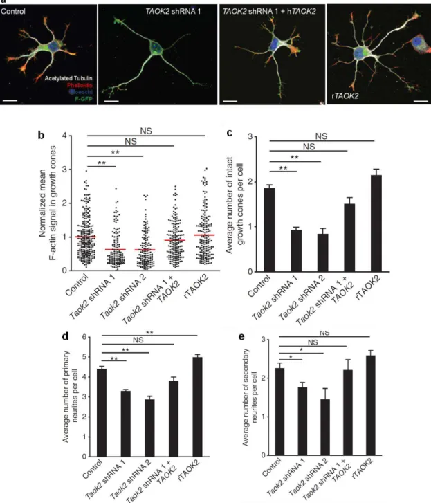
Figure 1.1.4.1. Distribution of TAOK2 and activated TAOK2 (pTAOK2) in cultured neurons.
(a) TAOK2 localizes to the growth cones (white arrowheads) of isolated cortical neurons. (b) TAOK2 (red) co-localizes with actin (green) in growth cones. (c) Activated TAOK2 (pTAOK2; green) localizes to the
neurite shaft of isolated cortical neurons. Scale bar: 10 μm (a), 200 μm(c).
We found that TAOK2 preferentially localized to growth cones (Figure 1.1.4.1a, b). The growth cone is a region where actin, but not microtubules, accumulates (Figure 1.1.4.1b) and where the actin cytoskeleton is the most dynamic (Bradke and Dotti, 1999). In contrast, TAOK2 activated by phosphorylation on Ser 181 (pTAOK2) localizes to the neurite shaft, where microtubules also accumulate (Figure 1.1.4.1c). This pattern of TAOK2 expression suggests that TAOK2 may act as a coordinator of actin and microtubule dynamics (King et al., 2011).
26
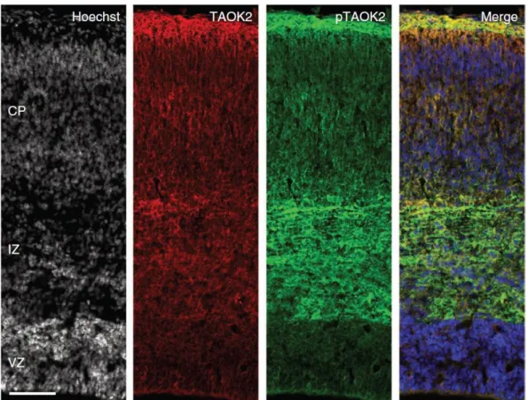
Figure 1.1.4.2. Distribution of TAOK2 and activated TAOK2 in the developing cerebral cortex.
TAOK2 and pTAOK2 are preferentially expressed in the IZ and CP of the developing cortex (E18). Scale bar: 200 μm.
Western blot analysis using whole-cell extracts from the cortices of mice at different embryonic and postnatal ages demonstrates that the long isoform of TAOK2 (TAOK2α; 140 KD) is expressed throughout the early cortical embryonic development. However, in perinatal (E19, P0) and adult mice, the TAOK2α isoform was considerably increased in expression. In contrast, the short isoform of TAOK2 (TAOK2β; 120KD) was only observed perinatally and in the adult brain (Figure 1.1.4.3).
Figure 1.1.4.3. Distribution of TAOK2 in the early cortical embryonic mouse brain.
27
TAOK2 impacts neuronal differentiation in cultured cortical neurons.
The remodeling of the actin-based cytoskeleton is an important regulatory step in axon and dendrite formation (Arimura and Kaibuchi, 2007; Barnes et al., 2008; Witte and Bradke, 2008). Since it has been shown that TAOK2 modulates the organization of the actin cytoskeleton in non-neuronal cells (Chen et al., 1999), and we find that TAOK2 expression is concentrated in actin-rich structures, it was asked whether TAOK2 loss-of- and gain-of-function affects neuronal differentiation. To examine the role of TAOK2 in brain development, three specific short-hairpin (sh)RNAs were designed, targeting different coding sequences of TAOK2, to acutely knock down the expression of TAOK2. The specificity of our shRNA constructs with respect to their ability to down-regulate endogenous neuronal TAOK2 was confirmed by immunohistochemistry (Figure 1.1.4.4a-d, and data not shown for shRNA 3). To this end, isolated cortical neurons at E17 from embryos were transfected by in utero electroporation at E15 with constructs expressing Taok2 shRNA- or control shRNA and membrane-bound GFP (F-GFP). Neurons were cultured for 48 hr before being processed for immunocytochemistry using antibodies against TAOK2 and acetylated tubulin (Figure 1.1.4.4a-d). Additionally, the specificity of our shRNA constructs was assessed for their ability to down-regulate endogenous TAOK2 by western blot analysis in Ht22 cells (Figure 1.1.4.4e, f, and data not shown for shRNA 3). These experiments show that Taok2 shRNAs efficiently down-regulate TAOK2 expression. shRNAs 1 and 2 were used for all subsequent experiments.
28
Figure 1.1.4.4. Taok2 shRNA’s specifically down-regulate TAOK2 expression in
dissociated cortical neurons and Ht22 cells.
29
Figure 1.1.4.5. TAOK2 down-regulation or overexpression affects the differentiation of isolated cortical neurons.
30
In addition, TAOK2 down-regulation decreased the number of neurites per neuron (Figure 1.1.4.5a, d; control: n=168 cells from three different cultures; Taok2 shRNA 1: n=149 cells from three different cultures; Taok2 shRNA 2: n=36 cells from two different cultures; P<0.0001 by one-way ANOVA and posthoc Dunnett test **P <0.01) and the number of secondary branches per cell, compared with control transfected neurons (Figure 1.1.4.5a, e; P=0.0102 by one-way ANOVA and posthoc Dunnett test *P <0.05).
TAOK2 autophosphorylation is known to play a role in TAOK2 activation (Chen et al., 1999; Zihni et al., 2007), thus the overexpression of TAOK2 increases levels of phosphorylated, active TAOK2 (Figure 1.1.4.4g, h). TAOK2 overexpression in cultured cortical neurons increased the number of primary, but not secondary, neuritis compared with control neurons (Figure 1.1.4.5a, d; rTAOK2: n=119 cells from three different cultures; P<0.0001 by one-way ANOVA and posthoc Dunnett test **P <0.01).
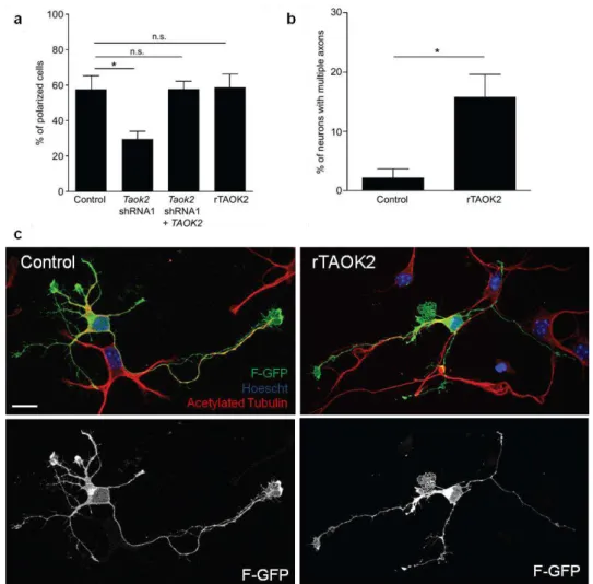
In addition, it was analyzed whether TAOK2 down-regulation or overexpression affects polarization in cultured neurons. TAOK2 downregulation impaired axon formation (Figure 1.1.4.6a; control: 57.57 ± 7.68% of neurons with a neurite longer than 40 μm, n=168 cells from three different cultures; Taok2 shRNA 1: 29.69 ± 4.3% of neurons with a neurite longer than 40
31
Figure 1.1.4.6. TAOK2 down-regulation or overexpression affects axon elongation of isolated cortical neurons.
(a) TAOK2 down-regulation decreases the number of neurons that elongate an axon (control: 57.57 ± 7.68% of neurons with a neurite longer than 40 μm, n=168 cells from three different cultures; Taok2 shRNA 1: 29.69 ± 4.3% of neurons with a neurite longer than 40 μm, n=149 cells from three different cultures; Taok2 shRNA 1 + TAOK2: n=48 cells from three different cultures; P=0.0044 by one-way ANOVA and posthoc Dunnett test *P <0.05). (b, c) rTAOK2 overexpression increases the number of neurons with multiple axons (control: 2.22 ± 1.42% of neurons with multiple neurites longer than 40 μm, n=168 cells from three different cultures; rTAOK2: 15.84 ± 3.79 % of neurons with multiple neurites longer than 40 μm, n=119 cells from three different cultures; *P=0.0386 by t test). Mean ± s.e.m. Scale bar: 10 μm.
32
TAOK2 affects basal dendrite formation and axon elongation in vivo.
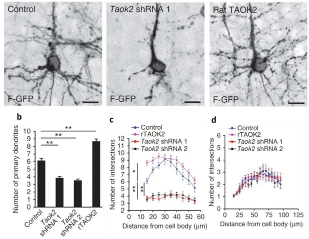
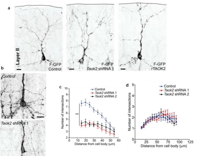
The previous results (Figure 1.1.4.5 and Figure 1.1.4.6) demonstrate the impact of TAOK2 upon the differentiation of cultured primary cortical neurons. We then sought to determine the effect of TAOK2 in the post-migratory differentiation of cortical neurons in vivo. E15 mouse embryos were in utero electroporated with Taok2 shRNA, control shRNA, or rTAOK2 (rat TAOK2 cDNA) plasmids together with F-GFP. Mice were sacrificed at postnatal day 7 (P7), and the dendritic morphology of control, Taok2 shRNA, and rTAOK2-expressing neurons was evaluated. It was found that both the knockdown and overexpression of TAOK2 disrupted cortical neuronal differentiation in vivo. In layer II-III of the in utero electroporated brains, the Taok2 shRNA-transfected neurons had significantly fewer primary dendrites compared to controls (Figure 1.1.4.7a, b; control: n=19 cells from three brains; Taok2 shRNA 1: n=23 cells from three brains; Taok2 shRNA 2: n=20 cells from two brains; P<0.0001 by one-way ANOVA and posthoc Dunnett test **P <0.01). TAOK2 overexpression, on the other hand, increased the number of primary dendrites (Figure 1.1.4.7a, b; rTAOK2: n=31 cell from three brains).
33
Figure 1.1.4.7. TAOK2 down-regulation or overexpression affects basal dendrite arborization in layer V neurons in the developing cortex.
(a) TAOK2 knockdown or up-regulation have opposite effects in basal dendrite development in vivo. (b) The number of primary dendrites decreases following TAOK2 knockdown and increases following rTAOK2 overexpression. (c) Sholl analysis of the dendritic arbor from upper cortical layer transfected neurons reveals significantly fewer and more dendritic process intersections at a distance between 15 μm and 55 μm from the cell soma in the TAOK2-silenced neurons or rTAOK2-overexpressed neurons, respectively, compared with control transfected neurons. (d) Sholl analysis of the apical dendrite does not show any differences between Taok2 shRNA-mediated down-regulation, rTAOK2 overexpression, and control conditions. Scale bar: 10 μm (a). *P < 0.05, **P < 0.01.
34
Figure 1.1.4.8. TAOK2 down-regulation or overexpression affects basal dendrite arborization in layer II-III neurons in the developing cortex.
35
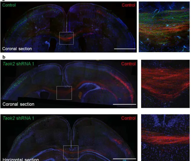
Figure 1.1.4.9. TAOK2 down-regulation affects callosal axon projection in the developing cortex.
(a, b) Taok2 shRNA-mediated down-regulation diminishes the number of callosal axons traversing the midline. (a) Control transfected neurons in both hemispheres (Venus: left hemisphere; mCherry: right hemisphere) project callosal axons that crossed the midline. Right panel: inset of the corpus callosum. (b) Taok2 shRNA-mediated knock-down in Venus-positive neurons prevents axons from these cells from crossing the midline, However, control transfected neurons positive for mCherry, from the contralateral hemisphere, project axons that cross the midline. Upper panel: coronal brain section. Lower panel: horizontal brain section. Right panels: inset of the corpus callosum from the coronal (upper panel) and horizontal section (lower panel). Scale bar: 500 μm (a, b).
36
Figure 1.1.4.10. TAOK2 overexpression affects axonal projection at the corpus callosum in the developing cortex.
rTAOK2 overexpression produces minor defects in axons crossing the corpus callosum. Some transfected axons deviate from the axonal tract (arrowheads). Mean ± s.e.m. Scale bar: 100 μm.
TAOK2 interacts with the Neuropilin 1 receptor to modulate neuronal differentiation.
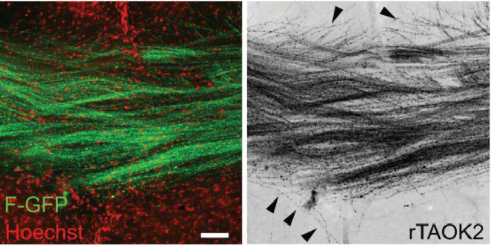
Previous studies have shown that the Sema3A – Nrp1/PlexinA4 signaling cascade controls basal dendritic arborization (Chen et al., 2008; Gu et al., 2003; Tran et al., 2009). Nrp1Sema- mice also develop axonal projection defects in the corpus callosum and the hippocampus (Gu et al., 2003). These defects vary from mild, in which some axons deviate from the axonal tract, to more severe phenotypes, where callosal axons defasciculate and do not cross the midline (Gu et al., 2003). We hypothesized that TAOK2 may collaborate with this pathway to modulate neuronal differentiation. To test this idea, coimmunoprecipitation experiments were performed to probe for an interaction between TAOK2 and Nrp1. TAOK2 and Nrp1-mCherry were co-expressed in HEK293 cells and it was found that TAOK2 interacts with Nrp1-mCherry (Figure 1.1.4.11a).
Figure 1.1.4.11. TAOK2 interacts with Nrp1.
37
we examined whether active TAOK2 (pTAOK2) co-localizes with Nrp1. In the mouse E19 cerebral wall, we found that pTAOK2 and Nrp1 co-localized preferentially in the IZ and lower CP, where axons elongate and deeper layer neurons begin to form dendrites (Romand et al., 2011) (Figure 1.1.4.12a, b).
Figure 1.1.4.12. TAOK2 interacts with Nrp1 at the intermediate zone and cortical plate in the developing cortex.
(a) Nrp1 and pTAOK2 are preferentially expressed in the IZ and CP of the developing cortex. (b) Inset
from (a), white box: Nrp1 and pTAOK2 co-localize in the developing cortex. Scale bar: 500 µm (a), 10 μm