Setembro de 2012
Universidade do Minho
Escola de Engenharia
Paula Alexandra da Silva Jorge
Preparation of a Standardized Methodology
for the Growth of Pseudomonas aeruginosa
Biofilms and Evaluation of their Response to
Antimicrobial Peptide Strategies
UMinho|20
12
P
aula Alexandra da Silva Jor
ge Preparation of a St andardized Met hodology for t he Growt h of Pseudomonas aeruginosa
Biofilms and Evaluation of t
heir R
esponse to Antimicrobial P
ep
Dissertação de Mestrado
Mestrado em Bioengenharia
Trabalho realizado sob a orientação da
Professora Maria Olívia Pereira
e da
Professora Anália Lourenço
Setembro de 2012
Universidade do Minho
Escola de Engenharia
Paula Alexandra da Silva Jorge
Preparation of a Standardized Methodology
for the Growth of Pseudomonas aeruginosa
Biofilms and Evaluation of their Response to
Antimicrobial Peptide Strategies
É AUTORIZADA A REPRODUÇÃO PARCIAL DESTA DISSERTAÇÃO APENAS PARA EFEITOS DE INVESTIGAÇÃO, MEDIANTE DECLARAÇÃO ESCRITA DO INTERESSADO, QUE A TAL SE COMPROMETE;
Universidade do Minho, ___/___/______
iii
Acknowledgments
I would like to acknowledge here the persons who supported me during my dissertation.
My first appreciation goes to my family and friends, which support and encourage me every day and without whom I could not have reached this stage of my life and academic journey.
Very special thanks to João for his help in every aspect of my life and specifically during this dissertation year with his advices and his encouragement and support in the stressful days.
To my two supervisors, Professor Olívia and Professor Anália, I give my great appreciation for their knowledge and support enwrapped in very good disposition. It has been a pleasure to work with them and hopefully we will carry it on in the future.
Finally, to my lab colleagues who fill the working days with laughs and friendship and make LMA1 a great place to work in.
The financial support from IBB-CEB and FCT and European Community fund FEDER, through Program COMPETE (FCT PTDC/SAU-SAP/113196/2009/ FCOMP-01-0124-FEDER-016012) is also gratefully acknowledged.
v
Abstract
Preparation of a Standardized Methodology for the Growth of Pseudomonas aeruginosa Biofilms and Evaluation of their Response to Antimicrobial Peptide Strategies
Novel antimicrobial products, such as antimicrobial peptides (AMPs), are being investigated to combat clinically relevant biofilms and their growing antimicrobial resistance. These relevant studies, however, lack protocol standardization. Acknowledging the importance of standardized methodologies to ensure the quality of experimental results, this dissertation initiates the establishment of a standardized operational procedure (SOP) for the growth of P.
aeruginosa biofilms. The proposed SOP was evaluated statistically, firstly in
terms of repeatability and ruggedness. The method showed a repeatability greatly influenced by between experimental variance. The variability of the live organism at study or other uncontrollable or yet unknown variable could be the cause. The ruggedness of the protocol showed that slight variations to some of the SOP conditions cause differences in the results, meaning that these variables are important in the biofilm growth and must be tightly controlled.
Furthermore, this thesis evaluated the P. aeruginosa biofilms’ response to prophylactic and therapeutic control approaches using the AMPs colistin, tachyplesin III and lactoferricin B, alone or combined. The analysis conjugates information from biomass, respiratory activity and cell viability measurements. Results show that the prophylactic approach revealed that combinations of colistin and tachyplesin III with lactoferricin B are capable of inhibiting biofilm formation at low concentrations. In the preliminary therapeutic tests, although the results were not as good, the colistin and lactoferricin B combination was able to eradicate most of the biofilm for the strain P. aeruginosa ATCC 10145.
vii
Resumo
Preparação de uma Metodologia Padronizada para o Crescimento de Biofilmes de Pseudomonas aeruginosa e Avaliação da sua Resposta a Estratégias de Controlo com Péptidos Antimicrobianos
Novos compostos antimicrobianos, tais como os péptidos antimicrobianos (AMPs), estão a ser investigados para combater biofilmes de relevância clínica e a sua crescente resistência a antimicrobianos tradicionais; no entanto, estes estudos carecem de protocolos padronizados. Reconhecendo a sua importância para garantir a qualidade dos resultados experimentais, esta dissertação inicia o estabelecimento de um procedimento operacional padronizado (SOP) para o crescimento de biofilmes de P. aeruginosa. O SOP proposto foi avaliado estatisticamente em termos de repetibilidade e robustez. O método mostrou uma repetibilidade muito influenciada pela variância entre experiências. A variabilidade do organismo vivo estudado ou outra variável incontrolável ou desconhecida pode ser a causa. Por sua vez, a robustez do protocolo mostrou que pequenas variações em algumas condições do SOP causam diferentes resultados, o que significa que estas variáveis são importantes no crescimento do biofilme e devem ser bem controladas.
Esta dissertação também avaliou a resposta dos biofilmes de P.
aeruginosa a abordagens profiláticas e terapêuticas usando os AMPs colistina,
taquiplesina III e lactoferricina B, isolada ou combinadamente. A análise conjugou informação da biomassa, da atividade respiratória e da viabilidade celular. Os resultados mostram que na abordagem profilática, as combinações de colistina e taquiplesina III com lactoferricina B são capazes de inibir a formação de biofilme em concentrações baixas. Nos ensaios preliminares terapêuticos, ainda que os resultados não sejam tão bons, a combinação de colistina e lactoferricina B foi capaz de erradicar a maior parte do biofilme para a estirpe P.
ix
Index
Acknowledgments ... iii Abstract ... v Resumo... vii Index ... ix Table Index ... xiFigure Index ... xiii
List of Abbreviations ... xv
1 Introduction ... 17
1.1 Context and Motivation ... 17
1.2 Objectives ... 19
1.3 Manuscript Structure ... 20
2 Biofilms ... 23
2.1 What are Biofilms? ... 23
2.2 Biofilm Related Infections ... 25
2.3 Biofilm Resistance ... 26
3 Antimicrobial Peptides ... 29
3.1 Sources of Antimicrobial Peptides ... 29
3.2 Action Mechanisms of Antimicrobial Peptides ... 30
3.3 Anti-Biofilm Antimicrobial Peptide Strategies ... 33
3.4 Biofilm Resistance to Antimicrobial Peptides ... 37
4 Standard Operating Procedures... 39
x
4.2 Repeatability ... 42
4.3 Ruggedness ... 43
4.4 Reproducibility ... 44
5 Materials and Methods ... 47
5.1 Microorganisms ... 47
5.2 Antimicrobial Peptides ... 50
5.3 Phenotypic Analysis of Biofilms ... 53
5.4 General Steps of the Statistical Analysis ... 55
5.5 Establishment of a Standardized Operating Procedure ... 59
5.6 Control of Biofilms with AMP Combinations ... 63
6 Results and Discussion ... 65
6.1 Repeatability of the SOP ... 65
6.2 Ruggedness of the SOP ... 69
6.3 Control of Biofilms with AMP Combinations ... 77
7 Conclusions and Future Work ... 89
7.1 Establishment of a Standardized Operating Procedure ... 90
7.2 Control of Biofilms with AMP Combinations ... 91
Bibliography ... 95
Annex I – Publications ... 105
Annex II – Antimicrobial Peptides ... 107
xi
Table Index
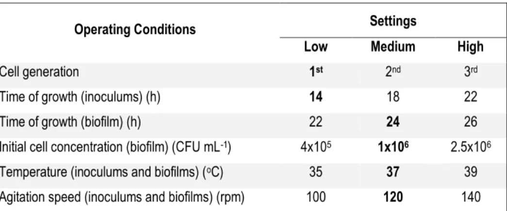
Table 5-1: The three settings for each of the six operating conditions studied in
the ruggedness test ... 61
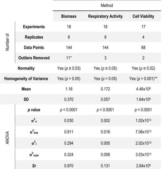
Table 6-1: Statistical evaluation of the repeatability of the proposed SOP ... 66
Table A: AMP sources and classes ... 107
Table B: Recent applications of AMPs on the control of biofilms ... 113
Table C: Results of the multiple comparisons Tukey’s test for the biomass dataset of the repeatability testing ... 117
Table D: Results of the multiple comparisons Tukey’s test for the respiratory activity dataset of the repeatability testing... 117
Table E: Results of the multiple comparisons Tukey’s test for the cell viability dataset of the repeatability testing ... 118
xiii
Figure Index
Figure 2-1: Life cycle of a bacterial biofilm ... 24 Figure 3-1: General statistics on the number of publications on AMPs and AMP
applications to biofilms in PubMed ... 31
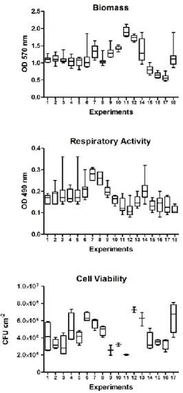
Figure 6-1: Data dispersion of the datasets for repeatability for the three
methods of analysis ... 68
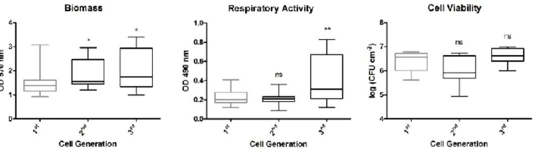
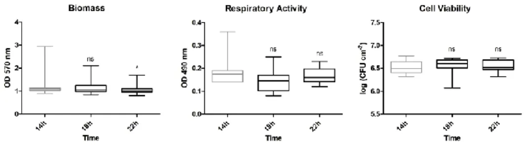
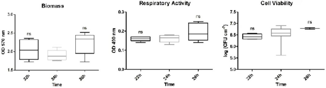
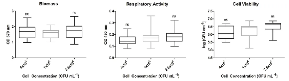
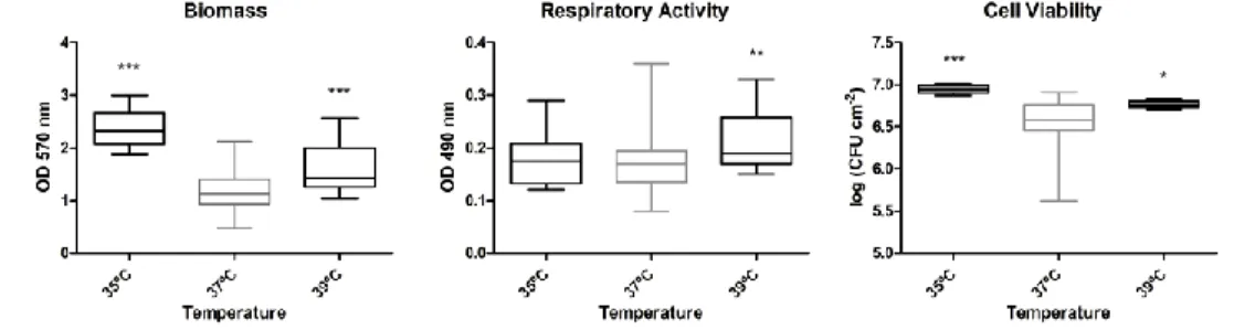
Figure 6-2: Influence of the cell generation on the biofilm phenotype ... 70 Figure 6-3: Influence of the time of inoculum growth on the biofilm phenotype 71 Figure 6-4: Influence of the time of biofilm growth on the biofilm phenotype .... 72 Figure 6-5: Influence of the initial cell concentration on the biofilm phenotype . 73 Figure 6-6: Influence of the temperature of inoculum growth on the biofilm
phenotype ... 73
Figure 6-7: Influence of the temperature of biofilm growth on the biofilm
phenotype ... 74
Figure 6-8: Influence of the rotation speed in the inoculum growth on the biofilm
phenotype ... 75
Figure 6-9: Influence of the rotation speed in the biofilm growth on the biofilm
phenotype ... 76
Figure 6-10: Inhibition of P. aeruginosa ATCC 10145, P. aeruginosa PAO 1 and P.
aeruginosa CGCT III biofilm formation with the AMPs colistin and lactoferricin B,
xiv
Figure 6-11: Inhibition of P. aeruginosa ATCC 10145, P. aeruginosa PAO 1 and P.
aeruginosa CGCT III biofilm formation with the AMPs tachyplesin III and
lactoferricin B, alone and combined. ... 80
Figure 6-12: Treatment of P. aeruginosa ATCC 10145, P. aeruginosa PAO 1 and P.
aeruginosa CGCT III biofilms with the AMPs colistin and lactoferricin B, alone and
combined, for 30 min and 2h ... 83
Figure 6-13: Treatment of P. aeruginosa ATCC 10145 , P. aeruginosa PAO 1 and P.
aeruginosa CGCT III biofilms for 2h with the AMPs colistin and lactoferricin B,
alone and combined ... 85
Figure 6-14: Treatment of P. aeruginosa ATCC 10145, P. aeruginosa PAO 1 and P.
aeruginosa CGCT III biofilms for 2h with the AMPs tachyplesin III and lactoferricin
xv
List of Abbreviations
AMP – antimicrobial peptide ANOVA – analysis of variance CBR – CDC biofilm reactor
CDC – centres for disease control and prevention CF – cystic fibrosis
CFTR – cystic fibrosis transmembrane regulator CV – crystal violet
CVC – central venous catheter DMMB – dimethyl methylene blue
EC50 – half maximal effective concentration e(DNA) – extracellular DNA
EPS – extracellular polymeric substance ESD – extreme studentized deviate FDA – fluorescein diacetate
GoF – goodness of fit
GraRS – glycopeptide resistance-associated two-component system h – hour
IC50 – fifty percent growth inhibitory concentration K-S – Kolmogorov-Smirnov
LPS – lipopolysaccharide
MBC – minimum bactericidal concentration
MBEC – minimum biofilm eradication concentration MDR – multidrug-resistant
xvi
MIC – minimum inhibition concentration min - minute
mPE – meta-phenylene ethynylene
MSSA – methicillin-sensitive Staphylococcus aureus MSSE – methicillin-sensitive Staphylococcus epidermidis MRSA – methicillin-resistant Staphylococcus aureus MRSE – methicillin-resistant Staphylococcus epidermidis NNISS – national nosocomial infection surveillance system OD – optical density
OM – otitis media
PMS – N-methyl dibenzopyrazine methyl sulphate QS – quorum sensing
QSI – quorum-sensing inhibitor ROS – reactive oxygen specie sad – surface attachment defective
SAMP – synthetic antimicrobial peptidomimetic SD – standard deviation
SOP – standard operating procedure
Sr – repeatability standard deviation
STAMP – specifically/selectively targeted antimicrobial peptide UTI – urinary tract infection
XTT –
17
1 Introduction
1.1 Context and Motivation
Biofilm control is a prominent research area as microbial adhesion onto surfaces and the subsequent formation of biofilms are critical concerns for many biomedical applications. It is well known that biofilm-forming bacteria account for about 80% of human bacterial infections [Fey, 2010]. In particular, biofilms cause most of the nosocomial infections and are resistant to traditional treatment with antimicrobials. The resistance towards antimicrobials is, in part, due to the biofilm matrix that acts as a protective shield against external pressures as human defences and antimicrobial agents [Hall-Stoodley et
al., 2009]. Particularly, Pseudomonas aeruginosa is one of the most
common microorganisms found in nosocomial scenarios, being highly related to infections in immune compromised patients due to its biofilm forming capabilities, and for which desirable treatment is still needed [Rodrigues, 2011].
Biofilm infections, along with the development of microbial drug resistance and drug-related toxicity, have encouraged the search of new alternatives to control these healthcare-associated infections. Natural compounds have emerged as an interesting approach to limit the emergence and the spread of resistant microorganisms [Spížek et al., 2010]. The main challenge is to find new natural prophylactic and therapeutic compounds with novel antimicrobial targets and
18 mechanisms of action. Because of this, antimicrobial peptides (AMPs) have gained widespread interest as good replacements for current antibiotics, particularly considering the fact that these compounds are less likely to induce the development of acquired resistance, due to their mechanism of action with low specificity, and that they have a broad spectrum of activity and a wide availability of sources [Zasloff, 2002; Beckloff et al., 2007; Splith et al., 2011; Wimley et al., 2011]. Recent studies have been reporting the successful control of biofilms based on AMP strategies. These positive outcomes seem to be linked to the AMPs’ dual capacity to act both on the cytoplasmic membrane and on intracellular targets, once entered the microbial cell. Moreover, interesting is that some studies have reported AMP activity against biofilms at lower concentrations than those required for planktonic cell killing [Lynch et al., 2008; Jorge et al., 2012]. The combination of innovative and/or conventional compounds may improve the prophylactic and therapeutic efficacy and lower drug dosage, reducing toxic side effects [Ncube et al., 2008; Wei et al., 2011]. Therefore, synergistic studies between AMPs are also a viable route for exploring the anti-biofilm efficacy of these peptides.
Current protocols for biofilm laboratory research lack standardization, and therefore validity, in some ways. The inexistence of standard protocols hampers the quality of the information generated by researchers and makes the reproduction of results in different laboratories difficult. Many times, studies target the same scenario but results are not comparable because they are performed using different methods of analysis, which return diverse biological data and lead to various conclusions [Jackson et al., 2001]. Standard operating procedures (SOPs), attending to ruggedness, repeatability and
19
1.2 Objectives
This dissertation tests the repeatability and ruggedness of a method to growth biofilms of P. aeruginosa ATCC 10145 in 96 well microtiter plates in order to initiate the establishment of a SOP. Additionally, this thesis evaluates the potential anti-biofilm capabilities of a group of AMPs, namely colistin, lactoferricin B and tachyplesin III, used alone and in combination, in biofilms of P. aeruginosa ATCC 10145,
P. aeruginosa PAO1 and P. aeruginosa CGCT III.
The preliminary developments of a SOP for the growth of biofilms of P. aeruginosa ATCC 10145 are presented since the lack of a standard protocol in this area is impairing the comparison and output of results. This evaluation addressed the most commons variables in the laboratory growth of biofilms, such as temperature, speed of agitation, time of growth, cell generation and initial cell concentration, to investigate if slight variations affected the resulted biofilm. This will make possible the establishment of the ruggedness of the protocol. All experiments with SOP conditions were repeated at least 17 times with at least 4 replicates, making possible to access the protocol’s
repeatability.
The AMP anti-biofilm evaluation accounts for both prophylactic and therapeutic approaches. First, the evaluation focused on AMP efficacy in inhibiting biofilm growth. Then, the evaluation centred on the capacity of AMPs to eradicate mature biofilms, which are very recalcitrant to treatment, due to the exopolymeric protective matrix, presence of tolerant (persister) cells, among other factors.
20
1.3 Manuscript Structure
This dissertation is divided into seven main chapters, excluding bibliography and annexes.
Chapter 1 – Introduction
This chapter exposes the context and motivation for the completion of this master’s thesis. Also, the objectives of the thesis are detailed and the manuscript’s structure is also described.
Chapter 2 – Biofilms
In this chapter, the concept of biofilms is explained by stating its definition and main characteristics. Furthermore, highlight is given to the biofilm role in infections and to its resistance mechanisms to antimicrobial treatment.
Chapter 3 – Antimicrobial Peptides
This chapter reviews the different sources and families of AMPs and their mechanisms of action. A detailed revision of the latest studies of biofilm control with AMPs is given and the biofilm resistance mechanisms towards AMPs are specified.
Chapter 4 – Standard Operating Procedures
The importance of the establishment of standardized protocols is highlighted in this chapter, along with a revision on the existing SOPs for biofilm studies. The main characteristics of a SOP are also detailed.
21
Chapter 5 – Materials and Methods
In this chapter, the materials to be used are described in detail, focusing on the microorganism to be studied, P. aeruginosa, and the AMPs to be used, colistin, tachyplesin III and lactoferricin B. Also, the methods used in the laboratory work for the growth, antimicrobial testing and analysis of the biofilms are explained. The steps for the statistical analysis of the data are detailed along with the tests used for the protocol standardization of the growth of P. aeruginosa biofilm.
Chapter 6 – Results and Discussion
This section presents the results derived from the tests for the establishment of the standard protocol for the growth of P. aeruginosa biofilm and for the control of these biofilms with the AMPs colistin, tachyplesin III and lactoferricin B, alone and combined.
Chapter 7 – Conclusions and Future Work
Finally, the main conclusions, retrieved from the results and discussion section, are summarized and some follow up work is pointed out.
23
2 Biofilms
2.1 What are Biofilms?
For a long time, the study of microorganisms was closed off to the study of planktonic, liquid cultures. However, nowadays it is evident that the activity of microorganisms in Nature is mainly associated with surfaces and, thus, research has been shifting its attention to the study of the sessile, adhered mode of life [Rodrigues, 2011]. These organized, structured consortia of microorganisms are known as biofilms, and they are viewed as a form of adaptation that allows bacteria and fungi (one or more species combined) to survive in hostile environments and colonize other areas by cell dispersion [Hall-Stoodley et al., 2009].
Biofilms influence a diverse range of disciplines, ranging from biotechnological and environmental industries to medical applications, and they can have both a positive and a negative effect on modern society. For example, bacterial biofilms can be positively applied to wastewater treatment processes as flocs or granules, yeast aggregates are capable of improving brewing processes, and other biofilms have been applied in bioremediation and fuel technology. However, biofilms also have damaging effects in industry and public health. Bacterial communities are involved in detrimental processes such as biofouling and biocorrosion and biofilm-related infections are a serious concern in modern medicine, representing 65% of the total hospital-acquired infections [Beech et al., 2005; Todar, 2008-2012].
24 Biofilm formation undergoes essentially four typical stages (Figure 2–1): i) adherence of planktonic cells to the tissue or abiotic surface; ii) accumulation of cells and production of the extracellular polymeric substance (EPS) matrix, which is constituted by proteins, polysaccharides and extracellular DNA (eDNA) resulting from autolysis, and whose presence will ensure structural stability and protection to the biofilm; iii) biofilm maturation with development of towers and water channels and specialized zones; and iv) dispersion of cells and/or parts of the biofilm with subsequent colonization of other locations [Costerton et al., 1999; Donlan et al., 2002; Fey, 2010]. Eventually, bacteria can use type IV pili to move through another bacteria’s biofilm and colonize it [Høiby et al., 2010].
Figure 2-1: Life cycle of a bacterial biofilm. (a) – Adherence; (b) –
Accumulation and production of extracellular matrix; (c) – Maturation; (d) – Dispersion [Fey, 2010].
The architecture of a mature biofilm can have many configurations that range from flat homogenous layers of cells to highly organized cell clusters, e.g. a mushroom shaped structure containing water-filled channels [Wimpenny et al., 2000]. A mature biofilm results from complex spatial and temporal differentiation of cells in response to environmental signals and cell to cell communications [Bridier et al., 2011]. These dynamic three-dimensional structures maintain a tight
25 organization through cell signalling. This allows biofilm cells to respond to environmental signals, sense cell density and perform quorum sensing (QS) [Hall-Stoodley et al., 2009]. For example, bacteria in biofilms sense when critical concentration of cells is reached and respond by producing virulence factors, like enzymes or toxins [Høiby et
al., 2010].
2.2 Biofilm Related Infections
The aging of worldwide population has increased the number of surgeries, joint replacements and immunosuppressive therapies and, implicitly, the incidence of biofilm-related infections. That is, biofilm infections are more and more related to morbidity and mortality and have become the main cause of emergence and dissemination of antibiotic resistance in the nosocomial scenery [Spížek et al., 2010].
Bacterial biofilms are the cause of among 80% of all bacterial infections, the most common being biomaterial-related infections [Harro et al., 2010]. Biomaterial nosocomial infections are majorly found on central venous and urinary catheters, prosthetic heart valves, orthopaedic devices, cardiac pacemakers, vascular, voice and ocular prostheses, cerebrospinal fluid shunts, contact lenses, among others [Stewart et al., 2001; Fey, 2010; Rodrigues, 2011]. For example, currently about 12% to 25% of mortality in hospitalized patients is due to catheter-related bloodstream infections [Estrela et al., 2010].
In non-surgical devices, such as catheters, the colonization of the surface may be originated by the migration of the microorganisms of the skin in the point of insertion and throughout the catheter. In the case of surgical devices, such as orthopaedic replacements, the
26 adhesion of the bacteria to the surface will compete with the integration of the material with the surrounding tissue. This last step must be concluded before the 6 h decisive period ends, which is indicated has the time in which the material is most susceptible to colonization [Rodrigues, 2011].
Biofilms are also associated to non biomaterial infections, such as chronic wounds, endocarditis [Stewart et al., 2001; Fey, 2010], periodontitis, chronic urinary tract infections (UTI), recurrent tonsillitis, chronic rhinosinusitis, chronic otitis media (OM) and cystic fibrosis (CF) pneumonia [Hall-Stoodley et al., 2009]. Many of the biofilm producing organisms are opportunistic pathogens, like P. aeruginosa or
Staphylococcus epidermidis [Stewart et al., 2001; Rodrigues, 2011], and
the biofilm-associated infections are usually coupled with a chronic condition [Hall-Stoodley et al., 2009]. Microorganisms involved in biofilm human infections are revised by Lynch and Robertson [2008].
2.3 Biofilm Resistance
The administration of antibiotics is the common treatment of infections. However, the widespread, and sometimes unnecessary, use of the antibiotics has led to the selection of multi-drug resistant (MDR) pathogens [Spížek et al., 2010]. Additionally, current antibiotics have been classically developed for treatment of planktonic bacterial populations in acute infection scenarios, being usually ineffective in biofilm (persistent) related infections [Lynch et al., 2010]. Compared to planktonic bacteria, the minimum inhibition concentration (MIC) can be hundreds or thousands of times higher and the resistance to the innate and adaptive immune system is also higher. For example, Escherichia
27 eradicated compared with the same strain in the planktonic state [Estrela et al., 2010].
To make matters worse, biofilms’ antibiotic resistance does not seem totally linked to the usual resistance mechanisms, and even the most susceptible planktonic bacteria can become a threat when grown in biofilm. The frequency of mutation in biofilms is higher than in planktonic bacteria and there is an increased horizontal gene transmission, which also explains the fast development of the biofilms’ antibiotic resistance. However, bacteria detached from biofilms can become rapidly susceptible to antibiotics, suggesting that sometimes no mutations or other genetic modifications are required [Stewart et al., 2001].
Although antibiotic penetration is still feasible, the biofilm matrix can delay it till the expression of resistance-related genes takes place. Also, polymers in the matrix may bind to antibiotics, hindering their action, and antibiotic-degrading enzymes may deactivate them. If the antibiotic is still able to cross the matrix and reach the cells, some biofilms will express efflux pumps in the presence of the antibiotic, preventing its intracellular action [Hall-Stoodley et al., 2009].
Indeed, the three-dimensional structure of a biofilm plays, along with the EPS matrix, a major role in biofilms’ resistance to antimicrobials [Bridier et al., 2011]. Researchers have observed that the concentration of oxygen is higher at the surface and lower in the centre of the biofilm, and that the protein synthesis and metabolic activity is higher at the surface and lower or absent in the centre of the biofilm. Due to these oxygen and nutrient gradients throughout the biofilms, nutrient depleted zones can appear and bacteria can enter in a stationary phase-like dormancy and not be affected by antibiotics. Finally, oxidative
28 stress, caused by an imbalance between the formation of reactive oxygen species (ROS) and the antioxidant system, increases mutability in biofilms and promote antibiotic resistance [Hall-Stoodley et al., 2009; Høiby et al., 2010].
Recent studies have detected that biofilms present resistance to some new alternative treatments as well, as phage resistance and low susceptibility to antibodies [Hall-Stoodley et al., 2009], and resistance to QS inhibitors (QSIs) [Høiby et al., 2010]. Overall, the conclusion is that new compounds with novel mechanisms of action are desired to treat biofilms more effectively, i.e. avoiding their natural predisposal to antimicrobial resistance. Most of the antimicrobial products that are being developed are derivatives of already known compounds and target the same resistance mechanisms, so their action can only be somewhat better. Now, the attention is drifting to the deployment of new antimicrobial discovery strategies. For example, the discovery of non-traditional sources of antimicrobials, microbial genome sequencing focused in antibiotic gene expression, metagenomics, and the re-examination of old compounds and investigation of new targets in pathogenic bacteria [Spížek et al., 2010].
Within this scope, natural products stand out because they have a much higher hit rate in high-throughput screens than the combinational libraries of traditional antimicrobials. Moreover, natural products are usually much more complex than synthetic products and present scaffolds with viable and biological validated starting points to design chemical libraries [Spížek et al., 2010]. One group of natural antimicrobial products that have been showing promising results is AMPs, which are discussed in the next section.
29
3 Antimicrobial Peptides
3.1 Sources of Antimicrobial Peptides
AMPs are short-length peptide antibiotics (between 15 and 30 amino acids), whose majority are cationic, amphipathic, gene-encoded and directed to the cell membrane [Rossi et al., 2008; Melo et al., 2009; Chau, 2010; Splith et al., 2011]. AMPs are usually co-expressed in groups that act together [Lai et al., 2009], but despite their similarities, AMP sequences vary greatly. Usually, AMPs are classified as α helical, β sheeted, extended and looped [Melo et al., 2009] or ribosomally and non-ribosomally synthesized [Rossi et al., 2008].
AMPs come from a variety of sources and in many forms. Their widespread distribution throughout the animal and plant kingdoms suggests that AMPs play a fundamental role in the evolution of complex multicellular organisms. Despite their ancient lineage, AMPs have remained effective defensive weapons [Zasloff, 2002]. Indeed, it has been proposed that AMPs and AMP-directed resistance mechanisms have co-evolved, leading to a host-pathogen balance that has shaped the existing AMP portfolio [Peschel et al., 2006].
AMPs are part of the innate immune system of animals and plants, but can also be found in microbes, like bacteria and fungi [Rossi
et al., 2008; Melo et al., 2009; Chau, 2010; Splith et al., 2011].
Mammalian AMPs are generally expressed and easily induced in epithelial surfaces to repel assault from bacteria, viruses, fungi and
30 parasites [Lai et al., 2009]. AMPs have also been found in glandular cells of amphibian skin, fishes and most classes of invertebrates. In plants, AMPs help in the adaptation to stressful environmental conditions: plant cells act as recognition sites against pathogen-derived metabolites (elicitors), leading to the accumulation of AMPs in the affected plant tissue [Kido et al., 2010]. Additionally, microbes, such as bacteria and fungi, produce AMPs as a defence mechanism and as a competitive advantage against other microorganisms, sometimes of the same species [Sang et al., 2008].
Synthetic AMPs, produced by de novo synthesis or by modification of existing AMPs, emerged as an alternative to reduce production costs [Wimley et al., 2011]. Recently, some reviews [Vooturi
et al., 2010; Giuliani et al., 2011] have reported the engineering of AMP
mimetics or peptidomimetics, non-peptide molecules which aim to retain and improve the basic features of AMPs [Giuliani et al., 2011].
A resume of the sources and classifications of the various AMPs is available in Table A of Annex I.
3.2 Action Mechanisms of Antimicrobial Peptides
AMPs have been recognized as promising candidates for replacing classical antibiotics due to their multiple mechanisms of action and low specificity in terms of molecular target, which reduces the chance of acquired resistance [Zasloff, 2002; Beckloff et al., 2007]. Moreover, compared with conventional antimicrobials, which are generally active only against bacteria or fungi, AMPs exert activity against a broad spectrum of microorganisms, such as both Gram-negative and Gram-positive bacteria, including drug-resistant strains,
31 parasites, enveloped viruses and even some cancer cells [Sang et al., 2008; Chau, 2010; Splith et al., 2011; Wimley et al., 2011]. AMPs are also cell specific and are able to distinguish host from non-host cells based on their charge [Beckloff et al., 2007]. Besides their antimicrobial action, AMPs can also influence processes in support of antimicrobial properties, like cytokine release, chemotaxis, antigen presentation, angiogenesis and wound healing[Lai et al., 2009].
Indeed, the number of published papers considering AMPs has risen considerably in the last years, including the studies of AMPs applied to biofilms (Figure 3–1).
Figure 3-1: General statistics on the number of publications on AMPs (black
bars) and AMP applications to biofilms (gray bars) in PubMed [Jorge et al., 2012].
Conventional antibiotics usually act by inhibition of cell wall, DNA, RNA and protein synthesis [Sang et al., 2008; Chau, 2010]. On the other hand, most AMPs permeabilize microbial membranes, inducing either a large-scale failure or small defects that dissipate the transmembrane potential, which results in cell death [Sang et al., 2008; Wimley et al., 2011]. This mechanism of action does not depend on the recognition of chiral targets and, therefore, all D-enantiomers are equally active, giving AMPs broad action spectrum [Podda et al., 2006].
32 AMP mechanisms of action are divided into pore and non-pore models [Wimley et al., 2011]. Pore models account for the formation of membrane-spanning pores, namely the barrel stave pore model [Rapaport et al., 1991] - in which AMPs interact to form a hydrophilic channel - and the toroidal pore model [Ludtke et al., 1996] - in which AMPs affect the curvature of the membrane. In turn, non-pore models comprise: the carpet model [Gazit et al., 1996], which is the most cited model and accounts for the parallel deposition of AMPs on the membrane, causing global bilayer destabilization due to a detergent-like effect; the detergent model [Ostolaza et al., 1993] that explains catastrophic collapse of the membrane using high concentrations of AMPs; the molecular shape models [Bechinger et al., 2006], in which AMP-lipid interactions can be portrayed with phase diagrams; the lipid
clustering model [Epand et al., 2009], in which AMPs induce lipid phase
separation; the sinking raft model [Pokorny et al., 2002], in which AMP activity is described in terms of binding, insertion and perturbation; and, the interfacial activity model [Rathinakumar et al., 2008], which is used to explain, predict and engineer the activity of AMPs. All aforementioned models imply the need to reach a certain threshold concentration of AMPs in the membrane prior to disruption [Melo et al., 2009].
Some AMPs act by alternative means, like binding to DNA, inhibiting cell wall, DNA, RNA and protein synthesis, autolysin and inhibiting enzyme activity [Sang et al., 2008]. The type of mechanism of AMPs can dictate their application fields. For example, it has been noticed that AMPs targeted to the membrane are better suited to be used in surface coating instead of AMPs that act at an intracellular level [Bagheri et al., 2012].More details about the mechanisms of action of AMPs are available in the reviews of Melo et al. [2009], Splith et al. [2011], Wimley et al. [2011], Park et al. [2011] and Nguyen et al. [2011].
33
3.3 Anti-Biofilm Antimicrobial Peptide Strategies
Biofilm control can be achieved in three ways: i) reduction of the planktonic population; ii) prevention of the initial adhesion of cells to the surface; and iii) removal of the established biofilm. Studies on biofilm-forming bacteria or yeasts in the planktonic state may open routes to the first strategy. However, in vivo application of the first strategy is quite complicated, given that the planktonic population in the body is widespread and identification of the presence of the planktonic biofilm-forming bacteria is difficult; so, microbial adhesion must be prevented as the next step.
Conceptually, the easiest method for preventing microbial attachment is by pre-treating the surfaces. This can be achieved by impregnating the surface with an antimicrobial agent or using functionalized coatings that allow a localized antimicrobial delivery [Zilberman et al., 2008; Shukla et al., 2010; Yala et al., 2011]. As reviewed by Glinel et al. [2012], several AMP-based coatings have been tested successfully.
When surface pre-treatments are not effective, biofilms may form. Then, strategies based on the administration of antimicrobials, to the infected live tissue (antibiotic treatments) or the non-living surface (disinfectant treatments), must be applied to kill the biofilm-growing microorganisms. A summary of the latest biofilm control studies using AMPs is depicted in Table B of Annex I.
The analysis of Table B showed that the AMPs tested on biofilms come from various natural sources, such as humans (AMP-IBP5; HBD3; LL-37; α-MSH), mammals (BMAP-28; cathelicidin WAM1), amphibians (aurein 2.5; magainin I; phylloseptin-1), fishes (chrysophsin-1;
34 pleurocidin), arthropods (tachyplesin III), bacteria (gramicidin A; lacticin 3147; nisin) and plants (Tn-AFP1). Non-natural AMPs are classified into mimetics (peptoid 1; peptoid 1-C134mer; (RW)4D) and synthetic (F2,5,12W; KSL; PTP-7; Tet213; SAMPs Ltx5, Ltx9 and Ltx10; omiganan pentahydrochloride; STAMPs C16G2, M8G2, C16-33, M8-33 and G10KHc). A substantial part of the tested AMPs is synthetic, which means that improving AMP optimal performance is nowadays becoming an important issue. Most of the microorganisms tested are bacteria probably due to their ubiquity in Nature and their frequent association with infectious diseases and biofilms.
It is also noteworthy that most biofilm-related studies, as seen in Table B, cover mainly biofilm growth in the presence of AMPs, i.e. prophylactic strategies meant to prevent biofilm formation, rather than testing AMPs against pre-established biofilms, i.e. therapeutic strategies meant to treat existing biofilms. This suggests that prevention of biofilm formation is possibly the current favourite research strategy in the combat of nosocomial infections. However, more work must be done in order to evaluate anti-biofilm efficacy of AMPs on mature biofilms.
One of the characteristics that seems to be linked to the anti-biofilm efficacy of some AMPs is their dual capacity to act both on the cytoplasmic membrane and on intracellular targets, once entered the cell. For example, it is thought that the synthetic AMP meta-phenylene ethynylene (mPE), designed based on magainin and active at nanomolar concentrations against Streptococcus mutans biofilms, acts both as an membrane-active molecule, inhibiting lipopolysaccharides (LPSs), similarly to magainin, and as an intracellular antibiotic by binding to DNA at equimolar ratios [Beckloff et al., 2007]. Another example is pleurocidin, which is thought to inhibit nucleic acid and protein synthesis without damaging E. coli cytoplasmic membrane at low
35 concentrations [Patrzykat et al., 2002], but it is able to cause membrane leakage and pore-like channels at higher concentrations [Mason et al., 2006].
Also, interesting is that some studies have reported AMP activity against biofilms at lower concentrations than those required for planktonic cell killing. This is the case of the synthetic AMP NA-CATH:ATRA1-ATRA1 and the natural AMP LL-37, both from the cathelicidin family, that are effective against Staphylococcus aureus and
P. aeruginosa biofilms, respectively. These AMPs are thought to act
internally on the bacteria, affecting gene expression essential for the development of biofilms [Overhage et al., 2008; Dean et al., 2011]. Actually, in P. aeruginosa, the AMP LL-37 alters the expression of biofilm related genes, such as type IV pili, rhamnolipid and Las QS systems, at sub-antimicrobial levels, and genes, associated with the assembly of flagella, involved in initial adherence during biofilm formation, were found to be down regulated [Overhage et al., 2008]. LL-37 is also capable of inhibiting initial biofilm attachment (58 - 62%), suggesting that peptides of this kind may be interacting with bacterial adhesins as part of their anti-biofilm mechanism [Dean et al., 2011]. Another study also showed that the AMP 1037 directly inhibits biofilms by reducing swimming and swarming motilities, stimulating twitching motility, and suppressing the expression of a variety of genes involved in biofilm formation in P. aeruginosa (e.g. PA2204) [de la Fuente-Núñez et al., 2012].
In fact, adhesion may be one of the great properties of anti-biofilm AMP abilities, which allows them to be used as an effective pre-treatment strategy. The AMP nisin, which is known to interfere with cell wall synthesis and form membrane pores [Peschel et al., 2006], retards biofilm formation without inhibiting S. aureus growth when immobilized
36 in multi-walled carbon nanotubes [Qi et al., 2011]. Another example is the cathelicidin-2 derived peptide, F2,5,12W, which suppresses S.
epidermidis biofilm formation at a concentration four times below the
MIC, which reflects decreased initial adhesion of the bacteria [Molhoek
et al., 2011].
Anti-attachment capabilities may be related to binding of DNA as well. DNA binding may facilitate the detachment or disruption of biofilm structures, since it has been reported that eDNA is involved in cell-cell attachment [Allesen-Holm et al., 2006]. This is the case for cationic AMPs and peptoids [Lobo et al., 2003; Otvos, 2005; Hale et al., 2007]. For example, the development of P. aeruginosa biofilms is disrupted by the enzyme DNase I [Whitchurch et al., 2002].
Matrix disruption in biofilms may also be a target for AMPs. It is thought that the peptoid 1-C134mer, which as a hydrophobic tail and is active against P. aeruginosa biofilms, interacts strongly with and disrupt the hydrophobic matrix due to its surfactant like nature, facilitating deeper penetration [Kapoor et al., 2011].
Additionally, AMPs have some organism-specific features. For example, lactoferrin inhibits biofilm formation in P. aeruginosa due to its iron-chelating properties, increasing surface motility and causing the bacteria to wander around the surface, forming thin and flat biofilms [Singh et al., 2002]. In turn, the inhibition of Porphyromonas gingivalis and Prevotella intermedia biofilms by lactoferrin is independent of the iron status of the protein. Lactoferrin may interact with the cell surface of these bacteria and interfere with their adherence [Wakabayashi et
al., 2009], since this peptide was already reported to interfere with the
binding of P. intermedia to subepithelial matrix proteins, as well as fibroblasts and epithelial cells [Alugupalli et al., 1994; 1995].
37
3.4 Biofilm Resistance to Antimicrobial Peptides
Although the development of resistance to AMPs is rare, some studies have reported this phenomenon. General mechanisms for microbial resistance to AMPs, which are valid both for planktonic and sessile states, include mutations that affect the structure and charge distribution of the cytoplasmatic membrane, modifications in the lipopolysaccharide structure of Gram-negative bacteria, and active pumping of the AMPs out of the cell [Altman et al., 2006]. Specifically, it has been reported that Gram-negative bacteria have evolved mechanisms to remodel the composition of the outer membrane through modification of the LPS molecules [Miller et al., 2005], which impairs LPS-binding AMPs.
Biofilm structure is another factor correlated with biofilm resistance to AMPs. For instance, the increased survival of E. coli biofilms when treated with colistin is not related directly with biofilm forming ability, but rather to the organization of the biofilm. Also, there is some evidence that biofilm formation in E. coli induces tolerance to AMPs due to changes in intra-biofilm physiochemical gradients [Folkesson et al., 2008].
AMP activity over intracellular targets is countered by genetic mutations. Interestingly, in S. aureus, the glycopeptide resistance-associated two-component system (GraRS), which is involved in up-regulation of biofilm production, was reported to mediate the resistance of the planktonic cells to AMPs [Herbert et al., 2007]. In CF, where P.
aeruginosa biofilms cause pneumonia, results show that colistin kills the
stalk subpopulation (deeper layer with low metabolic activity) preferentially, whereas the metabolically active cap-forming subpopulation in the upper layer becomes colistin resistant due to the
38 up-regulation of the pmr and mexAB-oprM genes [Haagensen et al., 2007; Pamp et al., 2008].
39
4 Standard Operating Procedures
Biofilms research has grown greatly in recent years, with an ever growing number of publications, but results and conclusions are sometimes contradicting. Results comparison is pivotal to validate individual experiments as well as consolidate research across laboratories. However, most of the current protocols for laboratory research lack standardization, and therefore validity, at some extent. Specifically, the inexistence of standard protocols hampers the quality of the information generated by researchers and makes the comparison of results produced in different laboratories difficult. Many times, studies focusing the same scenario are not comparable because they are performed using different methods of biofilm growth or analysis, which output very different biological data and may lead to various conclusions [Jackson et al., 2001]. In the case of biofilm-related studies,
e.g. methodologies for cell growth, antimicrobial susceptibility and final
biomass/cell activity vary from paper to paper, making it impossible to compare results conveniently.
To enable inter-laboratory evaluations and ensure results’ transparency, some validated protocols, the so-called SOPs, have been presented for biofilm growth, biofilm detection and quantification and for antimicrobial testing on biofilms
The SOPs proposed for laboratory biofilm growth are specific for: growth of mix biofilms of P. aeruginosa ATCC 700829, Pseudomonas
fluorescens ATCC 700830 and Klebsiella pneumoniae ATCC 700831,
40 a flat-plate, open channel reactor [Jackson et al., 2001]; growth of S.
epidermidis ATCC 35984 biofilms on polycarbonate coupons in the CDC
biofilm reactor (CBR) [McLeod et al., 2010]; and the growth of P.
aeruginosa biofilms using the CBR [Goeres et al., 2005; EPA/OPP, 2011].
Biofilm detection and quantification SOPs are available for: quantification of P. aeruginosa biofilm grown with high shear and continuous flow using CBR [ASTM, 2007b]; quantification of a P.
aeruginosa biofilm grown using a drip flow biofilm reactor with low
shear and continuous flow [ASTM, 2008]; colorimetric microtiter model for the detection of S. aureus biofilms [Toté et al., 2008]; quantification of microbial biofilms grown in microtiter plates (CV, Syto9, fluorescein diacetate (FDA), resazurin, XTT and dimethyl methylene blue (DMMB) assays) [Peeters et al., 2008]; harvesting and disaggregating steps [Hamilton et al., 2009]; optimized quantification of enterococci biofilms using microtiter-plates [Extremina et al., 2011]; and quantification of P.
aeruginosa biofilm grown with medium shear and continuous flow using
rotating disk reactor [ASTM, 2012].
Currently, no SOP is available for anti-biofilm AMP-based strategies. However, there are some works on general antimicrobial testing on biofilms: evaluating: resistance of S. aureus biofilms cells to disinfectants [Luppens et al., 2002]; biofilms susceptibility using a microplate alamar blue assay for S. epidermidis [Pettit et al., 2005]; and disinfectant efficacy on a laboratory hot tub model on planktonic bacteria and biofilms [Goeres et al., 2007].
41
4.1 Desired Characteristics for a SOP
A well-established protocol, like a SOP, has to respect several conditions that attest its validity, namely: reasonableness, relevancy,
validity, ruggedness, repeatability and reproducibility [Hamilton, 2010]:
i) A SOP is reasonable if it can be conducted within practical limitations, such as time, labour and material and if it is easily understandable and requires conventional and inexpensive laboratory material.
ii) In biological test methods, relevancy correlates with the capacity to emulate the real-world environment where the biological phenomenon in study occurs. Specifically, in Microbiology, test
relevancy has motivated a shift in testing, from planktonic
microbes to biofilms.
iii) A SOP is valid if the data results are unbiased, which means that the observed values equals (or approximates greatly) the true values. In Microbiology, however, the true values are usually unknown (e.g. log reductions, MICs, ODs, etc.).
iv) A SOP is repeatable if the results within the same experiment and between experiments have low variance.
v) Ruggedness of a SOP is encountered when results are not
affected by small deviations from the SOP conditions.
vi) Finally, a SOP is reproducible if similar results are obtained by different operators and in different locations/laboratories [Hamilton, 2010].
Next, a detailed explanation of repeatability, ruggedness and
reproducibility is given because they will be assessed in this dissertation
42
4.2 Repeatability
The term repeatability accounts for random errors of the measurements and includes the contributions from any part of the procedure that varies within a run, e.g. gravimetric and volumetric errors, heterogeneity of the test material and variation in the chemical treatment stages of the analysis [Thompson et al., 2002]. For the establishment of repeatability, the following conditions need to be satisfied: same measurement procedure; same observer; same measuring instrument, used under the same conditions; same laboratory; and procedure repetition over a short period of time [Taylor
et al., 1994].
Typically, errors in repeatability can be detected by inspecting replicate dispersions, but formal methods should be employed to assert these errors. Dispersion analysis can be done recurring to box plots. These plots show minimum to maximum whiskers, the bottom and top of the box are always the 25th and 75th percentile (lower and upper quartiles, respectively), and the band near the middle of the box is always the 50th percentile (median). In the case of formal methods, generally, the analysis of variance (ANOVA) is the selected method for quantifying repeatability [Engineered Software, 1999]. The ANOVA assesses the variance (σ2) within experiments and between experiments. The two variances given in the output table (designated mean square (MS)) are: between experiment/group variance (σ2btw),
which shows the differences between group means; and within group variance (σ2e), which shows the differences among data within the same
group. The first variance, σ2btw, can be explained by a systematic
variation due to treatment or by chance due to non-systematic individual differences in data or experimental error. The second variance, σ2e, can only be explained by the later explanation. The
43
repeatability standard deviation (Sr) was calculated as follows:
. Sr is interpreted as the difference between a single
experiment and the mean across many independent, identical experiments. Small Sr values indicate good repeatability. Sr can further be analysed in terms of the percentage of influence that each variance (σ2btw and σ2e) had. It is known that the between group variance is
estimated by , in which σ2t is the variance due to
treatment effect and whose value we can determine. Total variance (σ2total) is the sum of σ2e and σ2t. The percentage of the error variance
and treatment variance relatively to the total variance can be now easily calculated [NIST, 2012b].
4.3 Ruggedness
Ruggedness is the resistance to change in the results produced by
an analytical method when minor deviations are made from the experimental conditions described in the procedure [Thompson et al., 2002]. The aim is to identify the factors that strongly influence a method’s measurements and estimate how tightly those factors need to be controlled, i.e. to determine the degree of control required for the experimental procedures [ASTM, 2007a]. Ruggedness testing is usually done within a single laboratory and on uniform material, so that the effects of changing only the factors are measured. Deliberately, small changes are introduced into the procedure and the effect on the results is examined. The factors to be tested can be quantitative (continuous, like pH or temperature), qualitative (discrete, like the manufacturer or batch of a reagent) or “mixture” factors (e.g. the fraction of solvents in a mixture) [Heyden et al., 2001]. Examples of the factors that a
44 instrument(s)’s calibration, the operator(s); the brand of reagent(s) or the concentration of reagents; the pH of solutions; the temperature of a reaction; and, the time allowed for completion of a process [Thompson
et al., 2002].
The factors are examined in an experimental design, which is selected as a function of the number of factors to investigate. The most common designs applied are the fractional factorial [Voelkel, 2004] and the Plackett–Burman [Plackett et al., 1946] designs. Ruggedness can be quantified by ANOVA [Goeres et al., 2005] and multiple comparison testing to assess if the variations in the factors cause statistical significant differences in the results. The results can be easily analyzed through box ploting. The significance from the post-hoc multiple comparison can be given in the plots to help graphic analysis.
4.4 Reproducibility
Reproducibility relates to the ability of the procedure to be
reproduced by others, namely in different laboratories. In reproducibility testing, the results of a collaborative study between laboratories are usually summarized by a reproducibility standard deviation (SR), which
can be no smaller, and is usually significantly larger, than Sr [Goeres et
al., 2005]. SR includes between-laboratory and within-laboratory
variations.
Between-laboratory variation arises from factors such as variation in calibration standards, differences between local interpretations of the protocol, changes in equipment or reagent source, or environmental factors, such as differences in average climatic conditions. Collaborative trials directly estimate the variance of
45 between-laboratory biases [Thompson et al., 2002; Annis et al., 2005; Wallmann et al., 2006; Huys et al., 2010]. This is important to identify the causes of the differences among laboratories so that they may be controlled. Otherwise they will be summed into SR [AOAC, 2002].
47
5 Materials and Methods
5.1 Microorganisms
5.1.1 P. aeruginosa Relevance in Biofilm Infections
P. aeruginosa is a member of the Gamma Proteobacteria class of
Bacteria. It is a Gram-negative, aerobic rod belonging to the bacterial family Pseudomonadaceae. The genus Pseudomonas is cleaved into eight groups and P. aeruginosa is the type species of its group, which contains 12 other members. P. aeruginosa is a free-living bacterium, commonly found in soil and water. However, it occurs regularly on the surfaces of plants and occasionally on the surfaces of animals [Todar, 2008-2012].
This bacterium is one of the favourite model organisms to study biofilm formation. Several reasons justify the preference for P.
aeruginosa. First, there are many molecular tools available for this
organism, including DNA arrays for genome studies. Second, the genus
Pseudomonas has simple requirements for growth, tolerating a wide
range of temperatures (4 - 42oC), facilitating laboratory experiments. Third, it is considered a representative organism for a diverse and important group of bacteria [Kjelleberg et al., 2007]. Fourth, it is highly related to nosocomial infections. Actually, according to data from the US Centres for Disease control and Prevention (CDC) and the National Nosocomial Infection Surveillance System (NNISS), P. aeruginosa is the
48 second most common cause of nosocomial pneumonia, third most common cause of urinary tract infections and seventh most common cause of nosocomial bacteraemia [Aksoy et al., 2008].
Specifically, biofilms of this bacteria are commonly related to cystic fibrosis pneumonia, an infection that afflicts patients with the recessive genetic disease CF [Costerton et al., 1999]. The genetic defect in CF leads to the loss of the CF transmembrane regulator (CFTR) chloride channel in the apical membranes of epithelial cells [Cutting et
al., 2005]. This defect leads to persistent bacterial infections of the
lungs. Most CF patients are colonized with P. aeruginosa, and eventually they succumb to the lung damage inflicted by the persistent bacterial infection, with a median life expectancy of about 30 years [Costerton et
al., 1999]. P. aeruginosa biofilms also cause dermatitis, soft tissue
infections, bone and joint infections, contact lenses infections, gastrointestinal infections and a variety of systemic infections, particularly in patients with severe burns and in cancer and AIDS patients who are immune suppressed [Todar, 2008-2012].
P. aeruginosa is intrinsically resistant to many antimicrobial
agents, including most β-lactams, the older quinolones, chloramphenicol, tetracycline, macrolides, co-trimoxazole and rifampin. The most important anti-pseudomonal agents include some β-lactams (ticarcillin, ureidopenicillins, piperacillin, cefoperazone, ceftazidime, cefepime, aztreonam, imipenem, and meropenem), aminoglycosides (gentamicin, tobramycin, netilmicin, and amikacin), and fluoroquinolones (of which ciprofloxacin remains the most active compound). Polymyxins, such as the AMP colistin, are also active, but they are usually considered only for MDR strains due to their higher toxicity [Aksoy et al., 2008].
49 Also, pandrug resistant P. aeruginosa isolates, which are defined as resistant to carbapenems or to all antibiotics available for clinical use, are being reported with growing frequency. Most cases are seen in patients who were previously infected with P. aeruginosa and had been treated with long courses of multiple anti-pseudomonal antibiotics [Aksoy et al., 2008].
Several traits of P. aeruginosa have been related to its capability of biofilm formation. The study of two P. aeruginosa mutants defective in the initial steps of biofilm formation, called “sad” (surface attachment defective) mutants, revealed that these bacteria need flagella, type IV pili and, therefore, motility to adhere well to the plastic surface used at the time [O'Toole et al., 1998; Costerton et al., 1999]. Indeed, it seems that the attachment itself can initiate the synthesis of the extracellular matrix. Studies with P. aeruginosa algC, algD, and algU::lacZ reporter constructs showed that the transcription of these genes, required for synthesis of the extracellular polysaccharide alginate, is activated after attachment to a solid surface [Davies et al., 1995; Costerton et al., 1999].
5.1.2 Strains, Preservation and Culture
In this work, there were used three strains of P. aeruginosa, namely: P. aeruginosa ATCC 10145, P. aeruginosa PAO1 and P.
aeruginosa CGCT III. The use of three different strains enhances the
study, comparing the response of each strain to antimicrobial treatment. All strains are conserved in a cryogenic stock at -80oC in Tryptic Soy Broth (TSB) media (30 g L-1 - Merck) with 20% glycerol. The strains were activated by streaking the cells in a Petri dish, or plate, containing TSA (TSB with Agar 12g L-1 - Merck). The plates were then incubated overnight at 37oC and then stored at 4oC. Each plate was used
50 for a week to pick colonies for the biofilm experiments. The colonies in TSA plates were not used as a stock and a fresh plate was grown each week from the cryogenic stock.
Cells were grown in TSB, whose composition comprehends: casein peptone (pancreatic), 17 g L-1; dipotassium hydrogen phosphate, 2.5 g L-1; glucose, 2.5 g L-1; sodium chloride, 5 g L-1; soya peptone (papain digest.), 3 g L-1. TSB is considered a nonselective rich media and is commonly used to grow bacterial biofilms. The media was prepared with distilled water and autoclaved at 121oC for 15 min. prior to use.
5.2 Antimicrobial Peptides
5.2.1 Tachyplesin III
Tachyplesins are a group of AMPs isolated from horseshoe crabs’ hemocytes. Tachyplesin III was first isolated from Southeast Asian horseshoe crabs Tachypleus gigas and Carcinoscorpius rotundicauda [Muta et al., 1990]. This peptide consists of 17 amino acids (KWCFRVCYRGICYRKCR-NH2) along with two disulfide bridges, six cationic residues and it has a unique arginine α-amide at the COOH terminal end. Tachyplesin III is considered a representative AMP with cyclic β-sheet and it exhibits broad-spectrum activity against both Gram-negative and Gram-positive bacteria, fungi and enveloped viruses, including extracellular HIV-1, at low concentrations. Because it is a potent and relatively small peptide, tachyplesin III is considered in structure/activity studies addressing novel infection therapeutics [Nakamura et al., 1988; Hirakura et al., 2002; Cirioni et al., 2007; Minardi et al., 2007]. Furthermore, tachyplesin III also plays a role in
51 pro-inflammatory response due to the formation of complexes with bacterial LPS [Nakamura et al., 1988]. Recently, Hong et al revealed that tachyplesin I acts on the membrane by binding to the interface of anionic lipid membranes and undergoes fast uniaxial diffusion to cause membrane defects [Hong et al., 2011].
5.2.2 Colistin
Polymyxins are non-ribosomally synthesized by the Gram-positive bacterium Bacillus polymyxa. They present themselves as pentabasic decapeptides and contain a cycloheptapeptide ring with a C9 or C10 hydrophobic fatty acid chain through a α-amide linkage [Mogi et
al., 2009]. Colistin (polymyxin E) was discovered in the late 1940s but
soon its popularity diminished due to its nephrotoxicity and neurotoxicity. Recently, colistin’s toxicity was dismissed and it has been proposed for the treatment of severe infections due to MDR Gram-negative bacteria [Li et al., 2006; Lim et al., 2010].
Commercial preparations of colistin consist of a mixture of colistins A and B, which differ from one another by their fatty acid residues [Landman et al., 2008]. Colistin is bactericidal and its mechanism of action consists in binding to the LPS in the outer membrane of Gram-negative bacteria, disrupting it [Martti, 2010]. Today, it is primarily used in the last resort treatment of infections caused by P. aeruginosa, Acinetobacter baumannii and K. pneumoniae [Lim et al., 2010]. Sadly, resistance to colistin has already been reported among all three of the mentioned organisms. Although the exact mechanism that causes colistin resistance is not clear, it is hypothesized that the PmrA-PmrB and PhoP-PhoQ genetic regulatory systems may play a role [Lim et al., 2010].
![Figure 2-1: Life cycle of a bacterial biofilm. (a) – Adherence; (b) – Accumulation and production of extracellular matrix; (c) – Maturation; (d) – Dispersion [Fey, 2010]](https://thumb-eu.123doks.com/thumbv2/123dok_br/17595428.819803/25.893.276.678.523.771/figure-bacterial-adherence-accumulation-production-extracellular-maturation-dispersion.webp)
![Figure 3-1: General statistics on the number of publications on AMPs (black bars) and AMP applications to biofilms (gray bars) in PubMed [Jorge et al., 2012]](https://thumb-eu.123doks.com/thumbv2/123dok_br/17595428.819803/32.893.252.684.488.750/figure-general-statistics-number-publications-applications-biofilms-pubmed.webp)