Universidade do Minho
Escola de Ciências
Tânia Soraia Vieira da Silva
outubro de 2015
The role of macroH2A1 in prostate
carcinogenesis
Tânia Soraia Vieira da Silva
The role of macroH2A1 in pros
tate carcinogenesis
UMinho|20
Universidade do Minho
Escola de Ciências
Tânia Soraia Vieira da Silva
outubro de 2015
The role of macroH2A1 in prostate
carcinogenesis
Trabalho efetuado sob a orientação da
Professora Doutora Carmen de Lurdes Fonseca Jerónimo
e da
Professora Doutora Ana Arminda Lopes Preto de Almeida
Dissertação de Mestrado
DECLARAÇÃO
Nome
Tânia Soraia Vieira da Silva
Endereço electrónico: soraia.silva92@gmail.com Telefone: 253382241
Número do Bilhete de Identidade: 14140514
Título tese
The role of macroH2A1 in prostate carcinogenesis
Orientadores:
Professora Doutora Carmen de Lurdes Fonseca Jerónimo Professora Doutora Ana Arminda Lopes Preto de Almeida
Ano de conclusão: 2015
Designação do Mestrado: Genética Molecular
É AUTORIZADA A REPRODUÇÃO INTEGRAL DESTA TESE APENAS PARA EFEITOS DE INVESTIGAÇÃO, MEDIANTE DECLARAÇÃO ESCRITA DO INTERESSADO, QUE A TAL SE COMPROMETE.
Universidade do Minho, 21 de Outubro de 2015
iii
“I’ve missed more than 9000 shots in my career. I’ve lost almost 300 games. Twenty-six times I’ve been trusted to take the game winning shot and missed. I’ve failed over and over and over again in my life. And that is why I succeed.”
v
AGRADECIMENTOS
Os últimos dois anos representaram uma fase muito importante na minha formação profissional, por todos os conhecimentos que adquiri e as técnicas que desenvolvi. A elaboração desta Tese de Mestrado representou, para mim, a minha evolução, não só a nível profissional como pessoal. E não teria sido possível sem um conjunto de pessoas, na qual sinto a necessidade de sobrescrever o meu profundo agradecimento.
Em primeiro lugar, agradeço à minha orientadora, Professora Doutora Carmen Jerónimo, por ter concordado que eu realizasse a minha Tese de Mestrado no Grupo de Epigenética e Biologia do Cancro (GEBC) do IPO-Porto. Obrigada por ter sempre acreditado em mim e lutado por este trabalho, mesmo quando surgiram contratempos. Agradeço-lhe por todos os conselhos e por se preocupar de forma tão maternal com todos os elementos do Grupo, sem distinções. Tudo o que me ensinou, todos os conhecimentos que partilhou, fez-me a crescer. Obrigada!
À Professora Doutora Ana Preto, que no seu papel de orientadora, sempre foi a minha ligação para a Universidade do Minho no último ano. Agradeço por todo o seu apoio e por toda a disponibilidade que demonstrou.
Ao Professor Doutor Rui Henrique, na qual foi um privilégio aprender tanto com ele. Obrigada pelo tempo que dispensou para acompanhar este projecto. Sinto-me grata pelo meu trabalho ter sido submetido às suas críticas e conhecimentos científicos. E ainda, como Director do Serviço de Anatomia Patológica, não posso deixar de reconhecer o papel crucial que esse grupo prestou para a realização desta tese.
Ao Director do Departamento de Genética e do Centro de Investigação do IPO-Porto, o Professor Doutor Manuel Teixeira, por estarem disponíveis todas as condições necessárias para a concretização deste trabalho no GEBC.
Agradeço ao João, que foi um pilar neste projecto. Foi quem sempre me acompanhou na parte prática, me apresentou a todas as técnicas de laboratório e me ajudou a desenhar as minhas linhas de trabalho. Tendo sido um ano tão atarefado para ele, agradeço-lhe que tenha encontrado tempo, paciência e dedicação para mim. Deixaste-me aprender com os meus erros e ensinaste-me a contornar rapidamente as dificuldades que foram surgindo. E, tal como o vinho, este projecto ganhou qualidade ao desenvolver-se no Carvalho.
vi
Às mulheres da ciência. À Inês, na qual talvez tenha sido abusivo da minha parte todas as vezes que a obriguei a ir para o lado negro, para revelar. Agradeço-lhe por ainda me ter acompanhado no crescimento de linhas celulares. A força que transmites é incalculável e se ri e chorei o teu lado, foi porque foste das pessoas que eu procurava quando mais precisava. Também te agradeço, principalmente, por me teres segurando nos momentos de maior agitação. O teu apoio e a tua amizade não têm um preço. À Sara, por me ter introduzindo à técnica laboratorial na qual tinha menos conhecimento e a toda uma nova gastronomia que o meu estômago desconhecia. Se realizo com eficácia e autonomia o método de detecção da expressão de proteínas em células de tecido, é a ti que o devo. E se estou mais nutrida, também. Obrigada pelo teu bom humor, pelos conselhos e a alegria que espalhas. E à Ana Luís, pelos seus conhecimentos de anatomia patológica e humor inteligente, agradeço-te por sempre me ajudares a refrescar as ideias.
À Catarina e à Daniela. Foi o vosso afecto que mais me deu forças e ânimo nesta jornada. Sinto-me grata por vos ter conhecido e termos partilhado tantos momentos juntas. Agradeço-vos por terem ouvido todos os meus “Ai, Senhor!”, as minhas histórias, os meus problemas e as minhas pequenas vitórias. Por sempre terem acreditado em mim, pelas conversas e pelos risos partilhados à qual apenas nós compreendíamos. É fácil perceber que nossa amizade é para sempre!
Aos restantes elementos do GEBC: à Francisca, à Eva, à Maria João, à Ana Catarina, à Sofia, à Micaela e ao Nuno. Agradeço-vos pelo “bullying”, pela boa disposição, por darem vida ao laboratório e pela divisão de tarefas. O trabalho tornou-se muito mais fácil com a vossa presença e só vos desejo sucesso pelos projectos que estão a lutar. Também quero agradecer ao meu colega de secretária, o Jorge, na qual, percorremos lado a lado, a mesma caminhada. E um agradecimento especial ao Pedro que, embora esteja num fuso horário diferente, esteve sempre a horas para responder a qualquer dúvida inesperada que me surgisse.
À Dona Luísa, porque sempre se certificou de que nada faltava no laboratório. O seu carinho, organização, cuidado e dedicação é sempre indispensável.
Às minhas colegas de Ponte de Lima na qual partilhei casa neste último ano: Marina, Laura e Reci. Obrigada pelas noites longas de conversas, pelos jantares deliciosos feitos em cima do joelho, pelas competições e sessões com pipocas. E por todas as vezes que me ouviram a narrar o meu dia no laboratório, por muito pouco que
vii entendessem. E às outras limianas, Andreia, Valentina e Francisca, que sempre lutaram, mais do que eu, pela distância que nos separa.
Aos meus colegas de Mestrado de Genética Molecular, tenho a agradecer a união que sempre tivemos. À Andrea, pelas longas conversas que partilhámos, onde dividíamos as preocupações e as saudades.
Aos meus irmãos. À Cátia, pelas boleias e por sempre me ouvir quando mais precisava. A tua boa disposição, os teus conselhos e o teu apoio foram indispensáveis. Ao Nuno e à Ana, por serem simplesmente o que são, crianças. Involuntariamente, sempre me ajudaram a descontrair de uma semana de trabalho e a, de vez enquanto, apreciar o mundo através de olhos de criança.
Ao meu pais, que sempre me apoiaram, financeiramente, para que nada me faltasse. São a eles quem eu mais tenho que agradecer por me terem permitido esta experiência. Agradeço-lhes pelo ensinamento, carinho, dedicação, amor, apoio e incentivo que sempre foram incondicionais. Ao meu pai, agradeço-lhe todas as vezes que abandonou a cama mais cedo por minha causa. Ao seu humor singular, paciência, dedicação e esforço que faz por querer estar sempre presente. À minha mãe que é, e sempre será, o pilar da minha vida e na qual, é impossível descrever o quanto lhe devo. Agradeço-lhe por tudo.
ix
THE ROLE OF MACROH2A1 IN PROSTATE CARCINOGENESIS
ABSTRACT
Prostate cancer (PCa) is the most common noncutaneous malignancy in men and the major cause of cancer-related morbidity and mortality worldwide. Due to genetic and epigenetic deregulations, prostate cancer is characteristically asymptomatic in early stages. Deeper understanding of this mechanisms strength the development of new and improved diagnostic and prognostic tools and, therefore, better treatment strategies.
The shuffle of canonical histones, an epigenetic mechanism, is highly conserved among species and expression alterations of these histones variants, such as macroH2A1, are related to cancer development. H2AFY gene codifies two isoforms of the H2A histone variant macroH2A1: macroH2A1.1 and macroH2A1.2. MacroH2A1.1 inhibits cell proliferation and cell migration, whilst macroH2A1.2 has opposite functions. To date, there were studies of this histone variant in several cancer types, but none in PCa. Thus, our aim was to assess whether macroH2A1 is implicated in prostate carcinogenesis.
In a large series of prostate samples from Portuguese Oncology Institute-Porto, we found that macroH2A1.1 transcript levels were downregulated in high-grade prostatic intraepithelial neoplasia (PIN) and primary PCa compared to normal prostatic tissues. Moreover, QKI, a splicing regulator that induces macroH2A1.1 expression, presented similar results. Compared with clinicopathological data, macroH2A1.1 and QKI expression were associated with Gleason Score and PSA blood levels. Both transcripts were able to discriminate cancerous from noncancerous prostate tissues.
MacroH2A1.1 in vitro overexpression in a PCa Cell line decreased cell viability. Thus, macroH2A1.1 seems to play a critical role in PCa development.
xi
O PAPEL DA MACROH2A1 NO CARCINOMA DA PRÓSTATA
RESUMO
O cancro da próstata é, mundialmente, a neoplasia não-cutânea mais comum do sexo masculino e a maior causa de morbilidade e mortalidade associada ao cancro. Com alterações genéticas e epigenéticas, o cancro da próstata é, inicialmente, assintomático. Uma melhor compreensão sobre estes mecanismos oferece o desenvolvimento de novas e aperfeiçoadas análises diagnósticas e, posteriormente, uma melhor aplicação de tratamentos.
A substituição das histonas canónicas, um mecanismo epigenético, encontra-se conservada ao longo da evolução. Alterações da expressão dessas variantes de histonas, como a macroH2A1, correlacionam-se com o desenvolvimento de cancro. O gene
H2AFY codifica duas isoformas da variante macroH2A1, da família H2A:
macroH2A1.1 e macroH2A1.2. Enquanto a macroH2A1.1 inibe a proliferação e a migração celular, a macroH2A1.2 tem consequências opostas. Até hoje, há registos desta variante de histona em diversos estudos de cancro, embora nenhum em cancro da próstata. Com base no que foi descrito, esta tese tem como principal objectivo determinar se a variante macroH2A1 está associada com o desenvolvimento do carcinoma da próstata.
Utilizando uma longa série de amostras de próstata do Instituto Português de Oncologia – Porto, descobrimos que os níveis de transcrito da macroH2A1.1 se encontravam mais baixos em neoplasias intraepiteliais prostáticas (PIN) de alto grau e tecidos primários de cancro da próstata, quando comparados com tecidos não-neoplásicos de próstata. Adicionalmente, o QKI, um regulador de splicing que induz a expressão da macroH2A1.1, demonstrou resultados semelhantes. Comparando com os dados clinico patológicos, a expressão dos genes macroH2A1.1 e QKI estão associados com o Gleason Score e níveis de PSA no sangue. Ambos os transcritos também discriminam significativamente tecidos primários de cancro da próstata de tecidos não neoplásicos.
A sobreexpressão de macroH2A1.1 numa linha de cancro da próstata diminuiu a viabilidade celular. Assim, a macroH2A1.1 parece desempenhar um papel relevante no desenvolvimento de cancro da próstata.
xiii
TABLE OF CONTENTS
I . INTRODUCTION………1
1. Prostate……….3
1.1. Prostate anatomy, histology and physiology……… 3
1.2. Non-cancerous prostate diseases………...4
1.3. Prostate cancer……….. 5
1.4. Epidemiology of prostate cancer: incidence and mortality……….. 6
1.5. Risk factors………... 8
1.6. Diagnostic tools for prostate cancer……… 8
1.7. Prognostic tools for prostate cancer………9
1.8. Prostate Cancers’ Clinical Mangement………...13
2. Epigenetics……….. 14
2.1. DNA methylation……… 14
2.2. Non-conding RNAs……… 15
2.3. Histone post-translational modifications……… 15
2.4. Histones variants………...17
2.4.1. MacroH2A1: the subdomains………...………..19
2.4.2. MacroH2A1: expression and nucleosome deposition regulations….. 20
2.4.3. MacroH2A1 targets………...21
3. The role of macroH2A1 in carcinogenesis……….23
I . AIMS………...…………...25
xiv
1. Patients and clinical samples………. 31
1.1. Patients and clinical samples collection………31
1.2. RNA extraction and quantification………...31
1.3. Quantitative reverse transcription PCR (qRT-PCR)………. 31
2. Immunohistochemistry………...33
3. Cell lines studies……….34
3.1. Prostate cancer cell lines………...34
3.2. RNA extraction………...35
3.3. cDNA synthesis………...36
3.4. Quantitative reverse transcription PCR (qRT-PCR)………. 31
4. Transfection studies ………...37
4.1. MacroH2A1.1 overexpression in LNCaP………...37
4.2. MacroH2A1.1 expression assay………..37
4.3. Protein extraction and quantification………..38
4.4. SDS-PAGE and western blot………..38
4.5 Cell viability assay…...………39
5. Statistical analysis………...………40
IV. RESULTS……….41
1. MacroH2A1 isoforms gene expression levels…..………..43
2. MacroH2A1 total gene expression in prostate……….. 44
xv
4. Correlations between macroH2A1 total and splice variants with the three major
splicing regulators expression………46
5. Expression levels of matched pin and prostate cancer samples……….47
6. Association between H2AFY or splicing regulators expression and clinico pathological parameters……….48
7. Evaluation of macroH2A1.1 and QKI as diagnostic biomarker………49
8. Evaluation of macroH2A1.1 immunoexpression in prostate tissues……….50
9. MacroH2A1 and splicing regulators expression levels in prostate cancer cell lines………52
10. Overexpression of macroH2A1 in LNCaP cell line………53
11. Preliminary in vitro studies: impact of macroH2A1.1 overexpression in cell viability………..54
V . DISCUSSION………..…...………...55
VI . CONCLUSIONS AND FUTURE PERSPECTIVES………..61
xvi
FIGURE LIST
Figure 1. Anatomic zones of the prostate described by McNeal. Adapted from
Hammerich et al, 2008 [8]. ... 4
Figure 2. Cellular progression of prostate cancer. Adapted from Witte, 2009 [26]. ... 6 Figure 3. Estimated incidence (A) and mortality worldwide of prostate cancer in 2012.7 Figure 4. Gleason Score: histological grading for prostate cancer. Grade 1 (well
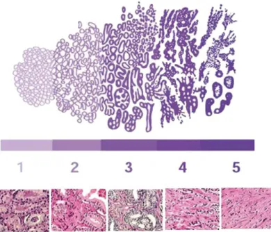
differentiated): closely packed, uniform shaped glands. Grade 2 (well differentiated): infiltration into the surrounding stroma, more variation in gland size and spacing. Grade 3 (moderately differentiated): irregular size and shape, separation of the glands, less defined boundaries and less intervening stroma. Grade 4 (poorly differentiated): fusion of the glands with a ragged invasive edge. Grade 5 (undifferentiated): complete absence of gland formation with sheets or clusters of cells. Adapted from Harnden et al, 2007 [52]. ... 10
Figure 5. The negative and positive crosstalk between histone post-translational
modifications. Adapted from Kouzadaries, 2007 [79]. ... 16
Figure 6. A. Human canonical and histone variants of H2A (yellow), H2B (red), H3
(blue), H4 (green). Unstructured amino- terminal tails are shown as black lines. Specific amino acid residues are depicted at key differences among variants of a common histone protein family. Different shades of color are used to indicate protein sequences that are highly divergent between canonical histones. B. Human canonical and histone variant linker H1. Unstructured amino- terminal tails are shown as light grey. Globular domains are shown in brown. Serine/threonine PXK phosphorylation sites targeted by cyclin-dependent kinases are indicated in magenta. Alternative names of variants are given in parentheses. aa, amino acid; mH2A1, macroH2A1.
Adapted from Maze and al, 2014 [97]. ... 18
Figure 7. Structure and subdomains of macroH2A1. ++ indicates a lysine-rich linker
region that resembles part of the C-terminal domain of histone H1, Zip indicates a region that resembles a leucine zipper, and the gray region shows the location of the region that is different between macroH2A1.1 and 1.2. The
xvii region C-terminal to the lysine-rich region is referred to as the non-histone region. Adapted from Pehrson and Fuji, 1998 [90]. ... 19
Figure 8. Schematic representation of the canonical histone H2A and histone variants
macroH2A1 isoforms with relative PTMs. Specific amino acids are depicted when they are found to be post-transnationally modified (PTMs are indicated by symbols as shown in the legend). Cylinders depict alpha-helical structures.
mH2A1.1, macroH2A1.1. mH2A1.2, macroH2A1.2. Adapted from
Vardabasso et al, 2013 [89]. ... 22
Figure 9. Primers used for expression quantification of total macroH2A1 and isoforms.
Specific-reverse primers for macroH2A1.1 and macroH2A1.2 are in blue and orange, respectively, and the forward primer used for both isoforms is in grey. The set of primers for macroH2A1 are in black. ... 33
Figure 10. Transcriptional status of isoforms macroH2A1.1 and macroH2A1.2 in
clinical samples. MacroH2A1.1 is progressively downregulated through PCa progression and macroH2A1.2 expression was not found statistically significant different between MNPT and PCa. Group analysis with Kruskal-Wallis test followed by a pairwise Mann-Whitney U test, **p<0.01 and ***p<0.001, ns = not significant. ... 44
Figure 11. (A) MacroH2A1 gene expression in MNPT, PIN and PCa samples. (B)
MacroH2A1 isoforms relative expression relative to total macroH2A1 in clinical samples. Group analysis with Kruskal-Wallis test followed by a pairwise Mann-Whitney U test, **p<0.01 and ***p<0.001, ns = not significant. ... 45
Figure 12. Ratio of isoforms macroH2A1.1 normalized for macroH2A1.1 expression in
clinical samples. Group analysis with Kruskal-Wallis test followed by a pairwise Mann-Whitney U test, ***p<0.001. ... 45
Figure 13. Transcripts levels of slicing regulators of H2AFY mRNA in MNPT, PIN and
PCa samples. Group analysis with Kruskal-Wallis test followed by a pairwise Mann-Whitney U test, **p<0.01 and ***p<0.001, ns = not significant. ... 46
Figure 14. Relative expression of macroH2A1.1 (A) and QKI (B) mRNA levels with
matched PCa and PIN lesions samples. ... 47
Figure 15. Clinicopathological Gleason Score associations with expressions levels of
xviii
macroH2A1.1 transcript levels (C). Pairwise Mann-Whitney U test, **p<0.01. ... 48
Figure 16. ROC curve analysis of macroH2A1.1 and QKI genes in a series of PCa
against MNPT samples. (AUC, area under curve; CI, confidence interval). . 49
Figure 17. Illustrative images of MacroH2A1.1 immunostaining in MNPT, PIN and
PCa samples………50
Figure 18. Distribution of macroH2A1.1 protein levels by percentage of positive cells
in prostate tissues………51
Figure 19. Expression levels of macroH2A1 isoforms (A and B), total (C) and splicing
regulators (D, E and F) in prostate cell lines (normalized to RWPE-1)…….52
Figure 20. MacroH2A1.1 overexpression in LNCaP was confirmed at mRNA (upper
panel), and at protein level (lower panel). MacroH2A1.2 transcript and protein levels were also assessed to confirm specific-variant transfection. *p<0.05, ns = not significant (Mann- Whitney U-test)………...53
Figure 21. Impact of MacroH2A1.1 overexpression in cell viability of LNCaP at 72h.
xix
TABLE LIST
Table 1. Criteria for low and high PIN. Adapted from Bostwick and Cheng, 2012 [16]. 5 Table 2. The AJCC/UICC TNM staging classification for PCa. Adapted from Edge and
Compton, 2010 [54]. ... 12
Table 3. Primers sequences for macroH2A1 isoforms and total, splicing regulators and
control primers [104, 105]. ... 32
Table 4. PCa cell lines used and the growth conditions. ... 35 Table 5. Clinical and histopathological data of patients. ... 43 Table 6. Spearman’s ρ correlations between total and splice variants of macroH2A1
with three splicing regulators.. ... 47
Table 7. Performance of macroH2A1.1 and QKI as diagnostic biomarker for PCa. .... 49 Table 8. Immunohistochemistry of macroH2A1.1 protein levels in MNPT, PIN and
xx
ABBREVIATIONS
APS – Ammonium persulfate AR – Androgen receptor AS – Active surveillance
AJCC – American Joint Committee on Cancer ATRX – α-thalassemia/MR, X-linked
APLF – Aprataxin-PNK-like factor BPH – Benign prostate hyperplasia BSA – Bovine serum albumin CDK8 – Cyclin-dependent kinase 8 DNMT – DNA methyltransferase
DDX5 – Deadbox 5 (also known as p68) DDX17 – Deadbox 17 (also known as p72) DRE – Digital Rectal Examination
ES cells – Embryonic stem cells
EMT – Epithelial mesenchymal transition ERBB2 – Receptor tyrosine-protein kinase FBS – Fetal bovine serum
FFPE – Formalin-fixed, paraffin embedded GUSβ – β-glucuronidase,
HAO1 – Hydroxyacid Oxidase 1
H3K04me3 – Tri-methylated histone H3 at lysine 4 (active transcription) H3K27me – Mono-methylated histone H3 at lysine 27 (repress transcription) H3K27me3 – Tri-methylated histone H3 at lysine 27 (repress transcription) HAT – Histone acetyltransferase
HDAC – Histone deacetylase HDM – Histone demethylase HGPIN – High-grade PIN
HMT – Histone methyltransferase LGPIN – Low-grade PIN
MET – Mesenchymal epithelial transition mH2A1 – MacroH2A1
xxi
mH2A1.1 – MacroH2A1.1 mH2A1.2 – MacroH2A1.2 miRNA – MicroRNA
MNPT – Morphologic normal prostate tissue MRI – Magnetic Resonance Imaging
mRNA – Messenger ribonucleic acid NAD+ – Nicotinamide adenine dinucleotide NC – Negative control
NRF-1 – Nuclear respiratory factor 1 PCa – Prostate cancer
PCR – Polymerase chain reaction
PIA – Proliferative inflammatory atrophy PIN – Prostate intraepithelial neoplasia PSA – Prostate-specific antigen
PAR – Poly-ADP-ribose
PARP-1 – Poly-ADP-ribose polymerase 1 PTM – Post-translational modification
qRT-PCR – Quantitative reverse transcriptase polymerase chain reaction QKI – Quaking
RP – Radical Prostatectomy SDS – Sodium dodecyl sulfate SOD3 – Superoxide Dismutase 3
SAHF – Senescence-Associated Heterochromatic Foci SWI/SNF – Switch/sucrose non-fermentable
TEMED – Tetramethylethylenediamine TNBC – Triple-negative breast cancer TRUS – Transrectal Ultrasound
TURP – Transurethral resection of the prostate TSS – Transcription start site
UICC – Union for International Cancer Control WW – Watchful waiting
____________________________________________________________I. INTRODUCTION
3
1. PROSTATE
1.1. PROSTATE ANATOMY, HISTOLOGY AND PHYSIOLOGY
Prostate, along with the seminal vesicles and the bulbourethral glands, constitute the male reproduction’s accessory glands [1]. The prostate is a walnut-shaped organ, which the size grow with age, around 28 to 47cm2 and is localized under the bladder, near the rectum, surrounding the beginning of the urethra [2, 3]. The function of the prostate is to segregate an alkaline fluid, where one of the components is a serine protease of the Kallikrein family, the prostate-specific antigen (PSA) [4].
The prostate is composed by acini and ducts, organized in lobules, and delimited by fibromuscular stroma. Acinus per se consists of epithelial (secretory and basal) and neuroendocrine cells, surrounded by fibroblasts and smooth-muscle cells [5]. Stromal and epithelial cells express androgen receptors (ARs), depending on androgens (i.e. testosterone) to proliferate [5]. A thin layer of connective tissue surrounds the prostate, being connect with nerves and other tissues, constituting the prostatic capsule [1].
The model of prostate anatomy has been puzzlingly discussed through time and culminate divided into lobes, based on laboratory animal’s analogy [6]. This concept was accepted until the decade of 1960s, when John E. McNeal start describing the most widely accepted anatomic divisions of the prostate: peripheral, central, transition and anterior fibromuscular stroma zones (Figure 1) [7, 8]. The peripheral zone is structured by a disc of tissue with radiated ducts laterally from the urethra lateral and distal, which constitutes 70% of the glandular prostate. The central zone is organized by ducts that follow the ejaculators ducts, constituting 25% of the prostate. The transition zone includes the prostatic urethra and arranges 5% of the glandular prostate. Lastly, the anterior fibromuscular forms the thick surface of the prostate and is responsible for sphincter functions [9, 10]. Cells of the transition zone proliferate dramatically throughout the puberty and later, after the age of 55 years, leading to the increase of the main zone of the glandular prostate, the peripheral zone [5]
THE ROLE OF MACROH2A1 IN PROSTATE CARCINOGENES______________________
4
1.2. NON-CANCEROUS PROSTATE DISEASES
Prostate disorders are commonly being more frequent in men with advanced age [11]. The most common non-cancerous prostate diseases include benign prostatic hyperplasia (BPH), proliferative inflammatory atrophy (PIA) and prostatic intraepithelial neoplasia (PIN).
BPH origins in the transition zone of the prostate and is described by an excess of glands and stroma [12]. The possible risk factors for BPH are heredity, gene polymorphisms, diet, metabolic syndrome, exercise and cigarette smoking [13].
A PIN lesion take place in the peripheral zone and is commonly characterized by neoplastic resembles with undetectable abnormal changes phenotypically and not raising the PSA levels [14]. PIN lesions were firstly characterized by Bostwick and Brawer [15] in low and high grade PIN (LGPIN and HGPIN, respectively), which differ by architectural and cytological characteristics (Table 1) [16]. PIN spread through prostatic ducts and is characterized by the conservation of the basal cell layer, while luminal cells are replaced by neoplastic cells [17]. Those neoplastic cells own a hyperchromatic nuclei and nucleoli enlargement [18].
____________________________________________________________I. INTRODUCTION
5 Table 1. Criteria for low and high PIN. Adapted from Bostwick and Cheng, 2012 [16].
LGPIN HGPIN
Architecture
Epithelial cells crowding and stratification, with irregular
spacing
Similar to low-grade PIN; More crowding and stratification; four patterns: tufting, micro papillary,
cribriform, and flat
Cytology Nuclei Enlarged, with marked size
variation
Enlarged; some size and shape variation
Chromatin Normal Increased density and clumping
Nucleoli Rarely prominent* Prominent
Basal cell layer Intact May show some disruption
Basement
membrane Intact Intact
*Fewer than 10% of cells have prominent nucleoli.
PIA usually originates in the peripheral zone and is described by the rapidly epithelial cells division without full differentiation [16]. Proliferative cell regeneration is induced by inflammation or external factors, as chemicals or bacteria [16]. PIN and PIA share similar alterations at key molecular pathways and originates in the same prostate glandular zone, suggesting that PIA can be a precursor of PIN [19].
1.3. PROSTATE CANCER
The most prevalent malignant disease in prostate is adenocarcinoma corresponding to approximately 95% of the cases [8]. Prostate cancer (PCa) is characterized by a heterogeneous low proliferate carcinoma, asymptomatic when confined in the organ (latent tumors) [20]. The only recognized putative precursor of PCa is HGPIN, which pre-dates the onset of PCa by 5–10 years and, along with PCa, disrupt both cell layers (Figure 2) [21]. Other prostate diseases keep the basal cell layer intact [16].
THE ROLE OF MACROH2A1 IN PROSTATE CARCINOGENES______________________
6
Approximately 9% of isolated HGPIN is found in biopsies, although the prevalence of HGPIN with PCa in biopsies vary with the number of cores and race, not surpassing 45% [16]. Irrefutably, HGPIN and PCa share the location [15] and have similar morphology [22], histology [23] and chromosomal abnormalities [24]. Consequently, HGPIN diagnosis could be used as tool for patients with PCa predisposition [18]. The heterogeneity, slow-growing behavior and no symptoms in early phases, turns PCa in a real challenge for patient management, triggering late diagnosis, and consequently compromising prognosis and target therapies [25]. Hence, it is important to comprehend underlying mechanisms and sequential pathways of PCa initiation and development [26].
1.4. EPIDEMIOLOGY OF PROSTATE CANCER: INCIDENCE
AND MORTALITY
PCa is a major health concern due to growth and ageing of global population. PCa is the fourth more frequent cancer considering overall population and, after lung cancer, is the most common cancer in men [27].
PCa incidence diverge drastically worldwide, thought could be related with the median-age and number of cases diagnosed per country. In the early 1990s, there was a dramatically increase of PCa incidence worldwide, due to the introduction and largely
____________________________________________________________I. INTRODUCTION
7 use of transurethral resection of the prostate (TURP) and PSA screening for cancer detection in developed countries [28]. Nonetheless, PCa incidence is higher in North America, Australia and Nordic countries, whereas lower incidence is found in Asia and Northern Africa (Figure 3A) [27]. In 2015, is expected that PCa would represent 25% of all new diagnosis cases in men [28].
Concerning PCa mortality, rates have been more constant through time. PCa mortality has been decreasing due to early diagnosis and therefore, the therapy is provided at earlier stages of the disease [29]. Currently, PCa constitutes the fifth cancer-related mortality worldwide, excluding non-melanoma skin cancer. PCa mortality rate is more prevalent in Africa and South America (Figure 3B) [30]. In Portugal, PCa is currently, the number one in incidence rates and second in mortality rate, among men [27].
Figure 3. Estimated incidence (A) and mortality worldwide of prostate cancer in 2012. Adapted from Globocan [27].
THE ROLE OF MACROH2A1 IN PROSTATE CARCINOGENES______________________
8
1.5. RISK FACTORS
To date, there are established three risk factors which represent the furthermost main influences that could lead to PCa: age, family history and ethnicity [31].
PCa patients’ average age is between 70 and 74 years, and it increases in older men [32]. Indeed, the likelihood of PCa development is 85% in men after 65 years old and higher than 90% in men with more than 90 years old [33]. It should be recalled that precursor lesions and PCa early phases are silence diseases, therefore, it may exist during years or decades before PCa is diagnosed [34].
Family history always represented a risk factor to develop cancer and PCa is no exception. Familial PCa represents 10-15% of all PCa diagnosed cases [35]. Additionally, it was observed that first-degree relatives of PCa patients have a higher risk to develop the disease. Furthermore, the number of affected members in a family and the early-onset of the disease increase even more the risk of prostate cancer [33]. Nevertheless, familial PCa and non-familial PCa are clinically and pathologically similar [31].
Lastly, ethnicity may justify PCa incidence divergence around the world. African-American men present 60% higher probabilities to develop PCa, even in younger ages, comparing to Caucasian American men [36]. However, immigration studies suggest that races with low PCa incidence, as Asians, increase dramatically the probabilities of develop PCa when immigrate to America [33, 34]. Hence, external factors, as environment, dietary habits, exercise, access to medical care and diagnosis tools and others [37, 38], might have an additional role in the likelihood of developing PCa [31].
1.6. DIAGNOSTIC TOOLS FOR PROSTATE CANCER
The efforts for development of tools for PCa detection is to effectively identify this disease while silently confined in the organ and, thus, curable. The two complemental detection tools available nowadays are digital rectal examination (DRE) and PSA screening.
Since PCa develops in the peripheral zone of the prostate gland and knowing the prostate proximity to the distal rectum, about 18% of all PCa can be detected by DRE
____________________________________________________________I. INTRODUCTION
9 [5, 39]. However, DRE lacks in sensibility and depends on professional experience [39]. Alternatively to DRE, PCa can be detected by Transrectal ultrasound (TRUS) or Transrectal magnetic resonance imaging (MRI), though the last is more utilized to verify PCa invasion to nearby tissues [40, 41].
Alternatively, the glycoprotein PSA is segregated in epithelial cells of the prostate and release in the blood circulation. PSA quantification was introduced as a diagnosis tool in the 1980’s, providing the identification of prostate diseases, with low levels of specificity and sensibility for PCa [42]. It is expected PSA levels between 0 to 4.0ng/ml in prostate-healthy men under 70 years old and slightly higher through age. Additionally, PSA levels can be influenced by obesity [43], cardiovascular disorders [44], type 2 diabetes [45] and other prostatic diseases besides PCa. This test demonstrates important limitations in specificity and sensibility but, to date, is the only available biomarker used for the detection and monitoring of treatment efficacy for prostate cancer [46].
Regardless limitations, the annual combination of DRE and PSA screening, in fact, diminish the number of advanced PCa patients [47]. If DRE and PSA screening results are PCa abnormal, is recommended a TRUS-guided systemic needle biopsies 3 to 6 months, with 12 or more small tissue cylinders (cores) removed each biopsy for analyzation [5, 48].
Moreover, it is important to take into account that PCa patients are, commonly, older than 50 years old and inaccurate regular diagnosis can lead to over diagnosis and over treatment of latent tumors and damage both physical and psychological. Therefore, it is advocated to avoid PSA screening in men over 75 years old [1]. All the points mentioned above strengthen the importance to develop specific non-invasive diagnosis methods for PCa [49].
1.7. PROGNOSTIC TOOLS FOR PROSTATE CANCER
Prognostics tools are designed to accurately distinguish clinically significant
from indolent PCa. Currently, Gleason Score and the TNM systems assist clinicians in
decision-making.
The heterogeneity of PCa is the main problem in prostate biopsies, since cores may not represent the complete tumor [50]. To decipher the glandular epithelial
THE ROLE OF MACROH2A1 IN PROSTATE CARCINOGENES______________________
10
architectural patterns, ignoring cytologic details, in 1966, Donald F. Gleason elaborate a histological grading system based on the sum of the two more frequent glandular histological patterns present in each tumor: the Gleason Score [51]. This system scores well-differentiated pattern as 1 and as 5 the most undifferentiated. Therefore, Gleason grading system increases with the tumor aggressiveness, in a 2 (1+1) to 10 (5+5) combined score scale (Figure 4) [51]. Although limited by the pathologist proficiency and the cores removed, accurate Gleason Score is critical, once, can differ in malignancy based on the most frequent pattern, for example, a Gleason Score 5+3 (n=8) represent a worst prognosis than a Gleason Score 4+4 (n=8) or 3+5 (n=8) [50].
Figure 4. Gleason Score: histological grading for prostate cancer. Grade 1 (well differentiated): closely packed, uniform shaped glands. Grade 2 (well differentiated): infiltration into the surrounding stroma, more variation in gland size and spacing. Grade 3 (moderately differentiated): irregular size and shape, separation of the glands, less defined boundaries and less intervening stroma. Grade 4 (poorly differentiated): fusion of the glands with a ragged invasive edge. Grade 5 (undifferentiated): complete absence of gland formation with sheets or clusters of cells. Adapted from Harnden et al, 2007 [52].
To recognize the dimension of PCa and the level of extension, in 1950s was establish a clinical and pathological staging system for solid tumors: the TNM (Tumor Node Metastasis) classification system. The American Joint Committee on Cancer (AJCC) and the Union for International Cancer Control (UICC) systematically update the staging system that allows distinguish primary tumors clinically (T) and pathologically (pT), regional lymph nodes status clinically (N) and pathologically (pN) and distant metastases (M) (Table 2) [53]. Clinical staging is only associated to the evaluation of cancer spread, being firstly obtained during diagnosis, before treatment. Pathological staging is related to histological data and is firstly determined after radical
____________________________________________________________I. INTRODUCTION
11 prostatectomy (RP) thus there is no pT1 classification [54]. Concerning PCa metastasis, tumors frequently spread to bones, lymph nodes, lungs, liver and brain [55].
The TNM classification, along with Gleason Score and PSA screening results, provides a complete PCa stage, classified to I to IV, increasing with the PCa aggressiveness [54].
THE ROLE OF MACROH2A1 IN PROSTATE CARCINOGENES______________________
12
Table 2. The AJCC/UICC TNM staging classification for PCa. Adapted from Edge and Compton, 2010 [54]. PRIMARY TUMOR (T)
CLINICAL
Tx Primary tumor cannot be assessed
T0 No evidence of primary tumor
T1 Clinically unapparent tumor neither palpable nor visible by imaging
T1a Tumor incidental histologic finding in 5% or less of tumor resected
T1b Tumor incidental histologic finding in more than 5% of tumor resected
T1c Tumor identified by needle biopsy
T2 Tumor confined within prostate gland
T2a Tumor involves one half of one side or less
T2b Tumor involves more than one half of one lobe but not both lobes
T2c Tumor involves both lobes
T3 Tumor extends through prostate capsule
T3a Extracapsular extension (unilateral or bilateral)
T3b Tumor invades seminal vesicle(s)
T4
Tumor is fixed or invades adjacent structures other than seminal vesicles, such as: external sphincter, rectum, bladder, levator muscles, and/or pelvic
wall PATHOLOGIC (PT)
pT2 Organ confined
pT2a Unilateral, one half of one side or less
pT2b Unilateral, involving more than one half of one lobe but not both lobes
pT2c Bilateral disease
pT3 Extraprostatic extension
pT3a Extraprostatic extension or microscopic invasion of bladder neck
pT3b Seminal vesicle invasion
pT4 Invasion of rectum, levator muscles and/or pelvic wall
REGIONAL LYMPH NODES (N)
CLINICAL
Nx Regional lymph nodes were not assessed
N0 No regional lymph node metastasis
N1 Metastasis in regional lymph node(s) PATHOLOGIC (pN)
pNx Regional nodes not sampled
pN0 No positive regional nodes
pN1 Metastasis in regional node(s)
DISTANT METASTASIS (M)
M0 No distant metastasis
M1 Distant metastasis
M1a Non-regional lymph node(s)
M1b Bone(s)
____________________________________________________________I. INTRODUCTION
13
1.8. PROSTATE CANCER’S CLINICAL MANAGEMENT
The main goal of treatment of clinically localized PCa (stage I and II) is the cancer eradication, while no curative treatment is available for advanced PCa (stage III and IV) and treatment is only palliative support. Early-stages PCa present about 90% of progression-free survival after 5 to 10 years [56]. In fact, it is more frequent a man die with PCa than from PCa. Nevertheless, all aggressive therapies could lead to different side-effects, as urine or bowel dysfunction, fatigue, increased risk of diabetes or heart attacks and others [57]. Therefore, age, life expectancy, comorbidities and quality of life of the patients are taken in consideration to select the better treatment approach.
To avoid inadequate treatments, PCa patients can be monitored by watchful waiting (WW) or active surveillance (AS). It is suggested WW to patients who are not advised to undergo aggressive treatment. These patients are followed on 6 months and
only are treated if PCa progress. AS is recommended for indolent tumors where therapies
are pointless: low Gleason Score grade, low PSA screening result and <50% presence of cancer in biopsies [58]. These patients are followed by systematically diagnosis procedures, evaluating the progression of PCa.
Clinically localized PCa can be treated with RP or radiotherapies. For early-stage PCa patients with good general conditions for surgical intervention and with 10 or more years of life expectancy, the most adequate treatment is ablation of the prostate gland and the seminal vesicles by RP [59]. Radiotherapy may be an alternative to RP, showing high rates of disease-free survival, either by noninvasive external-beam radiation therapy or interstitial radiation therapy (brachytherapy), in which radioactive seeds with a life-time of 60 days are placed near the tumor [60, 61].
For advanced PCa patients, the treatment option is suppress the action or inhibit the production of testosterone, decreasing the prostate hormone-response. Androgen-deprivation therapy can be achieved by surgical castration (orchiectomy) or chemical castration, a combination of gonadotropin-releasing hormone analogues with antiandrogens (i.e. bicalutamide) [57]. These therapies may be used along with early-advanced PCa treatments. Unluckily, AR mutations lead to castration-resistance after 18-30 months of treatment [62]. The therapies available for metastatic castration-resistant PCa patients only provide supportive care.
THE ROLE OF MACROH2A1 IN PROSTATE CARCINOGENES______________________
14
2. EPIGENETICS
The nucleus of a human cell compacts the three billion base pair genome: DNA bonded to proteins, forming the chromatin [63]. When chromatin is strongly compacted is named heterochromatin and when is lesser condensed is designated euchromatin which is associated with transcription, DNA replication or repair and recombination processes [64]. Epigenetic mechanisms play a key role in chromatin dynamics and therefore in expression regulation.
The term “epigenetic” use the Greek prefix epi- which means over, beyond – genetics and was defined by Conrad Waddington, in the 1940s, as the branch of science of embryonic development studies, through experimental analysis. [65]. The “epigenetic landscape” was the explanation of cellular differentiation: how totipotent cells develops into all the different cells types in an organism with the same genome [66].
Epigenetic definition has been changing through time and currently, is defined as the heritable changes that occur in a gene regulation/function without alter the DNA sequence [67]. Epigenetics studies explain, for example, the differences among monozygotic twins or, in females, the silence of one X chromosome [66].
Epigenetic mechanisms are divided in four different main groups: DNA methylation, non-coding RNAs, post-translational modifications (PTMs) of histones and histone variants, which will be slightly described below. Alterations in epigenetic mechanisms affect innumerous cells processes, being implicated in several diseases, including cancer.
2.1. DNA METHYLATION
In mammals, DNA methylation refers to the addition of a methyl group, by DNA methyltransferases (DNMT), in a cytosine next to a guanine, known as CpG dinucleotides. CpG dinucleotides clusters are designed as “CpG islands” and are generally found in promoters, introns, repetitive sequences or untranslated sequences of the genome [66]. The latter are globally methylated in the genome being important to maintain DNA stability [68].
____________________________________________________________I. INTRODUCTION
15 Promoters with low level of methylation are related with active gene expression, whereas heavy hypermethylated promoters are associated with stable silenced genes, as in the inactive female X chromosome [69]. DNA methylation inhibits gene expression directly by avoiding the binding of transcription factors [70] or indirectly by the recruitment of chromatin remodeling complexes [71].
DNA methylation is the most studied epigenetic mechanism in cancer. A cancer cell is characterized by hypermethylation of tumor suppressor genes promoters and by global hypomethylation of the genome [72].
2.2. NON-CODING RNAS
Nearby 90% of all RNAs transcribed are non-coding RNAs that do not codifiy proteins [73]. Non-coding RNAs, as ribosomal RNAs, are grouped according to size; microRNAs (miRNAs) are 18-30 nucleotides, 30–300nt are denominated small RNAs and non-coding RNAs with larger 300nt are considered long RNAs [73]. Non-coding RNAs are described as key players in gene regulation [73]. From these, miRNAs are the most well studied in cancer [73, 74].
MiRNAs are synthetized and processed in the nucleus and are transported to the cytoplasm to bind complementary mRNAs, repressing their function by degradation or by translation inhibition [74]. Interestingly, miRNAs could also be involved in the up-regulation of translation during the cell cycle [75].
Different mRNAs can be regulated by the same miRNA, the same way as different miRNA can target the same mRNAs [74]. About 30% of the human genes are regulated by time and tissue-specific miRNAs [76], interfering with several cellular pathways as differentiation, proliferation, apoptosis, and stress response [77].
In cancer, upregulated miRNAs target tumor suppressor genes and downregulated miRNAs target oncogenes [74]. Gene amplification, deletion, mutation and other epigenetic mechanisms can alter the miRNAs expression [74].
2.3. HISTONE POST-TRANSLATIONAL MODIFICATIONS
Eukaryotic DNA is packaged by histones, positively-charged proteins that easily bind with the negatively-charge DNA [78]. Eight histones, one pair of each H2A, H2B,
THE ROLE OF MACROH2A1 IN PROSTATE CARCINOGENES______________________
16
H3 and H4, constitute a protein complex designed nucleosome that is wrapped by a core DNA 1.7 times and sealed by one H1 [79, 80], along with numerous hydrogen, electrostatic and hydrophobic bonds [81].
Histones are dynamic proteins responsible for DNA support and chromosomal remodel [82]. All histones share a similar structural architecture with α-helices bonded by short loops and a flexible undefined N-terminal tail where is more susceptible to occur covalent histone modifications (post-translational modifications), such as acetylation, methylation, phosphorylation or ubiquitination which impact on chromatin condensation and globally constitute the so-called histone code (Figure 5). [79, 82, 83]. These modifications are “written”, “read” and “erased” by different histone modulating enzymes [84, 85].
Regarding histone acetylation, gene transcriptional activity is balanced due to alterations of electrostatic charge in the nucleosomes [86]. Therefore, hyperacetylation is characteristic of euchromatin by decreasing the histone-DNA affinity and allowing gene transcription, whereas hypoacetylation is related with heterochromatin [79]. Histone acetylation is “written” by histone acetyltransferases (HATs) and “erased” by histone deacetylases (HDACs) [84].
Histone methylation promotes transcription activation or repression depending on the residue and the number of methylation molecules added (mono-, di- or tri-) [87]. Indeed, tri-methylation of lysine 4 of H3 (H3K4me3) promotes active transcription while mono- and tri-methylation of lysine 27 of H3 (H3K27me and H3K27me3) inhibits gene transcription. The writers of histone methylation are histone methyltransferases (HMT) and the erasers are histone demethylases (HDM) [79, 82]. Figure 5. The negative and positive crosstalk between histone post-translational modifications. Adapted from
____________________________________________________________I. INTRODUCTION
17 Histone modifying enzymes expression are disrupted in cancer and the
imbalance between writers and erasers affect the PTMs’ profile [88]. Moreover, DNMTs
are directly recruited by HMTs to inhibit genes’ expression and recruit HDACs to increase the gene silencing. This interplay between DNA methylation and PTMs is also impaired in cancer [88].
2.4. HISTONES VARIANTS
The less studied epigenetic mechanism is the shift of canonical histones by sequential similar non-allelic histones variants [89]. Among species, histone variants are the mostly conversed proteins and have been considered functionally irreplaceable [90, 91].
On one side, canonical histones are genomically organized by clusters lacking introns [92]. The transcription is DNA replication-dependent and therefore, exclusive to the S phase of the cell cycle, and the mRNA obtained contains a unique 3′ stem loop [93]. On the other side, histone variants are orphan genes with introns and the mRNA translated holds a polyadenylated tail [81]. Although they are present throughout the cell cycle, variants are tissue and temporal-specific [94, 95]. Variants are named “replacement histones” because they substitute the canonical histones during development and differentiation, establishing cell identity [81].
The slightly sequential differences, along with unique PTMs of histone variants, result in nucleosome-DNA stability differences [96] and alters the efficiency of protein complexes responsible from histone deposition and displacement in the nucleosome. These adjustments change the accessibility of transcription factors into the chromatin, regulating the gene expression.
To date, histone variants have been described for all canonical histones, excluding H4 (Figure 6) [97]. H2A family is the largest histone family with the most structurally diverse histone variants: H2A.X, H2A.Z, macroH2A, H2A.Bbd [98]. Variants of H2A are described by distinguish length, sequence and genome distribution [81]. Mis-regulation or mutations in these H2A histone atypical variants have been implicated in cancer initiation and progression [89].
THE ROLE OF MACROH2A1 IN PROSTATE CARCINOGENES______________________
18
A
B
Figure 6 A. Human canonical and histone variants of H2A (yellow), H2B (red), H3 (blue), H4 (green). Unstructured amino- terminal tails are shown as black lines. Specific amino acid residues are depicted at key differences among variants of a common histone protein family. Different shades of color are used to indicate protein sequences that are highly divergent between canonical histones. B. Human canonical and histone variant linker H1. Unstructured amino- terminal tails are shown as light grey. Globular domains are shown in brown. Serine/threonine PXK phosphorylation sites targeted by cyclin-dependent kinases are indicated in magenta. Alternative names of variants are given in parentheses. aa, amino acid; mH2A1, macroH2A1.
____________________________________________________________I. INTRODUCTION
19
2.4.1. MACROH2A1: THE SUBDOMAINS
Currently, there are two described macroH2A histones, macroH2A1 and macroH2A2, which are encoded by H2AFY and H2AFY2 genes, respectively [99]. The vertebrates-exclusive macroH2A1 is one of the most distinctive histone variant [81], composed by 370 amino acids with 40kD of molecular weight in a tripartite structural organization, three times larger than canonical H2A.
The first 122 amino acids of the N-terminal histone-like region shares 64% homology with canonical H2A [99]. The last 209 amino acids from the C-terminal histone domain establish a strongly folded macro domain of 30kDa with a random coil without any similarity to other histones [99]. This non-histone region prolongs out from the asymmetric nucleosome, disturbing the transcription factors binding, and contains a putative leucine-zipper motif responsible for nucleosomes interactions [90]. A lysine-rich linker sequence connects the H2A-region with the macro domain that increases nucleosome stability (Figure 7) [100].
Chakravarthy et al. demonstrated, by in vitro studies, that the presence of macroH2A in the nucleosomes increases DNA-nucleosome stability [101]. MacroH2A1 is responsible for chromatin compaction by affecting the dynamic properties of nucleosomes and even by strengthen the H1-DNA interactions [100, 102]. Overall, nucleosomes containing macroH2A1 are more stable and establish DNA-histone interactions in the entry and exit of the DNA into the nucleosomes leading to formation of heterochromatin regions.
Figure 7 Structure and subdomains of macroH2A1. ++ indicates a lysine-rich linker region that resembles part of the C-terminal domain of histone H1, Zip indicates a region that resembles a leucine zipper, and the gray region shows the location of the region that is different between macroH2A1.1 and 1.2. The region C-terminal to the lysine-rich region is referred to as the non-histone region. Adapted from Pehrson and Fuji, 1998 [90].
THE ROLE OF MACROH2A1 IN PROSTATE CARCINOGENES______________________
20
H2AFY (5q31.1) codifies for macroH2A1.1 and macroH2A1.2 isoforms by
alternative splicing of a single exon. The difference of these isoforms is a sequence of 10 amino acids in the macro domain of macroH2A1.1 and an alternative exon of 11 amino acids in the macroH2A1.2 [103].
2.4.2.
MACROH2A1:
EXPRESSION
AND
NUCLEOSOME
DEPOSITION REGULATIONS
MacroH2A1 can be regulated, post-translationally, by alternative splicing or by nucleosome deposition and chromatin localization.
Concerning expression regulation, macroH2A1 isoforms may occur by pre-mRNA splicing regulators, as the Quaking (QKI) or RNA helicases deadbox 5 (DDX5 or p68) and deadbox 17 (Ddx17 or p72) [104, 105]. The RNA-binding protein QKI is a 37kDa RNA-binding protein known to upregulate macroH2A1.1 through binding at the intron upstream of macroH2A1.2-specific exon [104]. RNA helicases DDX5 and DDX17, which share a highly degree of homology, were also reported to control macroH2A1 splicing [105].
Regarding nucleosome regulation, ATP-dependent nucleosome remodeling complexes, such as switch/sucrose non-fermentable (SWI/SNF), rearrange or mobilize nucleosomes and allow the shuffle of histones [106]. Specifically, SWI/SNF helicase ATRX (α-thalassemia/MR, X-linked) acts as negative regulator of macroH2A1 in chromatin-free state, monitoring macroH2A1 deposition. Indeed, increased nucleosomes deposition of macroH2A has been reported for ATRX syndrome patients [107]. Moreover, the Aprataxin-PNK-like factor (APLF), a poly(ADP-ribosyl)ation-regulated protein, acts as DNA damage-specific histone chaperone, increasing the macroH2A1 nucleosome deposition [96, 108].
For nucleosome stability, nuclear respiratory factor 1 (NRF-1) interacts with macroH2A1, avoiding unnecessary gene expression [109]. Nonetheless, macroH2A1 plays a complex role in transcription by either positive or negative gene’s regulation [110]. The switch key between repressor/activator roles of macroH2A1 remains uncertain, though PTMs, splicing alternative or the transcription factors nature may be predict the macroH2A1 function [110].
____________________________________________________________I. INTRODUCTION
21
2.4.3. MACROH2A1 TARGETS
Firstly found in the female X inactivation chromosome, by Costanzi and Pehrson in 1998, macroH2A1 were generally described as an autosomal transcriptional repressive histone [111, 112] implicated in female X chromosome inactivation [113-115]. Nonetheless, macroH2A1 is found in about 25% of the genome, being similarly expressed in female and male mammals [116].
MacroH2A1 is present in Senescence-Associated Heterochromatic Foci (SAHFs) [117] and upstream and downstream of transcription start sites (TSSs) of genes implicated in cell cycle [110], pluripotency [118] and development [119]. As previously described, a subset of genes can be positively regulated by this histone variant [120] or even be recruited for DNA repair [121].
MacroH2A1.1 expression is generally limited to differentiated cells, whereas macroH2A1.2 is highly expressed in proliferating cells [122]. Embryonic stem (ES) cells exclusively express macroH2A1.2 and macroH2A1.1 only during development increases [123].
Regarding PTMs, ubiquitylation of macroH2A1.2 at lysines115 and 116 (K115 K116) were associated with the X chromosome inactivation and phosphorylation at serine 137 (S137) of both macroH2A1.1 and macroH2A1.2 were implicated in cell cycle regulation (Figure 8) [89]. Remarkably, macroH2AS137ph is excluded from female X chromosome inactivation and enriched during mitosis, suggesting phosphorylation as a key regulation for critical interactions of macroH2A1 with effector molecules [115, 120]. Additionally, PTMs without a known function were also described for macroH2A1.2, specifically, methylation at lysines 17, 122 and 238 (K17, K122, K238) and phosphorylation at threonine 128(T128) (Figure 8) [98].
THE ROLE OF MACROH2A1 IN PROSTATE CARCINOGENES______________________
22
MacroH2A1.1-specific exon allows macroH2A1.1 to exclusively bind NAD+-derived ligands, including the poly-ADP-ribose (PAR) produced by poly-ADP-ribose polymerase 1 (PARP-1), disturbing several pathways [124]. Indeed, while macroH2A1.2 deposition is related with the repressive H3K27me3, the splicing isoforms exchange, leads PARP-1 to recruit HATs, through macroH2A1.1-PAR binding, promoting H2B K12ac and H2B K120ac thus, activating gene transcription [124].
In addition to PARP-1downregulation, macroH2A1.1 negatively regulates cellular proliferation and cell cycle progression genes, such as cyclin-dependent kinase 8 (CDK8) and c-Fos, [104, 110, 125] and upregulates redox metabolism genes [98]. On the other hand, macroH2A1.2 is known for promoting the receptor tyrosine-protein kinase (ERBB2) expression, inducing proliferation [126].
Figure 8. Schematic representation of the canonical histone H2A and histone variants macroH2A1 isoforms with relative PTMs. Specific amino acids are depicted when they are found to be post-transnationally modified (PTMs are indicated by symbols as shown in the legend). Cylinders depict alpha-helical structures. mH2A1.1, macroH2A1.1. mH2A1.2, macroH2A1.2. Adapted from Vardabasso et al, 2013 [89].
____________________________________________________________I. INTRODUCTION
23
3. THE ROLE OF MACROH2A1 IN CARCINOGENESIS
Expression alterations of macroH2A1 has been studied in several types of cancer, including those of breast [127], lung [128] colon [103], bladder [129] and melanoma [125].
MacroH2A1 expression levels alterations in cancer might occur due to altered pre-mRNA alternative splicing. QKI is reported to be downregulated in several tumors, leading to macroH2A1.1 decreased expression levels [104]. Consequently, macroH2A1.2 expression increases in relation to macroH2A1.1. In breast cancer, DDX5/DDX17 inhibits the expression of redox metabolism related-genes as SOD3 (Superoxide Dismutase 3) and HAO1 (Hydroxyacid Oxidase 1), through macroH2A1.1 downregulation [98, 105].
In non-metastatic breast cancer cells, overexpression of macroH2A1.1 activates SOD3 gene that inhibits cell migration/invasion, whereas in metastatic breast cancer cells, high levels of macroH2A1.2 represses SOD3, increasing cell migration/invasion [105].
Moreover, in breast cancer, macroH2A1.2 overexpression induces ERBB2 overexpression, promoting uncontrolled cell growth and proliferation [126]. However, in triple-negative breast cancer (TNBC), without estrogen and progesterone receptors and absence of amplification of ERBB2, macroH2A1.1 overexpression associated with poor survival [127].
Overall, macroH2A1.1 variant is commonly accepted as a pleiotropic tumor suppressor, by repressing cell proliferation and cell migration, whereas macroH2A1.2 seems to play an oncogenic role, contributing to a metastatic phenotype, and promoting cell proliferation through ERBB2 [98, 105, 121].
_____________________________________________________________________II. AIMS
27
AIMS
PCa is one of the most prevalence cancers in men and cause of cancer-related morbidity and mortality worldwide. Characteristically asymptomatic in early stages, diagnostic and prognostic tools for PCa are fallible and for PCa at late-stage, the therapeutic options are limited. To avoid unnecessary treatments in indolent tumors and choose the better options for PCa patients is important to fully understand the biology of this neoplasm.
Histone variants, the less studied epigenetic mechanism, have been implicated in cancer, regulating several important cellular pathways. Specifically, the histone variant isoforms macroH2A1.1 and macroH2A1.2 were found to be deregulated in some neoplasms; however no data is yet available for prostate cancer. Thus, the major goal of this Master Thesis is to evaluate whether macroH2A1 is deregulated in prostate cancer and whether it plays a critical role in prostate tumorigenesis. To achieve this, several objectives were established:
One. Determine the H2AFY expression levels, both total and isoforms, in a cohort of primary prostate tumors (n=197), prostate intraepithelial neoplasia (n=45) and in normal prostatic tissue (n=14).
Two. Study putative regulators of H2AFY.
Three. Correlate the H2AFY transcripts with clinical pathological variables.
Four. Assess the protein expression of H2AFY in prostatic tissues.
Five. Determine the H2AFY transcripts expression levels (total and isoforms) in a benign prostatic cell line (RWPE-1) and several PCa cell lines (22Rv1, DU145, LNCaP, PC-3 and VCaP).
Six. Evaluate the phenotypic impact of H2AFY shRNA or forced expression in PCa cell lines.
________________________________________________III. MATERIAL AND METHODS
31
1. CLINICAL SAMPLES
1.1
PATIENTS AND CLINICAL SAMPLES COLLECTION
Prostate samples of 197 primary tumors and 45 HGPIN (from here simply designated PIN) lesions were prospectively collected from patients diagnosed with the disease and primarily treated with RP, form 2001 and 2006, at the Portuguese Oncology Institute, Porto, Portugal. Samples of 15 morphological normal prostate tissues (MNPT), used as control, were collected from the peripheral zone of prostates not harboring PCa, obtained from radical cystoprostatectomy for bladder cancer. Immediately after surgery, all tissue specimens were frozen at -80ºC. Thick frozen sections were obtained from frozen tissues for stain identification and after, the tissue block was trimmed to maximize the yield of target cells (>70% of target cells). Histological slides from formalin-fixed, paraffin embedded (FFPE) tissue fragments were also obtained from the same surgical specimens for histopathological examination: Gleason Score and pathological staging evaluations. Relevant clinical data were acquired from clinical registers and these studies were approved by the institutional review board (Comissão de Ética para a Saúde – CES 019/2008) of Portuguese Oncology Institute - Porto, Portugal.
1.2
RNA EXTRACTION AND QUANTIFICATION
Samples were homogenized in Trizol® Reagent (Invitrogen, Carlsbad, CA, USA) and the total RNA were extracted from all 257 samples using PureLinkTM RNA Mini Kit (Invitrogen, Carlsbad, CA, USA), following manufacturer’s instructions. All genomic DNA were eliminated with TURBO DNA-free (Ambion, Applied Biosystems), according to manufacturer’s instructions. The concentration, purity ratios and quality of each sample were determined using a Nanodrop ND-1000 (ThermoScientific, Wilmington, DE, USA) and by an agarose gel electrophoresis. RNA samples were then stored at -80ºC.
![Figure 1. Anatomic zones of the prostate described by McNeal. Adapted from Hammerich et al, 2008 [8].](https://thumb-eu.123doks.com/thumbv2/123dok_br/17597558.819917/27.892.208.655.101.447/figure-anatomic-zones-prostate-described-mcneal-adapted-hammerich.webp)
![Table 1. Criteria for low and high PIN. Adapted from Bostwick and Cheng, 2012 [16].](https://thumb-eu.123doks.com/thumbv2/123dok_br/17597558.819917/28.892.127.776.133.522/table-criteria-low-high-pin-adapted-bostwick-cheng.webp)
![Figure 2. Cellular progression of prostate cancer. Adapted from Witte, 2009 [26].](https://thumb-eu.123doks.com/thumbv2/123dok_br/17597558.819917/29.892.210.701.101.430/figure-cellular-progression-prostate-cancer-adapted-witte.webp)
![Figure 3. Estimated incidence (A) and mortality worldwide of prostate cancer in 2012. Adapted from Globocan [27].](https://thumb-eu.123doks.com/thumbv2/123dok_br/17597558.819917/30.892.207.716.560.1021/figure-estimated-incidence-mortality-worldwide-prostate-adapted-globocan.webp)
![Table 2. The AJCC/UICC TNM staging classification for PCa. Adapted from Edge and Compton, 2010 [54].](https://thumb-eu.123doks.com/thumbv2/123dok_br/17597558.819917/35.892.120.773.114.1167/table-ajcc-uicc-staging-classification-adapted-edge-compton.webp)


![Table 3. Primers sequences for macroH2A1 isoforms and total, splicing regulators and control primers [104, 105]](https://thumb-eu.123doks.com/thumbv2/123dok_br/17597558.819917/55.892.117.779.783.1042/table-primers-sequences-isoforms-splicing-regulators-control-primers.webp)
