Analysis of the Performance of 11 Formulae for Fetal
Weight Estimation in Preterm Fetuses with Abnormal
Doppler Velocimetry
–
A Retrospective Multicenter
Study
Análise do desempenho de 11 fórmulas de estimativa de peso
fetal em conceptos prematuros com Dopplervelocimetria
alterada
–
um estudo retrospectivo multicêntrico
Alessandra Martins Heringer de Lima
1Paulo Roberto Nassar de Carvalho
1,2Saint Clair Gomes Junior
2Ana Carolina Costa Carioca
2José Maria de Andrade Lopes
1,21Diagnostic Center, Clínica Perinatal Laranjeiras, Rio de Janeiro, RJ,
Brazil
2Fetal Medicine Center, Instituto Nacional Fernandes Figueira,
Fundação Oswaldo Cruz, Rio de Janeiro, RJ, Brazil
Rev Bras Ginecol Obstet 2018;40:580–586.
Address for correspondence Alessandra Martins Heringer de Lima, Master, Centro Diagnóstico, Clínica Perinatal Laranjeiras, Rua das Laranjeiras, 445, 22240-002, Laranjeiras, Rio de Janeiro, RJ, Brazil (e-mail: [email protected]).
Keywords
►
fetal weight
►
birth weight
►
premature birth
►
ultrasound
►
prenatal ultrasound
►
doppler ultrasound
►
fetal growth
retardation
►
placental
insuf
fi
ciency
Abstract
Objective
To assess 11 formulae commonly used to estimate fetal weight in a
population of premature fetuses who had abnormal Doppler velocimetry due to
early-onset placental insuf
fi
ciency. The performance of each formula was evaluated
in subgroups of fetuses with expected growth and intrauterine growth restriction.
Methods
Data were collected from fetuses and mothers who delivered at three Brazilian
hospitals between November 2002 and December 2013. We used the following formulae:
Campbell; Hadlock I, II, III, IV and V; Shepard; Warsof; Weiner I and II; and Woo III.
Results
We analyzed 194 fetuses. Of these, 116 (59.8%) were considered appropriate
for gestational age (AGA), and 103 (53.1%) were male. The amniotic
fl
uid volume was
reduced in 87 (44.8%) fetuses, and the umbilical artery Doppler revealed absence or
inversion of diastolic
fl
ow in 122 (62.9%) cases, and the analysis of the ductus venosus
revealed abnormal
fl
ow in 60 (34.8%) fetuses. The Hadlock formulae using three or four
fetal biometric parameters had low absolute percentage error in the estimated fetal
weight among preterm fetuses with abnormal Doppler studies who were born within
5 days of the ultrasound evaluation. The results were not in
fl
uenced by the clinical and
ultrasound parameters often found in early-onset placental insuf
fi
ciency.
Conclusion
In this study, the formulae with the best performance for fetal weight
estimation in the analyzed population were Hadlock I and IV, which use four and three
fetal biometric parameters respectively to estimate the weight of preterm fetuses with
abnormal Doppler studies.
received
January 22, 2018
accepted
June 12, 2018
published online
September 20, 2018
DOI https://doi.org/ 10.1055/s-0038-1670643.
ISSN 0100-7203.
Copyright © 2018 by Thieme Revinter Publicações Ltda, Rio de Janeiro, Brazil Original Article
Introduction
The accuracy of fetal weight estimation is very important for patients who have intrauterine growth restriction (IUGR). When IUGR occurs at the threshold of neonate viability, accurate fetal weight estimation represents a valuable predictive factor to assess the probability of perinatal survival. Precise weight prediction before birth may minimize the perinatal morbidity and mortality associated with lower intrauterine growth.1
The normal development of the placenta, which occurs in the early stages of gestation, is dependent on the adequate invasion of trophoblastic cells in the decidual and myome-trial segments of the spiral uterine arteries, which represent the most important source of irrigation in the uterine body region.2–4Impaired trophoblast invasion (or abnormal pla-cental implantation) is associated with elevated vascular resistance in the fetal-placental and uteroplacental circula-tion, and, consequently, with the development of preeclamp-sia and IUGR, a condition called placental insufficiency.5,6
The reduction of fetal systemic blood flow due to fetal compensatory mechanisms in placental insufficiency leads to a decrease in the fetal growth rate.7When these events lead to gestation in the second or early third trimesters, we call this early-onset placental insufficiency, and its severity is directly proportional to gestational age (GA), estimated fetal weight (EFW) and alterations found in Doppler mapping.7 Intrauterine growth restriction is a major consequence of placental insufficiency, and constitutes a significant public health problem, increasing the rates of neonatal morbidity
and mortality and late postnatal consequences.3,4In general, this fetal pathology is a common clinical issue, present in 7 to 15% of all pregnancies.8
The perinatal outcome of fetuses affected by placental insufficiency is broadly dependent on the severity of the growth restriction, and EFW below the third percentile and/ or abnormalfindings in the umbilical artery (UA) represent the greatest risks for adverse perinatal results.8–11 Other important prenatal determinants for the perinatal outcome are GA at birth and birth weight (BW), which is traditionally used as a predictive parameter of neonatal survival.11
Most EFW ultrasound formulae have been evaluated in multiple clinical conditions, but there is criticism of the indis-criminate use of these models in situations such as IUGR triggered by early-onset placental insufficiency. Only the Had-lock formula with four fetal biometric parameters (head cir-cumference [HC], abdominal circir-cumference [AC], femur length [FL] and biparietal diameter [BPD]) was tested in the population with altered Doppler velocimetry and high risk for IUGR.12
The present study aimed to assess the performance of 11 ultrasound formulae used to estimate fetal weight in prema-ture fetuses with arterial and venous blood flow changes identified through Doppler velocimetry.
Methods
Data were collected from a cohort of women and their fetuses submitted to ultrasound and Doppler velocimetry examina-tions who delivered at one of three maternity hospitals in the
Resumo
Objetivo
Avaliar o desempenho de 11 fórmulas comumente utilizadas para
estima-tiva de peso fetal em uma população de fetos prematuros com dopplervelocimetria
alterada devido a insu
fi
ciência placentária de início precoce. O desempenho de cada
fórmula foi avaliado em subgrupos de fetos com crescimento adequado e com
crescimento intrauterino restrito.
Métodos
Foram coletados os dados de mães e fetos cujos partos foram
acompa-nhados em 3 instituições brasileiras entre novembro de 2002 e dezembro de 2013. As
fórmulas selecionadas para análise foram: Campbell; Hadlock I, II, III, IV e V; Shepard;
Warsof; Weiner I e II; e Woo III.
Resultados
Foram analisados os pesos de 194 fetos, dos quais 116 (59,8%) foram
considerados adequados para a idade gestacional, 103 (53,1%) eram do sexo
mascu-lino, em 87 (44,8%) havia redução do volume de líquido amniótico, em 122 (62,9%) o
Doppler de artéria umbilical demonstrou ausência ou inversão do
fl
uxo na diástole, e
em 60 (34,8%) a análise do duto venoso indicou
fl
uxo anormal. A média do erro
percentual absoluto (EPA) demonstrou que as fórmulas de Hadlock que utilizam 3 ou 4
parâmetros biométricos fetais apresentaram o melhor desempenho. Os resultados
obtidos para essas fórmulas não sofreram in
fl
uência dos parâmetros clínicos e
ultrassonográ
fi
cos frequentemente encontrados na insu
fi
ciência placentária de início
precoce.
Conclusão
O presente estudo demonstrou o melhor desempenho das fórmulas de
Hadlock que contêm 3 ou 4 parâmetros da biometria para estimativa de peso de fetos
prematuros com anormalidades ao mapeamento Doppler.
Palavras-chave
►
peso fetal
►
peso ao nascer
►
parto prematuro
►
ultrassonogra
fi
a
►
ultrassonogra
fi
a
pré-natal
►
ultrassonogra
fi
a
doppler
►
retardo do
crescimento fetal
Rio de Janeiro metropolitan area: Instituto Fernandes Figueira, Clínica Perinatal Barra, and Clínica Perinatal Laranjeiras, between November 2002 and December 2013.
The inclusion criteria were: women on the 24th to 33rd weeks of pregnancy, calculated according to the date of the last menstrual period and confirmed through obstetric ul-trasonography performed by the 20th week of pregnancy; presence of Doppler velocimetry levels compatible with fetal bloodflow redistribution (increase of the pulsatility index [PI] of the UA above the 95th percentile for GA; presence of brain sparing reflex, with PI of the middle cerebral artery [MCA] below the 5th percentile for GA; AU with zero or reverse diastole in the UA); interval between the last ultra-sound assessment of fetal biometry and birth not longer than five days; interval between last Doppler velocimetry exam and delivery not longer than 24 hours; and absence of signs of infection.
The exclusion criteria were multiple pregnancies, presence of fetal malformation assessed through prenatal care and/or confirmed on physical examination immediately after birth, and lack of reliable or available data to satisfy data collection. Doppler velocimetry and ultrasonography were per-formed using the following devices: General Electric Voluson E6, and General Electric Voluson S6 (Boston, MA, US). All of the ultrasound examiners had at least two years of experi-ence in obstetric examinations, and were certified by the Brazilian College of Radiology (CBR, in the Portuguese acro-nym) and the Brazilian Federation of Societies of Gynecology and Obstetrics (FEBRASGO, in the Portuguese acronym).
The fetal biometry measurements were BPD, HC, AC, and FL, which were based on previously described methodolo-gies.13–15The amnioticfluid index (AFI) was used to estimate the fluid volume, and was categorized as a dichotomous variable according to whether the values were normal or abnormal.16Intrauterine growth restriction was defined in
the present study as weight two standard deviations below the average BW for each GA.
The assessment of blood flow measurements of the resistance index (RI) and PI of the UA, MCA, and ductus venosus (DV) were obtained using a previously described methodology.17–19Brain sparing reflex was considered when the PI of the MCA was below the 5th percentile for GA. The ratio between ventricular systole and atrial contraction (S/A ratio) on the DV was considered abnormal when the values exceeded 3.6, according to the local curve.20
Fetal weight was estimated with eleven different formu-lae using different numbers and combinations of BPD, HC, AC and FL obtained in the literature and available on the ultra-sound equipment.
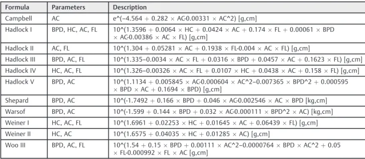
The EFW formulae listed were selected because they are widely used equations in the clinical practice, and because they are represented by fetal biometrics parameters avail-able in routine ultrasound examinations. Formulae de-scribed for small fetuses, such as those of Mielke I and II, were not selected because they required the measurement of the transverse diameter of the abdomen, which is not part of the patterns used in the selected health units. The selected formulae are described in detail in►Table 1.21–24
The Hadlock formulae are often interchanged in previous studies, according to the understanding of the authors. In the present article, these equations were used according to the detailed description and numbering in►Table 1. Following the birth, neonatologists immediately assisted the newborns. After initial care, birthweights were obtained and registered in scales of 5 g (Filizola, model BP Baby, São Paulo, SP, Brazil). The Local Ethics Committee approved this study under registration CAAE number 36546014.7.0000.5269.
The descriptive data analysis considered the mean and standard deviation. The formulae for fetal weight estimation listed in►Table 1 were compared in terms of means for
Table 1 Formulae for fetal weight estimation
Formula Parameters Description
Campbell AC e^(–4.564þ0.282AC-0.00331AC^2) [g,cm]
Hadlock I BPD, HC, AC, FL 10^(1.3596þ0.0064HCþ0.0424ACþ0.174FLþ0.00061BPD
AC-0.00386ACFL) [g,cm]
Hadlock II AC, FL 10^(1.304þ0.05281ACþ0.1938FL-0.004ACFL) [g,cm]
Hadlock III BPD, AC, FL 10^(1.335–0.0034ACFLþ0.0316BPDþ0.0457ACþ0.1623FL) [g,cm]
Hadlock IV HC, AC, FL 10^(1.326–0.00326ACFLþ0.0107HCþ0.0438ACþ0.158FL) [g,cm]
Hadlock V BPD, AC 10^(1.1134þ0.005845AC-0.000604AC^2–0.007365BPD^2þ0.000595 BPDACþ0.1694BPD) [g,cm]
Shepard BPD, AC 10^(-1.7492þ0.166BPDþ0.046AC-0.002546ACBPD [kg,cm]
Warsof BPD, AC 10^(-1.599þ0.144BPDþ0.032AC-0.000111BPD^2AC) [kg,cm]
Weiner I HC, AC, FL 10^(1.6961þ0.02253HCþ0.01645ACþ0.06439FL) [g,cm]
Weiner II HC, AC 10^(1.6575þ0.04035HCþ0.01285AC) [g,cm]
Woo III BPD, AC, FL 10^(1.54þ0.15BPDþ0.00111AC^2–0.0000764BPDAC^2þ0.05 FL-0.000992FLAC [g,cm]
absolute percentage error (APE¼|[Estimated weight–Birth weight]100/Birth weight|). All analyses were performed using the Statistical Package for the Social Sciences (SPSS, IBM Corp., Armonk, NY, US) software, version, 20, and the R (R Foundation for Statistical Computing, Vienna, Austria) software, version 2.15.1, with a significance level of 0.05 as reference.
A bivariate analysis was performed to compare the mean values for fetal weight estimation obtained with the different formulae with the following variables: growth pattern (ap-propriate for gestational age [AGA]/IUGR), GA at birth (< 28 weeks, 28–32 weeks,>32 weeks), sex (male/female), AFI (normal/abnormal), UA status (normal/abnormal), and DV status (normal/abnormal). These analyses were performed to identify, for each of the equations used, which were related to the observed variations, considering a level of significance of 0.05.
Results
In total, 194 patients met the research inclusion criteria. One case was excluded due to inconsistent registration of BPD in the medical record. All fetuses were delivered through cesarean section, except for the stillbirths, which were delivered vaginally. The descriptive analysis of the studied population is summarized in►Table 2.
The average fetal weight estimated through each formula is shown in►Table 3.
For APE, the formulae that demonstrated the best per-formance in the studied population were Hadlock I, II, III, and IV and Warsof, which had the lowest median values (►Table 4).
The Hadlock IV (HC, AC, FL), Hadlock I (BPD, HC, AC and FL), and Hadlock III (BDP, AC, FL) formulae had the lowest median APE values: 8.17, 8.32, and 8.74 respectively. A mean APE value<10 was considered an indicator of satis-factory performance for each formula according to previous reports.
The cohort was divided into 2 groups per fetal growth: AGA,n¼116 (59.8%), and IUGR,n¼78 (40.2%). Intrauterine growth restriction was defined in the present study as weight two standard deviations below the average BW for each GA.
Regarding the mean APE in AGA fetuses, Hadlock I, II, III, IV, and Warsof formulae demonstrated the best performance (8.02, 8.95, 8.41, 7.87, and 8.72 respectively). Hadlock IV (HC, AC, FL) had the lowest APE value. The same performance was observed in the IUGR fetus population, with a mean APE of 8.77, 10.18, 9.23, 8.62, and 10.26 respectively, as shown in►Fig. 1. There was no significant difference between the median APE values of the AGA and IUGR groups, which were calculated using the Hadlock I (BPD, HC, AC and FL) and IV (HC, AC, FL) formulae.
The 11 formulae were also compared in groups defined per GA (24–28, 28–32 and 32–34 weeks), sex of the newborn (male or female), AFI (<5 cm or5 cm), UA Doppler status (PI>95th percentile or absent/reverse diastole), and DV Table 2 Characteristics of the studied sample
Maternal age (years), meanSD 316.2
Birth weight (grams), meanSD 918361.1
Gestational age at delivery (weeks), SD 28,82.3
BW<3rd percentile, frequency (percentage)
78 (40.2%)
Male sex frequency (percentage) 103 (53.1%)
Abnormal AFI frequency (percentage) 87 (44.8%)
Doppler UA AREDV frequency (percentage)
122 (62.9%)
Doppler DV abnormal S/A frequency (percentage)
60 (34.8%)
Stillborn frequency (percentage) 7 (3.6%)
Abbreviations: AFI, amnioticfluid index; AREDV, absent and reversed end-diastolic velocity; BW, birth weight; DV, ductus venosus; S/A, ventricular systole and atrial contraction ratio; SD, standard deviation; UA, umbilical artery.
Table 3 Representation of the 95% confidence interval of the mean estimated fetal weigth in grams using the eleven formulae
n Minimum Maximum Mean Standard deviation
Campbell 194 282.31 3,312.29 981.6658 450.65413
Hadlock I 193 318.5 2,032.9 910.569 368.9429
Hadlock II 194 305.0 2,382.2 909.611 375.9173
Hadlock III 194 320.7 2,127.4 918.754 374.0832
Hadlock IV 193 317.7 2,042.4 909.641 366.9738
Hadlock V 194 345.4 2,246.0 983.673 409.3975
Shepard 194 373.8 2,264.5 1,008.021 419.2746
Warsof 194 359.6 1,966.5 920.546 370.5401
Weiner I 193 348.4 1,657.7 840.630 298.8425
Weiner II 193 312.6 1,862.8 883.734 307.7843
(normal or altered). The Hadlock formulae demonstrated no statistically significant difference in performance in the subgroup analysis per neonate sex, GA at birth, AFI, UA, or DV status (►Table 5). The resulting table shows the existence or not of statistical association, considering a level of signifi -cance of 0.05.
Discussion
This study compared eleven formulae for fetal weight esti-mation in the clinical context of placental insufficiency observed on Doppler scan. This is a frequent pathology in pregnancy that is associated with an increased risk of growth restriction and low BW.2,3,8,11It is an original study in Brazil.
The formulae with the best performance for fetal weight estimation in the analyzed population were Hadlock I (BPD,
HC, AC and FL) and IV (HC, AC and FL), which use four and three fetal biometric parameters respectively, based on the mean APE. Thisfinding is consistent with what has already been documented in the medical literature for specific populations such as very premature fetuses and those with growth restriction.21,22,24–35 The formulae with the poorest performance in our population were Campbell and Shepard.
The Hadlock formulae did not significantly differ in per-formance in the analyses according to fetal growth, fetal sex, GA at birth, AFI, UA, and DV status.22Thisfinding is similar to those obtained by studies that assessed cases of extreme prematurity for any reason and pregnancies with specific conditions such as preeclampsia.21,22
Thefinal fetal biometry measurement for the ultrasound weight estimation was obtained at least five days before birth. The last Doppler velocimetry measurement was Table 4 Performance of the eleven formulae for fetal weight estimation, expressed as the mean absolute percentage error (APE)
n Minimum Maximum Mean APE Standard deviation
Campbell 194 0.01 99.54 13.8099 12.81752
Hadlock I 193 0.02 42.94 8.3193 7.32916
Hadlock II 194 0.05 47.86 9.4487 8.56320
Hadlock III 194 0.01 46.86 8.7386 7.69347
Hadlock IV 193 0.11 41.88 8.1745 7.23123
Hadlock V 194 0.02 48.81 11.5152 9.73483
Shepard 194 0.22 47.98 13.4033 10.14293
Warsof 194 0.00 45.49 9.3412 8.05797
Weiner I 193 0.05 33.24 9.7163 6.66733
Weiner II 193 0.01 37.21 9.5477 7.40668
Woo III 194 0.27 44.35 10.0275 8.15756
Table 5 Influence of the clinical/ultrasonographic parameters on the average of the absolute percentage error for each formula
Formula Growth pattern
GA at birth
Sex AFI UA status
DV status
Campbell No Yes Yes No Yes No
Hadlock I No No No No No No
Hadlock II No No No No No No
Hadlock III No No No No No No
Hadlock IV No No No No No No
Hadlock V No No No No Yes No
Shepard No No No No Yes No
Warsof No No No No No No
Weiner I Yes No Yes No No No
Weiner II No No No No No No
Woo III No No No No No No
Abbreviations: AFI, amnioticfluid index; DV, ductus venosus; GA, gestational age; UA, umbilical artery.
performed in all patients no more than 24 hours before birth. This can be considered a particular strength of the study, because these parameters are more accurate than what is commonly found in the literature, which is a range of up to seven days between the last ultrasound examination and delivery.21,22,25,26 Another quality that should be empha-sized is the fact that the present study included a relatively large cohort, composed of 194 patients.
The study has some limitations that should be addressed. Only formulae based on BPD, HC, AC, and/or FL with circum-ferences measured by ellipsis were included; thus, the present findings cannot be extrapolated to formulae that use other parameters.27We did not make adjustments for the interval between EFW and BW because this period was not longer than five days. This is a retrospective study; therefore, it carries a risk of loss of information due toflaws in medical records.
More studies, preferably with prospective designs and larger sample sizes, are necessary to corroborate thefindings presented here, minimizing possible biases and enabling the extrapolation of thefindings.
Conclusion
The present study demonstrated the variability of perfor-mance among eleven different formulae for weight estima-tion in premature fetuses who experienced changes in bloodflow. Our results indicate that the Hadlock formulae that use three (HC, AC and FL) or four (BPD, HC, AC and FL) biometric fetal parameters have the best results for this specific fetus population. Despite the reports of formulae designed specifically for premature and/or IUGR fetuses in the literature, the Hadlock I (BPD, HC, AC and FL) and IV (HC, AC and FL) formulae had fewer errors regarding BW in our study population. In addition, this better performance was not influenced by the clinical and ultrasound factors fre-quently present in early-onset placental insufficiency. Thus, considering the possible biases of this type of study design, our results indicate that the Hadlock I (BPD, HC, AC and FL) and IV (HC, AC and FL) formulae can be applied with satisfactory performance for fetal weight estimation in a population of fetuses with early-onset severe placental insufficiency.
Conflict of Interests
The authors have none to declare.
References
1 Ricci AG, Brizot MdeL, Liao AW, Nomura RM, Zugaib M. [Ultra-sonographic accuracy of fetal weight estimation and influence of maternal and fetal factors]. Rev Bras Ginecol Obstet 2011;33(09): 240–245. Doi: 10.1590/S0100-72032011000900004
2 Abuhamad AZ. The role of Doppler ultrasound in obstetrics. In: Callen PW, ed. Ultrasonography in Obstetrics and Gynecology. 5th
ed. Philadelphia, PA: Saunders Elsevier; 2011:794–807
3 Unterscheider J, Daly S, Geary MP, et al. Optimizing the definition of intrauterine growth restriction: the multicenter prospective PORTO Study. Am J Obstet Gynecol 2013;208(04):290.e1–290.e6. Doi: 10.1016/j.ajog.2013.02.007
4 Nardozza LMM, Araújo Junior E, Vieira MF, Rolo LC, Moron AF. Estimativa de peso ao nascimento utilizando a ultrassonografia bidimensional e tridimensional. Rev Assoc Med Bras (1992) 2010; 56(02):204–208. Doi: 10.1590/S0104-42302010000200020
5 Baschat AA. Doppler application in the delivery timing of the preterm growth-restricted fetus: another step in the right direction. Ultra-sound Obstet Gynecol 2004;23(02):111–118. Doi: 10.1002/uog.989
6 Nelson DB, Ziadie MS, McIntire DD, Rogers BB, Leveno KJ. Pla-cental pathology suggesting that preeclampsia is more than one disease. Am J Obstet Gynecol 2014;210(01):66.e1–66.e7. Doi: 10.1016/j.ajog.2013.09.010
7 Baschat AA, Gembruch U, Harman CR. The sequence of changes in Doppler and biophysical parameters as severe fetal growth restriction worsens. Ultrasound Obstet Gynecol 2001;18(06): 571–577. Doi: 10.1046/j.0960-7692.2001.00591.x
8 Moreira Neto AR, Córdoba JCM, Peraçoli JC. Etiologia da restrição do crescimento intrauterino (RCIU). Comun Ciênc Saúde 2011;22:21–30
9 Robson SC, Martin WL, Morris RK. The investigation and manage-ment of the small-for-gestational-age fetus. London: RCOG; 2013
10 Seravalli V, Baschat AA. A uniform management approach to optimize outcome in fetal growth restriction. Obstet Gynecol Clin North Am 2015;42(02):275–288. Doi: 10.1016/j.ogc.2015.01.005
11 Unterscheider J, O’Donoghue K, Daly S, et al. Fetal growth restric-tion and the risk of perinatal mortality-case studies from the multicentre PORTO study. BMC Pregnancy Childbirth 2014;14:63. Doi: 10.1186/1471-2393-14-63
12 Carvalho PRN, Sá RAM, Gomes SC Jr, Lopes LM, Moreira MEL. Evaluation of Hadlock’s formula in premature fetuses with severe Doppler abnormalities. J Perinat Med 2011;39:1–5
13 Campbell S, Wilkin D. Ultrasonic measurement of fetal abdomen circumference in the estimation of fetal weight. Br J Obstet Gynaecol 1975;82(09):689–697. Doi: 10.1111/j.1471-0528.1975.tb00708.x
14 O’Brien GD, Queenan JT, Campbell S. Assessment of gestational age in the second trimester by real-time ultrasound measure-ment of the femur length. Am J Obstet Gynecol 1981;139(05): 540–545. Doi: 10.1016/0002-9378(81)90514-7
15 Shepard M, Filly RA. A standardized plane for biparietal diameter measurement. J Ultrasound Med 1982;1(04):145–150. Doi: 10.78 63/jum.1982.1.4.145
16 Phelan JP, Smith CV, Broussard P, Small M. Amnioticfluid volume assessment with the four-quadrant technique at 36-42 weeks’
gestation. J Reprod Med 1987;32(07):540–542
17 Arduini D, Rizzo G. Normal values of Pulsatility Index from fetal vessels: a cross-sectional study on 1556 healthy fetuses. J Perinat Med 1990;18(03):165–172. Doi: 10.1515/jpme.1990.18.3.165
18 Rizzo G, Capponi A, Talone PE, Arduini D, Romanini C. Doppler indices from inferior vena cava and ductus venosus in predicting pH and oxygen tension in umbilical blood at cordocentesis in growth-retarded fetuses. Ultrasound Obstet Gynecol 1996;7(06):401–410. Doi: 10.1046/j.1469-0705.1996.07060401.x
19 Wladimiroff JW, Tonge HM, Stewart PA. Doppler ultrasound assess-ment of cerebral bloodflow in the human fetus. Br J Obstet Gynaecol 1986;93(05):471–475. Doi: 10.1111/j.1471-0528.1986.tb08656.x
20 Sá RAM, Chaves Netto H, Amim J Jr, et al. Ductus venosus velocimetry in normal pregnancy. Int J Gynaecol Obstet 2000; 70(S1):A28. Doi: 10.1016/S0020-7292(00)82042-1
21 Abele H, Hoopmann M, Wagner N, Hahn M, Wallwiener D, Kagan KO. Accuracy of sonographic fetal weight estimation of fetuses with a birth weight of 1500 g or less. Eur J Obstet Gynecol Reprod Biol 2010;153(02):131–137. Doi: 10.1016/j.ejogrb.2010.07.007
22 Geerts L, Widmer T. Which is the most accurate formula to estimate fetal weight in women with severe preterm preeclamp-sia? J Matern Fetal Neonatal Med 2011;24(02):271–279. Doi: 10.3109/14767058.2010.485232
24 Blumenfeld YJ, Lee HC, Pullen KM, Wong AE, Pettit K, Taslimi MM. Ultrasound estimation of fetal weight in small for gestational age pregnancies. J Matern Fetal Neonatal Med 2010;23(08):790–793. Doi: 10.3109/14767050903387052
25 Hadlock FP, Harrist RB, Sharman RS, Deter RL, Park SK. Estimation of fetal weight with the use of head, body, and femur measure-ments–a prospective study. Am J Obstet Gynecol 1985;151(03): 333–337. Doi: 10.1016/0002-9378(85)90298-4
26 Anderson NG, Jolley IJ, Wells JE. Sonographic estimation of fetal weight: comparison of bias, precision and consistency using 12 different formulae. Ultrasound Obstet Gynecol 2007;30(02):173–179
27 Smulian JC, Ranzini AC, Ananth CV, Rosenberg JC, Vintzileos AM. Comparison of three sonographic circumference measurement techniques to predict birth weight. Obstet Gynecol 1999;93(5 Pt 1):692–696. Doi: 10.1016/S0029-7844(98)00517-1
28 Dudley NJ. A systematic review of the ultrasound estimation of fetal weight. Ultrasound Obstet Gynecol 2005;25(01):80–89. Doi: 10.1002/uog.1751
29 Kurmanavicius J, Burkhardt T, Wisser J, Huch R. Ultrasonographic fetal weight estimation: accuracy of formulas and accuracy of examiners by birth weight from 500 to 5000 g. J Perinat Med 2004;32(02):155–161. Doi: 10.1515/JPM.2004.028
30 Medchill MT, Peterson CM, Kreinick C, Garbaciak J. Prediction of estimated fetal weight in extremely low birth weight neonates (500-1000 g). Obstet Gynecol 1991;78(02):286–290
31 Burd I, Srinivas S, Paré E, Dharan V, Wang E. Is sonographic assessment of fetal weight influenced by formula selection? J Ultrasound Med 2009;28(08):1019–1024. Doi: 10.7863/ jum.2009.28.8.1019
32 Jouannic JM, Grangé G, Goffinet F, Benachi A, Carbrol D. Validity of sonographic formulas for estimating fetal weight below 1,250 g: a series of 119 cases. Fetal Diagn Ther 2001;16(04):254–258. Doi: 10.1159/000053923
33 Shamley KT, Landon MB. Accuracy and modifying factors for ultrasonographic determination of fetal weight at term. Obstet Gynecol 1994;84(06):926–930
34 Siemer J, Egger N, Hart N, et al. Fetal weight estimation by ultrasound: comparison of 11 different formulae and examiners with differing skill levels. Ultraschall Med 2008;29(02):159–164. Doi: 10.1055/s-2007-963165