PERINATAL FACTORS
AFFECTING HUMAN DEVELOPMENT
PAN AMERICAN HEALTH ORGANIZATION
Pan American Sanitary Bureau, Regional Office of the
WORLD HEALTH ORGANIZATION
PERINATAL FACTORS
AFFECTING HUMAN DEVELOPMENT
Proceedings of the Special Session
held during the Eighth Meeting
of the
PAHO
Advisory
Committee on Medical Research
10 June 1969
Scientific Publication No. 185
PAN AMERICAN HEALTH ORGANIZATION
Pan American Sanitary Bureau, Regional Office of the
WORLD HEALTH ORGANIZATION
525 Twenty-third Street, N.W.
Washington, D.C. 20037, U.S.A.
NOTE
At each meeting of the Pan American Health Organization Advisory Committee on Medical Research, a special session is held on a topic chosen by the Committee as being of particular interest. At the Eighth Meeting, which convened in June 1969 in Washington, D.C., the session surveyed some of the factors which may act on the fetus during pregnancy and labor interfering with its normal development or causing irreversible damage. Their influence on perinatal morbidity and mortality as well as their, long-term consequences on the surviving child received special attention. The basis for early diagnosis, prevention and trcatment was carefully reviewed. This volume records the papers presented and the ensuing
PAHO ADVISORY COMMITTEE ON MEDICAL RESEARCH
Dr. Hernán Alessandri
Ex-Decano, Facultad de Medicina Universidad de Chile
Santiago, Chile
Dr. Otto G. Bier
Director, PAHO/WHO Immunology Research and Training Center Instituto Butantan
Sao Paulo, Brazil
Dr. Roberto Caldeyro-Barcia Jefe, Departamento de Fisiopatología Facultad de Medicina
Universidad de la República Montevideo, Uruguay
Dr. Philip P. Cohen
Chairman, Department of Physiological Chemistry
The University of Wisconsin Madison, Wisconsin, U.S.A.
Dr. René Dubos Member and Professor The Rockefeller University New York, New York, U.S.A.
Dr. Herman E. Hilleboe
Director, Division of Public Health Practice
School of Public Health and Administrative Medicine Columbia University New York, New York, U.S.A.
Dr. Bernardo A. Houssay Director, Instituto de Biología
y Medicina Experimental Buenos Aires, Argentina
Dr. Robert Q. Marston
Director, National Institutes of Health Bethesda, Maryland, U.S.A.
Dr. Walsh McDermott
Chairman, Department of Public Health Cornell University Medical College New York, New York, U.S.A.
Dr. James V. Neel
Chairman, Department of Human Genetics University of Michigan Medical School Ann Arbor, Michigan, U.S.A.
Professor Roger Revelle Center for Population Studies Harvard University
Cambridge, Massachusetts, U.S.A.
Dr. Marcel Roche
Director, Instituto Venezolano de Investigaciones Científicas Caracas, Venezuela
Dr. John C. Waterlow Director, Tropical Metabolism
Research Unit
University of the West Indies Kingston, Jamaica
Professor Abel Wolman Emeritus Professor of Sanitary
Engineering and Water Resources The Johns Hopkins University Baltimore, Maryland, U.S.A.
Dr. Salvador Zubirán
Director, Instituto Nacional de la Nutrición
México, D.F., Méx;co
Secretary
Dr. M. Martins da Silva Chief, Department of Research
Development and Coordination Pan American Health Organization Washington, D.C., U.S.A.
Special Session on
PERINATAL FACTORS AFFECTING HUMAN DEVELOPMENT
Moderator: Dr. Roberto Caldeyro-Barcia
PARTICIPANTS
Dr. Karlis Adamsons Department of Obstetrics and
Gynecology
College of Physicians and Surgeons of Columbia University New York, New York, U.S.A.
Dr. Omar Althabe
Laboratorio de Fisiopatología Obstétrica
Instituto de Maternidad "Pedro A. Pardo"
Buenos Aires, Argentina
Dr. Ileinz Berendes Perinatal Research Branch National Institute of Neurological
Diseases and Stroke National Institutes of Health Bethesda, Maryland, U.S.A.
Dr. Joseph Bieniarz Department of Obstetrics and
Gynecology
Michael Reese Hospital and Medical Center
Chicago, Illinois, U.S.A.
Dr. Herbert G. Birch Department of Pediatrics
Albert Einstein College of Medicine New York, New York, U.S.A.
Dr. Donald F. Caldwell Division of Psychobiology Lafayette Clinic Detroit, Michigan, U.S.A.
Dr. John A. Churchill Section on Pediatric Neurology National Institute of Neurological
Diseases and Stroke National Institutes of Health Bethesda, Maryland, U.S.A.
Dr. Geoffrey S. Dawes Department of Physiology The Nuffield Institute for Medical
Research Oxford, England
Dr. Joseph S. Drage
Section on Pediatric Neurology National Institute of Neurological
Diseases and Stroke National Institutes of Health Bethesda, Maryland, U.S.A.
Dr. Elio García-Austt Instituto de Neurología Hospital de Clínicas Montevideo, Uruguay
Dr. Peter Gruenwald Department of Pathology Veterans Administration Hospital Philadelphia, Pennsylvania, U.S.A.
Dr. Louis M. Hellman Department of Obstetrics and
Gynecology
State University of New York Downstate Medical Center Brooklyn, New York, U.S.A.
Dr. Wilbur Lawrence Holley Section on Pediatric Neurology National Institute of Neurological
Diseases and Stroke National Institutes of Health Bethesda, Maryland, U.S.A.
Dr. Edward H. Hon Department of Obstetrics and
Gynecology
LAC/USC Medical Center Los Angeles, California, U.S.A.
Dr. L. Stanley James Department of Anesthesiology College of Physicians and Surgeons
of Columbia University New York, New York, U.S.A.
Dr. Leon 1. Mann Behavioral Unit
Dr. Carlos Méndez-Bauer Servicio de Fisiología Obstétrica Hospital de Clínicas
Montevideo, Uruguay
Dr. Kamrran S. Moghissi Department of Gynecology and
Obstetrics
WAayne State University School of Medicine
Detroit, Michigan, U.S.A.
Dr. Ronald E. Myers
Laboratory of Perinatal Physiology National Institute of Neurological
Diseases and Stroke National Institutes of Health San Juan, Puerto Rico
Dr. Margaret Ounsted
The Nuflield Department of Obstetrics and Gynecology
Radcliffe Infirmary Oxford, England
Dr. Serafín V. Pose
Servicio de Fisiología Obstétrica Hospital de Clínicas
Montevideo, Uruguay
Dr. Juan J. Poseiro
Servicio de Fisiología Obstétrica Hospital de Clínicas
Montevideo, Uruguay
Dr. Mortimer G. Rosen
Department of Obstetrics and Gynecology The University of Rochester School of
Medicine and Dentistry Rochester, New York, U.S.A.
Dr. Arthur L. Rosenbaum Section on Pediatric Neurology National Institute of Neurological
Diseases and Stroke National Institutes of Health Bethesda, Maryland, U.S.A.
Dr. J. K. Russell
Department of Midwifery and Gynecology Princess Mary Maternity Hospital The University of Newcastle upon Tyne Newcastle upon Tyne, England
Dr. Ricardo L. Schwarcz Laboratorio de Fisiopatología
Obstétrica
Instituto de Maternidad "Pedro A. Pardo" Buenos Aires, Argentina
Dr. Z. K. gtembera
Research Institute for the Care of Mother and Child
Prague-Podolí, Czechoslovakia
Dr. William Weiss Office of Biometry
National Institute of Neurological Diseases and Stroke
National Institutes of Health Bethesda, Maryland, U.S.A.
Dr. William F. Windle
Institute of Rehabilitation Medicine New York University Medical Center New York, New York, U.S.A.
Dr. Myron Winick Department of Pediatrics The New York Hospital New York, New York, U.S.A.
Dr. Stephen Zamenhof
Department of Medical Microbiology and Immunology
University of California School of Medicine
Los Angeles, California, U.S.A.
Dr. M. Martins da Silva (Secretary) Department of Research Development
and Coordination
CONTENTS
Page
Opening Statement Roberto Caldeyro-Barcia ... 1
Effects of Protein and Zinc Nutrition on Behavior in the Rat Donald F. Caldwell and D onald O berleas . . . . . . ... ... 2
DNA Content of Placenta and Fetal Brain Myron Winick, Elba Velasco, and Pedro Rosso ... 9
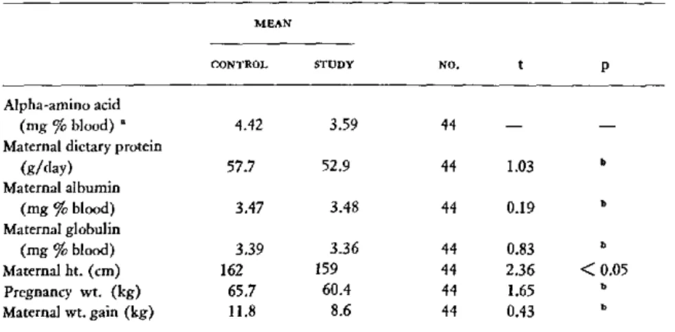
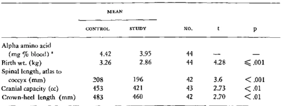
Relationships of Maternal Amino Acid Blood Levels to Fetal Development K. S. Moghissi,
1.
A. Churchill, and C. Frohman ... 16Discussion Stephen Zamenhof ... ... ... 20
General Discussion ... ... 22
Physical and Mental Deficits of Twinning W. L. Holley and
1.
A. Churchill ... 24Intelligence of Children Whose Mothers Had Acetenuria During Pregnancy John A. Churchill and Heinz W . Berendes .... ... . 30
Maternal Factors and Mental Performance in Children
1.
K. Russell and D. G. M illa r ... . . . . 3 6 Effect of Rapid Succession of Pregnancy W. L. Holley, A. L. Rosenbaum, and1.
A . C h u rch ill . ... .... .. . .. ... . .. ... . .. .. .. .. .. . ... . . .. .. . .. ... .. . ... 4 1 Perinatal Factors in Mental Subnormality Herbert G. Birch . ... 45Discussion John A. Churchill ... ... ... 46
General Discussion . . ... ... ... 47
The Problem of Prematurity and of Intrauterine Growth Retardation Peter Gruenwald ... ... 51
laternal Factors Affecting Birth Weight William Weiss and Esther C.
Jackson
. 54 Familial Factors Affecting Fetal Growth Margaret Ounsted ... 60Discussion . . ... . .. . . .. .... ... . 68
The Sonographic Depiction of the Growth and Development of the Human Fetus Louis M. Hellman, Mitsunao Kobayashi, Lewis Fillisti, and Ellen Cromb ... 70
Fetal Tolerance to Maternal Exercise Hypoxia Z K. Stembera ... 105 D iscussion . . . . .. .... .... .... ... . . .... . . 111
Pressure Exerted by Uterine Contractions on the Head of the Human Fetus During Labor R. L. Schwarcz, G. Strada-Sdenz, O. Althabe,
1.
Fernández-Funes, ahd R. Caldeyro-Barcia . ... ... . ... 115 Effects of Uterine Contractions on the EEG of the Human Fetus During LaborE lio G arcía-A ustt ... 127 The Human Fetal Electroencephalogram: 2. Characterizing the EEG During Labor
Mortimer G. Rosen and Joseph
J.
Scibetta ... 137 Influence of the Rupture of Membranes on Compression of the Fetal Head DuringLabor O. Althabe, G. Aramburú, R. L. Schwarcz, and R. Caldeyro-Barcia . . 143 Discussion Leon 1. Mann ... 150
General Discussion ... ... .... ... ... 157 Effect of Uterine Contractions on Maternal Blood Flow Through the Placenta
1. J.
Poseiro, C. Mendez-Bauer, S. V. Pose, and R. Caldeyro-Barcia ... 161 Factors Influencing the Acid-Base State of the Fetus During Labor K. Adamsons,H. O. Morishima, and A. C. Comas-Urrutia ... 172
Changes in Fetal Heart Rate Associated with Acute Intrapartum Fetal Distress
C. Méndez-Bauer,
J.
Monleón, G. Guevara-Rubio, C. Casacuberta, R. Bustos, G. Giussi, L. Escarcena, R. Yabo, and R. Caldeyro-Barcia ... 178 The Fetal Effects of Umbilical Cord Compression Edward H. Hon ... 188 Cardiovascular Adjustment of the Fetus During Asphyxia: The AorticChemore-ceptors G. S. Dawes ... 199 D iscussion . . ... . . . ... 202 Fetal Asphyxia and Perinatal Brain Damnage Ronald E. Myers ... 205 Asphyxial Brain Damage at Birth, with Reference to the Minimally Affected Child
William F. Wlndle ... 215 The Apgar Scores and Four-Year Psychological Examination Performance Joseph
S. Drage, Heinz W. Berendes, and Pearl D. Fisher ... 222 Fetal Distress: Its Significance in Neurological and Mental Impairment of Childhood
Heinz W. Berendes ... 228 D iscussion ... . ... ... 234 Administration of Oxygen, Glucose, and Alkali to Mother and Newborn L. Stanley
lames ... 239 A New Approach to the Treatment of Acute Intrapartum Fetal Distress R.
Caldeyro-Barcia, 1. M. Magaña,
J.
B. Castillo,1. 1.
Poseiro, C. Méndez-Bauer, S. V. Pose, L. Escarcena, C. Casacuberta,J.
R. Bustos, and G. Giussi ... 248OPENING STATEMENT
Roberto Caldeyro-Barcia, Moderator
Moderator:
One of the main objectives of the
health sciences today is to promote full, normal
human development. This approach includes the
somatic, sexual, neurological, and psychological
aspects. Unfortunately, there are many factors
that may interfere with this normal development.
At this meeting we have a distinguished group
of scientists from widely different disciplines,
all of which are interested in studying the factors
that may affect normal development. We shall
focus on some agents that may act during
preg-nancy, labor, and the early stages of the infant's
life.
The first series of papers will be devoted to
factors affecting the intrauterine development
of the fetus during pregnancy and the
conse-quences for the newborn and adult-maternal
nutrition, placental circulation, diabetes,
hyper-tension, rapid succession of pregnancies, familial
factors, weight of the mother before pregnancy
and weight gained during pregnancy, and others.
All of these may affect the growth of the fetus
during pregnancy. Special attention will be
devoted to newer methods recently developed
to evaluate the condition of the fetus during
pregnancy. The information supplied by these
methods will be most useful for deciding the
therapy to be applied in each case.
Later we shall study the more acute effects of
labor on the fetus, starting with one aspect that
has received little attention in recent years: the
compression received by the fetal head and the
possible resulting damage to the brain. Fetal
electroencephalography is a very promising
method for the study of this topic.
We shall then turn our attention to the more
fashionable subject of intrapartum fetal asphyxia
and acidosis resulting from acute failure of
pla-cental exchanges in labor, and also to the
cor-responding defensive reactions of the fetus. The
long-term neurological consequences of fetal
asphyxia will be discussed from experimental
studies in primates and from clinical data.
Finally, the methods for the treatment of acute
intrapartum fetal asphyxia will be discussed;
both the currently accepted procedures and some
recent proposals will be presented.
EFFECTS
OF PROTEIN AND ZINC NUTRITION ON
BEHAVIOR IN THE RAT'
Donald F. Caldwell and Donald Oberleas
2Although many factors may account for the paucity of research dealing with the effects of malnutrition on development of the nervous sys-tem and behavior patterns, a significant one is the doctrine of "brain sparing." To a great extent, sparing is a valid phenomenon in con-sidering the effect, or absence of effect, of some form of stress applied to the mature nervous system. However, with the discovery of "criti-cal" periods during the process of growth and development, numerous investigators have shown the effects of malnutrition during these periods to affect brain morphology profoundly
(6, 7, 15, 16, 17, 18, 19, 20, 21, 22, 23, 31). Of prime importance has been current research on whether various states of malnutrition when ap-plied prenatally, perinatally, or neonatally result in impaired performance (2, 3, 4, 5, 8, 9,
11,
12,
13, 14).
Initially, we attempted to compare the be-havioral effects in progeny from fifteen 80-to 90-day-old Harlan-Wistar rats fed either a 7.20 per cent or a 23.93 per cent protein diet administered throughout the total gestation period. We were unable to obtain a single test-able offspring from the low-protein group, al-though maternal weight data and autopsy results confirmed that most of the subjects had conceived. The experiment was repeated
' Supported by USPHS Grant AM-08142 and HD-01335-03, Detroit General Hospital Research Corpora-tion, Veterans AdministraCorpora-tion, and Gerber Products Company.
2 Presented by Dr. Caldwell.
beginning from the eleventh day of gestation through parturition (8). Our choice of this
period was based mainly on observations in the first study indicating a large percentage of preg-nancy terminations during the period of major organogenesis. In addition, we were interested in the interval during which the brain undergoes rapid maturation. Although pair-feeding was not instituted, daily ration weights were meas-ured for each subject and not found to differ significantly between experimental and control groups.
Table 1 presents the biometric data from this
TABLE 1. Biometric data for protein-depleted and con-trol diet groups
PROTEIN-DEPLETED CONTROL DIET GROUP DIET GROUP
Litters bred 10 10
Mean number born per litter 11.14 9.55
(alive and stillborn) (9 to 13) (4 to 13)
Mean number stillborn per 1.43 1.11
litter (3 litters eaten completely at birth)
Mean gestation days 22.89 21.67
(21 to26) (21 to23)
Mean weight (alive) of 4.13 gb 6.88 g
standardized litter at birth
Mean litter size at weaning 4.67 7.50
Mean weaning weight 43.02 g 51.70 g
p -= 0.05.
b p < 0.001.
-Protin Deple.d .2
.C.,.tr. 230
£
DAY 3 i
.'.o-. 1
DAY 2
¡ ¡ 1 U ¡ 1 I I
Z rr
T,¡.|.
m1 It",.%.
=,-al
1 3 mFIGURE 1. Mean latency to traverse Lashley 111 water maze on each of three trials for three consecutive test days. From eleventh day of gestation to parturition, sub-jects received either a 7.20 per cent protein diet (pro-tein-depleted group, N = 25) or a standard laboratory chow diet containing 23.93 per cent protein (control group, N = 58). Testing began at 30 days of age. Each test trial was 10 minutes.
--- otei. {-u0d.-~J
z-…_____ j_" (rattle...}I... 1 23.9,
111
111
\ 1
1
. 1
z u = I u m 3 mZ
TmIALS ?lll&tS T~IALS
FIGURE 2. Mean number of cul-de-sac and retrace errors in Lashley III water maze for each of three daily test trials administered for three consecutive days to subjects fed from eleventh day of gestation to parturition either 7.20 per cent (N = 25) or 23.93 per cent (N = 58) protein diet. Testing began at 30 days of age. Each test trial was 10 minutes.
study for the period from birth to weaning at 21 days of age. The gestation period was signifi-cantly longer and the birth weight lower for subjects in the low-protein (7.20 per cent) treatment. Similarly, the weaning weight was lower and the preweaning mortality greater for the protein-deficient subjects-differences that may in part reflect, since cross-fostering was not applied in this study, a detrimental influence of the maternal protein deficiency on early lactation. At parturition, the subjects in the low-protein group were placed on the 23.93 per cent
protein diet.
Beginning at 30 days of age, all animals were tested for learning ability with the Lashley III
TABLE 2. Comparison of first and third trials for each maze of 23.93 and 7.20 per cent protein groups
water maze. Three test trials were administered daily for three consecutive days, and the results for mean response latency are shown in Figure 1. The low-protein subjects took significantly
longer to solve the maze, but only for the first day of testing (mean= 126.02 and 133.82 sec-onds for the 23.93 per cenrt and 7.20 per cent protein groups, respectively; t=1.97, p<0.02 5).
However, the high-protein group showed, and the low-protein group did not, a significant intra-day reduction in response latency for each test day (see Table 2). Figure 2 presents the graph of mean number of errors for the two groups on the maze test. Analyses for differences between treatments in rate of improvement revealed that only for combined cul-de-sac errors on day 1 of
test day for mean response latency on Lashley III water
MEAN, TEST DAY I MEAN, TEST DAY 2 MEAN, TEST DAY 3
DIET IST TRIAL 3RD TRIAL 1ST TRIAL 3RD TRIAL 1ST TRIAL 3RD TRIAL
23.93'% protein 157.21 94.84 87.12 b 66.95 54.53 * 40.27
(N = 58)
7.20% protein 133.64 e 134.00 94.76 ' 80.76 73.16 ' 54.84
(N = 25)
p < .0005. b < .025. ' Not significant.
11 11
11
' ., 1
TABLE 3. Comparison of first and third trials for each test day for mean cul-de-sac and retrace errors on Lash-ley III water maze of 23.93 and 7.20 per cent protein groups
MEAN, TEST DAY 1 MEAN, TEST DAY 2 MEAN, TEST DAY 3
DIET IST TRIAL 3RD TRIAL IST TRIAL 3RD TRIAL lST TRIAL 3RD TRIAL
23.93% protein Retrace Cul-de-sac 7.20% protein
Retrace Cul-de-sac
2.22 1 9.00 a
2.08 c 8.32 b
.46 3.95
1.12 5.48
.1o b
4.91 '
1.24 d
5.72 d
'o
.u9 2.86
.72 4.00
.83 b
2.97
.52 d
3.04 d
.4!
1.65
.56 2.60
· p < .0005.
bp < .01.
p < .05.
d Not significant.
test difl
cul-7.2(
t=
con dec (cu wh
can (se
F
'4
'3-'0-9 ,,w
za
i;
,,: r
ing were the two group means significantly
groups on the pole-jump conditioned-avoidance
.erent (mean=5.05 and 2.84 reduction in test at 50 to 55 days of age. Avoidance responses
-de-sac errors, for the 23.93 per cent and were noted only for the high-protein subjects,
0 per cent protein groups, respectively;
and the over-all mean latency scores for the two
5.89, p<.0001). However, intra-treatment
groups were significantly different (mean=
nparisons showed that the high-protein group
5.99 and 9.19 seconds for the 23.93 per cent and
reased significantly in both types of errors 7.20 per cent protein groups, respectively; F=
l-de-sac and retrace) within each test day,
4.52, df=l
1 and 19, p<.01).
ereas the low-protein group showed a signifi-
Histological examinations of a random
sam-at decrease only within the first day of testing pie of nine protein-deficient and seven control
e Table 3).
subjects at 70 to 90 days of age revealed no gross
Figure 3 depicts the performance of the cerebral malformations, hemorrhagic
demyelina-tion, or other forms of destructive lesions.
How-ever, the brains of the protein-deficient rats were
observed to have a narrower cortex, fewer large
pyramidal cells, satellitosis, clumping of
cyto-plasmic chromatin, and neuronal shrinkage.
Currently we are conducting research on the
\---. - *.
,role
of zinc in the diet and its relationship with
level and type of dietary protein and behavior.
Our findings to date suggest a need for
reap-praisal of the importance accorded both quantity
and quality of dietary protein
per se
in the
, , , , , , ,
, , ,
development of the organism and its behavior.
I 2 3 4 5 6 7 S TIAL LSOCKS (lOtll.lblk)
FIGURE 3. Mean response latency on pole-jump
condi-tioned-avoidance test at 50 to 55 days of age of subjects administered either 7.20 per cent (N= 10) or 23.93 per cent (N = 58) protein diet from eleventh day of gesta-tion to parturigesta-tion. Condigesta-tioned stimulus interval was 5 seconds.
For years, biochemists and nutritionists have
cautioned us on the use of plant protein as a
substitute for animal protein in the diet, using
reduced growth rate as their prime evidence of
the inferiority of plant protein. In 1957 O'Dell
and Savage (27) discovered that zinc was less
available to animals fed plant-seed protein. In
4
21
1960 they demonstrated (28) that phytate, a normal constituent of plant foodstuffs gener-ally found in highest concentration in the seed portion and in some roots and tubers, decreased availability.
More recently, Oberleas and Prasad (26)
compared the growth of weanling rats placed for 10 weeks on either a zinc-supplemented (55 mg/kg) or a nonsupplemented diet of 4, 8, 12, 16, or 20 per cent soy assay protein. Corn oil (10 per cent), minerals (5 per cent), calcium carbonate (2 per cent), vitamins and methionine, and glucose monohydrate to make up the balance were used for all diets, and phytate was equalized at 1 per cent. Figure 4 shows the mean relationship between growth (weight gain for 3 weeks) and level of dietary protein as affected by zinc. The correlation between protein level and growth for the zinc-deficient diets probably reflects, in part, the basal levels of zinc in these diets, since no attempt was made to free the soy protein of zinc in the nonsupplemented diets. When compared to the growth curves for comparable non-zinc-supple-mented casein protein diets, the data indicated that soy protein supplemented with zinc is similar in quality to casein. This finding
con-firmed earlier observations (25, 28, 30).
It has been shown that zinc insufficiency
40 t
120
y 100
r')
E 80
gá 60
o
E 40
20-) 4 8 12 16 20
Dietory protein (%)
FIGURE 4. Mean relationship between growth (weight gain for three weeks) and five levels of dietary soy pro-tein either supplemented with 55 mg/kg zinc carbonate or nonsupplemented (basal level).
results in decreased net synthesis of DNA, RNA, and protein (29). Concomitant with this would be decreased activity or concentration or an increased turnover of enzymes. However, the biochemical interrelationship of zinc and pro-tein has only recently become the object of inves-tigation and still remains unclear.
We have recently begun studies to determine the behavioral effects of varying zinc loads in the diet (24). Table 4 summarizes the results of an investigation designed to test the effects on the behavior of offspring of rats fed 18 per cent soy protein with or without zinc supplemen-tation beginning ten weeks prior to breeding and throughout gestation and lactation. The supplemented diets contained 70 ppm zinc, the nonsupplemented 10 to 14 ppm, which was considered to constitute a mild zinc deficiency. At weaning, all offspring were placed on the zinc-supplemented 18 per cent protein regimen. Although the females on the nonsupplemented diet appeared normal throughout the duration of the study, prepartum ruptured uteri or post-partum death occurred in 56 per cent of them. Furthermore, only one litter survived to weaning at 21 days of age. An absence of typical maternal behavior (nest-building, cleaning of pups, con-sumption of placenta, retrieval, and so forth)
TABLE 4. Biometric data for zinc-supplemented and
nonsupplemented 18 per cent soy protein diet groups
NON-SUPPLE- SUPPLE-MENTED MENTED DIET DIET
(70 PPM (10-14 PPM
ZINC) ZINC)
Number of gravid females 15 16 Per cent with total litter
stilborn 0 32 Mean weight of liveborn at
birth (g) 6.40 * 4.85 Mean number liveborn per
litter 10.13 b 7.73
Per cent litters with total
preweaning mortality 0 93.7
* p < .0005. .05 < p < .02.
was observed consistently in all the zinc-deficient females. A similar observation has recently
been reported by Apgar (1).
Beginning at 45 days of age, the nine sur-viving progeny from the nonsupplemented treat-ment and a randomly selected litter of nine pups
fron
the supplemented, treatment were tested for behavior differences. Figure 5 shows the clear superiority of the subjects from zinc-supplemented mothers over the nonsupple-mented animals on the Lashley III water maze. Performance on the platform avoidance-condi-tioning test is shown in Figure 6; statistical analyses again revealed a significant superiority for subjects in the zinc-supplemented treatment (mean=5.07 and 3.76 seconds for nonsupple-mented and zinc-supplenonsupple-mented groups,respec-tively; F=4.58, df=l and 16, p<.0
5). The
results of an analysis computed to determine whether the groups differed for number of con-ditioned responses versus escape responses showed a significant increase in CR's for zinc-supplemented subjects (xz=17.72, p<.001). Finally, after a single five-minute exposure in the open field, a significantly higher activity score was manifested by animals in the zinc-supplemented treatment, a result interpreted as indicating lower emotionality than the perform-ance of the nonsupplemented subjects (mean=
PRENATAL ZINC PROJECT L-sley I!! Water Mae.
a
e
300
llOO
90 60 so 70
-40 40-30 -20
10
-IIl* nomal Ss
kq, ~" zinc deficient S s
... %%jLIJl.p ith*J ai_
iii1- ii i*{ iiII1 I tl
15 14 13 12
'ol
9 >.7 05 4 3 2PRENATAL ZINC PROJECT
Platform rno
I*lB normeal s
lar
zin deficienit . .. ,.,_. ... ,.
-1;*lalttit s.l
I 1 1 L Il L I I I L
1 2 3 4 5 o 7 a 9 lu 11 12
Trial BlIcks
(Five trial por blak)
FIGURE 6. Mean response latency on one-way condi-tioned-avoidance test of progeny of females fed 18 per cent soy protein diet supplemented with zinc (N = 9)
and without supplementation (N =- 9).
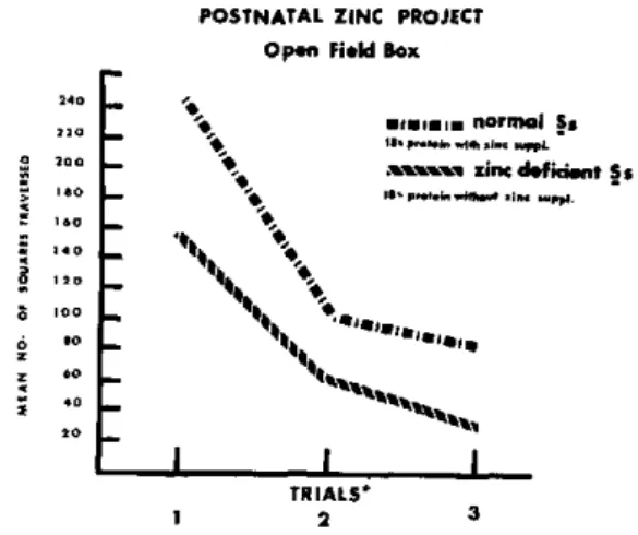
195.0 and 278.0 squares traversed in one five-minute trial for nonsupplemented and zinc-sup-plemented subjects, respectively; t = 2.45, p <.05 ). We next tested the behavior of normal labora-tory-reared male rats after being placed on an 18 per cent soy-protein zinc-deficient (8 ppm) diet for 48 days beginning at 30 days of age (10). Pair-fed control animals received an iden-tical diet that contained 70 ppm zinc. The diets contained 60 per cent glucose, 10 per cent corn oil, vitamins, and minerals; dietary phytate was equalized at 1 per cent for both. Figure 7 shows the significantly greater mean response latencies by the 12 nonsupplemented subjects in the Lashley III maze (mean= 161.22 and 102.72
POSTNATAL ZiNC PROJECT
Lashlay IIl Watear Maze
180 leo 160 * 140
, 120 100
80 60
40 20
1 2 3
TRIALS
FIGURE 5. Mean latency to traverse Lashley III water maze on each of three trials for three consecutive test days of progeny of females fed 18 per cent soy protein diet supplemented with zinc (N -9) and without sup-plementation (N - 9).
*i.i raiin ial Sa -sX P in de ficient Sa
",,1.. -i_ i- -r'
sarasa,.,
'Co
1 2 3
TRIALS
FIGURE 7. Mean latency to traverse Lashley III water maze on each of three trials for three consecutive test days. Subjects received either zinc-supplemented (N = 12) or nonsupplemented (N = 12) 18 per cent soy pro-tein diet for 48 days commencing at time of weaning.
_ _
15 14 13 12 10
9
7a
6
5 4
3
2
POSTNATAL ZINC PROJECT
Platform Box
- -.... 4 .l 1e
--ttt XInc defidi.n 5.
¡ 1 E
1 7 3 4 5 6 7 8 1 11 12
Tl,¡ial BIocks
(Five tr;als p.r block)
FIGURE 8. Mean response latency on one-way
condi-tioned-avoidance test of subjects receiving either
zinc-supplemented (N = 12) or nonzinc-supplemented (N= 12) 18 per cent soy protein diet for 55 days commencing at time of weaning.
seconds for nonsupplemented and zinc-supple-mented groups, respectively; F = 6.80, df = 1 and 33, p<.025). The differences between the groups for performance on the conditioned-avoidance test were also statistically significant (Figure 8) and again indicate superior learning by zinc-supplemented subjects (mean=5.19 and 4.06 seconds for nonsupplemented and zinc-supple-mented groups, respectively; F=15.32, df=l and 110, p<.001). A significantly larger pro-portion of conditioned responses was made by the zinc-supplemented animals (X2=21.04, p< .001). Finally, reduced emotionality levels were observed for zinc-supplemented subjects for the three days of testing in the open-field maze
REFEI
1. APGAR, J. Effects of zinc deficiency on
parturi-tion in the rat. Amer. J. Physiol. 215:160, 1968.
2. BARNES, R. H. Experimental animal approaches
to the study of early malnutrition and mental de-velopment. Fed. Proc. 26:144, 1967.
3. BARNES, R. H., S. R. CUNNOLD, R. R.
ZIMMER-MAN, J. SIMMONS, R. B. MAcLEOD, and L. KROOK. Influence of nutritional deprivations in early life on learning behavior of rats as measured by
perform-ance in a water maze. 1. Nutr. 89:399, 1966.
4. BARNES, R. H., A. V. MOORE, I. M. REID, and W. G. POND. Learning behavior following
nutri-G
O
S:
POSTNATAL ZINC PROJECT
Opon Field Box
**
res pimmn n
MIpL.ote vi~
z $._18iP.*i
ill aSii lii
TRIALS*
1 2 3
ormal Ss
Mi;~ pp. '
nc deficint Ss . i. .i..p.
*Five minute exploration period per trial -one trial por day
FIGURE 9. Mean number of squares traversed for each of three daily five-minute test periods on open field test for subjects receiving either zinc-supplemented (N = 12) or nonsupplemented (N= 12) soy protein diet for 55 days commencing at time of weaning.
(mean = 82.52 and 144.58 squares traversed in one five-minute trial per day for three consecu-tive days for nonsupplemented and zinc-supple-mented groups, respectively; F=14.88, df=l 1 and 33, p<.001), as is seen in Figure 9.
Research has demonstrated zinc to be an indispensable trace element for proper protein utilization and consequent growth. Further-more, for phytate-rich plant diets, zinc supple-mentation appears crucial. The present series of studies has shown for both prenatal and post-natal nutrition that even a mild zinc deficiency has a profound influence on behavior potential despite an apparently adequate protein level in the diet.
RENCES
tional deprivation in early life. /. Amer. Dietet. Assoc. 51:34, 1967.
5. BARNES, R. H., A. V. MOORE, I. M. REID, and
W. G. POND. Effects of food deprivation on beha-vioral patterns. In N. S. Scrimshaw and J. E. Gor-don (eds.), Malnutrition, Learning, and Behavior.
Cambridge, Massachusetts Institute of Technology Press, 1968.
6. BENTON, J. W., H. W. MOSER, P. R. DODGE,
and S. CARR. Modification of the schedule of mye-lination in the rat by early nutritional deprivation.
Pediatrics 38:801, 1966.
7
2.. 1 1. 2. . 1.. ,.o
1 1 .
.o 1.
7. CABAK, V., and R. NAJDANVlC. Effects of un-dernutrition in early life on physical and mental development. Arch. Dis. Childh. 40:532, 1965.
8. CALDWELL, D. F., and J. A. CHURCHILL.
Learn-ing ability in the progeny of rats administered a protein deficient diet during the second half of gestation. Neurology 17:95, 1966.
9. CALDWELL, D. F., and J. A. CHURCHILL.
Learn-ing impairment in rats administered a iipid free diet during pregnancy. Psychol. Rep. 19:99, 1966.
10. CALDWELL, D. F., D. OBERLEAS, A. S. PRASAD,
and J. CLANCY. Learning ability and emotionality
in rats administered a zinc deficient diet. In prepa-ration, 1969.
11. COWLEY, J. J., and R. D. GRIESEL. Some effects of low protein diet on a first filial generation of white rats.
1. Genet. Psychol.
95:187, 1959.12. COWLEY, J.
J.,
and R. D. GRIESEL. Pre- and post-natal effects of a low protein diet on the be-havior of the white rat. Psychol. Africana, 9:216, 1962.13. COWLEY, J. J., and R. D. GRIESEL. The
devel-opment of second-generation low-protein rats. ].
Genet. Psychol. 103:233, 1963.
14. COWLEY, J. J., and R. D. GRIESEL. The effect on growth and behavior of rehabilitating first and second generation low protein rats. Anim. Behav.
14:506, 1966.
15. CRAVIOTO, J., E. R. DELICARDIE, and H. G.
BIRCH. Nutrition, growth and neurointegrative de-velopment: an experimental and ecologic study.
Pediatrics, Suppl. 2, Part II, 38:319, 1966.
16. CULLEY, W. J., and E. T. MERTZ. Effect of
restricted food intake on growth and composition of preweaning rat brain. Proc. Soc. Exp. Biol. Med.
118:233, 1965.
17. DAVIDSON, A. N., and J. DOBBING. Myelination
as a vulnerable period in brain development. Brit. Med. Bull. 22:40, 1966.
18. DICKERSON, J. W. T., and J. DOBBING.
Pre-natal and postPre-natal growth and development of the central nervous system of the pig. Proc. Roy. Soc. Biol. 166:384, 1967.
19. DICKERSON, J. W. T., and J. DOBBING. The
effect of undernutrition early in life in the brain and spinal cord in pigs. Proc. Nutr. Soc. 26:5, 1967.
20. DICKERSON, J. W. T., J. DOBBING, and R. A.
MCCANCE. The effect of undernutrition on the
post-natal development of the brain and cord in pigs.
Proc. Roy. Soc. Biol. 166:396, 1967.
21. DOBBING, J. The influence of nutrition on the
development and myelination of the brain. Proc.
Roy. Soc. Biol. 159:503, 1964.
22. DOBBING, J. Effects of experimental
under-nutrition on development of the nervous system.
In N. S. Scrimshaw and J. E. Gordon (eds.), Malnu-trition, Learning, and Behavior. Cambridge,
Massa-chusetts Institute of Technology Press, 1968. 23. DOBBEING, J. Vulnerable periods in developing
brain. In A. N. Davison and J. Dobbing (eds.),
Applied Neurochemistry. Oxford, Blackwell Scien-tific Publications, 1968.
24. OBERLEAS, D., D. F. CALDWELL, A. S. PRASAD,
and J. CLANCY. Postnatal behavior impairment in
progeny of rats administered a prenatal zinc defi-cient diet. In preparation, 1969.
25. OBERLEAS, D., M. E. MUHRER, and B. L.
O'DELL. The availability of zinc from foodstuffs. In A. S. Prasad (ed.), Zinc Metabolism. Springfield, Illinois, Charles C Thomas, 1966.
26. OBERLEAS, D., and A. S. PRASAD. Growth as
affected by zinc and protein nutrition. Am.
1. Clin.
Nutr., in press, 1969.27. O'DELL, B. L., and J. E. SAVAGE. Symptoms
of zinc deficiency in the chick. Fed. Proc. 16:394, 1957.
28. O'DELL, B. L., and J. E. SAVAGE. Effects of phytic acid on zinc availability. Proc. Soc. Exp. Biol. Med. 103:304, 1960.
29. PRICE, C. A. Control of processes sensitive to zinc in plants and microorganisms. In A. S. Prasad (ed.), Zinc Metabolism. Springfield, Illinois, Charles
C Thomas, i966.
30. SMITH, W. H., M. P. PLUMLEE, and W. M. BEESON. Effects of source of protein on zinc
require-ment of the growing pig.
J.
Anim. Sci. 21:399, 1962. 31. STOCH, M. B., and P. M. SMYTHE. Doesunder-nutrition during infancy inhibit brain growth and
subsequent intellectual development? Arch. Dis.
DNA CONTENT OF PLACENTA AND FETAL BRAIN
Myron Winick, Elba Velasco, and Pedro Rosso
1Growth may be defined simply as an increase in the size of an animal or an individual organ. This increase in organ size is the result of a continuous accretion of protein and in some cases lipids. Hence the rate of net protein syn-thesis determines the rate of growth. When protein synthesis and protein degradation reach equilibrium, growth ceases. Since net protein synthesis is linear in all organs in utero and for a varying period postnatally, growth would ap-pear to be a homogeneous process. However, this is only one aspect of growth. The protein in any organ is packaged within cells. The same total organ may be contained in numerous smnall cells or in a few larger cells. The constant amount of DNA within the diploid nucleus of any cell in a particular species makes possible an accurate determination of the total number of cells by chemical methods (2).
During normal growth, total organ DNA con-tent or cell number increases linearly, then be-gins to decelerate, and finally reaches a maxi-mum long before the organ size, as determined by net protein accretion, has reached its maxi-mum (16). As a consequence of this pattern, three phases of growth can be described: pro-portional increase in weight, protein, and DNA content (hyperplasia); an increase in DNA slower than the increase in protein and weight (hyperplasia and concomitant hypertrophy); and finally no further increase in DNA content, with net protein and weight continuing to increase at the same rate (hypertrophy).
1 Presented by Dr. Winick.
The growing period, then, is not a homo-geneous process when viewed in cellular terms. It is possible that stimuli that retard or accelerate growth may result in different effects, depending on whether they influence the rate of cell division (DNA synthesis) or the subsequent increase in cell size.
It has been shown in rats that early malnu-trition will curtail the rate of cell division if it occurs during the period of hyperplasia. In brain, this period is prior to weaning. Neonatal undernutrition not only will curtail the rate of cell division but will result in a permanent deficit in brain-cell number (17). Malnutrition after weaning will prevent the subsequent in-crease in cell size-an effect that can be reversed by later rehabilitation. During the period of rapid cellular proliferation, undernutrition will
DNA MALNUTRITION, PER CENT OF NORMAL
80
60
20
0
¿
CE EBRUM HI PPOCAMPUS CEREBELLUM
FIGURE 1. Etect of neonatal caloric restriction in the rat on total DNA content of various brain regions.
affect the rate of cell division in any brain area
where cells are dividing and in any cell type
in the process of division
(14).
Figure 1
dem-onstrates that in animals malnourished from
birth, cerebellum, where cells are most rapidly
dividing, is affected earliest and most severely.
Figure 2 focuses on the cell types involved. In
cerebral cortex, neurons are not dividing
post-natally and hence are not affected by
under-nutrition. The number of glia, however, is
reduced. In cerebellum all cell types are reduced.
The areas under the lateral ventricle and under
the third ventricle are other areas in which
primitive neuronal division is curtailed. Thus
discrete brain areas are disproportionately
af-fected by neonatal undernutrition. The
magni-tude of the effect is proportional to the rate of
cell division in a particular area. In contrast,
if increased nutrition occurs during the
pro-liferative phase, it will accelerate cell division
and result in a permanent increase in brain-cell
number (15,
19).
Thus, within certain limits, the number of
cells ultimately present in the adult brain may
be programmed during early life-during that
period in postnatal life when brain cells are
still dividing. Can this type of programming
take place during prenatal life? Certainly all
fetal organs are in the proliferative growth
phase and therefore would appear susceptible
to permanent cellular effects. However, the fetus
M NORMAL E2 MALNUTRITION
251
= l! It
2'I
Cerebral cortex
_
FIGURE 2. Effect of neonatal caloric restriction on vari-ous cell types in rat brain.
is protected from the environment by the mother
and the placenta. It may therefore be asked,
Can the mother and/or the placenta protect the
growing fetus from adverse environmental
stim-uli such as maternal malnutrition, maternal
dis-ease, or insufficient uterine vascular supply?
In animals it is possible to monitor the effects
of various maternal stresses on the cellular
growth of both placenta and fetal organs. In
the human the fetus is not available for organ
analysis of this kind, and therefore the placenta,
which is accessible after birth, is the only organ
that can be examined when the fetus is born
in a viable condition. Thus if changes in the
pattern of cellular growth in placenta may be
correlated with the cellular growth of fetal
or-gans in animals, perhaps similar changes in
human placenta will give us a clue to the effects
of similar maternal stresses on the human fetus.
Normal placental growth, both in the rat and
in the human, proceeds in the same way as was
described above for the organs of the rat. In
the rat placenta, DNA synthesis and hence cell
division stop at 17 days of a 21-day gestation
(18).
In the human placenta, cell division
con-tinues until around the fourth to
thirty-sixth week of gestation (20). In both cases net
protein synthesis continues to term. Thus the
same three phases of cellular growth may be
described as for other organs of the rat.
Stim-uli imposed prior to the seventeenth day or
thirty-sixth week, respectively, should lead to a
permanent reduction in placental cell number,
whereas stimuli that are active only after cell
division has stopped should affect the size of
individual cells but not their total number.
Hence examination of the ultimate cellular
make-up of the placenta should give us an idea
of when, during the course of growth, a stimulus
has been active.
determine the time during pregnancy when these stimuli are active.
Maternal protein restriction in rats will retard both placental and fetal growth. In placenta, cell number (DNA content) was reduced by 13 days after conception, cell size (protein/DNA) remained normal, and the RNA/DNA ratio was markedly elevated. Retardation in fetal growth first became apparent at 15 days. After this there was a progressive decrease in cell number in all the organs studied. By term there were only about 85 per cent as many brain cells as in control animals (Table 1). These data agree with previous data of Zamenhof, which showed a similar reduction in total brain-cell number in term fetuses whose mothers were exposed to a slightly different type of nutritional deprivation (22). Thus the cellular changes produced by severe prenatal food restriction are reflected in placenta even earlier than in the fetus, but a retardation of cell division in all fetal organs including brain can be clearly
dem-onstrated.
By employing radioautography after injecting the mother with tritiated thymidine, cell divi-sion can be assessed in various discrete brain regions (1). Differential regional sensitivity can be demonstrated in this way by the sixteenth day of gestation in the brains of fetuses of pro-tein-restricted mothers (Figure 3). The cerebral white and gray matter are mildly affected. The area adjacent to the third ventricle and the subiculum are moderately affected, whereas the cerebellum and the area directly adjacent to the lateral ventricle are markedly affected. These
TABLE 1. Effect on fetal growth of rats caused by mater-nal protein restriction
O NORMAL CONTROL
TISSUE WEIGHT PROTEIN RNA DNA
Whole animal 87 81 83 81
Brain 91 85 82 84
Heart 84 84 79 81
Lung 82 85 85 89
Liver 82 80 85 85
Kidney 84 81 82 85
25
20
-- , NORMAL
15
-10 _
¡M AiTERNAL POUIEIN
RESTRICTION
Cerebrum Cerebrum Third Subiculum Cerebellum Laleral
(grayl (whitel ventricle ventricle
FIGURE 3. Effect of maternal protein restriction on re-gional DNA content of fetal brain.
data again demonstrate that the magnitude of the effect produced on cell division is directly related to the actual rate of cell division at the time the stimulus is applied. Moreover, they demonstrate that the maternal-placental barrier in the rat is not effective in protecting the fetal brain from discrete cellular effects caused by maternal food restriction.
The subsequent course of these animals born of protein-restricted mothers can be examined. Chow has reported that, even if they are raised normally on foster mothers, their ability to utilize nitrogen is permanently impaired (3). Data from our own laboratory demonstrate that if they are nursed on normal foster mothers in normal-sized litters, they will remain with a deficit in total brain-cell number at weaning. Thus we can again see early programming of the ultimate number of brain cells. This pro-gram, moreover, is written in utero in response to maternal nutrition.
them to prenatal undernutrition will reverse the
cellular effects, this may not actually be so in
specific brain areas.
Perhaps the situation most analogous to that
in humans is exposing these pups, malnourished
in utero,
to subsequent postnatal deprivation.
Animals reared on foster mothers in groups of
18 show a marked reduction in brain-cell
num-ber by weaning. This effect is much more
pronounced than that of either prenatal or
post-natal undernutrition alone. As has been pointed
out, animals subjected to prenatal malnutrition
alone show a 15 per cent reduction in total
brain-cell number at birth. Animals subjected
only to postnatal malnutrition show a similar
15 to 20 per cent reduction in cell number at
weaning. In contrast, these "doubly deprived"
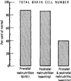
animals demonstrate a 60 per cent reduction
in total brain cell number by weaning (Figure
4). These data demonstrate that malnutrition
applied constantly throughout the entire period
of brain-cell proliferation will result in a
pro-found reduction in brain-cell number-greater
than the sum of effects produced during various
parts of the proliferative phase. The duration
of malnutrition as well as the severity during
1001
801-EE
o
CL
15~
oi-
601-
40-
20!-0'
-
TOTAL
BRAIN CELL NUMBERPrenatal
mainutrition
(birth)
Postnatal
mainutrition
(weaning)
Prenatal
& postnatal
mainutrition
(weaning)
FIGURE 4. Comparison of caloric restriction after birth, protein restriction during gestation, and "combined" pre-natal and postpre-natal restriction.
this early critical period would appear to be
extremely important in determining the ultimate
cellular make-up of the brain.
It has been shown in rats that clamping the
uterine artery supplying one horn of the uterus
will curtail fetal growth
(11).
The
accompany-ing cellular events can be examined. Regardless
of when the clamping is performed, cell division
is curtailed in all fetal organs except brain
(Table 2). In contrast, the DNA content of
placenta is reduced only if the clamping is done
before the seventeenth day; after this time the
cell size (protein/DNA) is reduced. The RNA/
DNA ratio in placenta increases following
uter-ine artery ligation at any time
(13).
The exact
significance of this is not known. An elevated
RNA/DNA ratio has been observed under a
variety of conditions involving tissue "stress,"
and is one of the first changes noted in cardiac
hypertrophy secondary to experimental aortic
ligation (6), in uterine hypertrophy induced by
estrogen (8), and in muscles exposed to repeated
nerve stimulation (7).
Organs of animals delivered on the
twenty-first day from ligated horns and then
foster-nursed to weaning do not attain the expected
number of cells by weaning. Thus again a
change produced
in utero
will persist throughout
life. In these animals, however, brain was
nor-mal at birth and remained nornor-mal at weaning.
Recently Zamenhof demonstrated that
arti-ficially reducing the number of fetuses
in utero
will result in offspring with an increased number
of brain cells
(10).
Certainly, then, in rats,
TABLE 2. Effect on cell division in rat fetuses of clamp-ing artery supplyclamp-ing one horn of uterus
% NORMAL CONTROL
TISSUE WEIGHT PROTEIN RNA DNA
manipulation of the maternal environment
dur-ing gestation will produce permanent cellular
changes in the offspring. The effects of similar
naturally occurring environmental stresses
dur-ing human pregnancies are more difficult to
demonstrate, but certain clear changes can be
noted.
Placentas from infants with "intrauterine
growth failure" show fewer cells and a higher
RNA/DNA ratio than controls
(12).
Fifty
per cent of placentas from an indigent
popula-tion in Chile showed similar findings, and
pla-centas from a malnourished population in
Guate-mala had fewer cells than normal (4). In a
single case of anorexia nervosa in which a
se-verely emaciated mother carried to term and
gave birth to a 2,500-gram infant, the placenta
contained less than 50 per cent of the expected
number of cells (Figure 5). Thus both vascular
insufficiency and maternal malnutrition will
curtail cell division in human placenta. The
cellular make-up of the placenta in both of these
situations strongly suggests that both stimuli
have been active for some time prior to the
thirty-fourth to thirty-sixth week of gestation.
The effects of these stimuli on the cellular
growth of the fetus are more difficult to assess.
In both situations fetal growth is retarded and
birth weight reduced (9). Indirect evidence
would suggest that cell division in the human
10C
80
60
40
20
0'
117
(20)
(1)
IUGF Indigent population Malnourished Anorexia
in Santiago, Chile population nervosa in Guatamala°
( ) Number of cases Data of Dayton et al.
MARASMUS
^^ (low birth weioht) lnormal MARASMUShirth weiMhl) KWASHIORKOR
1UO -- ' .... ... ... ...
· 50
FIGURE 6. Total DNA content in brains of children who died of malnutrition.
fetus might be retarded by maternal under-nutrition. If the available data on infants who died after exposure to severe postnatal malnu-trition are examined, three separate patterns emerge: Breast-fed infants malnourished during the second year have a reduced protein/DNA ratio but a normal brain DNA content. Full-term infants who subsequently died of severe food deprivation during the first year of life had a 15 to 20 per cent reduction in total brain-cell number. Infants weighing 2,000 g or less at birth who subsequently died of severe under-nutrition during the first year of life showed a 60 per cent reduction in total brain-cell number (Figure 6) (21). It is possible that these chil-dren were deprived in utero and represent a clinical counterpart of the "doubly deprived" animal. It is also possible that these were true premature infants and that the premature is
BRAIN STEM
50
40 DNA
(mg) 30
20
10 m , ¡- 1 ,1 ,1 . ,
,~
,
~
~
,
i
, i ,
i
i
0 4 8 12 16Age in months
20 24 28 FIGURE 5. Placental cell number in various types of ma- FIGURE 7. Regional growth of human brain in normal
ternal undernutrition. and marasmic children.
E
E
rX
7.
CEREBRAL CORTEX
DNA
(mg)
A
Age in months Age in montAs
FIGURE 8. Regional growth of human brain in normal FUR 9. Regional growth of human brain in normal
and marasmc children. and marasmic children.
much more susceptible to postnatal malnutrition than the full-term infant.
Regional growth in the human brain is some-what different from in the rat brain. Cell divi-sion stops at about the same time (eight to ten months) in cerebellum, cerebrum, and brain stem. Severe malnutrition during this prolifera-tive phase retards cell division in all three of these regions (Figures 7, 8, 9). The rate of cell division is about the same in cerebrum and in cerebral cortex, and both are severely affected by malnutrition. In comparison to the rat (5), cell division in human cerebellum is much more rapid during postnatal growth, and human cerebrum is much more affected by postnatal malnutrition. Thus postnatal malnutrition
cur-tails cell division in human brain as it does in rat brain. Prenatal stimuli affect human pla-centa in much the same way as rat plapla-centa.
Although at this time human fetal data are still sketchy, the suggestion that cell division in fetal organs can be curtailed by maternal undernutrition appears to be valid enough to require further studies. In view of the tremen-dous public health implications of this possi-bility, and in view of the evidence in animals that these changes are permanent, it would seem to us that every eflort should be made to quickly confirm or rule out the possibility that undernutrition of the mother may permanently reduce the number of brain cells in her offspring.
REFERENCES
1. ALTMAN, J., and G. DAS. Autoradiographic
and histological studies of postnatal neuronogenesis. I. A longitudinal investigation of the kinetics, mi-gration and transformation of celís incorporating tritiated thymridine in infant rats with special ref-erence to postnatal neurogenesis in some brain regions.
J.
Comp. Neurol. 126:337, 1966.2. BolviN, A., R. VENDRELY, and C. VENDRELY.
L'acide desoxyribonucleique du noyau cellulaire, dépositaire des caracteres hereditaires; arguments d'ordre analytique. Compt. Rend. Acad. Sci. 226: 1061, 1948.
3. CHOW, B. Presented at Gordon Research Con-ference on Nutrition, 1967, New London, New Hampshire.
4. DAYTON, D. H., L. J. FILER, and C. CANOSA. Reported at 53rd Annual Meeting, FASEB, Atlantic City, New Jersey, April 13-18, 1969.
5. FisH, I., and M. WINICK. Cellular growth in various regions of the developing rat brain. Pediat.
Res. 3:407, 1969.
6. GLUCK, L., N. J. TALNER, H. STERN, T. H.
GARDNER, and M. V. KULOVICH. Experimental car-diac hypertrophy; concentration of RNA in the
ventricles. Science 144:1244, 1964.
7. LOGAN, J. E., W. A. MANNELL, and R. ROSSITER. Chemical studies of peripheral nerve during
wal-lerian degeneration. Biochem. J. 52:482, 1952.
8. MOORE, R. J., and T. H. HAMILTON.
Estrogen-induced formation of uterine ribosomes. U.S. Nat. Acad. Sci. 52:439, 1964.
DNA
(mg)
9. SMITH, C. A. Effects of maternal undernutri-tion upon the newborn infant in Holland (1944-45).
1.
Pediat. 30:229, 1947.10. VAN MARTHENS, E., and S. ZAMENHOF.
De-oxyribonucleic acid of neonatal rat cerebrum in-creased by operative restriction of litter size. Exp.
Neurol. 23:214, 1969.
11. WIGGLEWORTH, J. S. Experimental growth
retardation in the fetal rat.
1.
Path. and Bact. 88:1, 1964.12. WINICK, M. Cellular growth of human pla-centa. IIl. Intrauterine growth failure.
1.
Pediat.71: 390-395, 1967.
13. WINICK, M. Diagnosis and Treatment of
Fetal Disorders. New York, Springer, 1969, 83-101.
14. WINICK, M. Proceedings of 53rd Annual Meeting, FASEB, Atlantic City, New Jersey, April
13-18, 1969.
15. WINICK, M., 1. FISH, and P. Rosso. Cellular recovery in rat tissues after a brief period of
neo-natal malnutrition. 1. Nutr. 95:623, 1968.
16. WINIcK, M., and A. NOBLE. Quantitative changes in DNA, RNA, and protein during
pre-natal and postpre-natal growth in the rat. Devel. Biol.
12:451, 1965.
17. WINICK, M., and A. NOBLE. Cellular response
in rats during malnutrition at various ages.
1.
Nutr.89:300, 1966.
18. WINICK, M., and A. NOBLE. Quantitative
changes in ribonucleic acids and protein during normal growth of rat placenta. Nature 212:34, 1966.
19. WINICK, M., and A. NOBLE. Cellular response
with increased feeding in neonatal rats. J. Nutr.
91:179, 1967.
20. WINICK, M., A. NOBLE, and A. CoscIA.
Cellu-lar growth in human placenta. 1. Normal placental growth. Pediatrics 39:248, 1967.
21. WINICK, M., and P. Rosso. The effect of
severe early malnutrition on cellular growth of hu-man brain. Pediat. Res. 3:181, 1969.
22. ZAMENHOF, S., E. VAN MARTHENS, and F. L.
MARGOLIS. DNA (cell number) and protein in
RELATiONSHIPS OF MATERNAL AMiNO ACiD BLOOD
LEVELS
TO FETAL DEVELOPMENT
K. S. Moghissi, J. A. Churchill, and C. Frohman
2Previous reports on the relationship between
low birth weight and intelligence quotient
sug-gest that maternal nutritional deficiencies may
produce intrauterine stunting and impairment
of fetal brain development (1,
3, 4).
Protein
deprivation in pregnant rats results in offspring
with decreased birth weight and impaired
learn-ing ability that persists after maturity (2).
Dobbing found that weanling rats given
low-protein and low-calorie diets had brains
differ-ent in chemical composition from those reared
on normal diets.
This study was designed to investigate the
effects of protein deprivation during human
gestation by comparing varying maternal blood
levels of free alpha-amino acids as related to
infant birth weight, length, and cranial volume.
Controlling and assessing the diets of pregnant
women is difficult. Hence, other measures of
nutrient supply were sought by determining
blood protein and amino acid levels. Transfer
of maternal amino acids to the fetus is known
to be free and proportional (8).
Study groups
All the patients selected for study were
ad-mitted to the hospital. All were in Social Class
V according to the Hollingshead-Redlich scale.
1 Supported by a grant from the Seyforth Foundation and grant PH-43-68-669 from the National Institute of Neurological Disease and Blindness, National Institutes of Health, Bethesda, Maryland, U.S.A.
2 Presented by Dr. Moghissi.