Rev Bras
Cineantropom
Hum
DOI: http://dx.doi.org/10.5007/1980-0037.2018v20n1p102
original article
Assessment of bone mineral density in young
female handball players
Avaliação da densidade mineral óssea em adolescentes
jogadoras de handebol
Tathyane Krahenbühl1,2
Juliano Henrique Borges2
Antonio de Azevedo Barros Filho3
Gil Guerra-Junior2,3
Ezequiel Moreira Gonçalves2,3
Abstract – Optimizing bone mass gain during childhood and adolescence may help pre-vent bone diseases in advanced ages. The aim of this study was to verify the bone mineral density (BMD) and bone mineral content (BMC) in female adolescent’s handball players. This is a cross-sectional study where 68 female adolescents (12–17 years) were allocated into two groups: handball players (n = 29) (HG) and control group (n = 39) (CG). BMC and BMD from total body (TB), total body less head (TBLH), lumbar spine (L1–L4), femoral neck (FN), Ward’s triangle (WT) and respectively Z-scores were measured using dual-energy X-ray absorptiometry (DXA). Sexual maturity, menarche, PHV, time of sun exposure, physical activity level and Calcium and vitamin D intake were assessed. The HG showed significantly higher BMC, BMD as well Z-scores values (p≤0.05) of total body, TBLH, femoral neck, hip and lumbar spine than the CG. When the values were adjusted for lean soft tissue (LST) the HG showed significantly higher BMC of femoral neck (p≤0.05), as well as BMD of TBLH and femoral neck (p≤0.05) and Z-score values all bone sites except hip, than the CG. We conclude that handball players have significantly higher bone mass values compared to group of girls of the same age.
Key words: Adolescent; Bone density; Woman; Sports.
Resumo – Otimizar o ganho da massa óssea durante a infância e adolescência pode auxiliar na prevenção de doenças ósseas em idades mais avançadas. Objetivou-se verificar a densidade mineral óssea (DMO) e conteúdo mineral ósseo (CMO) em adolescentes do sexo feminino. Foi realizado um estudo transversal com 68 meninas adolescentes (12 a 17 anos), divididas em dois grupos: jogadoras de handebol (HG: n=29) e grupo controle (CG: n=39). DMO e CMO de corpo inteiro (TB), corpo total menos cabeça (TBLH), coluna lombar (L1-L4), colo do fêmur (FN) e triângulo de Ward’s (WT) e respectivos escore Z foram medidos usando a absorciometria por dupla emissão de raios-x (DXA). Também foi avaliada a maturidade sexual, menarca, PVC e ingestão de cálcio e vitamina D. As jogadoras de handebol mostraram valores de CMO, DMO e respectivos escores Z do corpo inteiro, TBLH, fêmur, quadril e coluna lombar significativa-mente maiores quando comparados ao grupo controle. Quando ajustados para a massa isenta de gordura o grupo HG apresentou valores maiores para o CMO do fêmur e DMO do fêmur e TBLH; e nos valores de escore Z de todos os sítios ósseos, exceto o quadril, quando comparadas ao CG. Concluímos que as jogadoras de handebol têm valores de massa óssea significativamente
1 Universidade Federal de Goiás. Fa-culdade de Educação Física e Dança. Goiânia, GO. Brasil.
2 Universidade Estadual de Campi-nas. Faculdade de Ciências Médicas. Centro de Investigação em Pediatria. Laboratório de Crescimento e Desen-volvimento. Campinas, SP. Brasil.
3 Universidade Estadual de Campi-nas. Faculdade de Ciências Médicas. Campinas, SP. Brasil.
INTRODUCTION
Osteoporotic fractures are associated with low bone mass and dependent on the amount of acquired bone mass throughout life, mainly during puberty and early adulthood1,2. Childhood and adolescence are important phases
for mineralization and bone mass improvements3. Thus, optimizing the
bone mass acquisition during childhood and adolescence is an interesting strategy to preventing bone diseases during old age1,4.
Sports participation influences bone mineral density (BMD) and can be an important tool for children and adolescents to practice activities that increase bone mass; however, according to Wolff’s Law, bone strain must exceed the modelling threshold range to provide osteogenic stimulus to increase bone strength2. Bone strain in sports involving variety rapid
direc-tional changes such as volleyball and gymnastics is associated with a higher BMD than sports involving only one direction of motion such as running1,5,6.
Therefore, bone responses are modulated by the local application of a mechanical load e.g., bone sites to which a mechanical load was applied demonstrated higher bone mass improvements than those sites without mechanical overload7. So, the handball practice requires physical and
technical abilities such as repeated high-intensity actions, changes in direc-tion, sprints, jumps, duels, contact, throwing, falls, and ball blocks during defensive actions providing mechanical overload on the upper and lower limbs7-9. Altogether, the mechanical overload induced by handball actions
may increase osteogenic properties on the axial and appendicular bones7,9.
The background of the effects of physical activity and sports on BMD and bone mineral content (BMC) during adolescence, e.g., bone maturation and support strategies to optimize peak bone mass and prevent early bone diseases in old age1,10. However, the osteogenic effects induced by mechanical
overload from sports participation on specific bone sites and systemic bone properties in pubertal and post-pubertal participants are currently unclear.
Thus, this study aimed to verify the BMD and BMC from the whole body and several bone sites in female adolescent’s handball players, using dual-energy X-ray absorptiometry (DXA).
METHODOLOGICAL PROCEDURES
Study Design
This is a cross-sectional study. Sixty-eight female adolescent handball players from three different teams (regional and state competition level) and healthy subjects of the same age and sex from three cities of São Paulo state, Brazil, participated in the study.
Handball players group was recruited from December 2014 to Decem-ber 2015, and girls from the control group were recruited from January 2015 to July 2016.
months (e.g., corticoids); 3) no physical limitation that prevented the meas-urement procedures; 4) experienced menarche before the measmeas-urements; and 5) currently be between Tanner stages III and V. Participants also answered a questionnaire survey of physical activity level, food frequency, self-assessment of pubertal stage, and measurement of anthropometrics, BMD and BMC. Measurements were performed at the Growth and De-velopment Laboratory of the University of Campinas, Brazil.
Twenty-nine participants, those with at least 6 months of participation in official competitions before the measurements, were included in the handball players group (HG). Participants answered a questionnaire survey about participation time (months), weekly frequency of training (session/week), and weekly hours of indoor training (time/session), the minimum to participate in the research is to have frequency in the training of two days a week, with minimum time of ninety minutes per session, totalling three hours per week. The handball teams were invited because of the proximity to the university and because they are competitive teams with representative results in the respective age categories. And 75% of the sample studied in public school.
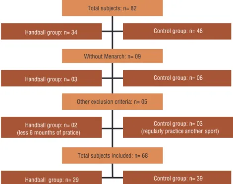
In the control group (CG), thirty-nine health adolescents without regular practice of physical activity or sport from four public schools were included. Several schools of Campinas (SP, Brazil) and cities in the region were contacted to participate in the study, however, only this four author-ized researchers to address students. Three participants were not included from the CG because participated in regular physical activity or sports in addition to physical education classes in their schools, observed in the interview with questionnaires. See figure 1.
Procedures were approved by the Research Ethics Committee of the University of Campinas (CAAE 37292814.9.0000.5404). Each participant provided written informed consent.
Maturity assessment
Pubertal status was assessed according to Tanner stages for females11 using
the secondary sexual characteristics (breast). Stages were determined by participant self-assessment in a private location after an explanation of the method was provided. Participants in stages III and IV were considered in the pubertal stage and those in stage V were in the post-pubertal stage. Sexual maturation was retrospectively analysed through the occurrence of menarche. We also measured somatic maturity predicting years from peak height velocity (PHV) from height, sitting height, leg length, weight, and age according to Mirwald et al.12.
Anthropometric measurements
Weight was measured using a Filizola digital scale (0–150 kg) with precision of 0.1 kg. Whole body height and sitting height were measured by Harpender stadiometer with precision of 0.1 cm. The body mass index (BMI) was calculated from weight (kg)/height (m2). The Z-scores of height (zHeight)
Figure 1. Flowchart of evaluations conducted with each group: handball group (HB) and control group (GC), with cross-sectional outline.
Bone Measurements
Bone status were measured by iDXA with enCORE™ 2011 version 13.60 software (GE Healthcare Lunar, Madison, WI, USA), the most accurate technique for measuring BMD in children, providing high reproduc-ibility, quick processing, low radiation exposure, and robust references in paediatric data1,10,13.
In accordance with the procedures recommended by the manufacturer, the equipment was calibrated daily. The same laboratory technician posi-tioned the subjects, performed the examinations and performed the analysis according to the operator’s manual using the standard analysis protocol. The reproducibility of the variables estimated by DXA was determined by the coefficient variation (CV%). The CV% of the laboratory was 0.28% (BMC and BMD) and 0.26% for LST.
T
he total body less head (TBLH) and spine are the preferred skeletal site for performing BMC and BMD in paediatric subjects13. Furthermore,we included other sites as femur and hip in this research, due to the large number of studies with these bone sites, so that it’s possible to compare it with the literature, even knowing that there are not the most suitable bone sites to be evaluated in this age group because of variability in skeletal development13. We also included BMC of arms, legs (rigth and left) and
trunk. Body composition was also performed for percentage of fat mass (%FM) and lean soft tissue (LST).
Sun Exposure, calcium, and vitamin D intake
A food frequency questionnaire was applied, estimating the daily intake of calcium (mg) and vitamin D (mg) according to the reference of daily nutrient intake (DRI) developed by the Institute of Medicine of the Na-tional Academies using the following formula:
D = (I-EAR) / √ (DpEAR2) + (Dpinter2 / n),
where I represents the daily nutrient value (mg) – from questionnaire, EAR is the estimated mean age requirement, Dinter = intrapersonal for age (respective values in specific tables), n =
number of days. Values of D ≥ 0 were considered a suitable daily nutrient intake.
All subjects in the sample had calcium and vitamin D intake within normal standards.
Sun exposure questionnaire was applied, which participants indicated how much time (<5 minutes, 5–30 minutes, or >30 minutes) they spend outdoors according to each day of the week, and the amount of skin that is exposed to the sun.
Physical Activity Level
A questionnaire survey of physical activity level was also applied as de-scribed by Baecke14. The questionnaire consists of three sections: 1) work,
2) sport (exercise), and 3) non-sport leisure activity.
Statistical analysis
SPSS version 16.0 (SPSS, Chicago, IL, USA) was used for the statistical analysis. To test data normality, we used the Shapiro-Wilk test. Variables of PHV, age, BMI, zBMI, LST, weight, calcium, vitamin D, zTBLH, and L1–L4 BMD were considered non-parametric. However, all BMC sites, all BMD sites (except L1–L4), all bone sites Z-score (except TBLH), Height, zHeight, %FM, Menarche, Sun exposure, and physical activity level were considered parametric variables. An unpaired Student’s t test or the Mann-Whitney U test was used to compare variables differences between the CG and HG groups. Multivariate analysis with Bonferroni correction was used to compare CG and HG adjusting the differences for LST, weight, BMI. The chi-squared x2 test was used to verify differences
in the proportion of participants according to pubertal status (Tanner stages). The coefficient of partial correlation was also used to verify the correlation between participation time of handball training and all analysed bone status variables using age as the controlled variable. The significance level was set at α ≤ 0.05.
RESULTS
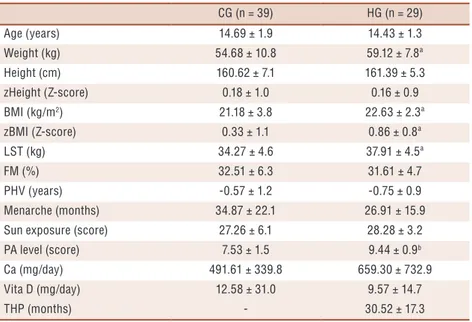
similar in most variables as age, height, zHeight, fat mass, maturity, sun exposure, calcium and vitamin D (p> 0.05); on the other hand, the handball players demonstrated significantly higher lean soft tissue (p= 0.00), weight (p= 0.01), BMI (p= 0.00), zBMI (p= 0.02), and PA level (p = 0.00) than the control group (Table 1).
In accordance with Brazilian obesity guidelines15 were found nine
sub-jects underweight (HG=03 and CG=06), 49 subsub-jects eutrophics (HG=23 and CG=26), seven with overweight (HG=02 and CG=05), three with pre-obesity (HG=01 and CG=02), and no obese in the sample.
Table 1. Subjects’ characteristics by groups (mean ± SD).
CG (n = 39) HG (n = 29)
Age (years) 14.69 ± 1.9 14.43 ± 1.3
Weight (kg) 54.68 ± 10.8 59.12 ± 7.8ª
Height (cm) 160.62 ± 7.1 161.39 ± 5.3
zHeight (Z-score) 0.18 ± 1.0 0.16 ± 0.9
BMI (kg/m2) 21.18 ± 3.8 22.63 ± 2.3ª
zBMI (Z-score) 0.33 ± 1.1 0.86 ± 0.8ª
LST (kg) 34.27 ± 4.6 37.91 ± 4.5ª
FM (%) 32.51 ± 6.3 31.61 ± 4.7
PHV (years) -0.57 ± 1.2 -0.75 ± 0.9
Menarche (months) 34.87 ± 22.1 26.91 ± 15.9
Sun exposure (score) 27.26 ± 6.1 28.28 ± 3.2
PA level (score) 7.53 ± 1.5 9.44 ± 0.9b
Ca (mg/day) 491.61 ± 339.8 659.30 ± 732.9
Vita D (mg/day) 12.58 ± 31.0 9.57 ± 14.7
THP (months) - 30.52 ± 17.3
Body Mass Index (BMI); Fat Mass (FM); Lean Soft Tissue (LST); Peak Height Velocity (PHV);Calcium (Ca);Vitamin D (Vita D); Physical Activity (PA); Time Handball Participation (THP). ªDifference between groups (Mann-Whitney U test, P ≤ 0.05). bDifference between groups (unpaired Student’s t test, P ≤ 0.001).
The handball players showed significantly higher BMC of total body (p= 0.00), TBLH (p= 0.00), femoral neck (p= 0.00), Ward’s triangle (p= 0.00), lumbar spine (p= 0.00), Arms (p= 0.00), Legs (p= 0.00), and Trunk (p= 0.00) as well as BMD of total body (p= 0.00), TBLH (p = 0.00), femoral neck (p= 0.00), Ward’s triangle (p= 0.00), and lumbar spine (p= 0.00) than the control group (Table 2).
All subjects in the sample had bone mass within the normal range. The handball players showed significantly higher BMD Z-scores for total body (p= 0.00), TBLH (p = 0.00), femoral neck (p = 0.00), Ward’s triangle (p = 0.02), and lumbar spine (p < 0.00) than the control group (Figure 2).
Table 2. Comparison of bone mineral content (BMC), bone mineral density (BMD) and Z-score from the whole body and by bone site between handball players and control group (mean ± SD).
CG (n = 39) HG (n = 29) (%) p* p** p***
BMC (g)
TB 2042.31 ± 312.5 2250.04 ± 289.7c 9.2 0.95 0.04 0.03
TBLH 1598.70 ± 264.3 1811.75 ± 250.7c 11.8 0.30 0.00 0.00
FN 4.46 ± 0.7 5.28 ± 0.8b 15.5 0.00 0.00 0.00
WT 1.95 ± 0.4 2.41 ± 0.5c 19.1 0.07 0.00 0.00
L1-L4 51.16 ± 10.3 58.18 ± 9.9c 12.1 0.50 0.04 0.02
Arms 239.27 ± 37.3 267.07 ± 39.0c 10.4 0.68 0.03 0.02
Legs 756.85 ± 122.0 861.00 ± 115.7b 12.1 0.14 0.00 0.00
Trunk 602.59 ± 113.7 683.67 ± 110.4c 11.9 0.74 0.02 0.02
BMD (g/cm2)
TB 1.04 ± 0.1 1.11 ± 0.0a 6.3 0.37 0.03 0.02
TBLH 0.92 ± 0.0 1.00 ± 0.0a 8.0 0.02 0.00 0.00
FN 1.06 ± 0.1 1.22 ± 0.2b 13.1 0.00 0.00 0.00
WT 0.99 ± 0.1 1.14 ± 0.2c 13.2 0.06 0.01 0.01
L1-L4 1.05 ± 0.1 1.17 ± 0.1a 10.3 0.04 0.00 0.00
Z-score
TB 0.29±1.0 1.23±1.0 76.4 0.02 0.00 0.00
TBLH -0.05±0.9 0.88±1.1 105.6 0.03 0.00 0.00
FN 0.64±1.1 2.04±1.8 68.6 0.00 0.00 0.00
WT 0.52±1.1 1.62±2.0 67.9 0.06 0.02 0.03
L1-L4 -0.21±1.1 1.04±1.5 100.2 0.00 0.00 0.00
Total Body (TB); Total Body Less Head (TBLH); Lumbar Spine (L1-L4); Femoral Neck (FN); Ward’s triangle (WT); Bone Mineral Density (BMD); Bone Mineral Content (BMC). ªDifference between groups (Mann-Whitney U test, p< 0.05). bDifference between groups (unpaired Student’s t test, p < 0.00). cDifference between groups (unpaired Student’s t test, p< 0.05). Adjustments analysis were performed using lean soft tissue*, weight**, body mass index*** as covariates (Bonferroni). Values in boldface indicate significant differences (p < 0.05).
Figure 2. Comparison of bone mineral density (BMD) Z-score of the total body (TB), total body
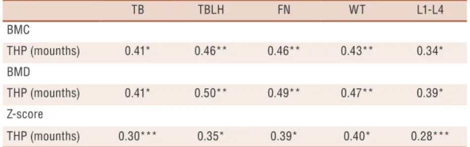
Table 3. Correlation between handball participation time and BMC and BMD from the whole body and bone sites adjusted for age in handball players.
TB TBLH FN WT L1-L4
BMC
THP (mounths) 0.41* 0.46** 0.46** 0.43** 0.34*
BMD
THP (mounths) 0.41* 0.50** 0.49** 0.47** 0.39*
Z-score
THP (mounths) 0.30*** 0.35* 0.39* 0.40* 0.28***
Total Handball Training (THP); Total Body (TB); Total Body Less Lead (TBLH); Lumbar Spine (L1-L4); Femoral Neck (FN); Ward’s triangle (WT); Bone Mineral Density (BMD); Bone Mineral Content (BMC).Coefficients of partial correlations were adjusted for age. * p< 0.01; **p< 0.00; ***p < 0.05.
DISCUSSION
In this study, handball players showed higher values of BMC and BMD in all measured bone sites when compared with the control group. The results showed associations between participation time in handball training with BMC and BMD in all bone sites.
These findings are in agreement with some cross-sectional studies, which found association between handball practice with increased physi-cal fitness, lean mass and bone mass9,16,17. Subjects in the handball group
had practice in this sport at least six months before the study presenting an average of participation over 30 months (2.5 years), that may strongly associate with the osteogenic effects on specific and whole-body BMD, which may explain these differences between groups.
However, great bone health in handball group may be explained by body composition. Although, subjects showed no differences between groups for %BF, they showed an average difference of ~3.5kg of LST and consequent differences in weight and BMI.
It is well established that muscle mass has a great influence on bone mass2. Accordingly, when we adjusted bone analysis for LST, weight and
BMI. There were no differences in results when adjusted for weight and BMI, but when he BMC and BMC were adjusted for LST the differences between the handball group and control group disappeared in some skeletal sites. Thus, greater muscle development in these subjects may support the better bone development18.
However, the differences for handball players maintained for BMD of TBLH, femur and lumbar spine after the adjustments for lean soft tissue. Total body (with or without the head) and lumbar spine are preferred for evaluation in the pediatric population, because they provide more informa-tion about the status of trabecular and cortical bone, being that the spine is the preferred site in pediatrics because of precision of measurements and easily identified bone landmarks13. So, our sample showed the handball
Added to this result, the Z-score data was maintained for all evaluated bone sites, except the hip, but according to the ISCD Pediatric Official Positions13, because of the variability in skeletal development in the hip,
it is not a preferred site for evaluation in growing children.
This adjusted for LST result is in agreement with the idea that sports participation also induces improvements in body composition, as we dem-onstrated in the present study; thus, high lean mass is also linked with bone status improvements13,19. So, handball participation is associated with both high
bone status and high lean mass9. The main mechanical stimulus to bone comes
from skeletal muscle contractions, which promote mechanical strain on bone structure to determine its hypertrophy as a consequence of bone remodelling20.
That is, the higher bone density found may be related to the greater lean mass and consequent action of muscle contraction, as explained by the concept of the “muscle–bone unit” which suggests that bone strength is mainly dependent on muscle actions that provide a high mechanical load and strain on bone structure2. The influence of physical activity on bone status
is related to mechanical tension and compression from skeletal muscle con-tractions induced by weight-bearing during movements21. Therefore, sports
with high muscle tension and high impact provide greater degrees of bone remodelling22, explaining the findings of the present study that higher bone
status may be strongly linked with the physical and technical demands of handball8, corroborating with our hypothesis that physical actions required
in handball such as defensive blocks, changes in direction, sprints, jumps, contact, throwing, falls, providing mechanical overload on the bone7-9.
Studies generally present absolute values of BMD and BMC, and here we demonstrated comparisons of Z-score between groups. Z-scores are calcu-lated by DXA software using the BMD and compared with reference values from healthy subjects of the same age, sex, and ethnicity10, adjusting data
according to these variables. Handball players demonstrated significantly higher Z-scores of BMD than control group in our study, even when the values were adjusted for the LST, demonstrated high bone status expected for the same age and sex in specific bone sites as well as systemic bone.
There are few studies relating the practice of handball and girls in ado-lescence9,16,17, but the scientific literature bring forward a variety sports, such
as gymnastics23, volleyball24, basketball25, and soccer26,27, might influence
bone status since subjects who participate in these sports have higher BMD measured by DXA than those who do not. Also, studies comparing the dominant and non-dominant sites of young athletes showed that the sport exerts influence on the members most used7,28. This supports the idea that
handball may be positively influencing the bone density of these adolescents. Practice of sports and physical activity for promoting bone health is important, but it is also important to have an optimal dietary intake of nutrients. Low calcium and vitamin D intake are associated with higher fracture risk at any age and sex, including childhood and adolescence10.
control group, minimizing the possible confusion of this variable.
Another factor that might influence BMC and BMD in adolescents is the maturational development. This factor was controlled, for example, only girls who have had menarche included in the study and the sample consisted of adolescents in the pubertal and post-pubertal stages (Tan-ner stages III–V). The proportions between groups of participants in the pubertal and post-pubertal stages could have biased the results16,29, but the
non-significant difference in the proportion between groups, demonstrated that the samples were homogeneously distributed since most subjects were in the pubertal stage (~82%).
However, we made the necessary adjustments and still demonstrated non-significant differences in age, growth (Height and zHeight), and maturity (PHV and menarche), because they are factors that may optimize improvements in bone status is maturity during the pubertal stage.
High bone status is important in quality of life related to preventing diseases such as osteoporosis and is associated with a lower prevalence of fractures in old age, demonstrating that the high impact induced by sports is beneficial to bone density modifications and osteogenic effects and protecting against fractures30. Bailey et al.3 showed in a longitudinal
study of children and adolescents of both sexes that the most active subjects presented higher bone mass peaks (9% and 17% greater BMC for males and females, respectively) than less active subjects, demonstrating the great effects of an active life on bone health.
Some studies demonstrate the importance to stimulation of muscle con-traction to bone formation2, may explaining our findings, that greater bone
health from handball players are linked with the sport characteristics, which has a high level demand of muscle actions from lower and upper limbs8.
This study has some limitations such as the intensity of the training sessions was uncontrolled, this was a cross-sectional study, and longitudinal studies are needed to verify the effects of handball training on bone status. And variables such as skin color and economic status were not evaluated.
Despite this, our study controlled some confound variables as height, PHV, age, maturity; did adjustments for variables as LST and weight and analysed Z-score data which brings reliability to the research.
So, the findings of the present study demonstrate the importance of sports participation such as handball on the body composition and bone health of female adolescents in the pubertal and post-pubertal stages.
We conclude that handball players had a great bone status when compared to control group, especially in Z-score values, and the handball participation time is also associated with higher muscle mass and bone status. Thus, participation in sports such as handball is a great path to improving bone heath during adolescence and can be a tool for preventing bone diseases in old age.
Acknowledgments
Laboratory from the University of Campinas for the academic support. We appreciate the financial support of CNPq: Edital Universal MCTI/ CNPq Nº14/2014 (462310/2014-0).
REFERENCES
1. Bailey CA, Brooke-Wavell K. Exercise for optimising peak bone mass in women. Proc Nutr Soc 2008; 67: 9-18.
2. Frost HM, Schonau, E. The “muscle-bone unit” in children and adolescents: a 2000 overview. J Pediatr Endocrinol Metab 2011;13(6):571-90.
3. Bailey DA, Mckay HA, Mirwald RL, Crocker PRE, Faulkner RA. A six-year longitudinal study of the relationship of physical activity to bone mineral accrual in growing children: The University of Saskatchewan Bone Mineral Accrual Study. J Bone Miner Res 1999;14(10):1672-9.
4. Matkovic V, Jelic T, Wardlaw GM, Ilich Z, Goel PK, Wright JK, et al. Timing of peak bone mass in caucasian females and its implication for the prevention of osteo-porosis. Inference from a cross-sectional model. J Clin Invest 1994;93(2):799-808.
5. Creighton DL, Morgan AL, Boardley D, Brolison PG. Weight-bearing exercise and markers of bone turnover in female athletes. J Appl Physiol 2011;90(2):565-70.
6. Nichols DL, Sanborn CF, Bonnick SL, Gench B, DiMarco N. Relationship of regional body composition to bone mineral density in college females. Med Sci Sports Exerc 1995;27(2):178-82.
7. Boshnjaku A, Dimauro I, Krasniqi E, Grazioli E, Tschan H, Migliaccio S, et al. Effect of sport training on forearm bone sites in female handball and soccer players. J Sports Med Phys Fitness 2016;56(12):1503-10.
8. Karcher C, Buchheit M. On-court demands of elite handball, with special refer-ence to playing positions. Sports Med 2014;44(6):797-814.
9. Vicente-Rodriguez G, Dorado C, Perez-Gomez J, Gonzalez-Henriquez JJ, Calbet JA. Enhanced bone mass and physical fitness in young female handball players. Bone 2004;35:1208-1215.
10. Bacharach LK, Gordon CM. Bone densitometry in children and adolescents. Pediatrics 2016;138(4): e20162398.
11. Marshall WA, Tanner JM. Variations in pattern of pubertal changes in girls. Arch Dis Child 1969;44(235):291-303.
12. Mirwald RL, Baxter-Jones AD, Bailey DA, Beunen GP. An assessment of maturity from anthropometric measurements. Med Sci Sports Exerc 2002;34(4):689-94.
13. Crabtree NJ, Arabi A, Bachrach LK, Fewtrell M, Fuleihan GEH, Kecskemethy HH, et al. Dual-energy X-ray absorptiometry interpretation and reporting in children and adolescents: the revised 2013 ISCD Pediatric Official Positions. J Clin Densitom 2014;17(2):225-42.
14. Guedes DP, Guedes JERP. Manual prático para avaliação em educação física. Barueri: Manole; 2006.
15. Godoy-Matos AF, Oliveira J, Guedes EP, et al. Diretrizes brasileiras de obesidade 2009/2010. Associação Brasileira para o Estudo da Obesidade e da Síndrome Metabólica (ABESO); 2009.
16. Ubago-Guisado E, Gomez-Cabello A, Sanchez-Sanchez J, Garcia-Unanue J, Gallardo L. Influence of different sports on bone mass in growing girls. J Sports Sci; 2015;33(16):1710-18.
17. Mrabet Bahri D, Selmi A, Abdelkéfi M, Mbarek M, Sahli H, Sellami S. Study of bone mineral density in adolescent handball players: a study of 20 cases. Tunis Med 2013;91(11):633-7.
18. Macdonald H1, Kontulainen S, Petit M, Janssen P, McKay H. Bone strength and its determinants in pre- and early pubertal boys and girls. Bone 2006;39(3):598-608.
CORRESPONDING AUTHOR
Tathyane Krahenbühl Faculdade de Educação Física e Dança, Universidade Federal de Goiás (FEFD/UFG) - Avenida Esperança s/n, Campus Samambaia- CEP: 74.690-900, Goiânia - Goiás – Brasil
E-mail: [email protected]/ [email protected] 20. Cordey J. Introduction: basic concepts and definitions in mechanics. Injury
2000;31(2):S-B1-13.
21. Strong WB, Malina RM, Blimkie CJR, Daniels SR, Dishman RK, Gutin B, et al. Evidence based physical activity for school-age youth. J Pediatr 2005;146(6):732-7.
22. Tenforde AS, Fredericson M. Influence of sports participation on bone health in the young athlete: a review of the literature. PM&R 2011;3(9):861-7.
23. Gruodyte-Raciene R, Erlandson MC, Jackowski AS, Baxter-Jones AD. Structural strength development at the proximal femur in 4- to 10-year-old precompetitive gymnasts: a 4-year longitudinal hip structural analysis study. J Bone Miner Res 2013;28(12):2592-600.
24. Alfredson HP, Nordstrom P, Pietila T, Lorentzon R. Long-term loading and regional bone mass of the arm in female volleyball players. Calcif Tissue Int 1998;62(4):303-8.
25. Rebaia H, Zarroukb N, Ghroubic S, Mouna S, Fatma A, Sofien B, et al. Long-term basketball playing enhances bone mass and isokinetic muscle strength. Isokinet Exerc Sci 2012;20(3):221-7.
26. Alfredson HP, Nordstrom P, Lorentzon R. Total and regional bone mass in female soccer players. Calcif Tissue Int 1996;59:438-442.
27. Plaza-Carmona M, Vicente-Rodríguez G, Gómez-Cabello A, Martín-García M, Sánchez-Sánchez J, Gallardo L, Ara I. Higher bone mass in prepubertal and peripubertal female footballer. Eur J Sport Sci 2016;16(7):877-83.
28. Ducher G, Bass SL, Saxon L, Daly RM. Effects of repetitive loading on the growth-induced changes in bone mass and cortical bone geometry: a 12-month study in pre/peri- and postmenarcheal tennis players. J Bone Miner Res 2011;26 (6):1321-9.
29. Krahenbühl T, Goncalves EM, Costa ET, Barros Filho AA. Factors that influence bone mass of healthy children and adolescents measured by quantitative ultrasound at the hand phalanges: a systematic review. Rev Paul Pediatr 2014;32(3):266-72.