ORIGIN
AL RESEAR
CH
Hospital Getúlio Vargas (HGV-UPE) – Recife (PE), Brazil.
1 Multidisciplinary resident in Urgency, Emergency and Trauma Department of the Hospital Getúlio Vargas of the Universidade de
Pernambuco (HGV-UPE) – Recife (PE), Brazil.
2 Intensivist physical therapist, PhD student in Biology Applied to Health of the Hospital Getúlio Vargas of the Universidade de
Pernambuco (HGV-UPE) – Recife (PE), Brazil.
3 Intensivist physical therapist, master in Intensive Therapy of the Hospital Getúlio Vargas of the Universidade de Pernambuco (HGV-UPE) – Recife (PE), Brazil.
251
Corresponding address: Jonathan Galvão Tenório Cavalcante – Avenida Padre Silvestre, 1063, c04, Barra Nova, Marechal Deodoro (AL) – Zip Code: 57160-000 – Phone: 55 (77) 98824-9155 – E-mail: [email protected] – Finance source: Nothing to declare– Conflict of interest: Nothing to declare – Presentation: Jul. 14th, 2017 –
Accepted for publication: Apr. 3th, 2018 – Approved by the Research Ethics Committee (CAAE 60149516.5.0000.5192) of the Institute of Biological Sciences of the Universidade
de Pernambuco (UPE) under opinion nº 60149516.5.0000.5192.
ABSTRACT | The expiratory muscles have functions throughout the respiratory cycle, but they are not often evaluated in the weaning from mechanical ventilation. Thus, reviews and consensus do not mention the maximal expiratory pressure (MEP) and the expiratory training. The aim of this study was to investigate the relationship of expiratory muscle strength with the spontaneous breathing of individuals on mechanical ventilation. This is a cross-sectional study with participants aged between 18 and 79 years. The groups satisfactory MEP (SMEPG) and low MEP (LMEPG) were formed according to the cut-off point of 55 cmH2O and compared to weaning parameters. The SMEPG (n=9) had better performance than LMEPG (n=21) in the rapid shallow breathing index (RSBI) (40.6±17.6 bpm/L and 75.3±44.1 bpm/L, respectively; p=0.022) and in the respiratory rate (RR) (19.1±6.2 bpm and 26.1±9.4 bpm; p=0.044). Prevalence of satisfactory MEP was low, as observed in the size of groups. In addition, although the MEP percentage of the predicted value was lower in LMEPG, as expected (67.2±15.4% vs. 45.8±14.7%; p=0.001), the percentage for maximal inspiratory pressure was not significantly different (82.4±21.8% vs. 67.8±18.4%; p=0.077). The MEP was moderately correlated with the RSBI (r=−0.406; p=0.026) and with the RR (r=−0.426; p=0.017). It was concluded that MEP≥55 cmH2O was associated with
better values in RSBI and RR and that the reduction of expiratory muscle strength was more prevalent and severe than that of inspiratory muscle strength.
Keywords | Abdominal Muscles; Muscle Weakness; Ventilator Weaning; Physical Therapy Modalities; Critical Care.
RESUMO | Os músculos da expiração têm funções em todo o ciclo respiratório, mas não são frequentemente avaliados no desmame da ventilação mecânica. Assim, revisões e consensos não mencionam a pressão expiratória máxima (PEmáx) e o treino expiratório. Objetivou-se investigar a relação da força muscular expiratória com a respiração espontânea de indivíduos ventilados mecanicamente. Trata-se de um estudo transversal com participantes de 18 a 79 anos de idade. Foram formados os grupos PEmáx satisfatória (GPES) e PEmáx baixa (GPEB) conforme o ponto de corte de 55cmH2O e comparados a parâmetros de desmame. O GPES (n=9) teve desempenho superior ao do GPEB (n=21) no índice de respiração rápida e superficial (IRRS) (40,6±17,6rpm/L e 75,3±44,1rpm/L, respectivamente; p=0,022) e na frequência respiratória (f) (19,1±6,2rpm e 26,1±9,4rpm; p=0,044). A prevalência de PEmáx satisfatória foi pequena, observada no tamanho dos grupos. Além disso, embora a PEmáx percentual do valor predito tenha sido menor no GPEB, como esperado
Analysis of expiratory muscle strength and
spontaneous breathing of individuals on mechanical
ventilation: a cross-sectional study
Análise da força muscular expiratória e respiração espontânea de indivíduos em ventilação
mecânica: estudo transversal
El análisis de la fuerza muscular espiratoria y la respiración espontánea de los individuos en
ventilación mecánica: el estudio transversal
Jonathan Galvão Tenório Cavalcante1, Rafael Dornelas e Silva1, Helga Cecilia Muniz de Souza2,
INTRODUCTION
Abdominal muscles (AM) are the main organs
responsible for the forced expiration1 and have other
respiratory functions. In inspiration, the tension of rest of AM limits the visceral expansion, allowing the diaphragm to raise the intra-abdominal pressure, which is transferred to the zone of apposition, thus expanding the thoracic diameter. In addition, the abdominal tone assists in the maintenance of the functional residual capacity (FRC) and of the shape of the dome of the diaphragm, which is crucial in the performance of such2.
The transverse abdominal muscle (TVM) is connected with the diaphragm in the last six ribs and, with other passive and contractile structures, stabilizes the rib cage3,4.
The TVM contracts at the end of the calm expiration and reduces the lung volume below the FRC, causing sudden diaphragmatic stretching that facilitates the subsequent inspiration. The AM can sustain the ventilation when keeping the expiration active, compensating the overload on inspiratory muscles5,6.
The fatigue of the AM decreases the exercise tolerance and their strengthening increases the lung volume and the stability of the torso7,4. Muscle inhibition caused by
abdominal surgery makes the cough little effective and
favors the accumulation of secretions in the airways8.
In the study by McCaughey et al.9, the electrostimulation
of the AM in tetraplegic patients improved lung function and reduced the time of weaning from mechanical ventilation (WMV).
The fault in the DVM has as one of the main causes
the respiratory muscle weakness10-12, commonly evaluated
by the maximal inspiratory pressure (MIP) and maximal
expiratory pressure (MEP)13; however, there are only a
few studies focusing on the function of expiratory muscles at weaning, whose focus in generally on the inspiratory ones, in particular on the diaphragm14-16. Thus, reviews and
guidelines do not mention the MEP and the expiratory muscle training10,12,17. On the other hand, recently the MEP,
the rapid shallow breathing index (RSBI) and the test of airway patency were the only independent predictors of success in 6,583 endotracheal extubation processes18.
Given this context, this study aimed to investigate the relationship of expiratory muscle strength with the spontaneous respiration of individuals on invasive mechanical ventilation (IMV).
METHODOLOGY
This is a cross-sectional study with convenience sampling conducted in an intensive care unit (ICU) of the Hospital
(67,2±15,4% vs. 45,8±14,7%; p=0,001), a pressão inspiratória máxima percentual não diferiu significantemente (82,4±21,8% vs. 67,8±18,4%; p=0,077). A PEmáx se correlacionou moderadamente com o IRRS (r=–0,406; p=0,026) e com a f (r=–0,426; p=0,017). Conclui-se que a PEmáx≥55cmH2O esteve associada à melhores valores no IRRS e na f, e que a redução da força muscular expiratória foi mais prevalente e severa que a da força muscular inspiratória.
Descritores | Músculos Abdominais; Debilidade Muscular; Desmame do Respirador; Modalidades de Fisioterapia; Cuidados Críticos.
RESUMEN | Los músculos de la espiración tienen funciones en todo el ciclo respiratorio, sin embargo, no son frecuentemente evaluados en el desmame de la ventilación mecánica. Así, revisiones y consensos no mencionan la tensión espiratoria máxima (PEmáx) y el entreno espiratorio. Se ha objetivado investigar la relación de la fuerza muscular espiratoria con la respiración espontánea de los individuos ventilados mecánicamente. Se trata de un estudio transversal con participantes de 18 a 79 años de edad. Han sido hechos los grupos PEmáx satisfactoria (GPES) y PEmáx baja (GPEB) de acuerdo con el
punto de corte de 55cmH2O y han sido comparados a parámetros de destete. El GPES (n=9) ha tenido el desempeño superior al del GPEB (n=21) en el índice de respiración rápida y superficial (IRRS) (40,6±17,6rpm/L y 75,3±44,1rpm/L, respectivamente; p=0,022) y en la frecuencia respiratoria (f) (19,1±6,2rpm y 26,1±9,4rpm; p=0,044). La prevalencia de PEmáx satisfactoria ha sido pequeña, ha sido observada en el tamaño de los grupos. Además de eso, aunque la PEmáx porcentual del valor predicho haya sido menor en el GPEB, como ha sido esperado (67,2±15,4% vs. 45,8±14,7%; p=0,001), la presión inspiratoria máxima porcentual no ha diferido significantemente (82,4±21,8% vs. 67,8±18,4%; p=0,077). La PEmáx se ha correlacionado moderadamente con el IRRS (r=–0,406; p=0,026) y con la f (r=–0,426; p=0,017). Se concluye que la PEmáx≥55cmH2O ha estado asociada a los mejores valores en el IRRS y en la f, y que la reducción de la fuerza muscular espiratoria ha sido más prevalente y severa que la de la fuerza muscular inspiratoria.
Getúlio Vargas, PE/Brazil. The sample calculation was conducted based on a finite population of known size. With an average of 64 admissions in 60 days (duration of data collection), half of this value (32 individuals) was considered the population, deducting losses and exclusions according to sectoral data. By assuming a 50% proportion, 95% reliability and 5% error, the calculation resulted in n=30. All patients already hospitalized or admitted in the period were assessed for inclusion. The study was approved by the Research Ethics Committee of the Universidade de Pernambuco (CAAE 60149516.5.0000.5192). Informed consent was obtained from patients or relatives.
For being an ICU of general scope, the inclusion criteria were: any ongoing clinical decompensation of non-traumatic origin with acute respiratory insufficiency; age between 18 and 79 years; and less than 48 hours on IMV. Exclusion criteria were: decompensated heart failure; acute cranial or thoracic injury; intolerance to the pressure support ventilation (PSV); respiratory rate (RR)
equal to or higher than 35 bpm; SpO2 lower than 90%
with positive-end expiratory pressure (PEEP) higher than
55 cmH2O or FiO2 equal to or higher than 0.5; heart
rate higher than 140 bpm; and systolic blood pressure equal to or higher than 180 or equal to or lower than 9019.
The groups satisfactory MEP (SMEPG) and low
MEP (LMEPG) were formed and compared considering
the parameters MEP and MIP (percentages of values
predicted20), RSBI, volume minute (VM), RR, absolute
(ATV) and relative (RTV) tidal volume, and absolute (AVC) and relative (RVC) vital capacity. MEP, MIP and
RSBI were considered satisfactory if ≥55 cmH2O, <−40
cmH2O and <68 rpm/L, respectively20.
Characterization of groups was determined by the variables: age, sex, type of artificial airway (AAW), time of hospitalization (T-Hosp), time in ICU (T-ICU), time on IMV (T-IMV), sepsis, comorbidities and postoperative care after abdominal and other surgeries.
Two physiotherapists, blind on the allocation, were trained in preestablished functions. Procedures were preceded by placement of individuals in semi decubitus position from 30° to 45°, adjustment of cuff pressure in 30
cmH2O21 and aspiration of airways. The protocol began
with a spontaneous breathing test (PEEP=5 cmH2O
and pressure support=7 cmH2O) interspersed with
disconnection of the ventilator for respiratory assessment. Measurements of respiratory pressures lasted from 40 to 60 seconds, having as subjective basis the patients’
comfort (adapted from Guimarães et al.22) and were only
repeated if deemed necessary. On MEP, a manovacuometer
(Comercial Médica – SP/Brazil, ±120 cmH2O) with
one-way valve that allowed only the inspiration necessary for the Valsalva maneuver was attached to the AAW: maximal expiratory effort in total lung capacity (TLC) with occlusive AAW. The reverse process was conducted for MIP (Muller’s maneuver): maximal inspiratory effort in residual volume (RV)13,23.
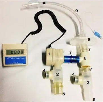
A ventilometer (AINCA-USA, model 00-295) recorded the VM, which was divided by RR, obtaining the mean ATV in mL, whose division by the predicted weight resulted in the RTV. The RSBI was obtained by dividing RR by ATV in liters24. As for the vital capacity
(VC), it was estimated by adapting the method of
Marini et al.25, arranging two one-way valves, a T-tube
and a ventilometer (Figure 1). The inspiratory valve was manually occluded and the patient exhaled until reaching the RV. Subsequently, only the expiratory valve was occluded and the volumes of consecutive inspiratory efforts were added up until ceasing the alterations in the ventilometer, indicating that the TLC had been achieved. The value resulting from this procedure was the AVC (RVC=AVC/predicted weight).
The height estimated was twice the distance from the center of the sternal furcula to the extremity of the middle finger, with the upper limb in elbow extension and 90°
of shoulder abduction26. Equations for predicted weight
were: men: 50+0.91 (height: 152.4); women: 45.5+0.91 (height: 152.4)27.
1. Ventilometer; 2. One-way inspiratory valve; 3. One-way expiratory valve; 4. Permanent occlusion; 5. Manual occlusion; 6. T-Tube; 7. Adapter; 8. Barrier filter; 9. Orotracheal tube.
Data were stored in the software Epi-info 7.2. The software SPSS 20.0 was used in descriptive analysis (mean, standard deviation, median, 25% and 75% percentile and 95% confidence interval). The Shapiro-Wilk test was used to evaluate the normality of data. The analysis of the heterogeneity between groups was obtained by the Student’s t-test for two independent samples (parametric) or by the Mann-Whitney U test (non-parametric). Contingency tables were evaluated by the Fisher’s exact test. Correlations were established through the Pearson’s (parametric) or Spearman’s rank (nonparametric) correlation coefficient. Associations were significant when p<0.05.
RESULTS
From the 68 admissions in the collection period (including those already hospitalized at the beginning), 8 exclusions occurred due to intolerance to the PSV and 30 others were not included due to: absence of AAW (12), extubation in less than 48 hours (10), and age above 79 years (8). Thus, 30 individuals were included: 9 (30%) allocated into the SMEPG and 21 (70%) into the LMEPG,
depending on the cut-off point of 55 cmH2O for MEP.
As expected, the MEP percentage (%MEP) of the SMEPG was greater than that of LMEPG (p=0.001), but there was no difference in MIP percentage (%MIP)
(0.077). In the intra-group subanalysis, the %MEP was lower than the %MIP in LMEPG (p<0.0001), which did not occur in the SMEPG (p=0.145). From the entire sample, only one patient presented unsatisfactory MIP (<40 cmH2O).
The SMEPG had superior performance than that of LMEPG in RSBI (p=0.022) and RR (p=0.044). The difference favoring the SMEPG on ATV (p=0.044) was not maintained in the RTV (0.312), a result similar to that of AVC (p=0.035) and RVC (p=0.227). The VM did not differ among groups (p=0.586). Table 1 shows the descriptive and analytical statistics of quantitative data.
The %MEP showed moderate correlation with RSBI (r=−0.406; p=0.026), RR (r=−0.426; p=0.017), T-IMV (r=−0.408; p=0.025) and T-ICU (r=−0.426; p=0.019). The %MIP was only correlated with the %MEP (r=0.676; p<0.001). As for the values measured, without considering the percentage, there was correlation of MEP (r=0.369; p=0.045) and MIP (r=−406; p=0.026) with the AVC; however, this phenomenon did not occur with the RVC, which had association with the RSBI (r=−0.640; p<0.001), RTV (r=0.673; p<0.001) and RR (r=−0.542; p=0.002).
There were more women in the LMEPG (71.4%) than in the SMEPG (22.2%, p=0.020). The satisfactory RSBI (<68 bpm/L) prevailed in SMEPG (88,89%) in relation to the LMEPG (47.62%, p=0.049). There were no differences in the other categorical variables (Table 2).
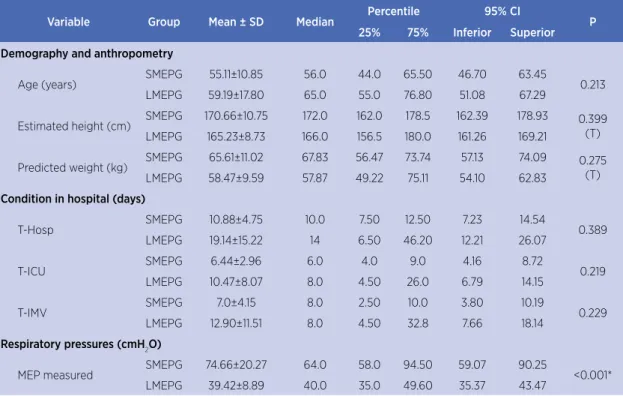
Table 1. Descriptive data of quantitative variables and the result of comparisons according to Student’s t- or Mann-Whitney U test
Variable Group Mean ± SD Median Percentile 95% CI P
25% 75% Inferior Superior
Demography and anthropometry
Age (years) SMEPG 55.11±10.85 56.0 44.0 65.50 46.70 63.45 0.213
LMEPG 59.19±17.80 65.0 55.0 76.80 51.08 67.29
Estimated height (cm) SMEPG 170.66±10.75 172.0 162.0 178.5 162.39 178.93 0.399
(T)
LMEPG 165.23±8.73 166.0 156.5 180.0 161.26 169.21
Predicted weight (kg) SMEPG 65.61±11.02 67.83 56.47 73.74 57.13 74.09 0.275
(T)
LMEPG 58.47±9.59 57.87 49.22 75.11 54.10 62.83
Condition in hospital (days)
T-Hosp SMEPG 10.88±4.75 10.0 7.50 12.50 7.23 14.54 0.389
LMEPG 19.14±15.22 14 6.50 46.20 12.21 26.07
T-ICU SMEPG 6.44±2.96 6.0 4.0 9.0 4.16 8.72 0.219
LMEPG 10.47±8.07 8.0 4.50 26.0 6.79 14.15
T-IMV SMEPG 7.0±4.15 8.0 2.50 10.0 3.80 10.19 0.229
LMEPG 12.90±11.51 8.0 4.50 32.8 7.66 18.14
Respiratory pressures (cmH2O)
MEP measured SMEPG 74.66±20.27 64.0 58.0 94.50 59.07 90.25 <0.001*
LMEPG 39.42±8.89 40.0 35.0 49.60 35.37 43.47
Variable Group Mean ± SD Median Percentile 95% CI P
25% 75% Inferior Superior
MIP measured SMEPG −86.28±22.70 −80.0 −69.0 −108.0 −68.77 −103.67 <0.001*
(t)
LMEPG −59.76±15.60 −58.0 −46.0 −81.60 −52.65 −66.86
MEP predicted SMEPG 112.74±22.34 119.94 111.84 128.42 98.14 127.35 0.021*
LMEPG 90.68±23.96 79.0 72.90 102.18 80.06 101.30
MIP predicted SMEPG 105.74±17.18 110.50 102.50 116.90 94.51 116.97 0.021*
LMEPG 89.56±18.82 81.0 76.10 99.62 81.22 97.89
MEP predicted (%) SMEPG 67.21±15.47 69.03 50.07 81.26 57.10 77.33 0.001*
(t)
LMEPG 45.88±14.77 44.30 34.23 58.50 39.33 52.42
MIP predicted (%) SMEPG 82.43±21.81 81.44 69.48 94.07 67.17 96.69 0.077
(t)
LMEPG 67.87±18.46 63.89 58.95 74.07 59.69 76.05
Ventilometry
RSBI (bpm) SMEPG 40.68±17.65 41.22 26.46 54.34 27.11 54.25 0.022*
LMEPG 75.37±44.09 69.23 40.27 152.29 55.29 95.44
ATV (mL) SMEPG 503.77±114.35 440.64 420.40 622.21 415.87 591.67 0.044*
LMEPG 398.02±123.80 400.0 306.36 572.08 341.66 454.37
RTV (mL/kg) SMEPG 7.83±2.09 7.74 6.42 8.66 6.45 9.20 0.312
(t)
LMEPG 6.91±2.28 6.64 5.54 8.28 5.90 7.92
RR (bpm) SMEPG 19.11±6.27 18 15.0 23.50 14.28 23.93 0.044*
LMEPG 26.14±9.46 24 19.50 42.20 21.83 30.44
VM (mL) SMEPG 9,273.3±2,499.9 8,900 7,020 11,430 7,351.7 11,194.9 0.586
(t) LMEPG 10,097.1±4,151.1 9,440 7,470 17,002 8,207.5 11,986.7
AVC (mL) SMEPG 1,917.22±610.21 1,865 1,425 2,370 1,448.1 2,386.2 0.035*
(t)
LMEPG 1,446.42±503.18 1,435 1,015 2,292 1,217.3 1,675.4
RVC (mL/kg) SMEPG 29.61±7.94 29.99 22.63 36.42 23.51 35.72 0.227
(t)
LMEPG 24.85±10.31 25.24 17.61 37.78 20.15 29.54
SD: Standard deviation; 95% CI: 95% confidence interval; T-IMV: Time on invasive mechanical ventilation; T-Hosp: Time of hospitalization; T-ICU: Time in intensive care unit; MEP: Maximal expiratory pressure; MIP: Maximal inspiratory pressure; RSBI: Rapid shallow breathing index; ATV: Absolute tidal volume; RTV: Relative tidal volume; RR: Respiratory rate; bpm: breaths per minutesize of groups.; VM: Volume minute; AVC: Absolute vital capacity; RVC: Relative vital capacity; SMEPG: Satisfying maximal expiratory pressure group; LMEPG: Low maximal expiratory pressure group; (t): Normal data evaluated by the Student’s t-test; * p<0.05.
(continues)
Table 1. Continuation
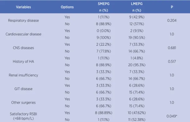
Table 2. Descriptive data of categorical variables and results of comparisons according to Fisher’s exact test.
Variables Options SMEPG LMEPG P
n (%) n (%)
Sex Male 7 (77.8%) 6 (28.6%) 0.020*
Female 2 (22.2%) 15 (71.4%)
Type of AAW OTT 8 (88.9%) 15 (71.4%) 0.393
TST 1 (11.1%) 6 (28.6%)
Sepsis Yes 3 (33.3%) 12 (57.1%) 0.427
No 6 (66.7%) 9 (42.9%)
Abdominal surgery Yes 4 (44.4%) 7 (33.3%) 0.687
No 5 (55.6%) 14 (66.7%)
DM Yes 0 (0.0%) 8 (38.1%) 0.067
No 9 (100%) 13 (61.9%)
SAP Yes 5 (55.6) 5 (23.8%) 0.115
DISCUSION
The LMEPG had better performance in the RSBI
and RR. There was no difference in RTV, RVC and VM.
Prevalence of satisfactory MEP was low, as observed in
size of groups. The MIP did not vary statistically among groups and only one patient had unsatisfactory MIP. Among the correlations observed, those that outstand
were observed only in %MEP with the RSBI, RR, T-IMV
and T-ICU and the correlations of RVC with the RSBI and its components.
The intra-group subanalysis of respiratory pressures shows that %MEP was lower than %MIP in both groups, with the significant difference in the LMEPG, suggesting that the expiratory weakness was more severe than the inspiratory one. This probably is an unprecedented finding on critical patients. However, changes of the complacency of the respiratory system are common in this population28 and
can culminate in lower TLC, reducing the MEP13. However,
the muscle weakness acquired in ICU (MWA-ICU) may
have influence on the phenomenon29.
The satisfactory RSBI of the SMEPG, even though it can be attributed to the action of both muscle groups, may have had decisive contributions for the expiratory muscles, given that: they modulate the respiratory control,
protecting the inspiratory muscles30; the AM ease the
diaphragmatic contraction2; the suitable MEP may
indicate greater effectiveness of cough and therefore greater airway permeability and less respiratory work8,31;
the LMEPG had a MIP of −59.76±15.60, which is higher
than the cut-off point of 40 cmH2O, but did not reflect
a good RSBI; only the %MEP was correlated with the RSBI; there was no correlation of the electrical activity
of the diaphragm with the RR or RSBI32; and usually the
inspiratory negativity of −5 cmH2O in pleural pressure was sufficient for inhalation of 500 mL of air33. The RSBI is
the predictive index most used in WMV17, but the design
of this study limits the interpretations. A longitudinal follow-up is required to verify the outcomes in WMV according to the MEP.
The similarity found in the VM was observed in other
studies18,30,32 and may have derived from the sample’s
tolerance to PSV. As the VM is the product of ATV by RR, the difference was found in those variables that tend to change inversely. Thus, the SMEPG had lower RR for presenting higher ATV as the LMEPG compensated the low ATV by increasing the RR. As the difference in the RTV was not significant, the largest contribution came
from RR, agreeing with Sugiura et al.30, in which the
expiratory fatigue triggers the fast and shallow pattern and, to a certain level of effort, the change in the ATV is not significant.
Variables Options SMEPG LMEPG P
n (%) n (%)
Respiratory disease Yes 1 (11.1%) 9 (42.9%) 0.204
No 8 (88.9%) 12 (57.1%)
Cardiovascular disease Yes 0 (0.0%) 2 (9.5%) 1.0
No 9 (100%) 19 (90.5%)
CNS diseases Yes 2 (22.2%) 7 (33.3%) 0.681
No 7 (77.8%) 14 (66.7%)
History of HA Yes 1 (11.1%) 1 (4.8%) 0.517
No 8 (88.9%) 20 (95.3%)
Renal insufficiency Yes 3 (33.3%) 7 (33.3%) 1.0
No 6 (66.7%) 14 (66.7%)
GIT disease Yes 3 (33.3%) 6 (28.6%) 1.0
No 6 (66.7%) 15 (71.4%)
Other surgeries Yes 3 (33.3%) 6 (28.6%) 1.0
No 6 (66.7%) 15 (71.4%)
Satisfactory RSBI (<68 bpm/L)
Yes 8 (88.89%) 10 (47.62%)
0.049*
No 1 (11.1%) 11 (52.38%)
SMEPG: Satisfying maximal expiratory pressure group; LMEPG: Low maximal expiratory pressure group; AAW: Artificial airway; OTT: orotracheal tube; TST: tracheal tube; DM: Diabetes mellitus; SAH: Systemic arterial hypertension; CNS: Central nervous system; HA: Heart arrest; GIT: Gastrointestinal tract; RSBI: Rapid shallow breathing index; *p<0.05.
The RVC did not vary statistically between groups, even when being correlated with the RSBI and its components in isolation, which, except for the RTV, showed difference in the comparison of SMEPG with the LMEPG, as well as the correlation with %MEP. It is possible that the use of one-way valves, aiming at leading individuals to exert greater efforts, promotes greater homogeneity even among those with difference of strength, since the volume of air is accumulated at each respiratory cycle. The findings
may also result from improper activation of AM1, since
the VC may increase with training3. In addition, apart
from relying on the neuromuscular respiratory function, the VC is influenced by the mechanical properties of the lung and thoracic system, which has similarities before distinct respiratory pressures34. The method used in the VC
was validated25, but its performance on individuals with
AAW still remains necessary. In validation, the authors found in healthy participants and those with ambulatory pneumonia, AVC of 4.63 L and 3.02 L, respectively. The values in this study were 1.91 L (SMEPG) and 1.44 L (LMEPG), acceptable numbers due to the need of IMV. Furthermore, correlations observed in this study indicate that the method has produced reliable data.
Expiratory muscles can be evaluated dynamically through the cough8. Individuals with a peak of expiratory
flow in cough below 60 L/min were five times more inclined to have unsuccessful extubation and 19 times more inclined to dye at the hospital35. There is interest
on indexes that predict combined results of WMV and extubation35,18. Although the need for ventilatory support
and AAW has different etiologies18, the expiratory function
seems useful in the evaluation of the discontinuity of these two factors and physical weakness.
Impairments in the diaphragm caused by critical illness polyneuromyopathy (CIPNM) are considered vital in
the prolongation of IMV36, but little is known about the
involvement of AM and its impact. The comparison of the transdiaphragmatic pressure in subjects with success and failure in the WMV showed no difference in the
establishment of fatigue in the diaphragm37. Results of
this study do not show significant reduction in %MIP between groups, being possible that the CIPNM primarily affects the expiratory muscles. However, the mechanism lacks research, because the poor performance of the AM
is also present on low back pain38, chronic obstructive
pulmonary disease39, multiple sclerosis40, and spinal cord
injury41.
CIPNM diagnosis requires invasive methods, of difficult interpretation and limited by conditions as low level of
consciousness, edema and previous neuropathy29. The
Medical Research Council Sum Score (MRC-SS) is used in the diagnostic hypothesis for identifying the MWA-ICU, but it also requires cooperation of the patient29,42. Tzanis
et al.43 found correlation between MIP and the MRC-SS,
showing that this could be an alternative. Similarly, there was correlation between the MRC-SS and the respiratory pressures, with greater significance on MEP (p<0.0001) in relation to the MIP (p=0.001)28.
The association between peripheral muscle weakness and duration of T-IMV is mediated by the concurrent
respiratory weakness28. In the absence of mechanical
disadvantage, the reduction of MEP may reflect a
generalized muscle weakness44. During a maximal
expiratory effort, the electrical activity of the AM is small compared to that recorded when the head and shoulders in dorsal positions stop touching a surface45. To
understand the expiratory muscle function in the WMV is essential in the therapeutic approach.
The existing correlation between %MEP and %MIP generates a questioning of the current consensus on intensive therapy of only training inspiratory muscles. It becomes necessary to compare the expiratory and inspiratory muscle training, separately and combined, as well as to test other strategies. In the case of neuromuscular electrical stimulation, the TVM, for being the deepest and most active muscle in forced expiration, followed by
the oblique and rectus abdominis muscles1, such a basis
can guide the arrangement of electrodes for effective neuromuscular stimulation and the monitoring of
the AM recruitment through ultrasonic images46. In
addition, active movements of the lower limbs may be
beneficial because they are preceded by TVM activation47
and it is possible that the increased VC is achieved by strengthening the inspiratory muscles on TLC and the expiratory ones in RV40.
CONCLUSION
The MEP≥55 cmH2O was associated to better values in
RSBI and RR. The reduction of expiratory muscle strength was more prevalent and severe than that of inspiratory muscle strength. There were no participants with normal MEP and low MIP at the same time, which limits the conclusions on the isolated function of expiratory muscles, but indicates that the inspiratory muscles are less affected and that the effects of expiratory muscle training must be investigated in the difficult weaning.
REFERENCES
1. Abe T, Kusuhara N, Yoshimura N, Tomita T, Easton PA. Differential respiratory activity of four abdominal muscles in humans. J Appl Physiol. 1996;80(4):1379-89. doi: 10.1152/jappl.1996.80.4.1379 2. Reid WD, Dechman G. Considerations when testing and
training the respiratory muscles. Phys Ther. 1995;75(11):971-82. doi: 10.1093/ptj/75.11.971
3. Kim E, Lee H. The effects of deep abdominal muscle strengthening exercises on respiratory function and lumbar stability. J Phys Ther Sci. 2013;25(6):663-5. doi: 10.1589/jpts.25.663
4. Silva KM, Sayers BM, Sears TA, Stagg DT. The changes in configuration of the rib cage and abdomen during breathing in the anaesthetized cat. J Physiol. 1977;266(2):499-521. doi: 10.1113/jphysiol.1977.sp011779
5. Gollee H, Hunt KJ, Fraser MH, McLean AN. Abdominal stimulation for respiratory support in tetraplegia: a tutorial review. J Automatic Contr. 2008;18(2):85-92. doi: 10.2298/ JAC0802085G
6. Cuello AF, Aquim EL, Cuello GA. Músculos ventilatórios: biomotores da ventilação: avaliação e tratamento. São Paulo: Andreoli; 2013.
7. Taylor BJ, Romer LM. Effect of expiratory muscle fatigue on exercise tolerance and locomotor muscle fatigue in healthy humans. J Appl Physiol. 2008;104(5):1442-51. doi: 10.1152/ japplphysiol.00428.2007
8. Suárez AA, Pessolano FA, Monteiro SG, Ferreyra G, Capria ME, Mesa L, et al. Peak flow and peak cough flow in the evaluation of expiratory muscle weakness and bulbar impairment in patients with neuromuscular disease. Am J Phys Med Rehabil. 2002;81(7):506-11. doi: 10.1097/00002060-200207000-00007 9. McCaughey EJ, Berry HR, McLean AN, Allan DB, Gollee H.
Abdominal functional electrical stimulation to assist ventilator weaning in acute tetraplegia: a cohort study. PLoS One. 2015;10(6):e0128589. doi: 10.1371/journal.pone.0128589 10. Boles JM, Bion J, Cannors A, Herridge M, Marsh B, Melot C,
et al. Weaning from mechanical ventilation. Eur Respir J. 2007;29:1033-56. doi: 10.1183/09031936.00010206
11. Appleton R, Kinsella J. Intensive care unit: acquired weakness. Contin Educ Anaesth Crit Care Pain. 2012:12(2):62-6. doi: 10.1093/bjaceaccp/mkr057
12. Associação de Medicina Intensiva Brasileira, Sociedade Brasileira de Pneumologia e Tisiologia. Diretrizes brasileiras de ventilação mecânica. São Paulo: AMIB, SBPT; 2013.
13. American Thoracic Society, European Respiratory Society. ATS/ ERS statement on respiratory muscle testing. Am J Respir Crit Care Med. 2002;166(4):518-624. doi: 10.1164/rccm.166.4.518 14. Caruso P, Carnieli DS, Kagohara KH, Anciães A, Segarra
JS, Deheinzelin D. Trend of maximal inspiratory pressure in mechanically ventilated patients: predictors. Clinics. 2008;63(1):33-8. doi: 10.1590/S1807-59322008000100007 15. Daniel Martin A, Smith BK, Davenport PD, Harman E,
Gonzalez-Rothi RJ, Baz M, et al. Inspiratory muscle strength training improves weaning outcome in failure to wean patients: a randomized trial. Crit Care. 2011;15(2):R84. doi: 10.1186/cc10081
16. Condessa RL, Brauner JS, Saul AL, Baptista M, Silva ACT, Vieira SRR. Inspiratory muscle training did not accelerate weaning from mechanical ventilation but did improve tidal volume and maximal respiratory pressures: a randomised trial. J Physiother. 2013;59(2):101-7. doi: 10.1016/S1836-9553(13)70162-0
17. Nemer SN, Barbas CSV. Parâmetros preditivos para o desmame da ventilação mecânica. J Bras Pneumol. 2011;37(5):669-79. doi: 10.1590/S1806-37132011000500016
18. Lai CC, Chen CM, Chiang SR, Liu WL, Weng SF, Sung MI, et al. Establishing predictors for successfully planned endotracheal extubation. Medicine (Baltimore). 2016;95(41):e4852. doi: 10.1097/MD.0000000000004852
19. Duan J, Liu J, Xiao M, Yang X, Wu J, Zhou L. Voluntary is better than involuntary cough peak flow for predicting re-intubation after scheduled extubation in cooperative subjects. Respir Care. 2014;59(11):1643-51. doi: 10.4187/respcare.03045
20. Neder JA, Andreoni S, Lerario MC, Nery LE. Reference values for lung function tests II: maximal respiratory pressures and voluntary ventilation. Braz J Med Biol Res. 1999;32(6):719-27. doi: 10.1590/S0100-879X1999000600007
21. Hamilton VA, Grap MJ. The role of the endotracheal tube cuff in microaspiration. Heart Lung. 2012;41(2):167-72. doi: 10.1016/j. hrtlng.2011.09.001
22. Guimarães FS, Alves FF, Constantino SS, Dias CM, Menezes SLS. Maximal inspiratory pressure evaluation among non-cooperative critical patients: comparison between two methods. Rev Bras Fisioter (São Carlos). 2007;11(3):233-8. doi: 10.1590/S1413-35552007000300010
23. Truwit JD, Marini JJ. Validation of a technique to assess maximal inspiratory pressure in poorly cooperative patients. Chest. 1992;102(4):1216-9. doi: 10.1378/chest.102.4.1216
24. Yang KL, Tobin MJ. A prospective study of indexes predicting the outcome of trials of weaning from mechanical ventilation. N Engl J Med. 1991;324(21):1445-50. doi: 10.1056/NEJM199105233242101 25. Marini JJ, Rodriguez RM, Lamb VJ. Involuntary breath-stacking:
an alternative method for vital capacity estimation in poorly cooperative subjects. Am Rev Respir Dis. 1986;134(4):694-8. doi: 10.1164/arrd.1986.134.4.694
27. Acute Respiratory Distress Syndrome Network. Ventilation with lower tidal volumes as compared with traditional tidal volumes for acute lung injury and the acute respiratory distress syndrome. N Engl J Med. 2000;342(18):1301-8. doi: 10.1056/ NEJM200005043421801
28. Jonghe B, Bastuji-Garin S, Durand MC, Malissin I, Rodrigues P, Cerf C, et al. Respiratory weakness is associated with limb weakness and delayed weaning in critical illness. Crit Care Med. 2007;35(9):2007-15. doi: 10.1097/01.ccm.0000281450.01881.d8 29. Confer J, Wolcott J, Hayes R. Critical illness polyneuromyopathy.
Am J Health Syst Pharm. 2012;69(14):1199-205. doi: 10.2146/ ajhp110343
30. Sugiura H, Sako S, Oshida Y. Effect of expiratory muscle fatigue on the respiratory response during exercise. J. Phys Ther Sci. 2013;25(11):1491-5. doi: 10.1589/jpts.25.1491
31. Kim J, Davenport P, Sapienza C. Effect of expiratory muscle strength training on elderly cough function. Arch Gerontol Geriatr. 2009;48(3):361-6. doi: 10.1016/j.archger.2008.03.006 32. Liu L, Liu H, Yang Y, Huang Y, Liu S, Beck J, et al. Neuroventilatory
efficiency and extubation readiness in critically ill patients. Crit Care. 2012;16(4):R143. doi: 10.1186/cc11451
33. Schellekens WJ, Van Hees HW, Doorduin J, Roesthuis LH, Scheffer GJ, Van der Hoeven JG, et al. Strategies to optimize respiratory muscle function in ICU patients. Crit Care. 2016;20(1):103. doi: 10.1186/s13054-016-1280-y
34. Vassilakopoulos T, Roussos C. Physiology and testing of respiratory muscles. In: Albert R, Spiro S, Jett J, editors. Clinical respiratory medicine. 3rd ed. Mosby Elsevier, Philadelphia: Mosby Elsevier; 2008. p. 135-146.
35. Smina M, Salam A, Khamiees M, Gada P, Amoateng-Adjepong Y, Manthous CA. Cough peak flows and extubation outcomes. Chest. 2003;124(1):262-8. doi: 10.1378/chest.124.1.262 36. Hermans G, Jonghe B, Bruyninckx F, Van den Berghe G. Clinical
review: critical illness polyneuropathy and myopathy. Crit Care. 2008;12(6):238. doi: 10.1186/cc7100
37. Laghi F, Cattapan SE, Jubran A, Parthasarathy S, Warshawsky P, Choi YSA, et al. Is weaning failure caused by low-frequency
fatigue of the diaphragm? Am J Respir Crit Care Med. 2003;167(2):120-7. doi: 10.1164/rccm.200210-1246OC
38. Hodges PW, Richardson CA. Inefficient muscular stabilization of the lumbar spine associated with low back pain. Spine. 1996;21(22):2640-50. doi: 10.1097/00007632-199611150-00014 39. Rochester DF, Braun NMT. Determinants of maximal inspiratory
pressure in chronic obstructive pulmonary disease. Am Rev Respir Dis. 1985;132(1):42-7. doi: 10.1164/arrd.1985.132.1.42 40. Gosselink R, Kovacs L, Ketelaer P, Carton H, Decramer
M. Respiratory muscle weakness and respiratory muscle training in severely disabled multiple sclerosis patients. Arch Phys Med Rehabil. 2000;81(6):747-51. doi: 10.1016/ S0003-9993(00)90105-9
41. Gounden P. Static respiratory pressures in patients with post-traumatic tetraplegia. Spinal Cord. 1997;35(1):43-7. doi: 10.1038/ sj.sc.3100336
42. Zhou C, Wu L, Fenghimg N, Ji W, Wu J, Zhang H. Critical illness polyneuropathy and myopathy: a systematic review. Neural Regen Res. 2014;9(1):101-10. doi: 10.4103/1673-5374.125337 43. Tzanis G, Vasileiadis I, Zervakis D, Karatzanos E, Dimopoulos
S, Pitsolis T, et al. maximal inspiratory pressure, a surrogate parameter for the assessment of ICU-acquired weakness. BMC Anesthesiology. 2011;11:14. doi: 10.1186/1471-2253-11-14 44. O’Neill S, McCarthy DS. Postural relief of dyspnoea in
severe chronic airflow limitation: relationship to respiratory muscle strength. Thorax. 1983;38(8):595-600. doi: 10.1136/ thx.38.8.595.
45. Campbell EJM, Green JH. The expiratory function of the abdominal muscles in man: an electromyographic study. J Physiol. 1953;120(3):409-18. doi: 10.1113/jphysiol.1953.sp004903 46. Larivière C, Gagnon, D, Oliveira JE Jr., Henry SM, Mecheri H,
Dumas JP. Reliability of ultrasound measures of the transversus abdominis: effect of task and transducer position. PM R. 2013;5(2):104-13. doi: 10.1016/j.pmrj.2012.11.002