1Museu de Zoologia da Universidade de São Paulo, Av. Nazaré, 481, 04263-000 São Paulo, SP, Brazil. (MP) pinna@ib.usp.br,
https://orcid.org/0000-0003-1711-4816 (corresponding author), (VA) vitorabrahao32@gmail.com, (VC) carvalhvinicius@gmail.com
2Instituto de Biologia, Universidade Federal da Bahia, Rua Barão de Geremoabo, 40170-115 Salvador, BA, Brazil. zanata.angela@gmail.com Introduction
The Chapada Diamantina is a large (over 30.000 square km) plateau composed of proterozoic terrain extending along the central portion of the State of Bahia, northeast-ern Brazil. Much of the plateau is over 1000m altitude, with some peaks reaching over 2000m and it includes headwaters of three major basins, rio São Francisco to the west, rio de Contas to the south and rio Paraguaçu to the east and north. The most emblematic aquatic component of the Chapada Diamantina is the Copionodontinae, a trichomycterid sub-family endemic to the formation (and therein only in rivers associated with the rio Paraguaçu drainage). Copionodon-tinae was described to include the genus Copionodon, with two species, and Glaphyropoma, with one (de Pinna, 1992). Since then, two additional species were described in Copi onodon (Campanario, de Pinna, 2000; de Pinna et al., 2018) and one in Glaphyropoma (Bichuette et al., 2008).
Copionodontines normally dwell on the upper reaches of rivers and are often restricted to a short sector therein. They
are frequently the only fishes in their habitat. One species of Characidae (Astyanax or Myxiops) and one Loricariidae co-occur in some localities. Up to three different species of copionodontines may occupy the same stream (de Pinna, 1992), though in distinct microhabitats. Because vast por-tions of the Diamantina plateau are accessible only on foot and with considerable difficulty, ichthyological sampling is still incipient in the region. Such fact, in combination with the narrowly endemic distributions of some species of co-pionodontines, suggests that new species of the subfamily await discovery.
Copionodontinae catfishes are of special phylogenetic interest because they display many plesiomorphic character-istics when compared to the majority of other trichomycter-ids. They seem to be the closest relative of the Trichogeninae from southeastern Brazil, the clade formed by the two sub-families being the sister group to all other trichomycterids (de Pinna, 1998; de Pinna et al., 2010). The Copionodonti-nae and its constituent genera are regarded as monophyletic groups (de Pinna, 1992; Bichuette et al., 2008).
Mario de Pinna
1, Vitor Abrahão
1, Vinicius Reis
1and Angela Zanata
2A new species of the Copionodontinae genus Copionodon is described from the riacho do Mosquito, tributary to rio Santo Antônio, rio Paraguaçu basin in the Diamantina Plateau, Bahia State, northeastern Brazil. This species represents the norther-nmost occurrence of the subfamily yet known and is a relictual population, apparently restricted to an underground sector of the stream and its immediate downstream exit. The new species can be readily recognized by the combination of the presence of opercular odontodes, the mostly uniform coloration of the body, and the lack of a free orbital rim. Despite several troglo-morphic traits shared between the new species and Glaphyropoma spinosum, the two species are not closest relatives and all such similarities are convergent adaptations to the subterranean habitat. A CT-Scan study of the holotype of the new species is presented and allows a view into the details of copionodontine anatomy with unprecedented clarity.
Keywords: Chapada Diamantina, Endemism, Rio Paraguaçu, Systematics, Taxonomy.
Uma nova espécie de Copionodon (Copionodontinae) é descrita do riacho do Mosquito, tributário do rio Santo Antônio, bacia do rio Paraguaçu, Chapada Diamantina, Bahia, Brasil. Essa espécie representa a ocorrência mais ao norte da subfamília até então conhecida como uma população relictual, aparentemente restrita a um setor subterrâneo do riacho e sua saída à jusante. A nova espécie pode ser reconhecida pela combinação de odontódeos presentes no opérculo, coloração uniforme do corpo e ausência de margem orbital livre. Apesar de alguns caracteres troglomórficos compartilhados entre a nova espécie e Glaphyropoma spinosum, as duas espécies não são proximamente relacionadas e todas essas similaridades são interpretadas como adaptações convergentes ao ambiente subterrâneo. É apresentado um estudo de tomografia computadorizada do holótipo da nova espécie que permite a visualização de detalhes da anatomia interna de copionodontinae com clareza sem precedentes.
New species of relictual Copionodon Neotropical Ichthyology, 16(4): e180049, 2018
2
Herein we report on a new species of Copionodon found in a small sector of the riacho do Mosquito, a tributary to the rio Santo Antônio, itself part of the rio Paraguaçu drainage. The population is apparently relictual, and specimens were se-cured only at the exit of a subterranean sector of the river. The underground portion itself was not explored, but supposedly includes the bulk of the population, with the specimens caught being just outlying individuals. Despite that, troglomorphic characteristics are only expressed mildly in specimens avail-able, with moderate reduction of eyes and integument pig-mentation. Distinguishing characters other than those associ-ated with subterranean adaptations, however, fully diagnose the new species. As part of our study, we provide anatomical data based on high-resolution X-ray computed tomography which allow a new view of various phylogenetically-infor-mative characters previously proposed for Copionodontinae.
Material and Methods
Morphometric data were taken with digital calipers fol-lowing the measurement definitions in de Pinna (1992). Os -teological information was taken from specimens cleared and counterstained for bone and cartilage according to the method of Taylor, Van Dyke (1985). Osteological informa -tion from addi-tional specimens, including vertebral counts, was taken from digital radiographs and/or CT-Scan im-ages (see below). Osteological nomenclature of braincase, lower jaw and caudal skeleton follows Patterson (1975), Nelson (1973) and Lundberg, Baskin (1969), respectively. Suspensorium nomenclature follows Arratia (1992). Ho-lotype counts are marked with asterisk in the descriptions. All morphometric data are shown in Tab. 1. Computerized
tomography imaging was based on Micro CT scans done with a Phoenix v|tome|x M – General Electric Company, using voxel size X = 0.02370301 microns, number of im-ages 5000, voltage 60Kv, and current 220mA. Photographs of skeletal preparations and whole specimens were made with a digital microphotographic system attached to an au-to-focus stereomicroscope. Drawings were prepared with a camera lucida attached to a stereomicroscope. Acronyms for collections follow Sabaj (2016).
Results
Copionodon exotatos, new species
urn:lsid:zoobank.org:act:2D56692E-B1F3-4557-89C6-0595CCE4CA9C
(Figs. 1-3)
Holotype. MZUSP 123522, 36.9 mm SL, Brazil, Bahia, Chapada Diamantina, Lençóis, exit of rock-enclosed sector of right-hand branch of riacho do Mosquito (immediately upstream from Cachoeira do Mosquito), itself a tributary of rio Santo Antônio, rio Paraguaçu basin 12°22’15.64”S, 41°22’15.85”W, M. de Pinna, V. Abrahão, V. Reis & A. Zanata, 4 Mar 2017.
Paratypes. MZUSP 121656, 5, 12.6-35.8 mm SL, collected with holotype.
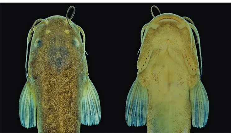
Diagnosis. Distinguished from all copionodontines, except Glaphyropoma spinosum, by the presence of opercular odon-todes (with presence variable; vs. opercular odonodon-todes enti-rely absent; Figs. 2-4). Further distinguished from all conge-ners by the small eye in mid- to large-sized specimens (over 35 mm SL, 11-12% HL; vs. 13% or more; Fig. 2a); and from all except C. elysium de Pinna, Burger & Zanata, 2018 by the lack of a free orbital rim (Fig. 2a). The combination of body coloration lacking conspicuous markings and lack of free or-bital rim distinguishes C. exotatos from all other congeners. Distinguished from C. elysium, C. pecten de Pinna, 1992 and C. orthiocarinatus de Pinna, 1992 by the color pattern composed of a tenuous continuous and straight midlateral stripe against a uniform scattering of dark chromatophores (Fig. 1; vs. color pattern more complex, including irregular dark markings dorsal and ventral to an irregular longitudinal stripe in C. elysium and C. pecten or absence of a midlateral stripe in C. orthiocarinatus). Further distinguished from C. elysium by the presence of a conical ossification cap in the anterolateral corner of the second hypobranchial (Fig. 5; vs. second hypobranchial entirely cartilaginous; this ossification is so far verified in a single c&s paratype of C. exotatos). Copionodon exotatos further differs from C. pecten by the smaller interopercular patch of odontodes, its anterior limit distant from lower lip (by distance equivalent to approxima-tely mouth width) and posteriorly not reaching base of first Tab. 1. Morphometric data for Copionodon exotatos, new
species. N = 6; SD = standard deviation.
Holotype Range Mean SD
Total length 44.78 30.9-41.8 39.2
-Standard length in mm 38.1 26.1-35.5 33.2 -Percents of standard length
Body depth 14.4 13.3-14.7 14.1 0.7
Predorsal length 58.7 54.6-58.2 57.2 2.2
Preanal length 63.8 62.5-63.2 63.2 0.6
Prepelvic length 48.6 48.8-51.7 49.7 1.7
Caudal peduncle length 20 19.6-20.3 19.9 0.3 Caudal peduncle depth 11.8 11.1-12.4 11.8 0.6 Dorsal-fin base length 9.3 9.7-11.5 10.2 1.1 Anal-fin base length 15.9 15.7-19.4 17 2.0
Head length 23.7 23-24.1 23.6 0.5
Percents of head length
Head width 87.1 86.5-90.8 88.1 2.3
Head depth 47.8 50-50.5 49.4 1.4
Snout length 42.5 38.9-43.9 41.8 2.5
Mouth width 38.9 37-37.4 37.8 0.9
Interorbital width 50 46.1-48.4 48.1 1.9
Length of interopercular patch of
odontodes 37.3 33.9-38.5 36.6 2.4
Fig. 1. Copionodon exotatos, holotype, MZUSP 120631, 45.0 mm SL, Brazil, Bahia, Chapada Diamantina, Município de Palmeiras, Riacho do Mosquito, immediately upstream from Cachoeira do Mosquito, rio Paraguaçu basin.
Fig. 2. Copionodon exotatos, holotype, MZUSP 120631, 45.0 mm SL. Left to right, dorsal and ventral views of head.
New species of relictual Copionodon Neotropical Ichthyology, 16(4): e180049, 2018
4
pectoral-fin ray (Fig. 2b; vs. interopercular patch of odon-todes large, its anterior limit near lower lip (separated by distance equivalent approximately to half of mouth width) and posteriorly reaching beyond pectoral-fin base) and lon -ger barbels, with maxillary barbel usually reaching
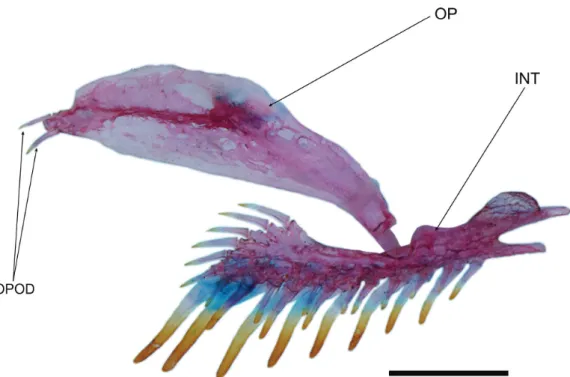
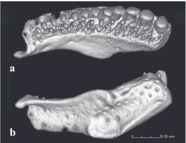
pecto-Fig. 4. Copionodon exotatos, paratype, MZUSP 121656, opercle and interopercle, right side, lateral view. Abbreviations: OPOD, opercular odontodes; OP, opercle; INT, interopercle. Scale bar = 1 mm.
Fig. 5. Copionodon exotatos, paratype, MZUSP 121656, basibranchials and hypobranchials, dorsal view. Grey areas represent cartilage. Abbreviations: BB2-4, basibranchials 2 to 4; HB1-3, hypobranchials 1 to 3. Scale bar = 1 mm.
ral-fin origin and rictal one extending beyond midlength of interopercular patch of odontodes (Figs. 1-2; vs. maxillary barbel reaching posterior ¼ of length of interopercular patch of odontodes and rictal reaching anterior ¼ of length of inte-ropercular patch of odontodes). From species of Glaphyro poma, the new taxon further differs by the broad first hypo -branchial (Fig. 5; vs. slender; cf. de Pinna, 1992, fig. 6 and Bichuette et al., 2008, fig. 6), the caudal fin bilobed (Fig. 1; vs. truncate), the toothed portion on dentary not reaching the coronoid process (Fig. 6; vs. reaching coronoid process), and the presence of asymmetrical spatulate teeth in jaws (Fig. 11; vs. all teeth symmetrically spatulate).
Description. Body elongate, trunk roughly as deep as wide in cross-section near head, gradually more compressed pos-terior to end of pectoral fin. Caudal peduncle compressed, tapering to caudal fin. Dorsal profile of body and head ne -arly straight from snout to dorsal-fin origin, slightly convex near head. Posterior region of body straight from endpoint of dorsal fin to caudal-fin origin. Ventral profile straight along head and gular regions, nearly parallel to longitudinal axis of body; convex or slightly convex from that point to pelvic-fin origin and nearly straight or slightly concave from pelvic-fin base to caudal-fin base. Dorsal profile of adipose fin nearly straight; fin barely developed, somewhat more visible pos -terior to vertical through end of anal-fin base. Adipose and caudal fins confluent (Figs. 1-3).
Fig. 7. Geographic distribution of Copionodon exotatos.
barbel. Lower lip with about same thickness as upper lip; continuous, not divided in lateral halves by median constric-tion, only slightly less convex. Angle of mouth with fleshy outgrowth just posterior and adpressed to rictal barbel (Figs. 2-3).
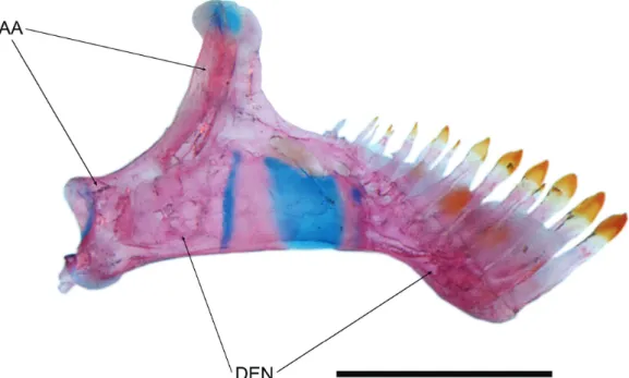
Fig. 6. Copionodon exotatos, paratype, MZUSP 121656. Lower jaw, left side, mesial view. Abbreviations: AA, anguloarticu-lar; DEN, dentary. Scale bar = 1 mm.
New species of relictual Copionodon Neotropical Ichthyology, 16(4): e180049, 2018
6
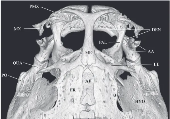
Fig. 9. Copionodon exotatos, holotype, MZUSP 120631, CT-scan image of anterior portion of skull, dorsal view. Abbrevia-tions: PMX, premaxilla; MX, maxilla; QUA, quadrate; PO, preopercle; ME, mesethmoid; AF, anterior fontanel; FR, frontal; DEN, dentary; PAL, palatine; AA, anguloarticular; LE, lateral ethmoid; HYO, hyomandibula.
Fig. 8. Collection site of Copionodon exotatos, right-hand branch of Riacho do Mosquito (trib. to rio Santo Antônio, rio Paraguaçu drainage), immediately upstream from Cachoeira do Mosquito (12º21’59.51”S, 41 ºS 22”19.37”W) at exit of rock-enclosed sector.
asymmetrical; spatulate portion not overlapping in lower jaw (Fig. 11). Distal border of teeth straight. In both jaws, second row of teeth also slightly expanded distally; third row conical. Teeth on lower jaw distributed over anterior two thirds of ossification and not reaching coronoid process.
Outer series of lower jaw with about six to eight spatulate larger anterior teeth followed by three to five conical teeth.
Fig. 10. Copionodon exotatos, holotype, MZUSP 120631, CT-scan images of right premaxilla and associated dentition: a. ventral view, b. dorsal view.
Fig. 11. Copionodon exotatos, holotype, MZUSP 120631, CT-scan image of jaws, anteroventral view. Abbreviations: AA, anguloarticular; DEN, dentary; PMX, premaxilla.
length, much closer to lateral border of head than to midline in dorsal view. Infraorbital latero-sensory canal complete, extending from sphenotic posteriorly to first infraorbital (la -crimal) anteriorly. Three infraorbital pores, one directly pos-terior to pospos-terior margin of eye, another directly ventral to
incomple-New species of relictual Copionodon Neotropical Ichthyology, 16(4): e180049, 2018
8
te posterolaterally, with appearance of two partly confluent flaps of skin. Three pairs of thin flat barbels, wide at base and gradually narrowing distally to fine tip. Maxillary barbel reaching slightly beyond base of pectoral fin. Rictal barbel inserted ventral to maxillary barbel, extending to middle of interopercular patch of odontodes. Nasal barbel originating on posterolateral region of anterior nares, reaching to middle of opercular patch of odontodes (or homologous position in specimens without latter) (Figs. 2-3). Nasal barbels shorter in juvenile specimens (12.6 and 13.1 mm SL), reaching to middle or posterior margin of eye. Interopercle with well--developed odontodes, visible in ventral aspect of head. Interopercular patch of odontodes extending from vertical through posterior border of eye anteriorly to slightly anterior to vertical through pectoral-fin base posteriorly. Odontodes arranged in two series, with those on mesial series larger than those on lateral one; odontodes gradually larger pos-teriorly in both series, with those pospos-teriorly on mesial row largest. Eight odontodes on mesial row and six or seven on lateral row. Interopercular odontodes not yet developed in two smallest specimens examined (12.6 and 13.1 mm SL). Opercular patch of odontodes variably present in two of four adults or subadult type specimens (odontodes not yet developed in two additional juvenile paratypes). Opercular odontodes vestigial in holotype, apparently lacking direct mineralized connection with opercle (Fig. 13) yet visible on surface of skin. Patch small and inconspicuous on surface of head, located anterodorsally to pectoral-fin base, with single
row of four or five small odontodes sunk in individual slits of integument. Entire patch surrounded by specialized rim of integument typical of that complex in most other trichomyc-terids. Specimens without opercular patch of odontodes with homologous region of head smooth, lacking both odontodes and associated integumentary modifications (Fig. 4).
Pectoral fin large, extending to middle of distance betwe -en occipital and pelvic-fin base, g-ently convex in distal pro -file, its base immediately posterior to vertical through pos -terior tip of interopercle (Fig. 1). Pectoral-fin rays i8i*(1), i9(1), or i9i(2). Pectoral-fin rays approximately same length except for short last one. Pelvic fin with round distal pro -file, its origin at or slightly posterior to middle of SL, and well anterior to vertical through dorsal-fin origin, entirely covering anal and urogenital openings, reaching or almost reaching anal-fin origin (Fig. 1). Pelvic-fin rays i5/i5i*(1) or i5i(3), first ray (unbranched) shorter than others. Dorsal fin smaller than anal fin, its distal profile gently convex. Dorsal-fin origin closer to base of caudal fin than to tip of snout (Fig. 1). Dorsal-fin rays i5i*(1), ii5(3). Anal fin with base longer than that of dorsal, and total area larger than those of all other fins except caudal, its distal profile strongly convex. Anal-fin origin slightly anterior to vertical through end of dorsal-fin base (Fig. 1). Anal-fin rays ii8*(1), iii9(1), or iii10(2). Caudal fin bilobed with round corners, with 8/9 principal rays. Upper lobe slightly longer than lower lobe in some specimens (Fig. 1). Six dorsal and ventral procurrent caudal-fin rays, plus one dorsal segmented non-principal ray dorsally and two ventrally. Adipose fin shallow, without clear anterior and posterior limits, extending between end of dorsal fin and caudal-fin origin (Fig. 1). Degree of deve -lopment of adipose fin ontogenetically variable, apparently inversely proportional do size of fish. Vertebrae 33*(2) or 34(2). First dorsal-fin pterygiophore immediately posterior to neural spine of 11th or 12th vertebra, first anal-fin ptery -giophore immediately posterior to neural spine of 14th or 15th vertebra.
Lateral line splitting immediately after leaving neurocra-nium, with ventral branch very short and with single termi-nal pore. Dorsal and main branch extending continuously to approximately a vertical through middle of pectoral fin and extending posteriorly along entire body length as series of independent tubules, progressively shorter and more spa-ced out posteriorly. Last tubule over complex caudal cen-trum and base of hypural plate. Lateral line ossicles absent. Pleural ribs 11*(4). Branchiostegal rays six* (4). Dorsal-fin pterygiophores seven* (4). Anal-fin pterygiophores 12(2); remaining specimens could not be examined with the availa-ble images. A single large and ossified proximal pectoral-fin radial and four (1) or six (1) cartilaginous distal pectoral-fin radials.
Coloration in alcohol. Dorsum and sides of body mostly uniformly colored by small dark chromatophores, dense dorsally (Figs. 1-3). In holotype, faint lateral dark band ex-tending midlaterally alongside of body, more pronounced Fig. 12. Copionodon exotatos, holotype, MZUSP 120631,
posterior to vertical through middle of dorsal-fin base. Band running exactly along midlateral line on the anterior half of SL, shifting to immediately dorsal to midline posteriorly to that. Middorsal line slightly lighter than rest of dorsum an-teriorly to dorsal fin in some specimens. Abdominal region mostly white, with sparse dark fields mesial to base of pec -toral fin (Fig. 2b).
Overall color of head similar to, and slightly denser than, that of dorsum. Region of head corresponding to skull roof darkest due to combination with underlying brain pigment. Laterosensory pores on head white. Nostrils whitish. Integu-ment slits of individual opercular odontodes, when present, white. Ventral surface of head with scattered minute mela-nophores (Fig. 2b). Fleshy outgrowth at angle of mouth whi-te. Upper and lower lips heavily darkly-pigmented, except for white anterior margin of lower lip and inner margin of upper one. Nasal barbel uniformly dark along its entire len-gth, on both sides. Maxillary barbel lighter than nasal barbel and gradually lighter distally, its dorsal surface darker than ventral one. Rictal barbel mostly white, with few dark fields near base. All rayed fins with sparse melanophores irregu -larly scattered alongside individual rays and segment limits. Pelvic and anal fins lighter than remaining fins. Adipose fin with uniform coating of melanophores similar to that on dorsal portion of body but looking darker because of dif-ferent underlying tissues. Base of pectoral fin with rounded dark fields. Region around base of pelvic fins white. Narrow vertical white area at base of caudal fin, clearly separating origin of fin from caudal peduncle (Figs 1-3).
Coloration of very small specimens (12.6 and 13.1 mm SL) similar to that of adults, but with a mostly white adipo-se fin and lower caudal peduncle fin-fold and no trace of a midlateral stripe.
Etymology. The specific epithet comes from the Greek exo tatos, meaning outermost, in reference to the outlying loca-lity of the new species.
Distribution. Known exclusively from the type-locality (Fig. 7).
New species of relictual Copionodon Neotropical Ichthyology, 16(4): e180049, 2018
10
(Fig. 8). That portion is the terminal part of the subterranean sector and is itself not exactly a cave, but rather a rock-enclo-sed portion at ground level on sloped terrain, with part of the enclosure apparently man-made and with evidence of past use of explosives. Such structures are locally known as “grunas” and are sites of past small-scale diamond mining. All speci-mens came from a small area some 10 cm deep, but which was the deepest portion of the stream in several square me-ters. The substrate was gravel, sand and solid rock. Collection and visual inspection of the riacho do Mosquito about three km both upstream and downstream from the subterranean sector revealed no copionodontines, but its uppermost sectors remain unsampled. Examination of stomach contents of one paratype (now c&s) of C. exotatos includes allochthonous and autochthonous items, composed mainly of aquatic insects.
Conservation status. Copionodon exotatos is known ex-clusively from a short (ca. 500m) and mostly underground sector of the riacho do Mosquito (tributary of the Upper rio Paraguaçu basin, within the Chapada Diamantina Domain). It is possible that during the rainy season it spreads further out in the epigean sector, but this is currently unknown. The reduced flow of water in the dry season makes that stream particularly vulnerable to any pollutants and is associated with the entrenched distribution of the species, which in turn results in an obviously vulnerable situation. The environment of the species has well oxygenated fast-running clear water running over nude rocky bottom with patches of sand. The apparently narrowly endemic nature of the species makes it (and remaining copionodontines) dependent on the preserva-tion the delicate headwaters they inhabit. The reduced flow of water in the dry season, and the use of water for human consumption and livestock in addition to deforestation of the banks constitute threats to the species, resulting in an obviously vulnerable situation. This, along with the absen-ce of studies on population biology and geographical range reductions for this species allow us to classify C. exotatos as Data Deficient (DD) according to the International Union for Conservation of Nature (IUCN) categories and criteria (IUCN Standards and Petitions Subcommittee, 2017).
Remarks. Because no specimens of C. exotatos were found either upstream or downstream of the subterranean sector of the riacho do Mosquito, it is likely that most of the popula-tion of the species resides inside the subterranean porpopula-tion, yet unsampled. In that case, the type specimens of C. exo tatos were probably rogue specimens roaming close to the limits of their subterranean habitat, at the beginning of the outflowing epigean sector. Collection of more abundant ma -terial of the species, from throughout its range, will require speleological exploration of the Mosquito Cave.
Discussion
The inclusion of the new species in the Copionodonti-nae is uncontroversial. Copionodon exotatos shares all the
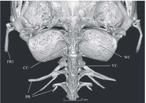
diagnostic features for the subfamily originally proposed (de Pinna, 1992) and which are clearly seen in the skele-tal preparation available. Those characters can be illustrated with unprecedented clarity in micro CT-scan images of the holotype, as follows: 1- lateral process of the lateral ethmoid directed posteriorly (Fig. 9); 2- maxilla articulating directly with the lower jaw, at limit dentary/anguloarticular of coro-noid process (Fig. 9); 3- maxilla and premaxilla linked by processes on each bone (Fig. 9; Fig. 10, for detail of process on premaxilla); 4- anterior margin of premaxilla with dorsal expansion partially surrounding anterior surface of corres-ponding mesethmoid cornu (Fig. 9); 5- spatulate jaw den-tition (Fig. 11); 6- scaphium with a hypertrophied articular process contacting complex centrum of Weberian complex (Fig. 12). An additional character, the dorsal odontode-bea-ring expansion of the interopercle, was also originally pro-posed as a synapomorphy of copionodontines, but was later found to be present also in a subsequently-described species of Trichogenes, T. claviger (de Pinna et al., 2010). Thus, this trait is probably synapomorphic for the clade Copionodon-tinae plus Trichogeninae, with a reversal in T. longipinnis. Large specimens of the latter species (e.g., largest two c&s specimens in MZUSP 48108), however, have small odonto-des in a homologous position on the interopercle, though not as conspicuously arranged and lacking an expanded bony base as in copionodontines and T. claviger. In any event, this trait is also nicely illustrated in micro CT-scan images avai-lable (Fig. 13). Some plesiomorphic conditions unique for Copionodontinae and Trichogeninae among trichomycterids are also well demonstrated in micro CT-scan images, such as the separation between the Weberian capsule and the poste-rior margin of the braincase (Fig. 14). The peculiar structure of the entopterygoid, broadly sutured with the palatine, is a condition synapomorphic for the two subfamilies which is revealed with particular clarity (Fig. 15).
The divided lower lip is also absent in the recently described C. elysium (de Pinna et al., 2018) and that state is probably a synapomorphy for a subgroup of Copionodon rather than for the entire genus.
A few other characteristics of C. exotatos, while not de-cisively synapomorphic for Copionodon, still match condi-tions in all other congeners, and not those in Glaphyropoma. The caudal fin in C. exotatos is clearly bilobed as in all other Fig. 14. Copionodon exotatos, holotype, MZUSP 120631, CT-scan image of posterior part of skull and Weberian capsule, dorsal view. Abbreviations: FR1, First Ray 1; CC, complex centrum; WC, Weberian capsule; VC, vertebrae centrum; PR, pleural ribs.
New species of relictual Copionodon Neotropical Ichthyology, 16(4): e180049, 2018
12
Copionodon species and not truncate with round corners or slightly concave as in species of Glaphyropoma. Similarly, vertebral number in C. exotatos, 33 or 34, also agrees with figures in Copionodon and not with the somewhat reduced value of 30 or 31 in species of Glaphyropoma. (de Pinna, 1992; Bichuette et al., 2008).
Further agreeing with its inclusion in Copionodon, C. exotatos does not share the only unambiguous synapomorphy known for Glaphyropoma: the slender first hypobranchial (cf. de Pinna, 1992, fig. 6 and Bichuette et al., 2008, fig. 6). The new species clearly has a robust version of that bone as seen in all other species of Copionodon and corresponding to the presumably plesiomorphic state in lower trichomycterids.
In view of the evidence discussed above, there is litt-le doubt that C. exotatos should be included in the genus Copionodon. The question of its closest relative within the genus, however, remains open. The lack of a free orbital rim present in C. exotatos matches the condition in the recently described C. elysium, from rio Mucugezinho subdrainage. All other species in Copionodon have free orbital rims. The condition in C. exotatos and C. elysium is unique in Copionodon, but its phylogenetic implications are uncer-tain, because a free orbital rim is also lacking in species of Glaphyropoma, Nematogenys and all trichomycterids other than some Copionodontinae and Trichogeninae, rendering its state optimization ambiguous.
Comparative material examined. All from Brazil, Bahia, rio Para-guaçu basin. Copionodon elysium de Pinna, Burger & Zanata, 2018: MZUSP 120631, holotype, 45 mm SL. MZUSP 120630, 15, 3 c&s, paratypes, 21.7-59.7 mm SL. Copionodon lianaeCampanario & de Pinna, 2000: MZUSP 42470, paratypes, 16, 2 c&s, 11.3-44.5 mm
SL.Copionodon orthiocarinatusde Pinna, 1992: MZUSP 42463,
holotype, 37.7 mm SL. MZUSP 100723, 58 (5 c&s), 23.3-68.5 mm SL. UFBA 6033, 25, 1 c&s, 25.2-56.8 mm SL. Copionodon pecten de Pinna, 1992: MZUSP 42462, 37, 5 c&s, paratypes, 31.4-62.2 mm SL. MZUSP 101352, 15, 2 c&s, 49.0-74.5 mm SL. Glaphyropoma
rodrigueside Pinna, 1992: MZUSP 42466, 13, 3 c&s, paratypes,
28.8-45.4 mm SL. Glaphyropoma spinosum Bichuette, de Pinna & Trajano, 2008: MZUSP 99742, holotype, 58.2 mm SL.MZUSP 99743, 4 (1 c&s), paratypes, 34.9-45.6 mm SL. UFBA 5298, 2, 1 c&s, 43.1-44.4 mm SL. Trichogenes claviger de Pinna, Helmer, Brit-ski & Nunes, 2010: MZUSP 105732, 6, 2 c&s, paratypes, 22.7-40.0 mm SL. Trichogenes longipinnis Britski & Ortega, 1983: MZUSP 16099, holotype; 64.0 mm SL. MZUSP 40238, 9, 2 c&s, 47,9-125.8 mm SL. MZUSP 48108, 6, 3c&s, 41.1-114.3 mm SL.
Acknowledgments
We thank Fernando Dagosta for help with photographs and Alberto Carvalho for expert technical support with the CT-scan facilities at MZUSP and image editing. Funding is provided by FAPESP (#2015/26804-4 to MdP, #2014/11397-1 to VA and #20#2014/11397-16/25467-7 to VC), CNPq (##2014/11397-1339#2014/11397-17/20#2014/11397-10-0 to AMZ) and CAPES (to VA and VC).
References
Arratia G. Development and variation of the suspensorium
of primitive catfishes (Teleostei: Ostariophysi) and their
phylogenetic relationships. Zoologisches Forshungsinstitut und Museum Alexander Koenig; 1992. (Bonner Zoologische Monographien; No. 32).
Bichuette ME, de Pinna MCC, Trajano E. A new species of Glaphyro poma: the first subterranean copionodontine catfish and the first occurrence of opercular odontodes in the subfamily (Siluriformes: Trichomycteridae). Neotrop Ichthyol. 2008; 6(3):301-06. Campanario CM, de Pinna MCC. A new species of the primitive
trichomycterid subfamily Copionodontinae from northeastern Brazil (Teleostei: Trichomycteridae). Ichthyol Explor Freshw. 2000; 11(4):369-76.
International Union for Conservation of Nature (IUCN). Red list of threatened species. Version 2017-3 [Internet]. 2017. Available from: http://www.iucnredlist.org
Lundberg JG, Baskin JN. The caudal skeleton of the catfishes,
order Siluriformes. New York: American Museum of Natural History; 1969. (American Museum Novitates; No. 2398). Nelson GJ. Relationships of clupeomorphs, with remarks on the
structure of the lower jaw in fishes. In: Greenwood PH, Miles RS, Patterson C, editors. Interrelationships of fishes. San
Diego: Academic Press; 1973. p.333-349.
Patterson C. The braincase of pholidophorid and leptolepid fishes,
with a review of the actinopterygian braincase. Phil Trans R Soc Lond B. 1975; 269(899):275-579.
de Pinna MCC. A new subfamily of Trichomycteridae (Teleostei, Siluriformes), lower locarioid relationships and a discussion on the impact of additional taxa for phylogeneticanalysis. Zool J Linn Soc. 1992; 106(3):175-229.
de Pinna MCC. Phylogenetic relationship of Neotropical Siluriformes
(Teleostei, Ostariophysi): historical overview and synthesis of
hypotheses. In: Malabarba LR, Reis RE, Vari RP, Lucena ZMS,
Lucena CAS, editors. Phylogeny and classification of Neotropical fishes. Porto Alegre: EDIPUCRS; 1998. p.279-330.
de Pinna MCC, Helmer JL, Britski HA, Nunes LR. A new species of
Trichogenes from the rio Itapemirim drainage, southeastern Brazil,
with comments on the monophyly of the genus (Siluriformes: Trichomycteridae). Neotrop Ichthyol. 2010; 8(4):707-17. de Pinna M, Burger R, Zanata AM. A new species of Copionodon
lacking a free orbital rim (Siluriformes: Trichomycteridae). Neotrop Ichthyol. 2018; 16(2):e170146[9p.].
Sabaj MH. Standard symbolic codes for institutional resource collections in herpetology and ichthyology: an online reference. Washington, DC: American Society of Ichthyologists and Herpetologists. Version 6.5 [Internet]. 2016 [16 August]..
Available from: https://asih.org/sites/default/files/documents/
symbolic_codes_for_collections_v6.5_2016.pdf
Taylor WR, van Dyke GC. Revised procedures for staining and
clearing small fishes and other vertebrates for bone cartilage.
Cybium. 1985; 9(2):107-19.