Polymorphism of exon 2-3 of bovine major histocompatibility complex class I
BoLa-A gene
Bharat Bhushan
1, Biswa Nath Patra
1, Pranab Jyoti Das
1, Triveni Dutt
2, Pushpendra Kumar
1, Arjava Sharma
1,
Umang
1, Satyabarta Dandapat
3and Shiv Pal Singh Ahlawat
11
Genetic Marker Laboratory, Animal Genetics Division, Indian Veterinary Research Institute,
Izatnagar, Bareilly, Uttar Pradesh, India.
2
Livestock Production and Management Section, Indian Veterinary Research Institute, Izatnagar,
Bareilly, Uttar Pradesh, India.
3
Division of Animal Biotechnology, Indian Veterinary Research Institute, Izatnagar, Bareilly,
Uttar Pradesh, India.
Abstract
The exon 2-3 region of bovine major histocompatibility complex (MHC) class I BoLa-A gene was investigated for polymorphisms in three breeds of cattle originated in the Indian subcontinent namely Sahiwal, Tharparkar, Hariana, as well as crossbred (Bos taurus x Bos indicus) cattle and Jersey, the exotic breed (Bos taurus). The PCR amplified fragment of 714 bp showed distinctDdeI-, TaqI- and HinfI- RFLP patterns, thus confirming a higher degree of poly-morphism in this region. To our knowledge this is the first report ofHinfI restriction patterns for BoLa-A exon 2-3. The sequencing results revealed a number of nucleotide substitutions in this region, which resulted in amino acid changes. The present investigation confirmed that MHC class I BoLa-A exon 2-3 is highly polymorphic in cattle.
Key words:MHC, BoLa-A polymorphism, cattle.
Received: November 28, 2005; Accepted: June 5, 2007.
The major histocompatibility complex (MHC) is a chromosomal region consisting of a series of closely linked loci or so called gene families. The MHC of cattle is known as bovine lymphocyte antigen (BoLa) located on chromo-some 23. Two molecularly well-defined classes of cell sur-face antigens are present among the gene products of the MHC region. Class I antigens are found on the surface of nearly all cell types and are in general involved in the cytotoxic T-cell response. These antigens are highly poly-morphic and their amino acid sequences are highly vari-able. These variations are concentrated in three to four discrete hypervariable regions within theα1 and α2
do-mains. The rest of the molecule is highly conserved and shows little sequence variation. At present, only one class I locus (BoLa-A) is internationally accepted on the basis of serological testing, although there is evidence for the exis-tence of a second class I locus (BoLa-B). Molecular analy-sis of MHC class I region suggested that this region may contain up to 15 genes. The BoLa-A locus has 32
serologi-cally defined alleles and at least four more putative alleles, in addition to a high frequency of null alleles (Tizard, 1998). Sequencing of the expressed class I genes from sero-logically typed animals showed that single BoLa-A types can be expressed up to three class I genes and that BoLa specificity is associated with one gene product (Elliset al., 1996, 1999). Thus, BoLa-A typing defines a class I haplo-type rather than an allele. This demonstrates that the limita-tions of serological typing go beyond its breed-specific nature and shows that there is need for methods that will al-low class I polymorphism to be analyzed directly,i.e. at DNA level (Sawhneyet al., 2001). Elliset al.(1998) de-scribed the DNA based typing of BoLa class I gene using sequence specific amplification of expressed class I alleles from cDNA for a few European haplotypes. The wider ap-plication of this method is limited by the lack of informa-tion on class I allele sequence variainforma-tion between breeds and by breed-specific differences in the class I types repre-sented (Bullet al., 1989). Analysis of MHC class II poly-morphism has benefited from the development of PCR-RFLP methods, which have improved the ease and re-liability of class II typing (van Eijket al., 1992; Ballingall
et al., 1997). However, the development of PCR based
typ-Send correspondence to Bharat Bhushan. Genetic Marker Labora-tory, Animal Genetics Division, Indian Veterinary Research Insti-tute, Izatnagar, 243 122 Bareilly, India. E-mail: [email protected] and [email protected].
ing techniques for class I genes is complicated by the ex-pression of multiple class I genes in most haplotypes and the lack of obvious sequence differences between these genes (Elliset al., 1999). Thus, designing of primer se-quences is limited to the available DNA sese-quences and is not locus-specific. The primers used for amplification were within the conserved regions at the start of exon 2 and the end of exon 3. As the primers incorporate all the polymor-phism observed in the class I sequences, they were ex-pected to amplify the majority of expressed class I genes. Considering the importance of BoLa-A gene in controlling immune responses, the present investigation was con-ducted to characterize the exon 2-3 region of this gene in cattle populations.
Investigations were undertaken on Sahiwal (n = 56), Tharparkar (n = 25), Hariana (n = 14) and crossbred {Bos taurus(Holstein Friesian)x Bos indicus(Hariana)} cattle (n = 70) of Indian origin and on an exotic (Bos taurus) Jer-sey (n = 18) cattle breed maintained at various organized livestock farms of the country. Genomic DNA was isolated from 5 mL of venous blood collected from the jugular vein by phenol-chloroform extraction method.
A 714 bp fragment encompassing exon 2-3 region of MHC Class I BoLa-A gene was amplified (Figure 1a) with the forward and reverse primers 5’-GTC CCC ACT CSM TGA GGT ATT-3’ and 5’-TCC AGG TAT CTG CGG AGC-3’, respectively, as previously described (Sawhneyet al., 2001). PCR was carried out in the final volume of 50µL
containing 100µM dNTPs mix, 200 ng of each primer,
5µL of 10x PCR assay buffer, 3µL MgCl2(2.5 mM), 1 unit
ofTaqDNA polymerase and 80-100 ng of genomic DNA. The amplification of the 714 bp product was carried out us-ing a thermal cycler (PTC-200, MJ Research) under the fol-lowing conditions: 94 °C for 3 min, followed by 35 cycles of 94 °C for 40 s, 58 °C for 40 s and 72 °C for 1 min. The fi-nal extension was done at 72 °C for 5 min. The PCR prod-ucts were checked by agarose gel electrophoresis. For each sample, 10µL of PCR product was digested with 5 units of
each restriction enzyme (DdeI,TaqI, andHinfI). Samples digested withTaqI were incubated at 65 °C, whereas sam-ples digested withDdeI andHinfI were incubated at 37 °C overnight. The DdeI and TaqI digested restriction frag-ments were resolved on 8% polyacrylamide gels and visu-alized by silver staining. The HinfI digested restriction fragments were resolved by 3% agarose gel electrophore-sis, stained with ethidium bromide and documented by means of the Gel Documentation System (Syngene, U.K.). Purified PCR products extracted from the gel were ligated into the cloning vector pTZ57R/T, according to the manu-facturer’s recommendations (MBI Fermentas), with some modifications. A 20-30 min extension at 72 °C resulted in a 3-4 folds higher yield of recombinant colonies of host cell
E. colistrain DH5α. The inserts were confirmed by colony
PCR and restriction digestion of plasmids with PstI and
EcoRI, yielding a fragment of 762 bp. Automated
sequenc-ing of the clones was carried out ussequenc-ing M13 forward and re-verse primers (ABI PRISM) and sequencing data were ana-lyzed by the program DNAstar (Thompsonet al., 1994).
DdeI digestion of the PCR-amplified 714 bp fragment produced five novel fragments, in addition to those already reported by Sawhneyet al.(2001). The fragment sizes of A10, A11, A18, A19, A20 and A31 BoLa-A types were 440, 380 bp; 380 bp; 440, 230, 170, 135 bp; 480, 380, 95 bp; 320, 300, 250 bp and 230, 170 bp, respectively (Fig-ure 1b). Two novel patterns with fragment sizes of 603, 99, 12 bp and 600, 60, 39, 12 bp in crossbred cattle, were later confirmed by cloning and sequencing. Digestion withTaqI also revealed the specific fragments of BoLa-A alleles as reported by Sawhneyet al.(2001), 300 and 250 bp for A10, 475 and 75 bp for A11; 70 bp for A18; 430 and 115 bp for A31, respectively (Figure 1c). Three new patterns with fragment sizes of 605,106 bp; 608, 68, 38 bp and 532, 106, 76 bp in crossbred cattle were confirmed by cloning and se-quencing. The 470 and 75 bp fragments of BoLa-A19 type reported by Sawhneyet al.(2001) were not observed after
TaqI digestion, indicating the absence of this restriction site for BoLa-A19 type in the present investigation.
Sawhneyet al.(2001) reported that theDdeI patterns with the largest number of bands revealed at least five BoLa-A types. The presence of conserved and polymorphic bands in all investigated animals supports the view that the functional,i.e.expressed and polymorphic class I genes, were produced by PCR-RFLP. Additionally, the pattern of
DdeI and TaqI conserved bands formed a framework within which the polymorphic bands could be easily identi-fied. Sawhneyet al. (2001) also observed that TaqI and
DdeI produced a number of BoLa-A type specific bands and unique combinations for most of the seven BoLa-A types. Elliset al.(1999) confirmed that the A11 BoLa-A type comprises multiple polymorphic class I genes and in-cludes the twoTaqI fragments assigned as A11 specific (470, 75 bp), but did not show the 380 bpDdeI fragment.
The frequencies of BoLa-A types in the present study ranged from 0.015 to 0.722, with wide variation among dif-ferent breeds of cattle (Table 1). These results revealed that the A10/A19, A10/A31, A18/A20 and A20/A31 BoLa-A types were similar to those reported by Sawhney et al.
(2001). Ten BoLa-A types were reported (A18/A31, A10/A19, A10/A31, A18/A20, A10/A11, A20/A31, A31/A32, A11/A20, A11/A32 and A19/A20) (Sawhneyet al. 2001) out of which A10/A19, A10/A31, A20/A31, A31/A32, A11/A20 and A11/A32 were already recognized in the 5th International Bovine Lymphocyte Antigen (BoLa) Workshop and were assigned the workshop ID numbers WK5-38, WK5-53, WK5-51, WK5-36, WK5-29 and WK5-48, respectively (Davieset al., 1994). Besides, other BoLa-A types (A18/A19 and A19/A31) revealed the high degree of polymorphism in cattle population.
B, C, D and E (Figure 1d) after validation by sequencing. It was observed that most of the cattle populations were het-erozygous. The fragment sizes of patterns A, B, C, D and E were 122, 212, 379 bp; 141, 193, 379 bp; 71, 122, 142, 379 bp; 379, 275, 60 bp and 335, 380 bp, respectively. To our knowledge this is the first report ofHinfI restriction pat-terns for BoLa-A exon 2-3.
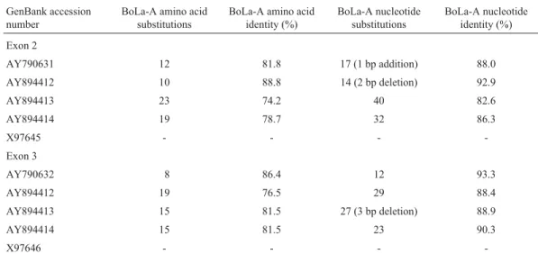
Sequencing revealed that the sizes of the exon 2-3 fragments ranged between 711 to 714 bp in crossbred cattle (GenBank accession numbers AY894412, AY894413 and AY894414). This variation in nucleotide number was due to the addition and deletion of bases. Exon 2 with 267 bp
(AY790631), encoding 89 amino acids, showed a large number of amino acid changes. It was also seen that the reading frame shifted because of two base pair deletions at positions 216 and 217, and maximum nucleotide changes occurred in three clusters. Polymorphism in exon 2 oc-curred mostly due to nucleotide base changes at the posi-tions from 61 to 72, 121 to 138 and 184 to 231, whereas nucleotides from 238 to 269 were conserved. Intron 2 fol-lowed by exon 2 with sequence GGTGAG had more fre-quent nucleotide substitutions than exon 2. Exon 3 initiated at nucleotide 468 and had the purine-rich conserved se-quence CGGGTCA (AY790632). More base changes
made this region highly polymorphic. The comparison of MHC exon 2 and 3 sequences of crossbred cattle (Bos taurusxBos indicus) and Holstein Friesian (Bos taurus) is shown in Table 2. The closest sequences of exon 2 and 3 of crossbred cattle differed by 10 and 8 amino acids, respec-tively in comparison to the Holstein Friesian sequence. The nucleotide substitutions and nucleotide identity ranged from 14 to 40 bases and 82.6% to 92.9%, respectively, for exons 2 and 12, to 29 bases and 88.4% to 93.3%, respec-tively for exon 3. The addition of two bases and the deletion of two bases in exon 2, as well as the deletion of three bases in exon 3 were also observed.
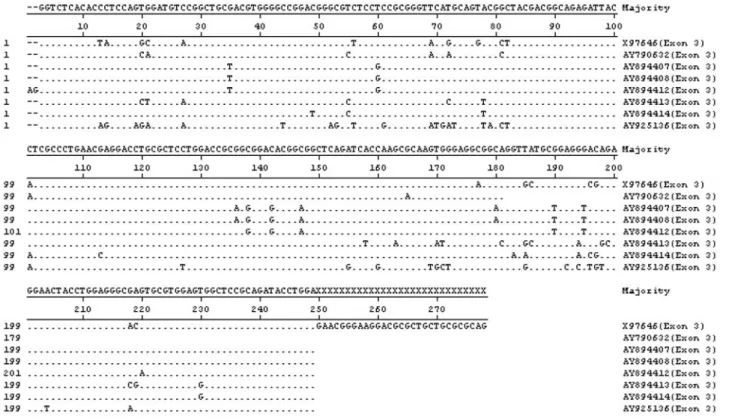
A phylogenetic tree analysis was performed by align-ing the sequences of the BoLa-A exon 2-3 (GenBank acces-sion numbers AY790631, AY790631, AY894412, AY894413 and AY894414) with BuLa-A exon 2-3 (GenBank accession numbers AY894407, AY894408 and AY925136) and Holstein Friesian sequences available in the GenBank (Accession numbers X97645 and X97646). The phylogenetic tree based on nucleotide sequences of exon 2 and 3 showed differences in proximity of the related
alleles (Figures 2a, 2b, 3a and 3b). The results further re-vealed that the nucleotide dissimilarity ranged from 1.1 to 17.9% for exon 2 and 0.0 to 20.9% for exon 3.
The phylogenetic tree analysis revealed that the exon 2 sequences of crossbred cattle (AY894412) and Murrah buffalo (AY894407) cluster together on one branch show-ing the close proximity between these two species. These two sequences of exon 2 were found closest to Holstein Friesian sequence (X97645). One exon 2 sequence of crossbred cattle (AY894413) was found in a completely separate cluster when compared to the other sequences of cattle and buffalo of this region. In the case of exon 3, two
Figure 2- Phylogenetic tree analysis using ClustalW methods with se-quence distance table in the Magalign program in DNAstar using MHC class I (BoLa-A) nucleotide sequences of crossbred cattle, Holstein Friesian cattle and Murrah buffaloes. (a) Phylogenetic tree analysis of exon 2; (b) phylogenetic tree analysis of exon 3.
Table 1- Frequencies of BoLa-A types of exon 2-3 in various cattle breeds.
Breed BoLa-A type
Frequency Breed BoLa-A type
Frequency
Hariana (n = 14)
A10 A11 A18 A19 A10/A11 0.055 0.055 0.055 0.722 0.111 Tharparkar (n = 25)
A10 A11 A18 A19 A31 A10/A19 A10/A20 A10/A31 A18/A19 A19/A31 0.037 0.037 0.148 0.259 0.111 0.111 0.074 0.037 0.037 0.148 Sahiwal (n = 56)
A10 A11 A19 A31 A10/A11 A10/A18 A10/A19 A10/A31 A11/A18 A18/A20 A19/A20 A19/A31 A20/A31 NEW* NEW** 0.054 0.036 0.304 0.054 0.018 0.054 0.214 0.018 0.036 0.018 0.018 0.054 0.071 0.018 0.036 Crossbred (n = 70)
A10 A11 A18 A19 A31 A10/A18 A10/A19 A11/A18 A11/A31 A18/A19 A19/A31 0.058 0.103 0.088 0.265 0.074 0.015 0.074 0.015 0.029 0.206 0.074 Jersey (n = 18)
A10 A11 A19 A10/ A11 A10/ A19 A10/ A31 0.315 0.368 0.105 0.105 0.052 0.052
sequences each of Murrah buffaloes (AY894407 and AY894408) and crossbred cattle (AY894412 and AY894413) formed the same cluster, showing the close-ness of this region in cattle and buffalo. In contrast, the
Hol-stein Friesian sequence (X97646) of exon 3 was found in a distinct branch, showing that in exon 2 Bos taurus was much closer to crossbred cattle and Murrah boffaloes (Bubalus bubalis) than in exon 3. One sequence of Murrah
Table 2- Comparison of MHC class I (BoLa-A types) exon 2 and 3 sequences of crossbred (Bos taurus x Bos indicus) and Holstein Friesian (Bos taurus) cattle.
GenBank accession number
BoLa-A amino acid substitutions
BoLa-A amino acid identity (%)
BoLa-A nucleotide substitutions
BoLa-A nucleotide identity (%)
Exon 2
AY790631 12 81.8 17 (1 bp addition) 88.0
AY894412 10 88.8 14 (2 bp deletion) 92.9
AY894413 23 74.2 40 82.6
AY894414 19 78.7 32 86.3
X97645 - - -
-Exon 3
AY790632 8 86.4 12 93.3
AY894412 19 76.5 29 88.4
AY894413 15 81.5 27 (3 bp deletion) 88.9
AY894414 15 81.5 23 90.3
X97646 - - -
buffalo (AY925136) was found on completely distant branch. Our finding of a lack of a polyphyletic lineage for the cattle and buffalo alleles does not support the trans-species persistence of allelic lineages in BoLa-A and BuLA-A. However, Busscheet al.(1999) reported trans-species persistence of allelic lineage in DRB alleles. The re-sults of the present study support the finding of Brunsberg
et al.(1996) of a characteristic patchwork pattern in DRB alleles, which can be explained as shared ancestral se-quences.
This study confirms the polymorphic nature of exon 2-3 of the MHC class I BoLa-A gene as revealed byDdeI and TaqI RFLPs, and describes the patterns of HinfI RFLPs. Sequencing revealed additions, deletions and sub-stitutions of bases in exons 2-3, producing a high degree of polymorphism in the gene.
Acknowledgments
The authors are thankful to the Director and Joint Di-rector (Academic) of the Indian Veterinary Research Insti-tute (IVRI), Izatnagar for providing the facilities to conduct the study. The help received from Incharge, Livestock Pro-duction Management Section, IVRI, Izatnagar, UP; Live-stock Research Center, GBPUA&T, Pantnagar, Uttaran-chal; Central Cattle Breeding Farm, Suratgarh, Rajasthan and College of Veterinary Sciences & Animal Husbandry, Mathura, UP, India for collection of the blood samples of cattle is also duly acknowledged.
References
Ballingall KT, Luyai A and Mckeever DJ (1997) Analysis of ge-netic diversity at the DQA locus in African cattle: Evidence for a DQA3 locus. Immunogenetics 46:237-44.
Brunsberg U, Lilja IE, Andersson L and Gustafsson K (1996) Structural and organization of pig MHC class II DRB genes: Evidence for genetic exchange between loci. Immunoge-netics 44:1-8.
Bull RW, Lewin HA, Wu MC, Peterbaugh K, Antczak D, Ber-noco D, Cwik Set al.(1989) Joint report of the third interna-tional bovine lymphocyte antigen (BoLA) workshop, Helsinki, Finland. Anim Genet 20:109-32.
Bussche RAV, Hoofer SR and Lochmiller RL (1999) Character-ization of MHC-DRB allelic diversity in white tail deer (Odocoileus virginianus) provides insight into MHC-DRB allelic evolution within cervidae. Immunogenetics 49:429-437.
Davies CJ, Joosten I, Bernoco D, Arriens MA, Bester J, Ceriotti G, Ellis S,et al.(1994) Polymorphism of bovine MHC class I genes. Joint report of the fifth international bovine lympho-cyte antigen (BoLA) workshop, Interlaken, Switzerland. Eur J Immunogenet 21:239-258.
Ellis SA, Holmes EC, Staines KA, Smith MJ, McKeever DJ, MacHugh ND and Morrison WL (1999) Variation in the number of expressed MHC genes in different cattle class I haplotypes.Immunogenetics 50:319-328.
Ellis SA, Staines KA and Morrison WL (1996) cDNA sequence of cattle MHC class I genes transcribed in serologically de-fined haplotypes A18 and A31. Immunogenetics 43:156-159.
Ellis SA, Staines KA, Stear MJ, Hensen EJ and Morrison WL (1998) DNA typing for BoLA class I using sequence-specific primers (PCR-SSP). Eur J Immunogenet 25:365-70. Sawhney SMS, Taylor DW and Russell GC (2001) Polymorphism of bovine major histocompatibility complex (MHC) class I genes revealed by polymerase chain reaction (PCR) and re-striction enzyme analysis. Anim Genet 32:27-31.
Thompson JD, Higgins DG and Gibson TJ (1994) CLUSTAL W: Improving the sensitivity of progressive multiple sequence alignment through sequence weighting, position-specific gap penalties and weight matrix choice. Nucleic Acids Res 22:4673-4680.
Tizard IR (1998) Veterinary Immunology: An Introduction. 5th edition. Harcourt Brace and Company, San Diego, 482 pp. van Eijk MJT, Stewart- Haynes JA and Lewin HA (1992)
Exten-sive polymorphism of the BoLA-DRB3 gene distinguished by PCR-RFLP. Anim Genet 23:483-496.
Internet Resource
GenBank: http://www.ncbi.nlm.nih.gov/Entrez, nucleotide se-quences (AY790631, AY790632, AY894412, AY894413, AY894414).