The role of KIR2DL3/HLA-C*0802 in Brazilian patients
with rheumatoid vasculitis
Wester Eidi Nishimura,I,* Zoraida Sachetto,ILilian Teresa Lavras Costallat,IMichel Alexandre Yazbek,I Ana Carolina Santos Londe,IEdilaine Gildo Guariento,IISilvia Barbosa Dutra Marques,II
Manoel Barros BertoloI
IUniversity of Campinas (UNICAMP), Department of Rheumatology, Campinas/SP, Brazil.IIUniversity of Campinas (UNICAMP), Department of Hematology, Campinas/SP, Brazil.
OBJECTIVES: Rheumatoid arthritis is a polygenically controlled systemic autoimmune disease. Rheumatoid vasculitis is an important extra-articular phenotype of rheumatoid arthritis that can result in deep cutaneous ulcers. The objective of this study was to establish a correlation between the frequency of major histocompatibility complex class I/II alleles and killer immunoglobulin-like receptor genotypes in patients with cutaneous rheumatoid vasculitis.
METHODS:Using the Scott & Bacon 1984 criteria to diagnose rheumatoid vasculitis and after excluding any other causes such as diabetes, atherosclerosis, adverse drug reactions, infection, and smoking, patients who met the criteria were selected. All of the selected rheumatoid vasculitis patients presented deep cutaneous ulcers. Identification of the major histocompatibility complex class I/II and killer immunoglobulin-like receptor genotypes was performed by polymerase chain reaction assays of samples collected from the 23 rheumatoid vasculitis patients as well as from 80 controls (40 non-rheumatoid vasculitis RA control patients and 40 healthy volunteers).
RESULTS: An association between the presence of the HLA-DRB1*1402 and HLA-DRB1*0101 alleles and
cutaneous lesions in rheumatoid vasculitis patients and a correlation between the inhibitor KIR2DL3 and the HLA-C*0802 ligand in rheumatoid vasculitis patients were found.
CONCLUSION:An association was found between the presence of the HLA-DRB1*1402 and HLA-DRB1*0101
alleles and the development of cutaneous lesions in rheumatoid vasculitis patients. Additionally, the HLA-C*0802 ligand protects these individuals from developing cutaneous lesions.
KEYWORDS: Rheumatoid arthritis; Receptors, KIR; Rheumatoid vasculitis; HLA-C antigens/HLA-DRB1 chains.
Nishimura WE, Sachetto Z, Costallat LT, Yazbek MA, Londe AC, Guariento EG, et al. The role of KIR2DL3/HLA-C*0802 in Brazilian patients with rheumatoid vasculitis. Clinics. 2015;70(6):408-412
Received for publication onAugust 27, 2014;First review completed onJanuary 12, 2015;Accepted for publication onMarch 17, 2015
E-mail: [email protected]
*Corresponding author
’ INTRODUCTION
Rheumatoid arthritis (RA) is a systemic autoimmune disease characterized by inflammatory polyarthritis (1,2). RA is polygenically controlled, with contribution from the major histocompatibility complex (MHC) (2–6). MHC class I
mole-cules are expressed on most nucleated human cells and comprise the principal human leukocyte antigens (HLA) A, B and C (3). MHC class II molecules are expressed on antigen-presenting cells (APCs; B lymphocytes, macrophages, and
dendritic cells) and include mainly HLA-DR and HLA-DQ (3). MHC class I molecule expression results in direct cytotoxicity from CD4+CD28- T cells or natural killer (NK) cells, and MHC class II molecule expression results in cytotoxicity via APCs (3). The polygenic characteristics of RA, which result in different phenotypic expressions, determine the heteroge-neous presentation of articular destruction and extra-articular manifestations (1,7). An important and potentially life-threatening extra-articular manifestation of RA is rheu-matoid vasculitis (RV), which affects the small- and medium-sized arterial vessels of the upper and lower extremities, peripheral nerves and occasionally other organs (8,10,11). RV is a rare complication that may affect approximately 1–5% of RA patients and that is diagnosed by ruling out
other possible clinical conditions such as diabetes, athero-sclerosis, adverse drug reactions, infections and neoplasms (1,9). The HLA-DRB1 allele (B1*0401 homozygotes, in particular) is more common in RV patients (12,13). In DOI:10.6061/clinics/2015(06)04
Copyright&2015CLINICS–This is an Open Access article distributed under the terms of the Creative Commons Attribution Non-Commercial License (http:// creativecommons.org/licenses/by-nc/3.0/) which permits unrestricted non-com-mercial use, distribution, and reproduction in any medium, provided the original work is properly cited.
addition to genetic factors, evidence of RA biomarkers such as CD4+CD28- T cells, which are uncommon in healthy subjects, has been reported (14). CD4+CD28
-T cells are increased in RV patients, and the role of these cells has been reported in arterial damage caused by inflammatory injury (15,16). Because CD4+CD28
-T cells do not express the CD40 ligand, they develop direct cytotoxic abilities and work in conjunction with the CD57 expressed on NK cells in RV patients, suggesting that both cells are involved in vascular endothelial damage (17,18). Regarding NK cells, the devel-opment of vascular damage is intrinsically dependent on the interaction of these cells with MHC class I molecules in RA patients (11,19). NK cells express activating and inhibiting receptors on their surface, which interact with the MHC antigens of vascular endothelial cells, resulting in NK cell activation or inhibition. On the NK cell surface, killer immunoglobulin-like receptors (KIRs), which recognize MHC class I molecules, are present (20). Increased cell surface expression of HLA-C ligands has been shown in RV patients. These ligands interact with KIR2DS2, which is present on CD4+CD28
-T cells, confirming the similarity of CD4+CD28 -T cell characteristics with NK cells, as both cell types are derived from the same hematopoietic stem cell (3,11,21).
The objective of this study was to establish a correlation between the frequency of MHC class I/II and KIR genotypes and cutaneous RV.
’ METHODS AND MATERIALS
Patient selection
We evaluated patients diagnosed with RA who were followed for a period of 2 years (2012–2013) in the
Rheumatology Department at the University of Campinas Teaching Hospital (HC-UNICAMP), a tertiary referral hospital located in Campinas, Sao Paulo State, Brazil. The clinical data of patients, who were all ethically unrelated, were obtained through a review of their records. The study was approved by the Ethics Committee, and the patients provided informed consent. The RA diagnosis was based on the American College of Rheumatology (ACR) 1987 Rheumatoid Arthritis classification criteria (22). Patients under 16 years of age or with overlap syndrome, diabetes, atherosclerosis, a history of adverse drug reactions, infection, or a history of smoking were excluded. RV was clinically diagnosed by the presence of deep cutaneous ulcers (DCU) on the lower limbs, by localized pain in areas of healthy skin that was suggestive of neuropathic involvement of small sensory fibers or by digital infarction of the toes (9). The diagnosis was based on the Scott & Bacon 1984 clinical criteria and required 1 of the following 3 characteristics: DCU, symptoms suggestive of mononeuritis multiplex (MNM), or digital infarction (9,23,24). All patients were evaluated for rheumatoid factor (RF) and citrullinated protein antibody (anti-CCP).
Control population
Patients diagnosed with RA from the Rheumatology Department at the HC-UNICAMP who were selected during the period that the RV cases were analyzed were included as controls (22). Blood donors from HC-UNICAMP were also included as healthy patient (HP) controls. Using the RV diagnostic criteria, some of the patients exhibiting evidence of the condition were excluded from the control groups. In the comparative analysis of case and control groups, factors such as gender and age were evenly matched across all 3 groups.
Deoxyribonucleic acid (DNA) extraction methodology
Whole blood samples were collected and subjected to DNA extraction using the GE Healthcare Illustra Blood GenomicPrep Mini Spin Kits
in accordance with the manufacturer’s guidelines.
MHC Class I/II Genotyping and Reading Methodology
DNA samples were processed using the high-resolution SSO/Luminexs
. The SSO/DNA method used oligonucleo-tide sequence-specific probes immobilized on fluorescently encoded microspheres. HLA alleles were identified in DNA samples amplified by polymerase chain reaction (PCR) using hybridization probes and flow cytometry analysis. The manufacturer’s guidelines were strictly followed for all methodological parameters.
KIR genotyping and reading methodology
Three reactions were analyzed for KIR genotyping to evaluate the genetic composition of exons 3–4 (group 1),
exon 5 (group 2), and exons 7–9 (group 3). DNA
amplifica-tion was performed by PCR with specific primer sequences, and the samples were aligned using the Luminexs
system. All steps were performed in accordance with the manufac-turer’s guidelines.
Statistical analysis
In addition to the use of graphs and one-dimensional scatter plots, all data collected were first descriptively analyzed by calculating various measurements and summa-ries, including the mean, median, minimum and maximum values, standard deviations, and absolute and relative frequency percentages. Pearson’s chi-squared (w2) test or an extension of Fisher’s exact test was used to compare group profiles according to gender, class I and II allelic frequencies, and the presence of diverse KIR genotypes (25). Analysis of variance (ANOVA) with one fixed factor was used to compare the age profiles of the groups (26). Because the study was exploratory, sample calculations were not performed due to the lack of data in the literature comparing the allelic frequencies in all 3 groups of interest. In all conclusions made through inferential analysis, the level of asignificance was equal to 5%. The data were compiled into a Windows Excel 2010 table to adequately manage the information. Statistical analyses were performed using R statistical software version 2.15.2.
Ethical standards
This study was approved by the ethics committee of the University of Campinas (UNICAMP) and was conducted in accordance with the Declaration of Helsinki of 1975, which was revised in 1983. All 103 subjects agreed to participate in this study by reading and signing the Informed Consent Form that was approved by the same committee.
’ RESULTS
and 3 (13%) men. Active ulcer cutaneous biopsies were performed in 3/23 case patients and demonstrated the standard histopathology of vessel wall fibrinoid necrosis with inflammatory cell infiltration and areas of occlusion with recanalization. All 23 case patients were undergoing treatment with Disease Modifying Anti-Rheumatic Drugs (DMARDs) and had also used high-dose corticosteroids at some point during the course of RV. Therapeutic doses of antiplatelet agents were introduced in cases of toe digital infarctions. The 40 RA control patients were also treated with DMARDs and occasional low doses of corticosteroids.
Of the 103 patients studied, 40 (38.8%) were in the HP control group, 40 (38.8%) were in the RA control group, and 23 (22.4%) were in the RV case group. The age and gender distributions of the groups are displayed in Table 1.
Notably, when comparing the gender and age profiles of the 3 groups, these groups are statistically equivalent with respect to gender (p=0.611) and age (p=0.927).
The frequency of class I alleles varied between 0.0% and 47.5% in the HP group, between 0.0% and 45.0% in the RA group, and between 0.0% and 43.5% in the RV group. The high frequencies of expression in all 3 groups can be explained by the presence of the A*0201 allele (p=0.949),
which was observed in all evaluated patient groups. The alleles with significantly different A*2601 (p=0.017)
and B*3906 (p=0.048) frequencies were more commonly
observed in the RV group than in the HP and RA groups. The B*5001 (p=0.024) and C*0602 (p=0.001) alleles were more
frequently observed in the HP group than in the RA and RV
groups. The B*1501 (p=0.048) and C*1502 (p=0.027) alleles
were more frequently observed in the RA group than in the other two groups. With respect to the C*0802 (p=0.019) allele,
it was less frequently observed in the RA group than in the HP and RV groups. In the HP and RV groups, there was a trend towards more frequent expression of the B*1402 allele (p=0.057) and less frequent expression of the C*0501 allele
(p=0.056) compared with the RA group. The other alleles
were observed equally in all 3 groups, without significant differences. These data are displayed in Table 2.
Higher allelic frequencies were observed for HLA-DRB1*0701 (p=0.428) and DQB1*0302 (p=0.740) in the HP
group, DQB1*0301 (p=0.752) in the RA group, and
DQB1*0501 (p=0.480) in the HP and RA groups. The
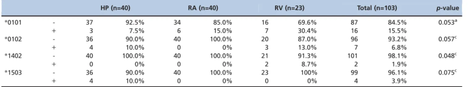
comparison of class II allelic frequencies between the groups suggests that almost all alleles were observed equally across the groups. The only exception was the DRB1*1402 allele (p=0.048), which was found more frequently in the RV group
than in the HP and RA groups. A trend towards a higher frequency of the DRB1*0101 (p=0.053) allele was found in the
RV group compared with the HP and RA groups, as well as a higher frequency of the DRB1*1503 (p=0.075) allele in the HP
group compared with the RA and RV groups. The other alleles were equally observed in the 3 groups, without significant differences. These data are displayed in Table 3.
Higher frequencies were observed for the KIR2DL2 genotype (p=0.529) in the RA and RV groups, as well as
for the KIR3DL2 and 3DL3 genotypes in all 3 groups (ap-value could not be calculated). Higher frequencies were Table 1 -General characteristics of the individuals in each group.
HP (n=40) RA (n=40) RV (n=23) Total (n=103) p-value
Gender
Female 31 77.5% 31 77.5% 20 87.0% 82 79.6% 0.611a
Male 9 22.5% 9 22.5% 3 13.0% 21 20.4%
Age (years)
Average 54.3 54.8 55.5 54.8 0.927b
Median 56.0 56.0 56.0 56.0
Minimum-maximum 25.0–77.0 17.0–78.0 32.0–71.0 17.0–78.0
Standard deviation 12.3 12.9 9.5 11.9
aPearson
’s chi-squared test,bANOVA with 1 fixed factor
Table 2 -Distribution of the HLA-A/B/C allelic frequencies among the patient groups.
HP (n=40) RA (n=40) RV (n=23) Total (n=103) p-value
A*2601 - 40 100.0% 38 95.0% 19 82.6% 97 94.2% 0.017c
+ 0 0% 2 5.0% 4 17.4% 6 5.8%
B*1402 - 36 90.0% 40 100.0% 20 87.0% 96 93.2% 0.057c
+ 4 10.0% 0 0% 3 13.0% 7 6.8%
B*1501 - 40 100.0% 35 87.5% 22 95.7% 97 94.2% 0.048c
+ 0 0% 5 12.5% 1 4.3% 6 5.8%
B*3906 - 40 100.0% 40 100.0% 21 91.3% 101 98.1% 0.048c
+ 0 0% 0 0% 2 8.7% 2 1.9%
B*5001 - 35 87.5% 40 100.0% 23 100.0% 98 95.1% 0.024c
+ 5 12.5% 0 0% 0 0% 5 4.9%
C*0501 - 37 92.5% 33 82.5% 23 100.0% 93 90.3% 0.056c
+ 3 7.5% 7 17.5% 0 0% 10 9.7%
C*0602 - 27 67.5% 38 95.0% 22 95.7% 87 84.5% 0.001a
+ 13 32.5% 2 5.0% 1 4.3% 16 15.5%
C*0802 - 35 87.5% 40 100.0% 19 82.6% 94 91.3% 0.019c
+ 5 12.5% 0 0% 4 17.4% 9 8.7%
C*1502 - 38 95.0% 31 77.5% 22 95.7% 91 88.3% 0.027c
+ 2 5.0% 9 22.5% 1 4.3% 12 11.7%
aPearson
’s chi-squared test,cextension of Fisher
observed for the KIR3DP1 genotype (p40.999) only in the
RV group. The various KIR genotypes showed comparable frequencies in all 3 groups; however, some trends suggested a higher frequency of 2DL3 in the HP and RV groups than in the RA group (p=0.096). A trend towards more frequent
expression of 2DL5 was observed in the HP group compared with the RA and RV groups (p=0.064). Finally, a trend
indicating higher frequencies of 3DL1 was observed in the HP group compared with the RA and RV groups (p=0.060).
The other genotypes were equally observed in the 3 groups, without significant differences. These data are displayed in Table 4.
’ DISCUSSION
Our study evaluated the frequencies of HLA class I and II alleles in 23 RV patients compared with HPs and RA patients without RV. KIR genotyping was performed in these groups, which was correlated with HLA class I antigens. This is the first work to perform correlation genotyping of HLA class I/ KIR antigens in Brazilian RV patients. Studies concerning HLA class I/KIR in RV patients are relevant from an etiopathogenic standpoint as an effort to identify patients with a severe disease.
Watts et al. described an annual RV incidence of 12.5 per million people (95% CI=8.5–17.2), with 15.8 per million (95%
CI=9.5–24.7) men and 9.4 per million (95% CI=4.8–16.4)
women (27). Alternatively, Symmons et al. found an annual incidence of 140 per million men and 360 per million women with RV in a similar population (28). RV affects approxi-mately 1–5% of RA patients, which emphasizes the relative
rarity of RV (1,9). We found 23 RV patients in a population of 700 RA patients over a period of 2 years, which was equivalent to a rate of 3.3%. Our findings are similar to previously reported studies.
RV typically presents in patients who have suffered from rheumatoid arthritis for more than 10 years (1,9). In this study, the average age in the RV group was 55.5 years, which is comparable to the global general RA patient population. RV was more frequent in females (87.0%), but this gender
predominance is not different from that of general RA patients without RV (1,9). In contrast to the findings of this study, gender has been reported to influence the disease phenotype, leading to more severe articular destruction and extra-articular manifestations, including RV, in men (29).
Cutaneous biopsies were performed on 3 patients with a confirmed RV diagnosis. In the remaining 20 patients, RV was diagnosed according to the Scott & Bacon clinical criteria because all patients had a previous RA diagnosis and all other causes of vasculopathy were excluded (9,22–24,30).
Before comparing significant allelic frequencies or analyz-ing statistical trends between groups for MHC class I and II antigens or KIRs, understanding the behavior of these alleles in the population studied was necessary. This was accom-plished using the Allele Frequency Net Database (AFND) to evaluate whether any associations that existed between the alleles examined could under- or overestimate the frequency of each allele in a particular geographic region of the population studied (31). When the search tool on the AFND site was utilized, no data on class I alleles were found for the Campinas region in Sao Paulo, Brazil. For the class II alleles, the same search tool was used for the region of Campinas, and data were found for the DRB1*0101, DRB1*0102, DRB1*1402 and DRB1*1502 alleles. Similarly, no association of these alleles with other alleles was found in this geographic region.
After confirming the allelic frequencies, their correlations with KIR frequency findings were analyzed. KIR2DL3 is an inhibitor that interacts with HLA-C (C*01, C*03, C*07, C*08, C*12, C*13, C*14 and C*16) ligands (32). KIR2DL5 is also an inhibitor, but its interactions with MHC class I molecules remain controversial (32). Therefore, correlating the fre-quency of KIR2DL5 with the findings from this class was not possible. KIR3DL1 is an inhibitor that interacts with HLA-A and HLA-B (A*24, B*13, B*27, B*37, B*44, B*51, B*52, B*53, B*57 and B*58) ligands (32). Nonetheless, the interaction of KIR2DL3 with the C*0802 ligand can disable the missing-self response; thus, in our RV patient population, C*0802 could be a protective allele against the development of cutaneous lesions from direct cytotoxicity (NK cells or CD4+CD28
-T
Table 3 -Distribution of HLA-DRB1 allelic frequencies among the patient groups.
HP (n=40) RA (n=40) RV (n=23) Total (n=103) p-value
*0101 - 37 92.5% 34 85.0% 16 69.6% 87 84.5% 0.053a
+ 3 7.5% 6 15.0% 7 30.4% 16 15.5%
*0102 - 36 90.0% 40 100.0% 20 87.0% 96 93.2% 0.057c
+ 4 10.0% 0 0% 3 13.0% 7 6.8%
*1402 - 40 100.0% 40 100.0% 21 91.3% 101 98.1% 0.048c
+ 0 0% 0 0% 2 8.7% 2 1.9%
*1503 - 36 90.0% 40 100.0% 23 100% 99 96.1% 0.075c
+ 4 10.0% 0 0% 0 0% 4 3.9%
aPearson
’s chi-squared test,cextension of Fisher
’s exact test, absent (-), present (+)
Table 4 -Distribution of KIR genotypes among the patient groups.
HP (n=40) RA (n=40) RV (n=23) Total (n=103) p-value
2DL3 - 5 12.5% 12 30.0% 3 13.0% 20 19.4% 0.096a
+ 35 87.5% 28 70.0% 20 87.0% 83 80.6%
2DL5 - 11 27.5% 20 50.0% 12 52.2% 43 41.7% 0.064a
+ 29 72.5% 20 50.0% 11 47.8% 60 58.3%
3DL1 - 7 17.5% 1 2.5% 1 4.3% 9 8.7% 0.060c
+ 33 82.5% 39 97.5% 22 95.7% 94 91.3%
aPearson
’s chi-squared test,cextension of Fisher
cells). This finding contradicts the results of other studies (11,19,21,33, 34).
Regarding the frequency results for class II alleles in the RV group, the frequency of DRB1*1402 (8.7%) was significantly different, and a statistical trend towards a difference in DRB1*0101 (30.4%) was observed compared with the other groups. Similarly to the findings of other studies (12,13), these findings suggest that these alleles are associated with the development of cutaneous lesions in our RV patient population because they are associated with APCs (B lymphocytes, macrophages and dendritic cells). For the DRB1*1503 allele (10.0%), a statistical trend towards a higher frequency was found in the HP group; this allele could be considered protective against RA and RV in our population.
An association exists between the presence of MHC class II alleles (HLA-DRB1*1402 and HLA-DRB1*0101) and the devel-opment of cutaneous lesions in RV patients. In other words, an association exists between APCs and vascular damage. The KIR2DL3 inhibitor was frequently found in RV patients, and its interaction with the HLA-C*0802 ligand suggests that this allele protects these individuals from cutaneous lesions caused by direct cytotoxicity. Our findings have not been previously described. Despite the rarity of RV, this study could be considered a baseline study for new genetic and multicenter descriptive studies to better our understanding of the phenotypic expression of this disease in Brazil.
’ ACKNOWLEDGMENTS
We would like to acknowledge the Research Assistance Foundation for the State of Sao Paulo (FAPESP), Gianni Mara Silva Santos, and MAppStats from the Federal University of Sao Paulo (UNIFESP) for statistical management.
’ AUTHOR CONTRIBUTIONS
Nishimura WE was responsible for recruiting RV patients and collecting biological materials. Sachetto Z, Costallat LTL, Yazbek MA, Marques SB and Bertolo MB were responsible for guiding the study. Londe CS and Guariento EG were responsible for analyzing the biological materials from RA patients.
’ REFERENCES
1. Turesson C, Matteson E L. Rheumatology. Extra-articular features of rheumatoid arthritis and systemic involvement. 5th ed. Philadelphia: Elsevier; 2011; p.9–27.
2. Ollier W, Winchester R. Current Directions in Autoimmunity. The Germline and somatic genetic basis for rheumatoid arthritis. Switzerland: Basel; 1999; p.166–93.
3. Gascoigne N, Wilson I. Immunobiology: the immune system in health & disease. Recognizing of antigen by receptors of cells B and T. 6th ed. New York: Garland Science; 2005; p.103–30.
4. Seldin MF, Amos CI, Ward R, Gregersen PK. The genetics revolution and the assault on rheumatoid arthritis. Arthritis Rheum. 1999;42(6):1071–9, http://dx.doi.org/10.1002/1529-0131(199906)42:6o41.0.CO;2-J. 5. Weyand CM, Goronzy JJ. Pathogenesis of rheumatoid arthritis. Med
Clin N Am. 1997;81(1):29–55, http://dx.doi.org/10.1016/S0025-7125(05) 70504-6.
6. Nepom GT. Major histocompatibility complex-directed susceptibility to rheumatoid arthritis. Adv Immunol. 1998;68:315–32, http://dx.doi.org/ 10.1016/S0065-2776(08)60563-5.
7. Weyand CM, Klimiuk PA, Goronzy JJ. Heterogeneity of rheumatoid arthritis from phenotypes to genotypes. Springer Semin Immunopathol. 1998;20(1-2):5–22, http://dx.doi.org/10.1007/BF00831996.
8. Turesson C, McClelland RL, Christianson TJ, Matteson EL. No decrease over time in the incidence of vasculitis or other extraarticular manifesta-tions in rheumatoid arthritis: results from a community-basea study. Arthritis Rheum. 2004;50(11):3729–31, http://dx.doi.org/10.1002/(ISSN) 1529-0131.
9. Genta MS, Genta RM, Gabay C. Systemic rheumatoid vasculitis: review. Semin Arthritis Rheum. 2006;36(2):88–98, http://dx.doi.org/10.1016/j. semarthrit.2006.04.006.
10. Turesson C, O’Fallon WM, Crowson CS, Gabriel SE, Matteson EL. Extra-articular disease manifestations in rheumatoid arthritis: Incidence trends and risk factors over 46 years. Ann Rheum Dis. 2003;62(8): 722–7, http:// dx.doi.org/10.1136/ard.62.8.722.
11. Yen JH, Moore BE, Nakajima T, Scholl D, Schaid DJ, Weyand CM, et al. Major histocompatibility complex class I-recognizing receptors are disease risk genes in rheumatoid arthritis. Exp Med. 2001;193(10):1159–67, http:// dx.doi.org/10.1084/jem.193.10.1159.
12. Weyand CM, Xie C, Goronzy JJ. Homozygosity for the HLA-DRB1 allele selects for extraarticular manifestations in rheumatoid arthritis. J Clin Invest. 1992;89(6):2033–9, http://dx.doi.org/10.1172/JCI115814. 13. Turesson C, Weyand CM, Matteson EL. Genetics of rheumatoid arthritis:
is there a pattern predicting extraarticular manifestations? Arthritis Rheum. 2004;51(5):853–63, http://dx.doi.org/10.1002/(ISSN)1529-0131. 14. Schmidt D, Goronzy JJ, Weyand CM. CD4+CD7 CD28 T cells are
expanded in rheumatoid arthritis and are characterized by autoreactivity. J Clin Invest. 1996;97(9):2027–37, http://dx.doi.org/10.1172/JCI118638. 15. Liuzzo G, Kopecky SL, Frye RL, O’Fallon WM, Maseri A, Goronzy JJ,
et al. Perturbation of the T-cell repertoire in patients with unstable angina. Circulation. 1999; 100(21):2135–9, http://dx.doi.org/10.1161/01. CIR.100.21.2135.
16. Liuzzo G, Goronzy JJ, Yang H, Kopecky SL, Holmes DR, Frye RL, et al. Monoclonal T-cell proliferation and plaque instability in acute coronary syndromes. Circulation. 2000; 101(25):2883–8, http://dx.doi.org/10.1161/ 01.CIR.101.25.2883.
17. Weyand CM, Brandes JC, Schmidt D, Fulbright JW, Goronzy JJ. Func-tional properties of CD4+CD28 T cells in the aging immune system. Mech Ageing Dev. 1998;102(2-3):131–47, http://dx.doi.org/10.1016/ S0047-6374(97)00161-9.
18. Namekawa T, Wagner UG, Goronzy JJ, Weyand CM. Functional subsets of CD4 T cells in rheumatoid synovitis. Arthritis Rheum. 1998;41 (12):2108–16, http://dx.doi.org/10.1002/(ISSN)1529-0131.
19. van Bergen J, Koning F. The tortoise and the hare: slowly evolving T-cell responses take hastily evolving KIR. Immunology. 2010;131(3):301–9, http://dx.doi.org/10.1111/imm.2010.131.issue-3.
20. Passweg JR, Koehl U, Uharek L, Meyer-Monard S, Tichelli A. Natural-killer-cell-based treatment in haematopoietic stem-cell transplantation. Best Pract Res Clin Haematol. 2006; 19(4):811–24, http://dx.doi.org/ 10.1016/j.beha.2006.06.004.
21. Turesson C, Schaid DJ, Weyand CM, Jacobsson LT, Goronzy JJ, Petersson IF, et al. Association of HLA-C3 and smoking with vasculitis in patients with rheumatoid arthritis. Arthritis Rheum. 2006;54(9):2776–83, http:// dx.doi.org/10.1002/(ISSN)1529-0131.
22. Arnett FC, Edworthy SM, Bloch DA, MacShane DJ, Fries JF, Cooper NS, et al. The American Rheumatism Association 1987 revised criteria for the classification of rheumatoid arthritis. Artrhitis Rheum. 1988;31(3):315–24, http://dx.doi.org/10.1002/(ISSN)1529-0131.
23. Scott DG, Bacon PA. Intravenous cyclophosphamide plus methylpredni-solone in treatment of systemic rheumatoid vasculitis. Am J Med. 1984;76 (3):377–84, http://dx.doi.org/10.1016/0002-9343(84)90654-5.
24. Ntatsaki E, Mooney J, Scott DG, Watt RA. Systemic rheumatoid vasculitis in the era of modern immunosuppressive therapy. Rheumatology (Oxford). 2014;53(1):145–52.
25. Agresti, A. Categorical data analysis. New York: Wiley; 1990.
26. Neter J, Kutner, M H, Nachtsheim CJ, Wasserman W. Applied linear statistical models. Boston: Irwin; 1996.
27. Watts RA, Carruthers DM, Symmons DP, Scott DG. The incidence of rheumatoid vasculitis in the Norwich Health Authority. Br J Rheumatol. 1994;33(9):832–3, http://dx.doi.org/10.1093/rheumatology/33.9.832. 28. Symmons DPM, Barret EM, Bankhead CR, Scott DG, Silman A J. The
incidence of rheumatoid arthritis in the United Kingdom: Results from the Norfolk Arthritis Register. Br J Rheumatol. 1994;33(8):735–9, http://dx. doi.org/10.1093/rheumatology/33.8.735.
29. Weyand CM, Schmidt D, Wagner U, Goronzy JJ. The influence of sex on the phenotype of rheumatoid arthritis. Arthritis Rheum. 1998;41(5):817– 22, http://dx.doi.org/10.1002/(ISSN)1529-0131.
30. Scope A, Hapern AC. Dermatology in General Medicine. Diagnostic procedures and devices. New York; 2007; p.42.
31. Allele*Frequencies in Worldwide Population [homepage on Internet]. [update 2014 Feb 08]. Available from: http://www.allelefrequencies.net/ default.asp.
32. Vilches C, Parham P. KIR: diverse, rapidly evolving receptors of innate and adaptive immunity. Annu Rev Immunol. 2002;20:217–51, http://dx. doi.org/10.1146/annurev.immunol.20.092501.134942.
33. Ljunggren HG, Kärre K. In search of the‘missing self’: MHC molecules and NK cell recognition. Immunol Today. 1990;11(7):237–44, http://dx. doi.org/10.1016/0167-5699(90)90097-S.