Molecular analysis of three
FUT3
gene single nucleotide polymorphisms
and their relationship with the lewis erythrocytary phenotype
in a human population of japanese-ancestry living in Tomé Açu,
a town in the Brazilian Amazon
Pablo Abdon da Costa Francez
1, Tereza Cristina de Oliveira Corvelo
1, Flávio Ricardo Leal da Silva
2and Sidney Emanuel Batista dos Santos
21
Laboratório de Imunogenética, Departamento de Genética, Universidade Federal do Pará, Belém,
Pará, Brazil.
2
Laboratório de Genética Humana e Médica, Departamento de Patologia,
Universidade Federal do Pará, Belém, Pará, Brazil.
Abstract
The Lewis blood group system involves two major antigens, Lea
and Leb
. Their antigenic determinants are not pri-mary gene products but are synthesized by the transfer of sugar subunits to a precursory chain by a specific enzyme which is the product of the FUT3 gene (Lewis gene). The presence of three FUT3 gene single nucleotide polymorphisms (SNPs) (59T > G; 508G > A and 1067T > A) was related to the Lewis phenotype of erythrocytes from 185 individuals of Japanese ancestry living in the town of Tomé-Açu in the Brazilian Amazon region. This relationship was detected using a serological hemagglutination test and theDot-ELISA assay along with the molecular technique PCR-RFLP. We found that the three SNPs investigated in this study only accounted for a proportion of the Lewis-negative phenotype of the erythrocytes.
Key words:Lewis blood groups,FUT3, Japanese, PCR-RFLPs, SNPs. Received: May 9, 2006; Accepted: September 13, 2006.
Introduction
TheLewis(FUT3orLe) gene is located on the short arm of chromosome 19 (Molliconeet al., 1990, 1992) and codes forα1-3/4-L-fucosyltransferase III (FucT III, also
called the Lewis enzime), an enzyme with 361 amino acids which catalyzes the transfer of a fucose unit to the subter-minal GlcNAc unit of type 1 precursory chains (De Vrieset al., 2001). The Lewis blood group system is comprised of two major antigens, one being the monofucosylated Lea an-tigen formed by the action of FucT III on a type 1 precursor and the other the difucosylated Lebantigen produced by the epistatic interaction between the FUT3 and secretor (FUT2) gene products on a type 1 precursor in tissues such as salivary glands, digestive mucosa and respiratory mu-cosa (Watkins, 1980; Oriolet al., 1986).
The Lewis antigens are not synthesized in erythrocyte progenitors, erythrocytes acquiring their Leaand Leb epito-pes by adsorbing Lewis antigenic glycosphingolipids from
plasma. Accordingly, typing of Lewis phenotypes is diffi-cult and is sometimes misjudged because of weak hema-gglutination as a result of the low specificities of anti-Lea and anti-Lebantibodies and the low number of antigens on red cells, resulting in low titers. (Nishiharaet al., 1993; Liu et al., 1996).
The situation is even more complex due to the pres-ence of FUT3 single nucleotide polymorphisms (SNPs) which can result in nucleotide mutations that generate en-zymes which may have different catalytic activities or even be inactive (Nishiharaet al., 1993,1994; Kudoet al., 1996; Cakiret al., 2002; Cooling and Gu, 2003; Soejimaet al., 2004; Jostet al., 2005). The most frequentFUT3SNPs are the202T>C,314C>T,508G>Aand1067T>A poly-morphisms (legenes) which affect and inactivate the cata-lytic domain of FucT III enzyme (Nishiharaet al., 1993, 1994; Kudoet al., 1996), while a further SNP,59T>G, is known to cause the substitution of an amino acid in the transmembrane region but its effect on enzymatic activity has not yet been defined (Elmgrenet al., 1996).
TheFUT3 202T> C;314C> Tand59T >G poly-morphisms have been reported in all populations in which
this gene has been studied (Elmgrenet al., 1993, 1996; Molliconeet al., 1994; Nishiharaet al., 1994; Kudoet al., 1996; Ørntoftet al., 1996; Liuet al., 1999; Cakir et al., 2002), while the508G>Aand1067T>Apolymorphisms have been identified at a high frequency in Japanese popu-lations (Kodaet al., 1993; Nishiharaet al., 1994).
In 1929, the first Japanese colonists to immigrate to the Northern region of Brazil arrived in the state of Pará and settled predominantly in areas near the town of Tomé Açu, there initially being 189 people divided into 43 families which settled permanently in the area supported by incen-tives provided by the state government for developing agri-cultural activities. As of 2006, about two percent of the inhabitants of Tomé Açu are of Japanese ancestry (SEPOF, 2006).
The aim of the present study was to establish the fre-quency of threeFUT3SNPs and investigate the expression of red blood cell Lewis antigens in a population of Japanese ancestry in the Northern region of Brazil.
Materials and Methods
Participants
Blood samples were obtained from 185 apparently healthy individuals randomly selected (134 females, 51 males; median age 46, range 20 to 88 years) of Japanese an-cestry living in the town of Tomé-Açu (02°25’0” S; 48°09’09” W) in the northeastern region of the Brazilian state of Pará. The participants in this study were either born in Japan or had both parents and all grandparents born in Ja-pan. There were no kinship ties between the participants and their paternal origin was from 22 different Japanese cit-ies situated on the three main japanese islands of Hokkaido, Honshu and Kyushu.
This study was approved by the NMT - Federal Uni-versity of Pará Ethics Research Committee (protocol num-ber 005/2000) and all individuals gave their informed consent to participate in the study.
Peripheral blood samples
From each participant we collected 5 mL of periph-eral blood in tubes containing EDTA. Erythrocytes were subjected to Lewis blood group typing, and genomic DNA was extracted from white blood cells using the QIAamp DNA Blood Mini kit (QIAGEN, Inc. Valencia, CA, USA).
Typing of Lewis red blood group phenotypes
Lewis blood group phenotypes of erythrocytes were determined using anti-Lea and anti-Lebmonoclonal anti-bodies (Ortho Diagnostic System, Raritan, NJ, USA) and hemagglutination tests according to the manufacturer’s in-structions. If a Lewis negative reaction had occurred in pre-vious hemagglutination tests, the phenotypic expression of Lewis antigen on erythrocytes was reexamined using the dot-blot-ELISA assay (Pflug et al., 1989) and the same
batches of monoclonal antibody used for the hemagglu-tination tests. The red cell suspensions were prepared at a concentration of 1:2, 1:4 and 1:10 in distilled water and 5µL of appropriate dilution were spotted onto
nitrocellu-lose membranes (Schleicher & Schuell, Dassel - Germany). Appropriate controls were used at all stages of testing.
SNP Screening
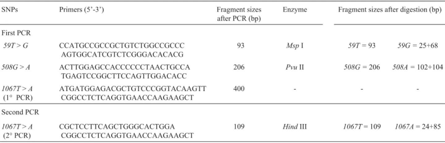
TheFUT3genotype was assigned based on the detec-tion of the59T>G,508G>Aand1067T>ASNPs by the PCR restriction fragment length polymorphism (PCRRFLP) method. The primers used (Invitrogen, São Paulo -Brasil) being listed in Table 1.
The DNA amplification were performed in a final volume of 25µL of LA Taq buffer (Perkin Elmer, USA)
containing 2.5 mM MgCl2, 20 mM of dNTPs, 25 pmol of each primer, 1µL ofTaqpolymerase (Perkin Elmer, USA),
and 40 ng of sample genomic DNA as template. The PCR cycles for the first, second and third set of primers in Table 1 consisted of initial denaturing at 94 °C for 5 min followed by 35 cycles of denaturing at 94 °C for 30 s, annealing at 65 °C for 30 s and extension at 72 °C for 1 min.
To detect theFUT3 1067T>ASNP, oneµL of the
product from the first PCR was used as the template for a nested PCR (denominated the ‘second PCR’ in Table 1) which was carried out under the same conditions the first PCR, except that the fourth primer set in Table 1 was used and the annealing temperature was 60 °C.
TheFUT3PCR products were digested by three dif-ferent restriction endonucleases:MspI for the59T>GSNP products; PvuII for the 508G > A SNP products; and HindIII for the1067T>ASNP products. TheFUT3 diges-tion products were separated on 7% (w/v) polyacrylamide gel (GibcoBRL, New York - USA) for the59T/59G and 508G/508ASNP products and 10% (w/v) polyacrylamide gel for theFUT3 1067T/1067ASNP products. The electro-phoresis was carried out using 10x TEB buffer (90 mM Tris; 2 mM EDTA, 90 mM boric acid pH 8.6), at 150 V, 25 mA, for 5 h and then silver stained (17% solution of sil-ver nitrate). The primers, restriction enzymes and fragment sizes produced after PCR and digestion are listed in Table 1.
Statistical analysis
Data was analyzed by calculating the Hardy-Weinberg Equilibrium and applying the chi-squared (χ2)
heterogeneity test using the CLUMP computer program (Sham and Curtis, 1995) and BioEstat 3.0 (Ayres et al., 2003). The frequencies of the haplotypes were estimated using the ARLEQUIN version 2.000 software.
Results
and1067T >ASNPs. In addition, we detected five differ-ent FUT3 haplotypes, estimated using the ARLEQUIN program (Table 2).
Our study of Lewis phenotype showed that 44 of the 185 individuals evaluated using the hemagglutination test and dot-ELISA assay were negative for Lewis antigen ex-pression in erythrocytes (Table 3).
The complexity of the polymorphism profiles ob-tained in our study is supported by the observation that 20 out of 44 of the individuals with the Lewis negative pheno-type had theFUT3 59Gallele associated with the FUT3 508AorFUT3 1067Aalleles inle59, 508/le59, 508,le59, 508/le59, 1067and
le59, 1067/le59, 1067genotypes. The others 24/44 indi-viduals with the Lewis negative phenotype presented the Le/Le,Le/le59, 508andLe/le59, 1067genotypes (Table 3).
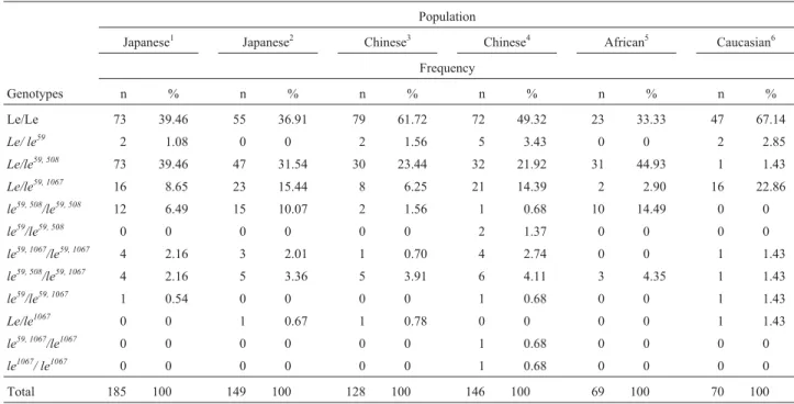
We also compared the FUT3 Lewis genotype fre-quencies found in the current study with those reported by other authors and found that the frequencies obtained by us in the present study (Table 4) were similar to the frequency distributions (χ24= 2.603; 0.90 > p > 0.80) reported by Liu et al.(1996) in a Japanese population, although the authors
did not cite the location of the population. But were signifi-cantly different to the frequencies reported by Liuet al. (1999) for two Chinese populations, one from Shenyang (χ24= 15.603; p < 0.05) and the other from Guangzhou (χ24= 15.993 p < 0.05).
In our study the observed Le/Le genotype frequency was 39.46% (Table 4), significantly lower (p < 0.001) com-pared with the 61.72% (Table 4) reported by Liu et al.
Table 1-FUT3gene single nucleotide polymorphism (SNP), PCR primer sequences and fragment sizes in base pairs (bp) before and after digestion by
each restriction enzyme. The1067T>ASNP was treated with restriction enzyme only after the second PCR.
SNPs Primers (5’-3’) Fragment sizes
after PCR (bp)
Enzyme Fragment sizes after digestion (bp)
First PCR
59T>G CCATGCCGCCGCTGTCTGGCCGCCC
AGTGGCATCGTCTCGGGACACACG
93 MspI 59T =93 59G =25+68
508G>A ACTTGGAGCCACCCCCCTAACTGCCA
TGAGTCCGGCTTCCAGTTGGACACC
206 PvuII 508G =206 508A =102+104
1067T>A
(1° PCR)
ATGATGGAGACGCTGTCCCGGTACAAGTT CGGCCTCTCAGGTGAACCAAGAAGCT
400 - -
-Second PCR
1067T>A
(2° PCR)
CGCTCCTTCAGCTGGGCACTGGA CGGCCTCTCAGGTGAACCAAGAAGCT
109 HindIII 1067T= 109 1067A= 24+85
Table 2-FUT3gene haplotypic frequencies and their standard deviations
(SD) identified using the Arlequin program.
Haplotypes Frequency ± SD
Le 0.6405 ± 0.0250
le59 0.0189 ± 0.0071
le59, 508 0.2622 ± 0.0023
le59, 1067 0.0676 ± 0.0131
le59, 508, 1067 0.0108 ± 0.0054
Total 1.0000
Table 3- Association between theFUT3gene SNP genotype frequencies and the Lewis (Le) erythrocyte phenotypes for a sample population (n = 185) of
Brazilians of Japanese ancestry. Thele59genotype represents the59Gallele whilele59, 508represents the association of the59Gand508Aalleles on the
same chromosome andle59, 1067the association of the59Gand1067Aalleles on the same chromosome.
Lewis genotype Frequency (n) of erythrocyte phenotypes Total
Le (a+b-) Le (a-b+) Le (a-b-) Frequency (n) %
Le/Le 10 57 6 73 39.46
Le/ le59 1 1 0 2 1.08
Le/le59, 508 12 44 17 73 39.46
Le/le59, 1067 4 11 1 16 8.65
le59, 508/le59, 508 0 0 12 12 6.49
le59, 1067/le59, 1067 0 0 4 4 2.16
le59, 508/le59, 1067 0 0 4 4 2.16
le59/le59, 1067 0 1 0 1 0.54
(1999) for the same genotype in a population from Shenyang, China.
Discussion
In our study, the genotype frequencies of theFUT3 59T>G,508G>Aand1067T>ASNPs was similar to data published for other Asiatic populations, especially Japa-nese populations (Table 4). This was clearly observable for theFUT3 59GandFUT3 508Aalleles, predominant among oriental populations (Liuet al., 1996), which were present at a high frequency in our population sample (Table 3), re-flecting the ethnic origin of the individuals sampled. The frequencies seen in our study were also different from the frequencies obtained for other populations, such as the Eu-ropean population studied by Panget al. (1998) and the Chinese population investigated by Liuet al.(1999).
TheFUT3 59Gallele isolated (le59), found in three in-dividuals in our study, was not described by Liu et al., (1996) in a study involving Japanese populations but was observed at a low frequency in European (Panget al., 1998) and Chinese (Liuet al., 1999) populations. Conversely, the FUT3 508Aallele isolated (le508) was not detected by us but was described by Liuet al.(1996) in an individual of Japa-nese origin. Because they are rare, both theFUT3 59Gand FUT3508A alleles, may be present in some populations but absent in others of the same ethnic group, due to the low frequency of these alleles, or alternatively, due to the
phe-nomenon of population isolation and other processes that restrict gene flow.
Based on previous studies (Nishihara et al., 1993, 1994; Kudoet al., 1996, Liuet al., 1999), we inferred that individuals who presented theFUT3 59Gallele associated with theFUT3 508AorFUT3 1067Aallele would present a cis-type chromosomal disposition forming the le59, 508or le59, 1067haplotypes. This is because isolatedFUT3 508Aor FUT3 1067Aalleles, although described, are uncommon, and their association with theFUT3 59Gallele in a trans ar-rangement would be extremely rare. When estimating al-lele and genotype frequencies, this possibility can be discarded, but to eliminate totally this risk direct DNA se-quencing is needed, which we did not carry out in our study. The fact that in our study all the individuals carrying genotypesle59, 508/le59, 508, le59, 508/le59, 1067andle59, 1067/le59, 1067presented a negative Lewis phenotype, confirms that
these mutations, especially those situated in the catalytic domain, eliminate enzymatic activity through changing the protein conformation of the enzyme, leading to the inactiv-ity of theFUT3gene product.Thele59/le59, 1067genotype identified by us in a Lewis-positive individual suggests that theFUT3 59Gallele alone is not capable of eliminating the enzymatic activity of the FUT3, although some studies (Elmgrenet al., 1993,1996; Kudoet al., 1996; Liuet al., 1996, 1999) suggest that the presence of this allele may re-sult in an increased susceptibility to the occurrence of the false Lewis-negative phenotype.
Table 4- Comparison of the genotype frequencies of the threeFUT3gene SNPs analyzed in the current study (n = 185) and in other populations described in the literature. Thele59genotype represents theFUT3 59Gallele whilele59, 508represents the association of theFUT3 59GandFUT3 508A
alleles on the same chromosome andle59, 1067the association of theFUT3 59GandFUT3 1067Aalleles on the same chromosome.
Population
Japanese1 Japanese2 Chinese3 Chinese4 African5 Caucasian6
Frequency
Genotypes n % n % n % n % n % n %
Le/Le 73 39.46 55 36.91 79 61.72 72 49.32 23 33.33 47 67.14
Le/ le59 2 1.08 0 0 2 1.56 5 3.43 0 0 2 2.85
Le/le59, 508 73 39.46 47 31.54 30 23.44 32 21.92 31 44.93 1 1.43
Le/le59, 1067 16 8.65 23 15.44 8 6.25 21 14.39 2 2.90 16 22.86
le59, 508/le59, 508 12 6.49 15 10.07 2 1.56 1 0.68 10 14.49 0 0
le59/le59, 508 0 0 0 0 0 0 2 1.37 0 0 0 0
le59, 1067/le59, 1067 4 2.16 3 2.01 1 0.70 4 2.74 0 0 1 1.43
le59, 508/le59, 1067 4 2.16 5 3.36 5 3.91 6 4.11 3 4.35 1 1.43
le59/le59, 1067 1 0.54 0 0 0 0 1 0.68 0 0 1 1.43
Le/le1067 0 0 1 0.67 1 0.78 0 0 0 0 1 1.43
le59, 1067/le1067 0 0 0 0 0 0 1 0.68 0 0 0 0
le1067/ le1067 0 0 0 0 0 0 1 0.68 0 0 0 0
Total 185 100 149 100 128 100 146 100 69 100 70 100
Comparison between the threeFUT3polymorphisms of individuals with Lewis-negative phenotype erythrocytes revealed that only 20 out of 44 presented thele59, 508and/or le59, 1067 haplotypes related to FUT3 inactivation in both FUT3alleles (Table 4). This implies that there were other FUT3 polymorphisms which were not detected in our study. However, on the other hand, such a discrepancy may be the result of the difficulty in characterizing the Lewis phenotype in erythrocytes, which may lead to false nega-tives because of specific biological conditions, as has been mentioned in this paper.
Our findings indicate that the frequency of the Lewis-negative trait, obtained by molecular analysis of the threeFUT3SNPs studied, was about 10.8% in the popula-tion sample studied, which is close to the serological phe-notype frequencies previously described for Asiatic popu-lations (Lin-Chuet al., 1988; Liuet al., 1996, 1999).
Lewis antigens are particularly important in kidney transplants because rejection is lower in transplants involv-ing Lewis-compatible donors and receptors (Oriolet al., 1978; Williamset al., 1978; Oriol and Danilovics, 1980; Myseret al., 1985; Blajchmanet al., 1985; Delmotteet al., 2002), and the molecular test described in this paper may be helpful in avoiding the false negative results sometimes ob-served whit the serological methods available for typing Lewis antigens.
Acknowledgments
The authors wish to thank the people who partici-pated in this study and the Brazilian agency CAPES for fi-nancial support.
References
Ayres M, Ayres MJ and Ayres DS (2003) Aplicação Estatística nas Áreas das Ciências Biológicas e Médicas. BioEstat 3.0. 3rd edition. Sociedade Civil Mamirauá, Belém, 259 pp. Blajchman MA, King DJ, Heddle NM, Singal DP, Walker IR and
Brain MC (1985) Association of renal failure with Lewis in-compatibility after allogeneic bone morrow transplantation. Am J Med 79:143-146.
Cakir B, Pankow JS, Salomaa V, Couper D, Morris TL, Brantley KR, Hiller KM, Heiss G and Weston BW (2002) Distribu-tion of Lewis (FUT3) genotype and allele: Frequencies in a biethnic United States population. Ann Hematol 81:558-565.
Cooling L and Gu Y (2003) Identification of two new single-nucleotide polymorphisms in FUT3 associated with the Lewis-null phenotype. Transfusion 43:1760-1761. Delmotte P, Degroote S, Lafitte JJ, Lamblin G, Perini JM and
Roussel P (2002) Tumor necrosis factorαincreases the ex-pression of glycosyltransferases and sulfotransferases re-sponsible for the biosynthesis of sialylated and/or sulfated Lewis x epitopes in the human bronchial mucosa. J Biol Chem 277:424-431.
De Vries T, Knegtel RMA, Holmes EH and Macher BA (2001) Fucosyltransferases: Structure/function studies. Glycobio-logy 11:119-128.
Elmgren A and Börjerson C (1996) DNA sequencing and screen-ing for point mutations in the human Lewis(FUT3) gene en-ables molecular genotyping of tire human Lewis blood group system. Vox Sang 70:97-103.
Elmgren A, Rydberg L and Larson G (1993) Genotype heteroge-neity among Lewis negative individuals. Biochem Biophys Res Comm 196:515-520.
Jost F, De Vries T, Knegtel RMA and Macher BA (2005) Muta-tion of amino acids in the alpha 1,3-fucosyltransferase motif affects enzyme activity and Kmfor donor and acceptor sub-strates. Glycobiology 15:165-175.
Koda Y, Kimura H and Mekada E (1993) Analysis fucosyltrans-ferases genes from the human gastric mucosa of Lewis-posi-tive and Lewis-negaLewis-posi-tive individuals. Blood 82:2915-2919. Kudo T, Iwasaki H, Nishihara S, Ando T and Narimatsu I (1996)
Molecular genetic analysis of human Lewis histo-blood group system. Am Soc Biochem Mol Biol 271:9830-9837. Liu Y, Koda Y, Soejima M, Uchida N, Wang B and Kimura H
(1996) PCR analysis of Lewis-negative gene mutations and the distribution of Lewis alleles in a Japanese population. J Forensic Sci 41:1018-1021.
Lin-Chu M, Broadberry RE and Chang FJ (1988) The distribution of blood group antigens and alloantibodies among Chinese in Taiwan. Transfusion 28:350-352.
Liu Y, Koda Y, Soejima M, Pang H, Wang B and Kimura H (1999) Lewis (FUT3) genotypes in two different Chinese populations. J Forensic Sci 44:82-86.
Mollicone R, Giband A, François A, Ratcliffe M and Oriol R (1990) Acceptor specificity and tissue distribution of three human α-3-fucosyltransferases. Eur J Biochem 191:169-176.
Mollicone R, Candelier JJ, Mennesson B, Couillin P, Venot AP and Oriol R (1992) Five specificity patterns of (1-3)-α -L-fucosyltransferase activity defined by use of synthetic oli-gosaccharide acceptors. Differential expression of the en-zymes during human embryonic development and in adult tissues. Carbohydrate Res 228:265-276.
Mollicone R, Reguigne I, Fletcher A, Aziz A, Rustam M, Weston BW, Kelly RJ, Lowe JB and Oriol R (1994) Molecular basis of plasma α(1,3) fucosyltransferase gene deficiency (FUT6). J Biol Chem 269:12662-12671.
Myser T, Steedman M, Hunt K, Strohm P, Williams M and Ken-nedy MS (1985) Linymphocytotoxic anti-Lea as seen in a bone marrow transplant patient. Transfusion 25:445. Nishihara S, Azawa S, Iwasaki H, Nakazato M, Kudo T, Ando T
and Narimatsu H (1993) α (1,3/1,4) fucosyltransferase (FucT-III) gene is inactivated by a single amino acid substi-tution in Lewis histo-blood type negative individuals. Biochem Biophy Res Com 196:624-631.
Nishihara S, Narimatsu H, Iwasaki H and Narimatsu I (1994) Mo-lecular genetic analysis of the human Lewis histo-blood group system. J Biol Chem 269:29271-29278.
Oriol R and Danilovics J (1980) Lymphocytoxic definition of combined ABH and Lewis antigens and their transfer from sera to lymphocytes. Hum Immunol 3:195-505.
Oriol R, Le Pendu J and Mollicone R (1986) Genetics of ABO, H, Lewis X and related antigens. Vox Sang 51:161.
Ørntoft TF, Vestergard EM, Holmes E, Jakobsen JS, Grunnet N, Mortensen M, Johnson P, Bross P, Gregersen N, Skors-tengaard K, Jensen UB, Bolund L and Wolf H (1996) Influ-ence of Lewisα1,3/1,4-L-fucosyltransferases (FUT3) gene mutations on enzyme activity, erythrocyte phenotyping, and circulation tumor marker sialyl-Lewis levels. J Biol Chem 271:32260-32268.
Pflug W, Bässler G and Eberspächer B (1989) ABO and Lewis typing of secretion stains on nitrocellulose membranes using a new dot-blot-ELISA technique. Forensic Science Interna-tional 43:171-182.
Pang H, Liu Y, Koda Y, Soejima M, Jia J, Schlaphoff T, Du Toit E and Kimura H (1998) Five novel missense mutations of Lewis gene (FUT3) in African (Xhosa) and Caucasian popu-lations in South Africa. Hum Genet 102:675-680.
Sham PC and Curtis D (1995) Monte Carlo tests for associations between disease and alleles at highly polymorphic loci. Ann Hum Genetic 59:97-105.
Soejima M, Kimura H and Koda Y (2004) Two novelFUT3 al-leles responsible for Lewis-null phenotypes in Sri Lanka. Transfusion 44:1534-1535.
Watkins WM (1980) Biochemistry and genetics of the ABO, Lewis and P blood group systems. Adv Hum Genet 10:1-136.
Williams G, Pegrum GD and Evans CA (1978) Lewis antigens in renal transplantation. Lancet 1:878.
Internet Resources
Arlequin - A software for population genetics data analysis, http://anthro.unige.ch/software/arlequin/.
SEPOF (Secretaria Executiva de Estado de Planejamento, Orça-mento e Finanças), http://www.sepof.pa.gov.br/estatistica/ ESTATISTICASMUNICIPAIS/Mesorr_Nordeste/ TomeAcu/TomeAcu.pdf (April 10, 2006).