A Single Histidine Is Required for Activity of Cytochrome c
Peroxidase from Paracoccus denitrificans*
(Received for publication, December 1, 1995, and in revised form, February 15, 1996)
Dermot F. McGinnity‡, Bart Devreese§, Susana Prazeres¶, Jozef Van Beeumen§, Isabel Moura¶, Jose J. G. Moura¶, and Graham W. Pettigrew‡i
From the ‡Department of Preclinical Veterinary Sciences, Royal (Dick) School of Veterinary Studies, University of Edinburgh, Summerhall, Edinburgh EH9 1QH, United Kingdom, the §Department of Biochemistry, Physiology and
Microbiology, Ledeganckstraat 35, 9000 Gent, Belgium, and the¶Departamento de Quimica, Faculdade de Ciencias e
Tecnologia, Universidade Nova de Lisboa, 2825 Monte de Caparica, Portugal
The diheme cytochrome c peroxidase from
Paracoc-cus denitrificans was modified with the
histidine-spe-cific reagent diethyl pyrocarbonate. At low excess of reagent, 1 mol of histidine was modified in the oxidized enzyme, and modification was associated with loss of the ability to form the active state. With time, the mod-ification reversed, and the ability to form the active state was recovered. The agreement between the spec-trophotometric measurement of histidine modification and radioactive incorporation using a radiolabeled rea-gent indicated little modification of other amino acids. However, the reversal of histidine modification ob-served spectrophotometrically was not matched by loss of radioactivity, and we propose a slow transfer of the ethoxyformyl group to an unidentified amino acid. The presence of CN2bound to the active peroxidatic site of the enzyme led to complete protection of the essential histidine from modification.
Limited subtilisin treatment of the native enzyme fol-lowed by tryptic digest of the C-terminal fragment (res-idues 251–338) showed that radioactivity was located in a peptide containing a single histidine at position 275. We propose that this conserved residue, in a highly con-served region, is central to the function of the active mixed-valence state.
Bacterial cytochrome c peroxidases (CCPs)1are diheme
en-zymes that oxidize monoheme Class I cytochromes c and reduce hydrogen peroxide to water. The most extensively character-ized example is from Pseudomonas aeruginosa and contains a high potential, electron-transferring heme and a low potential, peroxidatic heme (1, 2). In the oxidized state, the enzyme is inactive (1, 3); the high potential heme is partially high spin, and the low potential heme is low spin and inaccessible to ligands (2). Reduction of the high potential heme causes the low
potential heme to become high spin and enables binding of hydrogen peroxide (4, 5). We have studied the enzyme from Paracoccus denitrificans and have found very similar charac-teristics (6, 7), although a distinctive difference is the Ca21 requirement in P. denitrificans CCP for the low to high spin transition at the peroxidatic heme (8).
The amino acid sequence of P. aeruginosa CCP suggested that the molecule was constructed in two domains, each having the general appearance of a monoheme Class I cytochrome c (9). This family of cytochromes is characterized by a heme attachment site and proximal histidine ligand near the N ter-minus and a distal methionine ligand near the C terter-minus. Ellfolk et al. (9) proposed that the C-terminal domain followed this pattern and acted as the high potential electron-transfer-ring site. The N-terminal domain appeared to lack an appro-priately placed methionine, and Ellfolk et al. proposed that the sixth iron ligand of this domain was a histidine donated from the C-terminal domain and conferring a low redox potential. In the mixed-valence form of the enzyme, this histidine would be released from the iron, the heme would become high spin, and hydrogen peroxide could enter the active site.
The amino acid sequence of P. aeruginosa CCP has been revised on the basis of the gene sequence determined by Ridout et al. (10), and the key histidine in the proposal of Ellfolk et al. (9) is His-261. We have determined the amino acid sequence of P. denitrificans CCP,2 which shows 61% identity to the P.
aeruginosa enzyme. The histidine in P. denitrificans CCP cor-responding to His-261 in P. aeruginosa CCP is at position 275. During the preparation of this paper, the crystallographic structure of the oxidized form of the P. aeruginosa enzyme was determined (11). In contrast to the original proposal of Ellfolk et al. (9), the ligand to the proposed N-terminal peroxidatic heme is, in fact, His-71 (position 85 in P. denitrificans CCP numbering), and His-261 is situated at the interface of the two heme domains.
This paper describes the modification of P. denitrificans CCP with the histidine-selective reagent diethyl pyrocarbonate. We show that modification of His-275 in the oxidized form of the enzyme completely abolishes the ability of the enzyme to form the active mixed-valence state.
EXPERIMENTAL PROCEDURES
Enzyme Purification—P. denitrificans (Laboratory of Microbiology,
Delft.) was grown and cytochrome c peroxidase was purified as de-scribed by Goodhew et al. (12). After purification, the protein displayed an A408 nm/A280 nmratio of.5 and gave a single band on SDS-polyacryl-amide gel electrophoresis. Concentrated stocks of the protein in 5 mM Mes, pH 6, 10 mMNaCl were stored at240 °C. The concentration of P.
denitrificans CCP was determined at 408 nm usinge 5 250 mM21cm21
* This work was supported by Junta Nacional de Investigacao Cien-tifica e Tecnologica Grants PMCT/C/BIO/885 and STRDA/BIO/359/92 (to I. M.) and STRDA/CEN/538/92 Programa CIENCIA (to J. J. G. M.), Grant 32001891 from the Belgian Fund for Joint Basic Research, Con-certed Research Action Grant 12052293 from the Flemish Government, a Ph.D. grant (to S. P.), a Wellcome Trust project grant (to G. W. P.), and a faculty research studentship (to D. F. M.). The British Council/ Junta Nacional de Investigacao Cientifica e Tecnologica provided sup-port for travel. The costs of publication of this article were defrayed in part by the payment of page charges. This article must therefore be hereby marked “advertisement” in accordance with 18 U.S.C. Section 1734 solely to indicate this fact.
iTo whom correspondence should be addressed. Tel.: 44-31-650-6135; Fax: 44-31-650-6576; E-mail: [email protected].
1The abbreviations used are: CCPs, cytochrome c peroxidases; Mes, 2-(N-morpholino)ethanesulfonic acid; DEPC, diethyl pyrocarbonate;
HPLC, high performance liquid chromatography. 2J. Van Beeumen and G. W. Pettigrew, unpublished results. © 1996 by The American Society for Biochemistry and Molecular Biology, Inc. Printed in U.S.A.
11126
by guest on September 9, 2019
http://www.jbc.org/
(12). Yeast cytochrome c peroxidase (EC 1.11.1.5) was purified by the method of Pettigrew and Seilman (13). The concentration of yeast cytochrome c peroxidase was determined at 408 nm usinge 5 95 mM21 cm21 (14). Horse heart cytochrome c (type VI) was purchased from Sigma and used without further purification.
Formation of the Different Redox States of Cytochrome c Peroxidase—
The enzyme was purified in the oxidized state. The mixed-valence low spin state was formed in the presence of 1 mMascorbate, 10mMdiamino durol (6). The same reductive treatment in the presence of 1 mMCaCl2 gave rise to the mixed-valence high spin state (6). The ability of the sample to form the latter state was measured by difference spectra of the mixed-valence enzyme after Ca21treatment minus those of the mixed-valence enzyme.
Diethyl Pyrocarbonate—Both DEPC and [carbonyl-14C]DEPC were
purchased from Sigma and stored desiccated at 4 °C. Stock solutions were made by dilution of DEPC into acetonitrile and assayed by reac-tion with 5 mM imidazole, pH 7.5 (e233 nm 5 3.0 mM21 cm21) (15). Although stock solutions of DEPC are quite stable in acetonitrile for several months, they did develop weak absorption in the region at 250 –300 nm and were then discarded. DEPC in 5 mMHepes, pH 7.5, was found to hydrolyze exponentially with a half-life of 20 min.
Modification with DEPC—Cytochrome c peroxidase was incubated
with an excess of DEPC, and the stoichiometry of ethoxyformylation of histidine residues was determined by following an increase in absorb-ance at 245 nm (e 5 3.2 mM21 cm21) (16). The reaction is complete within 20 min. Alternatively, stoichiometry of modification was deter-mined using [14C]DEPC (labeled at both carbonyl groups with an over-all specific activity of 2.6 mCi/mmol). Excess reagent was removed by gel filtration on a Sephadex G-25 column in 5 mM Hepes, pH 7.5. Scintillation counting (.90% efficiency) was performed in 5 ml of Pack-ard Insta-Gel with a maximum of 250 ml of aqueous sample in a Packard Tri-Carb 1900CA scintillation counter. For the calculation of moles of radioactive uptake into protein, the fact that only one of the two carbonyl groups is incorporated must be taken into account (the other is lost as CO2). One nmol of [14
C]DEPC corresponds to 5838 dpm, and therefore, 1 nmol of incorporated ethoxyformyl group corresponds to 2919 dpm.
Assay of Cytochrome c Peroxidase—The activity of mixed-valence
cytochrome c peroxidase, in the presence of added Ca21, was measured by following the decrease in absorbance of thea-band of ferrocyto-chrome c as it was oxidized (7). Assays were initiated by addition of the enzyme (1 nM) to a cuvette containing 5 mMHepes, pH 7.5, 10 mMNaCl, 7mMhorse heart ferrocytochrome c, 1 mMCaCl2, and 18mMhydrogen peroxide. The percent activity of each sample in Fig. 3b was calculated on the basis of their respective turnover numbers, with the unmodified preactivated control turnover number representing 100%. Turnover numbers were calculated using the initial gradient of cytochrome c oxidation (7).
Spectroscopy—All spectra in the UV region were recorded on a Cary
219 spectophotometer. Difference spectroscopy in the visible region was performed with a single beam Philips PU8700 spectophotometer. Spec-tra were collected at 23 °C using 1-cm path length quartz cuvettes. EPR measurements were performed using a Bruker ESP 380 spectrometer with an Oxford Instruments ESR-9 continuous flow cryostat. The rela-tive intensities of the EPR signals were determined using software that simulates the experimentally determined spectra using the values for
gmax, gmed, and gminand integrates the resultant signals (17).
Peptide Separation and Mass Spectrometry—The Glu-1–Thr-250 and
Arg-251–Met-338 peptides were purified as described in the legend to Fig. 7. They were desalted by passage through a Superdex 75 column (Pharmacia Biotech Inc.), equilibrated in 0.1Mammonia, and lyophi-lized. The tryptic peptides of Arg-251–Met-338 were separated by HPLC on a reverse-phase Techsphere 50 DS column (25 cm3 4.6 mm) using a gradient of 0 –70% acetonitrile in 0.1% trifluoroacetic acid. The column eluate was monitored at 214 and 280 nm.
Mass spectrometry was performed using a BioQ triple quadrupole mass spectrometer equipped with a pneumatically assisted electrospray source (Fisons, Altrincham, United Kingdom) operating in positive ion mode. The resolution of the mass spectrometer allows for isotopic res-olution of peptides under Mr;3000. By averaging the mass of elemen-tal isotopes according to their natural distribution and thus determin-ing the average mass of individual residues, the theoretical Mrvalues of peptides over 3000 were calculated.
Secondary Structure Predictions—We used the secondary structure
prediction methods of Gibrat et al. (18), Levin et al. (19), Deleage and
Roux (20), and Geourjon and Deleage (21) contained in the package “SOPMA.”3
RESULTS
Extent of Modification of P. denitrificans CCP by Diethyl Pyrocarbonate—Diethyl pyrocarbonate reacts with both free imidazole in solution and histidyl residues in proteins, yielding the ethoxyformyl derivatives. The reaction may be followed spectrophotometrically by a gain of absorbance between 230 and 250 nm. Fig. 1 compares difference spectra (230 –300 nm) of the modification of P. denitrificans CCP (at 2 and 20 min), imidazole, and yeast CCP.
The difference spectrum of the modified P. denitrificans CCP displays an initial gain of absorbance at 245 nm (Fig. 1A, panel i), followed by slow formation of a trough between 265 and 300 nm (panel ii). Modification of free imidazole generated the ethoxyformylimidazole derivative, which gave a peak maxi-mum at 233 nm, but no corresponding changes at higher wave-lengths (Fig. 1B). As will be discussed below, the loss of ability to form the active state of P. denitrificans CCP is correlated
3
Available on the World Wide Web at http://www.ibcp.fr.
FIG. 1. UV difference spectra of the modification of imidazole,
P. denitrificans CCP, and yeast CCP with diethyl pyrocarbon-ate. A: panel i, P. denitrificans CCP (3.5mM) in 5 mMHepes, pH 7.5, was modified with DEPC to a final concentration of 20mM. After 2 min, a modified CCP minus unmodified CCP difference spectrum was taken and showed 0.57 mol of histidine modified per mol of protein, based on e245 nm5 3.2 mM21cm21for ethoxyformylhistidine. Panel ii, the reac-tion of DEPC with P. denitrificans CCP was complete within 20 min and showed 1.0 mol of histidine modified per mol of protein. B: 5 mM imidazole in 5 mMHepes, pH 7.5, was incubated with 4mMDEPC. The difference spectrum obtained at 2 min showed 3.8mM ethoxyformylim-idazole (e233 nm5 3.0 mM21cm21). The spectrum did not change in the subsequent 20 min. C: yeast CCP (3.8mM) in 5 mMHepes, pH 7.5, was modified with DEPC to a final concentration of 20mM. After 15 min, a difference spectrum was taken and showed 1.1 mol of histidine modified per mol of protein. The wavelengths of individual peak maxima are indicated on the respective spectra.
by guest on September 9, 2019
http://www.jbc.org/
with absorbance gain at 245 nm and not with the presence of the 265–300 nm features.
The modified yeast CCP difference spectrum (Fig. 1C) is very similar to the modified P. denitrificans CCP spectrum. For both proteins, a DEPC/protein ratio of 20mM:4mMresulted in
mod-ification of;1 mol of histidine/mol of protein. This was calcu-lated using the extinction coefficient of ethoxyformylhistidine at 245 nm of 3.2 mM21cm21(16). A possible source of error for
this calculation is the effect of spectral features near the peak at 245 nm. For example, the trough at 265–300 nm in the modified difference spectra of bacterial and yeast CCPs may have a “pulling down” effect on the 245 nm peak. An independ-ent check on the extindepend-ent of modification was made by measuring the incorporation of [14C]DEPC. These experiments have
de-termined that, in general, there is good agreement between the two methods of quantitation, with radioactive labeling giving slightly less (;10%) than spectroscopic measurements. This indicates first thatDA245 nmis a reliable measurement of
his-tidine modification and second that there is little uptake into amino acids other than histidine.
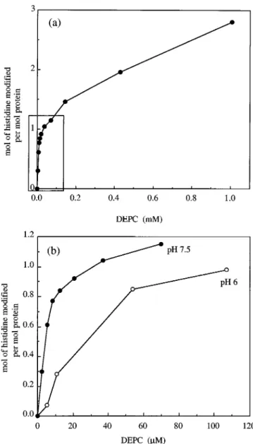
Fig. 2a shows the extent of modification of P. denitrificans CCP as DEPC concentration was raised. At each DEPC con-centration, the modification was allowed to proceed until the absorbance maximum at 245 nm was obtained (;20 min). The biphasic character of the plot of DEPC demonstrates the exist-ence of an easily modifiable histidine. Eventually, at high DEPC concentrations, an apparent maximum of four histidines are modified (data not shown). We expected a maximum of three histidines to be available for modification since the re-maining three are assumed to be coordinated to heme (two as proximal ligands and one as a distal ligand for the peroxidatic heme). The discrepancy that we see in our measured maximum value of four may be due to bis-modification of histidine (22). A similar biphasic pattern was obtained by following the extent of modification at a single high DEPC concentration (data not shown). A rigorous interpretation of the results was compli-cated by the fact that the final extent of modification is the result of the rate of modification, the rate of ethoxyformylhis-tidine reversal, and the rate of hydrolysis of the reagent. Fig. 2b compares the extent of modification at pH 6 and 7.5, and it indicates that much higher DEPC concentrations are required at pH 6 to achieve modification of 1 mol of histidine, a result consistent with the requirement that histidine must be unpro-tonated to be modified (23, 24). We chose pH 7.5 for subsequent studies.
The Most Easily Modified Histidine Is Essential for Activi-ty—It is worth re-emphasizing that the active form of the enzyme is achieved by reduction of the electron-transferring heme and the Ca21-dependent appearance of the high spin state at the peroxidatic heme. Thus, to assess the effect of modification on the oxidized enzyme, the protein must be re-duced with ascorbate, and the appearance of either activity or the high spin band at 380 nm must be monitored on addition of Ca21.
Fig. 3a demonstrates the loss of ability to form the high spin 380 nm band in the mixed-valence enzyme after modification of histidine in the oxidized state. These data are plotted in Fig. 3b along with loss of catalytic activity upon modification of histi-dine. Thus, modification of a single histidine in P. denitrificans CCP abolishes both the ability to form the high spin state and activity. Although a single histidine was also readily modified in yeast CCP at similarly low DEPC/protein ratios, modifica-tion did not cause loss of activity.
A further spectroscopic indicator of the active enzyme is the Ca21-dependent appearance of a gmax5 2.89 signal in the EPR
spectrum of the mixed-valence enzyme, which can be equated
with the high spin peroxidatic heme at room temperature (Fig. 4a). Because of the high protein concentrations needed for EPR spectroscopy, endogenous Ca21 allows partial appearance of
this gmax 5 2.89 signal in the spectrum of the mixed-valence
without added Ca21. The enzyme modified at a single histidine cannot undergo the transition to the active form (Fig. 4b) and is mostly trapped in a mixed-valence low spin form with gmax5 3.00.
Our difference spectrum due to modification at pH 7.5 showed the presence of a shallow trough between 265 and 300 nm (Fig. 1A, panel ii). We wanted to be certain that this was not an indicator of modification of aromatic acids such as tyro-sine or tryptophan, which may affect the activity of the protein. A difference spectrum (Fig. 1A, panel i) obtained after modifi-cation for 2 min showed no trough between 265 and 300 nm, and yet the histidine modified correlated well with the propor-tion of protein unable to form the high spin state (data not
FIG. 2. Extent of modification of P. denitrificans CCP with
increasing concentrations of DEPC. a, P. denitrificans CCP (4mM) in 5 mMHepes, pH 7.5, was treated with increasing concentrations of DEPC, and the reactions were allowed to proceed to completion at each concentration. The moles histidine modified were calculated using e245 nm5 3.2 mM21cm21. At a concentration of 8 mMDEPC, an apparent maximum of four histidines were modified (data not shown on figure). The boxed area indicates a subset of points used in b. b, shown is a comparison of histidine modification of CCP at pH 6 and 7.5. Modifica-tion of CCP (4mM) at pH 6 was carried out in 5 mMMes.
by guest on September 9, 2019
http://www.jbc.org/
shown). Also, during reversal of modification, the 245 nm peak is gradually lost, but the 265–300 nm trough remains (Fig. 5a). Again, the loss of histidine modification correlates well with the recovery of the ability to form the active high spin state (Fig. 5b). The reversal of modification does not follow a simple exponential; in 5 mMHepes, pH 7.5, 50% of the label is lost in
6 h. Taken together, these results indicate that the appearance of the 280 nm trough is not associated with changes in the activity of the enzyme and that those changes we observed are due to histidine modification.
Modification of the Different Redox States of P. denitrificans CCP and the Protective Effect of CN2—The susceptibilities to modification of the three redox states of the enzyme are very
different and not as we originally predicted. Fig. 6 shows that the oxidized enzyme at 4mMloses all ability to form the active
spin state at 20 mM DEPC, while this only occurs with the
mixed-valence low spin and mixed-valence high spin states at higher concentrations of reagent. For the two mixed-valence forms of P. denitrificans CCP, histidine modification cannot be measured spectroscopically because ascorbate interferes with UV measurements. [14C]DEPC was used to determine the
ex-tent of modification in the oxidized and mixed-valence states (Fig. 6). The results indicate that the histidine modified in the oxidized enzyme at low levels of DEPC is not modified in the mixed-valence low spin state until higher levels of reagent are used. Thus, the essential histidine is more susceptible to mod-ification in the oxidized state than in the mixed-valence low spin state. Similarly, the essential histidine is more susceptible to modification in the mixed-valence low spin state of the en-zyme than in the mixed-valence high spin state. The continu-ation of the experiment in Fig. 6 shows that modificcontinu-ation of the CN2adduct of the mixed-valence high spin enzyme, with levels of DEPC that completely inactivate the same state in the absence of CN2, yields an enzyme that retains almost all ac-tivity. When CN2is removed by molecular exclusion (a process that also reoxidizes the enzyme), the modified protein, at 4mM, FIG. 3. Modification of a single histidine from P. denitrificans
CCP abolishes catalytic activity and the ability of the enzyme to form the mixed-valence high spin state. a, loss of ability to form the
characteristic high spin 380 nm band on modification of a single histi-dine. CCP (4mM) in 5 mMHepes, pH 7.5, was modified for 20 min with DEPC at the following concentrations: 0, 1.5, 2.8, 5.6, 8.4, 12.2, 16.4, and 24.6mM. The extent of modification for each DEPC concentration was calculated to be 0, 0.12, 0.27, 0.46, 0.58, 0.68, 0.86, and 1.00 mol of histidine modified per mol of protein, respectively. The ability of each sample to form the mixed-valence high spin state was tested by differ-ence spectroscopy as described under “Experimental Procedures.” Each difference spectrum with decreasing DA380 nm correlates to the in-creased extent of modification as listed above. b, loss of ability to form the 380 nm band in the mixed-valence enzyme and loss of catalytic activity as histidine modification increases. The percent high spin of each sample was calculated from the changes at 380 nm in a. Aliquots of each sample were assayed, and activity was determined (see “Exper-imental Procedures”).
FIG. 4. EPR spectroscopy of modified and native P.
denitrifi-cans CCPs. a, EPR spectra of native CCP in oxidized, mixed-valence,
and mixed-valence1 Ca21states. Nine ml of CCP (4mM) in 5 mMHepes, pH 7.5, were concentrated to 180ml, and spectra were recorded from this 200 mM sample. b, EPR spectra of modified CCP in oxidized, mixed-valence, and mixed-valence1 Ca21states. To 9 ml of CCP (4mM) in 5 mMHepes, pH 7.5, was added DEPC to a final concentration of 20 mM, which resulted in 1.1 mol of histidine modified per mol of protein. The solution was concentrated, and spectra were recorded as described for the native sample. Experimental conditions were as follows: tem-perature, 7.5 K; microwave frequency, 9.65 GHz; microwave power, 2 milliwatts; modulation, 1 millitesla; and gain, 1.63 105
. g values for selected resonances are shown.
by guest on September 9, 2019
http://www.jbc.org/
can be further modified with a 5 3 excess of DEPC to give complete loss of the ability to form the high spin state. Addition of CN2to the oxidized and mixed-valence low spin states has no effect on the modifiability of the enzyme. These results show that an essential histidine is much less susceptible to modifi-cation when CN2is bound at the peroxidatic heme.
Identification of the Site of Modification in the Amino Acid Sequence—Subtilisin digestion of the native mixed-valence ho-loenzyme released two fragments that could be separated by molecular exclusion after dissociation in SDS (Fig. 7). SDS-polyacrylamide gel electrophoresis analysis of the elution pro-file of the molecular exclusion column showed complete resolu-tion of the larger from the smaller peptide (Fig. 7). However, the remaining undigested whole protein, which accounted for
7% of the total protein by densitometry of the Coomassie Blue-stained gel, coeluted with the larger peptide. Although the level of undigested protein could be lowered by more intense diges-tion, this tended to increase the amount of fragments of the larger peptide that coeluted with the smaller peptide (data not shown).
Mass spectrometry established that the larger fragment (Mr 27,950.9) corresponded to the N-terminal 250 amino acids con-taining both heme groups (Mrpredicted from the amino acid
sequence of 27,950.93). The smaller fragment (Mr 9581.38)
accounted for the remaining non-heme C-terminal 87 amino acids (Mrpredicted from the amino acid sequence of 9579.87).
Thus, subtilisin cleavage was limited to the peptide bond be-tween Thr-250 and Arg-251.
Subtilisin digestion of the mixed-valence holoenzyme that had been radiolabeled in the oxidized state to the extent of 1 mol of histidine/mol gave the same peptide pattern as shown in Fig. 7. A ratio of 2.2:1 for radioactive disintegrations in the Arg-251–Met-338 peptide relative to the Glu-1–Thr-250 pep-tide was obtained, and if the contribution of undigested protein to the Glu-1–Thr-250 fraction was allowed for, this ratio be-came 2.9:1. This ratio is a strong indication that the single essential histidine resides in the Arg-251–Met-338 fragment. However, the close correlation between modification of a single histidine and loss of activity shown in Fig. 3 had led us to expect an even cleaner pattern of label distribution. We found that a [14C]DEPC-labeled protein sample left for 24 h after
modification had completely lost its ethoxyformylhistidine based on spectroscopic measurements at 245 nm, yet had re-tained almost all radioactivity. Cleavage and separation of the two subtilisin fragments yielded an Arg-251–Met-338/Glu-1– Thr-250 radiolabel ratio of 1:5. Therefore, over the 24 h, label was transferred from a specific site on the Arg-251–Met-338 peptide to the Glu-1–Thr-250 peptide. We propose that a par-tial transfer of this type is the reason for the ratio of 2.9:1 (rather than higher) that we observed for analysis completed within;2 h.
As the Arg-251–Met-338 peptide contains both His-275 and His-322, unequivocal identification of the essential histidine in CCP required isolation of a fragment containing a single
histi-FIG. 5. Loss of ethoxyformylhistidine with time correlates
with regain of active enzyme. a, difference spectra of modified minus
unmodified CCP were taken at different times after DEPC addition. CCP (12mM) in 5 mMHepes, pH 7.5, was modified with DEPC to a final concentration of 45mM. The absorbance at 245 nm was maximal at 20 min (1.0 mol of histidine modified per mol) and fell thereafter due to slow hydrolysis of ethoxyformylhistidine. Times shown are hours after this maximal extent of modification. b, the loss of ethoxyformylhistidine after a given time was obtained from measurements at 245 nm and correlated with the extent of the ability to form the high spin state as described in the legend of Fig. 3.
FIG. 6. Relative susceptibility to DEPC modification of the different forms of P. denitrificans CCP. The different redox and spin states of CCP (4mMin 5 mMHepes, pH 7.5) were formed as described under “Experimental Procedures.” The cyano-mixed-valence enzyme was formed by the addition of 100mMNaCN to the mixed-valence high spin state. DEPC was added to each form of CCP until the enzyme’s ability to form the high spin state was abolished. The DEPC/CCP ratio required to abolish the ability to form the high spin state is noted next to each form of the enzyme. *, at a ratio of 330 DEPC:1 CCP, the cyano-mixed-valence enzyme retained 90% of its ability to form the high spin state. In a separate experiment, samples of CCP (4mM) in 5 mMHepes, pH 7.5, in the oxidized and mixed-valence high spin states were treated with [14C]DEPC to final concentrations of 20mM(5 DEPC:1 CCP) and 1.2 mM(300 DEPC:1 CCP). After 20 min, these two solutions were desalted on Sephadex G-25 columns equilibrated in 5 mMHepes, pH 7.5, and 200-ml aliquots of the desalted material were added to 4 ml of scintillation fluid. Moles of [14 C]ethoxy-formylhistidine/mole of enzyme were calculated as described under “Experimental Procedures.”
by guest on September 9, 2019
http://www.jbc.org/
dine. Therefore, the Arg-251–Met-338 peptide was subdigested with trypsin, generating six smaller fragments (labeled A–F) (Fig. 8) that were isolated by reverse-phase HPLC and identi-fied by mass spectrometry. Mrvalues were within 0.5 of those
predicted from the amino acid sequence. His-275 and His-322 were located in peptides E and F, respectively.
The essential histidine of CCP was radiolabeled with [14C]DEPC; the modified Arg-251–Met-338 peptide was
iso-lated and digested; and the resultant tryptic peptides were separated as described for the native protein. The HPLC profile for the modified tryptic digest displayed an extra peak E2, the appearance of which correlated with the diminution of the original peak E1. Almost all the label was contained in peptide E2 (Fig. 8). Thus, we conclude that peptide E contains the essential histidine, which, when ethoxyformylated, increases the retention time of the peptide. This was confirmed by ob-taining an elution profile that matched that of the unmodified
protein after reversal of the modification in 0.2 M ammonia solution (data not shown). Also, the Mr of peptide E2 was determined to be 2472.62 by mass spectrometry. This corre-sponds well with the theoretical mass of 2400.12 for peptide E (Asn-266 –Lys-287) plus 72 for an ethoxyformyl group. Thus, His-275 is the single histidine modified at low DEPC/CCP ratios that is essential for activity.
DISCUSSION
Modification of a Single Essential Histidine—Spectrophoto-metric and radioactive labeling measurements are consistent with the modification of a single histidine at low DEPC con-centrations. Although the trough between 265 and 300 nm resembles that expected for O-ethoxyformylation of tyrosine (25), the extent of the absorbance change would imply a mod-ification of at least 1 mol of tyrosine/mol of protein (e278 nm5 1.3 mM21cm21), and this is certainly not supported by
meas-urement of radiolabeling. A similar spectroscopic feature is present in modified yeast CCP and other proteins modified with DEPC (25, 26).
Modification of a single most reactive histidine is associated with loss of activity (Fig. 3), and activity is regained as the modification slowly hydrolyzes (Fig. 5). In yeast CCP, 1 mol of
FIG. 7. Identification of the Arg-251–Met-338 peptide as the
site of modification. CCP (20mM) in 10 mM Hepes, pH 7.5, was modified to an extent of 0.9 mol of histidine modified per mol of protein with [14C]DEPC (100m
M). The modified sample was desalted through a small Sephadex G-25 column (123 1 cm), equilibrated in 10 mMHepes, pH 7.5, before being digested with subtilisin. To CCP (4 –20mM) in 10 mMHepes, pH 7.5, were added 1 mMascorbate, 10mMdiamino durol, and 1 mMCaCl2. Subtilisin (Carlsberg) was added to give a substrate/ enzyme ratio of;500:1 (w/w). After 60 min at 0 °C, subtilisin was inhibited by 1 mMphenylmethanesulfonyl fluoride, 10 mMEGTA. The digest was made to 1% in SDS and loaded onto a Pharmacia Superdex 75 column equilibrated in 20 mMTris, pH 7.3, 100 mMNaCl. The eluate was monitored at 280 nm, and the resultant two main peaks were collected. The protein composition of the two peaks was determined by Coomassie Blue staining after SDS electrophoresis on a 15% polyacryl-amide gel. The remainder (two-thirds) of the two peaks was used for radioactive counting. The distribution of label between the two fractions is shown in the histogram. The contribution of labeled undigested protein to the fraction containing the Glu-1–Thr-250 peptide was de-ducted to give a corrected dpm. Lane 1, Mrstandards; lane 2, undigested CCP; lane 3, subtilisin digest; lane 4, peak 1 from the Superdex G-25 column; lane 5, peak 2.
FIG. 8. Identification of His-275 as the radiolabeled amino
acid. The unmodified purified Arg-251–Met-338 peptide was prepared
as described in the legend of Fig. 7. Trypsin was added to give a substrate/enzyme ratio of 50:1 (w/w). After 10 min at 23 °C, trypsin was inhibited with 0.5 mMphenylmethanesulfonyl fluoride. The resultant peptides were separated by HPLC as described under “Experimental Procedures” and gave rise to the 214 nm profile labeled Unmodified. Six peptides were collected (A–F) and identified by mass spectrometry (Mr values in parentheses). CCP modified to an extent of 0.9 mol of histidine modified per mol of protein with [14C]DEPC was cleaved with subtilisin, and the resultant radiolabeled peptide (Arg-251–Met-338) was purified as described in the legend of Fig. 7. The digestion of this peptide with trypsin yielded the HPLC profile labeled Modified. The unmodified peptide is labeled E1, while the modified peptide is labeled E2. The distribution of radiolabel was determined by radioactive counting and is shown in the histogram below each peak.
by guest on September 9, 2019
http://www.jbc.org/
histidine is also readily modifiable at low concentrations of DEPC, but in this case, modification was not associated with loss of activity (data not shown). This is consistent with the results of Bosshard et al. (27), who found that three histidines could be modified with no loss of activity. Similarly, in horse-radish peroxidase, two histidines were modified by DEPC, but it is the more slowly modified residue that is essential (26).
Identification of the Essential Histidine as His-275—If we assume that two histidines that remain unmodified at high DEPC concentrations are the conserved proximal ligands to the two heme groups, His-85 and His-275 are the only remaining conserved histidines, and therefore, one or the other (or both) is likely to be essential for activity. Because of the relatively rapid loss of the ethoxyformyl group, few studies have attempted location of a modified histidine in a peptide digest, although there are some exceptions (28). In our case, there was the additional problem that c-type cytochromes tend to be resistant to proteolysis unless the heme is removed, but this procedure requires conditions (0.1MHCl, 8Murea) that would be likely to accelerate loss of ethoxyformyl groups. However, we were able to exploit a remarkable single bond cleavage by subtilisin in the native state, which releases a C-terminal fragment containing the conserved His-275 and one other nonconserved histidine and an N-terminal fragment containing the conserved His-85 and one other nonconserved histidine. This cleavage between Thr-250 and Arg-251 does not reflect limitations in the action of subtilisin, but rather the limited access of protease to the native substrate.
Purification of the C-terminal fragment by molecular exclu-sion chromatography followed by tryptic digestion and peptide separation by reverse-phase HPLC allowed localization of ra-dioactive labeling in the tryptic peptide containing His-275 within 4 h of modification. Over a longer time scale (e.g. 24 h), the radioactive label was retained on the protein, but attached to the N-terminal region (Glu-1–Thr-250). We are unaware of a precedent for such an intramolecular transfer of an ethoxy-formyl group, but we propose that it is consistent with attack on ethoxyformylhistidine 275 by a lysine that is adjacent in the folded protein, but resides in the N-terminal sequence (Glu-1– Thr-250). Ethoxyformyllysine is not susceptible to nucleophilic attack (15). This proposal will be tested by identification of the labeled residue in the Glu-1–Thr-250 peptide.
Role of His-275—We have shown that His-275 is essential for activity. Consistent with this is its position in a highly con-served region of the cytochrome c peroxidase molecule. Ridout et al. (10) have corrected parts of the P. aeruginosa CCP se-quence, and in this region (P. denitrificans Asp-255–Trp-280 and P. aeruginosa Asp-241–Trp-266), 25 out of 26 residues are identical.4
In the crystallographic structure of the P. aeruginosa enzyme (11), His-261, the counterpart of His-275, is placed at the in-terface between the N-terminal domain (proposed to contain the peroxidatic heme) and the C-terminal domain (containing the electron-transferring heme). Fig. 7 of Ref. 11 shows the histidine as part of a hydrogen-bonded network involving the juxtaposed heme propionates of both heme groups. Our finding that His-275 is the most easily modified histidine in the oxi-dized state would not have been consistent with the original role as heme ligand proposed by Ellfolk et al. (9), but it is compatible with the position determined by x-ray crystallography.
Fulop et al. (11) proposed that this histidine may form part of an electron transfer route between the two heme groups. Our finding that its modification abolishes activity would be
con-sistent with disruption of this electron transfer route. However, Fulop et al. realized that proposals concerning the active mixed-valence enzyme but based on the oxidized form must be regarded with skepticism. There are none of the likely catalytic residues near the proposed peroxidatic heme that one would expect by analogy with eukaryotic active sites (29, 30), and histidine 71 must leave the heme before a hydrogen peroxide-binding site can be formed. It is therefore entirely possible that major conformational changes take place during formation of the active mixed-valence enzyme. Indeed, one possibility is that the conserved region containing Arg-265, His-275, and Trp-280 moves to form the distal side of a restructured peroxidatic site. Although this sequence is nota-helical in the x-ray structure of the oxidized form (11), the consensus of the four prediction methods used revealed somea-helix in this region. For exam-ple, the method of Gibrat et al. (18) predictsa-helix between Asp-255 and Ala-268 in the P. denitrificans CCP sequence. In response to the criticism that such major conformational changes would be likely to be slow, it must be realized that the oxidized form that has been crystallized is almost certainly not part of the catalytic cycle and represents a dead-end conforma-tion into which the protein relaxes to protect the catalytic site in the absence of reductant.
Susceptibility of His-275 to Modification in Different Redox States—The much lower susceptibility to modification in the mixed-valence states may be due to a changed environment of His-275 that affects either its accessibility to the reagent or its protonation state. The presence of CN2 bound at the peroxi-datic heme further protects His-275 from modification. Again, this may be due to an effect on the protonation state of the essential histidine. Such an effect has been demonstrated in Arthromyces ramosus peroxidase (31) by showing the presence of a hydrogen bond donated by the distal His-56 to CN2bound to the iron. This suggests that CN2binding raises the pK of the histidine and would have the effect of making it less reactive with DEPC (23, 24). Alternatively, CN2may lock the protein in a conformation corresponding to the active state and with a concealed His-275, whereas in the absence of CN2, the mixed-valence protein is known to be in a temperature-dependent, high spin-low spin equilibrium (6).
Model for the Active Mixed-valence Form of the Enzyme— Because of the likelihood of substantial conformational change accompanying reduction of the enzyme, we will only have a reliable picture of the active site and the electron transfer routes when the crystallographic structure of the mixed-va-lence enzyme has been solved. The simplest model of the mixed-valence enzyme would be one in which His-71 (position 85 in P. denitrificans CCP) has dissociated from the N-terminal peroxidatic heme, but remains close by as a distal catalytic ligand. Another possibility alluded to above is that His-261 (position 275 in P. denitrificans CCP) and its accompanying conserved sequence move to form the distal environment of the N-terminal peroxidatic heme. Such a form would resemble the active enzyme proposed by Ellfolk et al. (9). We should keep our minds open to a third intriguing possible arrangement, and that is that His-261 and accompanying residues move to form the distal side of the C-terminal heme and that that heme becomes the peroxidatic center. In this heme switch model, the N-terminal heme would be ligated by the conserved Met-115 (position 129 in P. denitrificans CCP) and would act as the storage point for the electron. We have already described the possibility of such a mixed-valence state (32).
REFERENCES
1. Ellfolk, N., Ronnberg, M., Aasa, R., Andreasson, L. E., and Vanngard, T. (1983)
Biochim. Biophys. Acta 743, 23–30
2. Foote, N., Peterson, J., Gadsby, P. M. A., Greenwood, C., and Thomson, A. J. (1984) Biochem. J. 223, 369 –378
4
G. W. Pettigrew and J. Van Beeumen, unpublished results.
by guest on September 9, 2019
http://www.jbc.org/
3. Ronnberg, M., Araiso, T., Ellfolk, N., and Dunford, H. B. (1981) Arch. Biochem.
Biophys. 207, 197–204
4. Ellfolk, N., Ronnberg, M., Aasa, R., Andreasson, L. E., and Vanngard, T. (1984)
Biochim. Biophys. Acta 784, 62– 67
5. Foote, N., Peterson, J., Gadsby, P. M., Greenwood, C., and Thomson, A. J. (1985) Biochem. J. 230, 227–237
6. Gilmour, R., Goodhew, C. F., Pettigrew, G. W., Prazeres, S., Moura, I., and Moura, J. J. (1993) Biochem. J. 294, 745–752
7. Gilmour, R., Goodhew, C. F., Pettigrew, G. W., Prazeres, S., Moura, J. J., and Moura, I. (1994) Biochem. J. 300, 907–914
8. Gilmour, R., Prazeres, S., McGinnity, D. F., Goodhew, C. F., Moura, J. J. G., Moura, I., and Pettigrew, G. W. (1995) Eur. J. Biochem., 234, 878 – 886 9. Ellfolk, N., Ronnberg, M., and Osterlund, K. (1991) Biochim. Biophys. Acta
1080, 68 –77
10. Ridout, C. J., James, R., and Greenwood, C. (1995) FEBS Lett. 365, 152–154 11. Fulop, V., Ridout, C. J., Greenwood, C., and Hajdu, J. (1995) Structure 3,
1225–1233
12. Goodhew, C. F., Wilson, I. B., Hunter, D. J., and Pettigrew, G. W. (1990)
Biochem. J. 271, 707–712
13. Pettigrew, G. W., and Seilman, S. (1982) Biochem. J. 201, 9 –18
14. Coulson, A. F., Erman, J. E., and Yonetani, T. (1971) J. Biol. Chem. 246, 917–924
15. Melchior, W. B., Jr., and Fahrney, D. (1970) Biochemistry 9, 251–258 16. Ovadi, J., and Keleti, T. (1969) Acta Biochim. Biophys. Acad. Sci. Hung. 4,
365–378
17. More, C., Gayda, J. P., and Betrand, P. (1990) J. Magn. Reson. 90, 486 – 499 18. Gibrat, J. F., Garnier, J., and Robson, B. (1987) J. Mol. Biol. 198, 425– 443 19. Levin, J. M., Robson, B., and Garnier, J. (1986) FEBS Lett. 205, 303–308 20. Deleage, G., and Roux, B. (1987) Protein Eng. 1, 289 –294
21. Geourjon, C., and Deleage, G. (1994) Protein Eng. 7, 157–164 22. Miles, E. W. (1972) Methods Enzymol. 25, 44 –55
23. Holbrook, J. J., and Ingram, V. A. (1973) Biochem. J. 131, 729 –738 24. Tudball, N., Bailey-Wood, R., and Thomas, P. (1972) Biochem. J. 129, 419 – 425 25. Burstein, Y., Walsh, K. A., and Neurath, H. (1974) Biochemistry 13, 205–210 26. Bhattacharyya, D. K., Bandyopadhyay, U., and Banerjee, R. K. (1992) J. Biol.
Chem. 267, 9800 –9804
27. Bosshard, H. R., Banziger, J., Hasler, T., and Poulos, T. L. (1984) J. Biol.
Chem. 259, 5683–5690
28. Hegyi, G., Premecz, G., Sain, B., and Muhlrad, A. (1974) Eur. J. Biochem. 44, 7–12
29. Finzel, B. C., Poulos, T. L., and Kraut, J. (1984) J. Biol. Chem. 259, 13027–13036
30. Poulos, T. L., Edwards, S. L., Wariishi, H., and Gold, M. H. (1993) J. Biol.
Chem. 268, 4429 – 4440
31. Fukuyama, K., Kunishima, N., Amada, F., Kubota, T., and Matsubara, H. (1995) J. Biol. Chem. 270, 21884 –21892
32. Prazeres, S., Moura, J. J. G., Moura, I., Gilmour, R., Goodhew, C. F., Pettigrew, G. W., Ravi, N., and Huynh, B. H. (1995) J. Biol. Chem. 270, 24264 –24269
by guest on September 9, 2019
http://www.jbc.org/
Jose J. G. Moura and Graham W. Pettigrew
Dermot F. McGinnity, Bart Devreese, Susana Prazeres, Jozef Van Beeumen, Isabel Moura,
Paracoccus denitrificans
Peroxidase from
c
A Single Histidine Is Required for Activity of Cytochrome
doi: 10.1074/jbc.271.19.11126
1996, 271:11126-11133.
J. Biol. Chem.
http://www.jbc.org/content/271/19/11126
Access the most updated version of this article at
Alerts:
When a correction for this article is posted
•
When this article is cited
•
to choose from all of JBC's e-mail alerts
Click here
http://www.jbc.org/content/271/19/11126.full.html#ref-list-1
This article cites 32 references, 15 of which can be accessed free at
by guest on September 9, 2019
http://www.jbc.org/