Chronic Hepatic Disease and Vitamin D
Lisbon Universitary Hospital reality
Catarina João Monteiro Ferreira
Número de aluno: 14023
Mestrado Integrado em Medicina
Unidade de Hepatologia do Hospital Santa Maria
Orientador: Professor Doutor Fernando Ramalho
Agradecimentos
Ao Sr. Prof. Doutor Fernando Ramalho pela disponibilidade em orientar este Trabalho Final do Mestrado Integrado em Medicina. Agrade¸co o apoio constante ao longo da sua execu¸c˜ao e especialmente as revis˜oes meticulosas com as quais aprendi bastante.
Aos meus pais pelo amor incondicional absolutamente fundamental para realizar mais um objectivo.
Ao Andr´e pela ajuda, apoio, amor e carinho inesgot´aveis.
Ao Francisco Macedo pela ajuda no tratamento estat´ıstico dos dados. Aos colaboradores do Arquivo de Gastrenterologia pela solidariedade e carinho, em especial ao ´Alvaro Branco pela paciˆencia que teve a arrumar todos os processos cl´ınicos utilizados na realiza¸c˜ao deste trabalho.
Resumo
Introdu¸c˜ao: Estudos anteriores sugerem que a Doen¸ca Hep´atica Cr´onica (DHC) pode estar relacionada com a deficiˆencia de vitamina D, independen-temente da etiologia. Baixos n´ıveis de 25(OH)D podem estar inversamente relacionados com a gravidade da doen¸ca.
Objectivos: 1) avaliar a prevalˆencia da deficiˆencia de vitamina D na DHC; 2) analisar a rela¸c˜ao entre os n´ıveis de 25(OH)D e etiologia da DHC, classi-fica¸c˜ao de Child-Pugh (CP) e classifica¸c˜ao Model for End-stage Liver Disease (MELD).
M´etodos: inclu´ıdos 85 doentes cirr´oticos internados na Unidade de Hep-atologia. Avalia¸c˜ao de 25(OH)D: d´efice ≤ 20ng/ml, insuficiente 21-29ng/ml; gravidade da doen¸ca avaliada por CP e MELD.
Resultados: maioria do sexo masculino (57%), m´edia de idades 59 ± 15 anos, 45% hepatite cr´onica viral. Valores m´edios de 25(OH)D: 17.1 ± 5.3 ng/ml; 79% tinha d´efice de vitamina D e 17% tinha n´ıveis insuficientes. N˜ao foram encontradas diferen¸cas entre n´ıveis de 25(OH)D e cirrose de etiologia viral (17.2 ± 8.1 ng/ml) e n˜ao viral (16.7 ± 5.7 ng/ml; P=0.838). Classe CP A tem n´ıveis mais elevados de vitamina D (20.2 ± 9.2 ng/ml) em compara¸c˜ao com as classes B e C (16.2 ± 6.3 ng/ml) (p=0.03); doentes com 20 pontos no MELD tˆem n´ıveis mais reduzidos de vitamina D (12.5 ± 3.5 ng/ml) que o grupo com 11-20 pontos no MELD (18.6 ± 8.2 ng/ml) (P=0.02).
Conclus˜oes: Existe uma elevada prevalˆencia de deficiˆencia de vitamina D na DHC. N´ıveis mais baixos de vitamina D est˜ao inversamente relacionados com gravidade da doen¸ca, mas n˜ao com etiologia da DHC.
Palavras-chave: Deficiˆencia de vitamina D, Doen¸ca Hep´atica Cr´onica, Cirrose, Classifica¸c˜ao Child-Pugh, Classifica¸c˜ao MELD, Gravidade da doen¸ca.
Abstract
Background: Previous studies suggest that Chronic Liver Disease (CLD) may be related to vitamin D deficiency, regardless of aetiology. Low 25(OH)D levels may be inversely related with disease severity.
Aims: 1) to evaluate the prevalence of vitamin D deficiency in CLD; 2) to analyse whether 25(OH)D levels are associated with disease aetiology, Child-Pugh (CP) score and Model for End-stage Liver Disease (MELD) score.
Methods: 85 cirrhotic patients admitted to our inpatient Liver Unit were enrolled. 25(OH)D measured: deficiency ≤ 20ng/ml, insufficiency 21-29ng/ml; disease severity was estimated by CP and MELD.
Results: a majority of patients were males (57%) and the mean age was 59 ± 15 years; the aetiology of 45% of patients was chronic viral hepatitis. The average levels of 25(OH)D was 17.1 ± 5.3 ng/ml and vitamin D deficiency was observed in 79% of patients and insufficiency in 17%. No significant difference was found in 25(OH)D levels between chronic viral cirrhosis (17.2 ± 8.1 ng/ml) and non-viral cirrhosis (16.7 ± 5.7 ng/ml; P=0.838). CP stage A (20.2 ± 9.2 ng/ml) had higher vitamin D levels than CP stages B plus C (16.2 ± 6.3 ng/ml)(p=0.03); patients with MELD 20 (12.5 ± 3.5) had lower levels of vitamin D than MELD 11-20 (18.6 ± 8.2)(P=0.02).
Conclusions: There is a high prevalence of vitamin D deficiency in CLD patients. Lower concentrations are inversely associated with disease severity, but it is not associated with CLD aetiology.
Keywords: Vitamin D deficiency, Chronic Liver Disease, Cirrhosis, Child-Pugh score, MELD score, Disease severity.
Nomenclature
BMD Bone Mineral Disease CLD Chronic Liver Disease CPR C Protein Reactive HBV Hepatitis B Virus
HCC HepatoCellular Carcinoma HCV Hepatitis C Virus
HO Hepatic Osteodystrophy
MELD Model for End-stage Liver Disease NASH NonAlcoholic SteatoHepatitis PTH ParaThyroid Hormone
RDA Recommended Dietary Allowance SD Standard Deviation
TIPS Transjugular Intrahepatic Portosystemic Shunt UVB UltraViolet B
Introduction
Chronic liver disease (CLD) is a process of long-term progressive destruc-tion and regeneradestruc-tion of the liver, evolving later to cirrhosis [1].
The complications of cirrhosis are basically the same regardless of the aetiology, nonetheless, it is useful to classify by the cause of their liver dis-ease. Patients can be divided into broad groups with alcoholic cirrhosis, cirrhosis due to chronic viral hepatitis, genetic disorders (haemochromato-sis, 1-antitrypsin deficiency, Wilson’s disease, cystic fibrosis), hepatic vein events (Budd-Chiari), non-alcoholic steatohepatitis (NASH), autoimmunity (primary biliary cholangitis, primary sclerosing cholangitis, autoimmune hep-atitis), drugs (eg amiodarone, methyldopa, methotrexate) and less common causes such as cardiac cirrhosis and cryptogenic cirrhosis [2].
For many years, the most common prognostic tool used in patients with cirrhosis was the Child-Pugh (CP) score. Patients can be categorized into CP grades A (5 to 6 points), B (7 to 9 points), or C (10 to 15 points). Epidemiologic work shows that the CP score may predict life expectancy in patients with advanced cirrhosis. A CP score of 10 or greater is associated with a 50% chance of death within 1 year [3].
While CP score was originally designed for assessing the prognosis of cir-rhotic patients undergoing surgical treatment of portal hypertension, Model for End-Stage Liver Disease (MELD) score was designed for assessing the prognosis of cirrhotic patients undergoing transjugular portosystemic intra-hepatic shunt (TIPS). Since 2002, liver transplant programs in the United States have used the MELD scoring system to assess the relative severity of patients’ liver disease. [4] [5].
As the liver is principally involved in the regulation of protein and en-ergy metabolism in the body, it is not surprising that patients with advanced
liver disease are commonly malnourished. On the other hand, osteoporosis is common in patients with chronic cholestatic liver disease because of mal-absorption of vitamin D and decreased calcium ingestion. The rate of bone resorption exceeds that of new bone formation in patients with cirrhosis re-sulting in bone loss [2].
Hepatic osteodystrophy (HO) is an important extrahepatic manifestation of advanced liver disease mimicking features of classical osteoporosis with an increased risk for fractures, which have a significant impact on morbidity, quality of life and even survival. HO is under-recognized and less attended complication of CLD. Multiple factors contribute to the development of HO. Newer diagnostic modalities have improved the detection of HO and vitamin D and calcium supplementation and bisphosphonates seem promising. [6] [7].
Recently, the role of vitamin D in CLD has received much attention, given its inherent activation process by the liver and the high prevalence of vitamin D deficiency in this patient group. Evidence is also beginning to unravel possible direct therapeutic benefits of vitamin D therapy. While clear evidence of an association between vitamin D and liver disease exists, it remains unknown whether vitamin D deficiency confers an enhanced risk to liver disease or whether liver disease causes vitamin D deficiency [8]. Vitamin D metabolism and functions
Vitamin D is a fat-soluble vitamin which can be obtained in dietary sources and supplements; however, the main source (more than 90%) is the sun, because vitamin D3 (cholecalciferol) is synthesized in the human skin from 7-dehydrocholesterol upon exposure to ultraviolet-B (UVB) radiation from sunlight. Vitamin D2 (ergocalciferol) is a vitamin D analogue photo-synthesized in plants, mushrooms and yeasts; vitamin D2 is also sometimes
used in vitamin D food fortification.
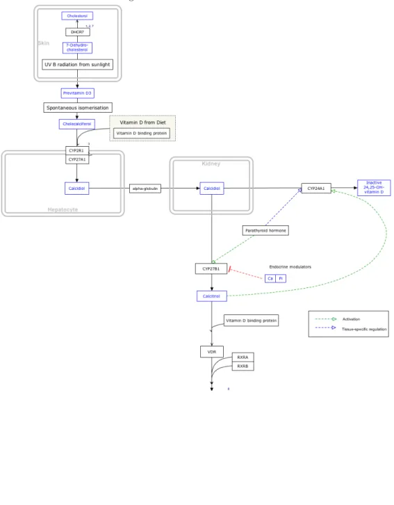
Cholecalciferol and ergocalciferol are biologically inactive precursors of vitamin D and must be converted to biologically active forms in the liver and kidneys. In fact, following dietary intake or synthesis, both forms of vitamin D enter the circulation and are transported to the liver by the vi-tamin D-binding protein (and to a lesser extent by albumin). In hepato-cytes, vitamin D is hydroxylated to form 25-hydroxyvitamin D (calcidiol; calcifediol). 25-hydroxyvitamin D constitutes the major circulating form of vitamin D, and the sum of 25-hydroxyvitamin D2 and 25-hydroxyvitamin D3 levels in serum is used as an indicator of vitamin D nutritional sta-tus. The renal 25-hydroxyvitamin D-1α-hydroxylase enzyme (also known as CYP27B1) eventually catalyzes a second hydroxylation that converts 25-hydroxyvitamin D to 1,25-di25-hydroxyvitamin D (calcitriol). The production of 1,25-dihydroxyvitamin D in the kidneys is regulated by several factors, in-cluding serum phosphorus, calcium, parathyroid hormone (PTH), fibroblast growth factor 23 (FGF-23), and 1,25-dihydroxyvitamin D itself. Most of the physiological effects of vitamin D in the body are related to the activity of 1,25-dihydroxyvitamin D (Figure A.1).
Both environmental factors and cultural practices result in variations in vitamin D status: environmental conditions (geographical locations, seasonal changes); concealed clothing style and sun safety measures. The efficiency of vitamin D synthesis, absorption, and metabolism also depends on a variety of biological factors: skin pigmentation; genetic variations; older age; chronic kidney disease; fat malabsorption syndromes; inflammatory bowel disease; obesity and magnesium deficiency [9].
The leading and most widely known physiological function of 1,25(OH)2D is to regulate mineral and skeletal homoeostasis. However, over the last
decades the functions of vitamin D have been broadened beyond those on skeletal tissue and calcium homoeostasis. Indeed, the finding of VDR ex-pression in a wide range of tissues such as the immune system (T and B cells, macrophages, and monocytes), the reproductive system (uterus, testis, ovary, prostate, placenta, and mammary glands), endocrine system (pan-creas, pituitary, thyroid and adrenal cortex), muscles (skeletal, smooth and heart muscles), and brain, skin, and liver, which has stimulated consider-able interest in understating the putative pleiotropic properties of vitamin D. This introduced the idea of a paracrine/autocrine role in regulating cell proliferation, differentiation and apoptosis as well as immune-cells regulation [10].
Sources of Vitamin D
Very few foods naturally contain vitamin D. Oily fish such as salmon (360 IU per 3.5-ounce serving), mackerel, and sardines are good sources of vitamin D3, as are irradiated mushrooms. Although egg yolks are reported to contain vitamin D, amounts are highly variable (usually no more than 50 IU per yolk), and the cholesterol content of egg yolks makes this a poor source of vitamin D. Cod liver oil, which has been considered for more than 3 centuries to be critically important for bone health, is an excellent source of vitamin D3. Fortified foods include milk (100 IU per 8-ounce serving), orange juice (100 IU per 8-ounce serving) and other juice products, and some breads and cereals. Studies reported that exposure of 20% of the body’s surface to either direct sunlight was effective in increasing blood concentrations of vitamin D3 and 25(OH)D3 among both young adults and older adults. [11]
The Recommended Dietary Allowance
In 2010, the Food and Nutrition Board of the Institute of Medicine set a Recommended Dietary Allowance (RDA) based on the amount of vitamin D needed for bone health. The RDA for vitamin D is listed in table B.2 by age and gender.
Assessing vitamin D status
The current cut-offs proposed by the Institute of Medicine are as fol-lows: deficiency as serum 25-hydroxyvitamin D values ≤ 12 ng/mL (30 nmol/L); insufficiency as serum 25-hydroxyvitamin D values of 12-19 ng/mL (30-49 nmol/L); and sufficiency as serum 25-hydroxyvitamin D values of 20 - 50 ng/mL (50 - 125 nmol/L). The dietary reference intakes (EAR, RDA) set by the Institute of Medicine are based on achieving circulating 25-hydroxyvitamin D concentrations (20-50 ng/mL) that are adequate to maintain bone health and optimal calcium absorption. Yet, considering the potential role of circulating levels lower than 30 ng/mL in the burden of many chronic diseases, the US Endocrine Society has suggested to define cut-off values as follows: deficiency as serum 25-hydroxyvitamin D values ≤ 20 ng/mL (50 nmol/L); insufficiency as serum 25-hydroxyvitamin D values of 21-29 ng/mL (51-74 nmol/L); sufficiency as serum 25-hydroxyvitamin D values of 30 - 100 ng/mL (75 - 250 nmol/L). Although this alternate target range is supported by some observational studies, it is not based on data from randomized controlled trials. With these latter cut-off values, studies from across the world have estimated that hypovitaminosis D is widespread. Data from supplementation studies indicate that vitamin D intakes of at least 800 - 1000 IU/day are required by adults living in temperate latitudes to achieve serum 25-hydroxyvitamin D levels of at least 30 ng/mL (75 nmol/L). Finally, total serum 25-hydroxyvitamin D concentrations may not always adequately
reflect vitamin D bioavailability and additional evidence is needed to im-prove the determination of vitamin D status in different ethnic populations [12] [13].
Prevalence of vitamin D deficiency in CLD and mechanisms
The global prevalence of vitamin D deficiency in the general population affects all age groups and ranges from 20 to 100% when referring to serum 25(OH)D concentrations < 20 ng/ml. In CLD, the prevalence of vitamin D levels < 20 ng/ml in CLD has been reported to range from 64 to 92% and is commonly inversely related to disease progression and severity. Some studies, however, have failed to find a difference in vitamin D status between patients with cirrhosis and those without, thus supporting the multi-factorial cause of this disease. Previously, vitamin D deficiency was thought to be pre-dominantly found in cholestatic liver disorders because of impaired intestinal absorption commonly observed in such patients. Accumulating evidence, however, supports its widespread presence in CLD, regardless of aetiology. Important potential mechanisms to consider in vitamin D deficiency in CLD are: 1) reduced exogenous exposure of patients to vitamin D sources (e.g. dietary, sunlight); 2) intestinal malabsorption of dietary vitamin D; 3) re-duced endogenous production of vitamin D binding protein and albumin in the liver, which are impaired in the presence of cirrhosis; 4) impaired hepatic hydroxylation of vitamin D to 25(OH)D; and 5) increased catabolic removal of 25(OH)D.
Aims
The aim of this study is: 1) to evaluate the prevalence of vitamin D deficiency in CLD patients; 2) to analyse the relation between 25(OH)D serum levels and the aetiology of CLD; 3) to evaluate whether 25(OH)D
serum levels are associated with CP score and MELD score in patients with cirrhosis.
Methods
It was requested to the planning and information office a list with the names and identification numbers of patients with cirrhosis that attended the inpatient Liver Unit at Hospital Santa Maria, Lisbon - Portugal, between January 2013 and July 2015. We analysed 117 clinical files and were consid-ered only those with serum vitamin D concentrations determined (n=85).
The sociodemographic data, the liver disease aetiology and co-morbidities, CP score, MELD score and laboratorial values of the first blood sample were used. Patients with ages ≥ 65 were considered elderly patients and < 65 were considered adult patients. Patients were split into three groups according to their MELD score: group 1, 0-10 points; group 2, 11-20 points; group 3, > 20 points.
Vitamin D status was assessed measuring serum concentrations of 25(OH)D using the ARCHITECT 25-OH Vitamin D automated immunoassay (CMIA technology - Chemiflex). Vitamin D levels were tiered according to the fol-lowing intervals: deficiency 0-20 ng/ml, insufficiency 21-29 ng/ml, sufficiency 30-100 ng/ml. All the included patients were not vitamin D supplemented. Two groups were created according to date of the blood sample collection. One, the Autumn/Winter group, where the samples were collected between September and March. The other, the Spring/Summer group, where the samples were collected between April and August.
Statistical Analysis
Data is presented for patients stratified into groups by CP stages, MELD score and tertiles of 25(OH)D levels. According to their distribution,
con-tinuous variables are either presented as means ± standard deviation (SD). Categorical variables are presented as percentages. Comparisons between groups are calculated by analysis of variance (ANOVA) with p for trend for continuous variables and with Fisher’s exact test for categorical variables. All P-values are reported two-sided. Analyses were performed using R Statistical Software r, version 3.2.2 .
Results
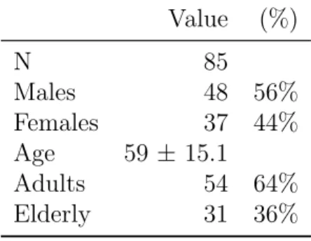
This study included 48 males (57%) and 37 females (44%) with an average of 59 ± 15 years (range: 21 - 88 years); 31 elderly patients (37%) and 54 adults (64%) (Table B.3). The main aetiology of cirrhosis was chronic viral hepatitis, as seen in 59 patients (69%): 48 (57%) HCV, 13 (27%) HBV; other aetiologies are: alcoholic plus chronic viral hepatitis - 19 patients (23%), 10 autoimmune (12%), 6 alcoholic (7%), 1 non-alcoholic fatty liver disease (2%), 4 genetic disorders (5%), 3 cardiac cirrhosis (4%) and 3 idiopathic (4%) (Table B.4). More than half, 47 (55%), had ascites, 22 (26%) encephalopathy and 18 (21%) hepatocellular carcinoma (HCC) (Table B.5).
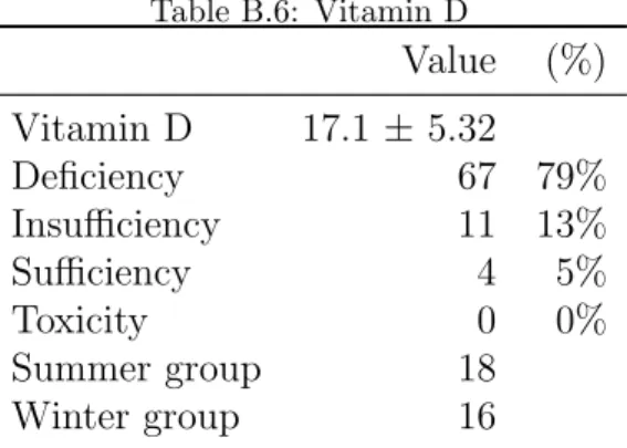
The average levels of 25(OH)D was 17.1 ± 5.3 ng/ml; 67 patients (79%) had vitamin D deficiency; 14 (17%) had insufficiency of vitamin D and only 4 (5%) had adequate vitamin D levels. The split of 25(OH)D measurements between Spring/Summer and Autumn/Winter was 46:39, respectively (Table B.6). No significant difference in vitamin D levels between these groups were observed (p=0.17). There was also no significant difference in 25(OH)D levels between alcoholic cirrhosis (17.7 ± 7.4 ng/ml) and non-alcoholic cirrhosis (16.8 ± 7.5 ng/ml; P = 0.403); as well as there was no significant difference in 25(OH)D levels between chronic viral cirrhosis (17.2 ± 8.1 ng/ml) and non-infectious cirrhosis (16.7 ± 5.7 ng/ml; P = 0.838). Clinical and laboratory
characteristics are presented according to CP stage A, B and C (Table B.8), according to MELD score (Table B.9) and according to 25(OH)D tertiles (Table B.10).
Patients with CP stage A (n = 20), B (n = 43), and C (n = 22) had 25(OH)D levels of 20.2 ± 9.2, 16 ± 7, and 16.4 ± 5.6, respectively (P = 0.118). Likewise, patients with MELD group 1 (n = 27), MELD group 2 (n = 47), and MELD group 3 (n = 11), had 25(OH)D levels of 16.3 ± 7.5, 18.6 ± 8.2 and 12.5 ± 3.5, respectively (P = 0.532). However, there was a significant difference comparing 25(OH)D levels of patients with CP stage A versus B plus C (p = 0.03). It was also shown a significant difference between 25(OH)D levels in MELD group 3 and MELD group 2 (P=0.02), but not between MELD group 3 and MELD group 1 (P=0.08).
Discussion
In this group of 85 patients with CLD, the main aetiology was chronic viral hepatitis (69%), contrasting with the most recent epidemiologic Por-tuguese study (63 910 patients included, between 2003 and 2012), where the most prevalent aetiology was alcoholic (76%). However, it was noticed a significant decline (P < 0.001) in admissions for alcoholic cirrhosis, whereas hospitalizations for cirrhosis caused by HCV or HCV plus alcohol increased by almost 50% (P < 0.001) [14].
In the US, alcoholic liver disease once was considered to be the predomi-nant source of cirrhosis, but hepatitis C has emerged as the nation’s leading cause of chronic hepatitis and cirrhosis. About 2-3% of Americans have NASH. It is estimated that 10% of patients with NASH will ultimately de-velop cirrhosis [15].
preva-lence (79%) of vitamin D deficiency in patients with CLD. Nevertheless, it would be useful to create a consensus about the definition of adequate vita-min D status. Otherwise, different studies will use different guidelines, which difficult the comparison between results [16] [17].
The association between vitamin D levels and the aetiology of CLD (al-coholic or chronic viral hepatitis) was not demonstrated (P = 0.403 and P = 0.838), which was also illustrated by Putz-Bankuti et al. [18] and Malham M et al. [16].
Furthermore, a significant (P = 0.03) inverse association was illustrated between vitamin D status and disease severity. Patients with CP class B plus C had significantly (P = 0.03) lower concentrations than patients in class A (16.2 vs. 20.2 ng/ml). These findings are supported by previous studies [19] [20] [21].
As in our study, Miroliaee et al. [20] shown that patients with CP class B plus C had significantly lower vitamin D levels than class A (P < 0.001); Fisher et al. [22] also demonstrated a significant difference between CP score C vs A (9 ± 4 vs. 18.3 ± 6.7, P < 0.001) [19] [20] [21].
Our results confirm and extend data from previous studies that have largely but not consistently demonstrated inverse correlations of 25(OH)D levels and liver dysfunction. Among various cirrhotic cohorts studied, few reports have linked vitamin D deficiency to higher degrees of liver dysfunction as estimated by the CP class and MELD score.
It is already known the liver plays an important role in the metabolism and pleiotropic functions of vitamin D, but the question is how vitamin D deficiency may be a contributor to the liver dysfunction. Regarding the rela-tion between vitamin D and disease severity, there is growing evidence that vitamin D is involved in the decrease of inflammation and fibrosis.
Proin-flammatory signals in monocytes and macrophages may regulate the local metabolism of vitamin D, auto-inducing the expression of CYP27B1 and the local production of 1,25(OH)2D, and thus controlling the excessive inflam-matory response [23] [24] [25].
On the other hand, recent reports identified VDR in hepatic stellate cells (HSCs) as an endocrine checkpoint for wound healing response in liver; VDR knockout mice develop spontaneous liver inflammation and fibrosis. Preliminary studies indicate that VDR is an important modulator of pro-inflammatory response in HSCs. Ligand-activated VDR represses a wide ar-ray of pro-inflammatory gene expression in HSCs through the direct genomic crosstalk with TGF-B and VDR-deficient HSCs exhibit a spontaneous pro-inflammatory response [26]. More studies suggest that a polymorphism in VDR is correlated with increased progression of liver fibrosis and evolution of cirrhosis [27] [28] [29].
However, this study has some limitations, due to the relatively small sample of patients and the observational nature of our study design which precludes conclusions regarding causality. Additional drawbacks of our work include missing data on some parameters related to vitamin D metabolism (e.g. parathyroid hormone) and the lack of 25(OH)D data in 32 excluded patients. We only included hospitalized patients in de-compensated status, which can influence the disease severity classification and vitamin D levels. Nevertheless, it is important to raise the awareness of physicians about this topic.
In a future work, it is important to corroborate the association of vitamin D status with the occurrence of hepatic de-compensation and the increase of mortality through longitudinal studies. We have poor evidence and conflict-ing results with longitudinal studies; Corey et al. [30] evaluated the impact
of vitamin D levels on the progression of CLD in a longitudinal nested case control study of vitamin D levels in subjects with and without liver histologic progression or clinical de-compensation. This study suggests that vitamin D levels do not influence the progression of CLD. There was found no differ-ence in mean vitamin D levels in patients with and without progressive CLD during any point over 45 months. Vitamin D levels declined over time in both groups consistent with the known effect of aging on vitamin D lev-els. However, patients with progression of liver disease did not experience a greater decline than those without disease progression. The authors specu-late that this specific observation may have resulted from supplementation, as detectable vitamin D2 levels were identified in 55% of the cohort.
On the other hand, in a study with 75 consecutive cirrhotic patients, Putz-Bankuti et al. [18] had shown a significant association of vitamin D levels with the degree of liver dysfunction. It was also suggested that lower vitamin D levels may predict hepatic de compensation and mortality in patients with chronic liver failure, after 4 years.
It is relevant to explore the impact of vitamin D supplementation in the severity disease and in the hepatic osteodystrophy progression. Intervention studies in which CLD patients receive vitamin D and/or calcium supplemen-tation have also yielded conflicting results [31]. One reason for these conflict-ing results may be attributed to the multi-factorial nature of osteodystrophy in CLD. Alternatively, one might argue that the heterogeneity amongst many studies hinders between study comparability, as bone measurements are car-ried out in various locations and different methods of BMD assessment are employed. In addition, we emphasize other issues such as small sample sizes and low statistical power of studies. Moreover, differences in vitamin D dosage and treatment duration hamper other study comparisons.
Combi-nation therapy is a potential confounder as very few studies evaluated the benefits of vitamin D therapy alone, but rather combined it with calcium and other medications known to benefit bone health. It also remains unknown whether vitamin D deficiency confers an enhanced risk to liver disease or whether liver disease causes vitamin D deficiency.
Conclusion
In conclusion, we have shown that vitamin D deficiency is highly preva-lent in patients with CLD; lower concentrations are inversely associated with disease severity, but it is not associated with CLD aetiology. Further studies, namely randomized controlled trials, are warranted to confirm our findings in this small Portuguese sample and to clarify whether vitamin D supple-mentation is effective in the prevention and treatment of liver dysfunction and improves survival in patients with CLD.
AppendixA. Figures
AppendixB. Tables
Table B.1: Sources of Vitamin D [11]
Food (naturally present) Vitamin D3: oily fish
(e.g. salmon, mackerel, tuna, sardines), egg yolk Vitamin D2: mushrooms
Food (Fortified) e.g. margarine, breakfast
cereals, milk (global variation in fortified foods) Oral supplements May contain vitamin D2 or vitamin D3
Sun exposure Photochemical conversion of
7-dehydrocholesterol to pre-vitamin D3
Table B.2: RDA of Vitamin D [12]
Age Males Females
mcg/day IU/day mcg/day IU/day
19-70 years 15 600 15 600
Table B.3: Sociodemographic data Value (%) N 85 Males 48 56% Females 37 44% Age 59 ± 15.1 Adults 54 64% Elderly 31 36% Table B.4: Aetiology Value (%)
Chronic Viral Hepatitis 59 69%
Alcoholic 6 7%
Autoimmune 10 12%
Chronic Viral Hepatitis / Alcoholic 19 22%
Metabolic 2 2%
NASH 2 2%
Chronic Viral Hepatitis / Alcoholic / Metabolic 2 2%
Idiopathic 3 4% Others 3 4% Table B.5: Complications Value (%) HCC 18 21% Encephalopathy 22 26% Ascites 47 55%
Table B.6: Vitamin D Value (%) Vitamin D 17.1 ± 5.32 Deficiency 67 79% Insufficiency 11 13% Sufficiency 4 5% Toxicity 0 0% Summer group 18 Winter group 16
Table B.7: Disease severity
Value (%) Child Pugh - A 20 24% Child Pugh - B 43 51% Child Pugh - C 22 26% MELD group 1 27 32% MELD group 2 47 55% MELD group 3 10 12%
Table B.8: Clinical and laboratory characteristics according to Child-Pugh stage
Variable Stage A Stage B Stage C P
N 20 43 22 Age (years) 59 ± 14 61 ± 16 57 ± 15 0.651 Males 12 23 13 0.880 Females 8 20 9 0.880 25(OH)D (ng/mL) 20.1 ± 9.2 16.0 ± 7.03 16.4 ± 5.6 0.118 25(OH)D (ng/mL) Winter 18.1 14.6 16.7 0.105 25(OH)D (ng/mL) Summer 21.6 17.2 16.2 0.105 Alcohol Abuse (%) 30 28 41 0.563
MELD score 10 ± 2.3 13 ± 3.9 19 ± 4.3 3.04e-11 Albumin (g/dL) 3.4 ± 0.5 2.9 ± 0.4 2.5 ± 0.5 1.57e-08 Bilirubin (mg/dL) 1.0 ± 0.7 2.8 ± 3.9 4.5 ± 2.9 0.001 INR 1.25 ± 0.2 1.34 ± 0.3 2.26 ± 2.0 0.003 Prothrombin time (s) 15.4 ± 5.7 15.8 ± 4.4 20.8 ± 6.1 0.001 Creatinine (mg/dL) 0.9 ± 0.3 1.2 ± 0.8 1.2 ± 0.6 0.127 CRP (mg/dL) 2.9 ± 5.6 3.8 ± 4.9 3.9 ± 5.1 0.538 Ascites (%) 10 60 86 0.000 Encefalopathy (%) 5 21 50 0.003
Table B.9: Clinical and laboratory characteristics according to MELD group 1 2 3 P N 27 47 11 Age (years) 58 ± 14 62 ± 15 55 ± 15 0.995 Males 13 29 6 0.995 Females 14 18 5 0.995 25(OH)D (ng/ml) 16.3 ± 6.4 18.6 ± 8.1 12.5 ± 3.4 0.532 25(OH)D (ng/ml) Winter 15.1 16.9 10.8 0.378 25(OH)D (ng/ml) Summer 17.2 20.4 13.2 0.378 Alcohol Abuse (%) 19 38 36 0.210 MELD 8 ± 1.1 15 ± 2.3 23 ± 1.91 2.2E-16 Albumin (g/dl) 3.2 ± 2.8 ± 0.4 2.4 ± 0.6 1.24E-05 Bilirubin (mg/dl) 1.2 ± 2.7 ± 2.1 7.4 ± 6.3 6.35E-07 INR 1.2 ± 1.4 ± 0.3 3.0 ± 2.6 9.79E-05 Prothrombin Time (s) 15.4 ± 16.7 ± 3.7 22.2 ± 7.5 0.002 Creatinine (mg/dL) 0.9 ± 1.2 ± 0.6 1.6 ± 1.2 0.035 CRP (mg/dL) 2.5 ± 4.1 ± 5.4 4.4 ± 4.3 0.210 Ascites (%) 60 65 32 0.399 Encefalopathy (%) 5 37 18 0.004
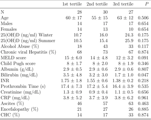
Table B.10: Clinical and laboratory characteristics according to 25(OH)D tertiles
1st tertile 2nd tertile 3rd tertile P
N 28 30 27 Age 60 ± 17 55 ± 15 63 ± 12 0.506 Males 14 17 17 0.654 Females 14 13 10 0.654 25(OH)D (ng/ml) Winter 10.7 16.0 24.3 0.175 25(OH)D (ng/ml) Summer 10.5 15.4 25.9 0.175 Alcohol Abuse (%) 18 43 33 0.117
Chronic viral Hepatitis (%) 68 73 67 0.874 MELD score 15 ± 6.0 14 ± 4.8 12 ± 3.2 0.091 Child Pugh score 8 ± 1.7 8 ± 2.0 8 ± 1.9 0.346 Albumin (g/dL) 2.9 ± 0.5 2.9 ± 0.6 2.9 ± 0.6 0.807 Bilirubin (mg/dL) 3.5 ± 4.8 3.2 ± 3.0 1.7 ± 1.0 0.047 INR 1.75 ± 1.8 1.55 ± 0.6 1.38 ± 0.2 0.218 Prothrombin Time (s) 17.4 ± 7.3 17.2 ± 5.4 16.4 ± 3.9 0.535 Creatinine (mg/dL) 1.3 ± 0.9 0.9 ± 0.4 1.1 ± 0.5 0.656 CRP (mg/dL) 3.8 ± 5.2 3.7 ± 3.9 3.8 ± 6.2 0.961 Ascites (%) 46 57 63 0.463 Encefalopathy (%) 21 27 26 0.885 CHC (%) 14 17 33 0.874
References
[1] V. Hernandez-Gea, S. L. Friedman, Pathogenesis of liver fibrosis, An-nual review of pathology: mechanisms of disease Vol. 6 (2011) 425–456 (2011).
[2] D. Longo, A. Fauci, D. Kasper, S. Hauser, Harrison’s Principles of In-ternal Medicine 19th edition, McGraw-Hill Professional, 2011.
[3] G. D’Amico, G. Garcia-Tsao, L. Pagliaro, Natural history and prognos-tic indicators of survival in cirrhosis: a systemaprognos-tic review of 118 studies, Journal of hepatology Vol. 44 (2006) p. 217–231 (2006).
[4] O. Procurement, Organ procurement and transplantation network, HRSA, DHHS 9.
[5] F. Durand, D. Valla, Assessment of the prognosis of cirrhosis: Child– pugh versus meld, Journal of hepatology 42 (1) (2005) 100–107 (2005).
[6] G. C. Cook, et al., Tropical gastroenterology., Oxford University Press., 1980.
[7] G. L´opez-Larramona, A. J. Lucendo, S. Gonz´alez-Castillo, J. M. Tenias, Hepatic osteodystrophy: An important matter for consideration in chronic liver disease, World Journal of Hepatology 3 (12) (2011) 300 (2011).
[8] C. S. Stokes, D. A. Volmer, F. Gr¨unhage, F. Lammert, Vitamin d in chronic liver disease, Liver International 33 (3) (2013) 338–352 (2013).
[9] Y.-P. Han, M. Kong, S. Zheng, Y. Ren, L. Zhu, H. Shi, Z. Duan, Vi-tamin d in liver diseases: from mechanisms to clinical trials, Journal of Gastroenterology and Hepatology 28 (S1) (2013) 49–55 (2013).
[10] M. Eliades, E. Spyrou, Vitamin D: A new player in non-alcoholic fatty liver disease?, World Journal of Gastroenterology: WJG 21 (6) (2015) 1718 (2015).
[11] M. F. Holick, Sunlight and vitamin d for bone health and prevention of autoimmune diseases, cancers, and cardiovascular disease, The Ameri-can Journal of Clinical Nutrition 80 (6) (2004) 1678S–1688S (2004). [12] A. A. Yates, S. A. Schlicker, C. W. Suitor, Dietary reference intakes:
the new basis for recommendations for calcium and related nutrients, B vitamins, and choline, Journal of the American Dietetic Association 98 (6) (1998) 699–706 (1998).
[13] M. F. Holick, N. C. Binkley, H. A. Bischoff-Ferrari, C. M. Gordon, D. A. Hanley, R. P. Heaney, M. H. Murad, C. M. Weaver, Evaluation, treatment, and prevention of vitamin D deficiency: an endocrine soci-ety clinical practice guideline, The Journal of Clinical Endocrinology & Metabolism 96 (7) (2011) 1911–1930 (2011).
[14] M. J. Silva, M. V. Rosa, P. J. Nogueira, F. Calinas, Ten years of hospital admissions for liver cirrhosis in portugal, European Journal of Gastroen-terology & Hepatology 27 (11) (2015) 1320–1326 (2015).
[15] L. A. Beste, S. L. Leipertz, P. K. Green, J. A. Dominitz, D. Ross, G. N. Ioannou, Trends in burden of cirrhosis and hepatocellular carcinoma by underlying liver disease in us veterans, 2001–2013, Gastroenterology (2015).
[16] M. Malham, S. P. Jørgensen, P. Ott, J. Agnholt, H. Vilstrup, M. Borre, J. F. Dahlerup, Vitamin d deficiency in cirrhosis relates to liver dysfunc-tion rather than aetiology, World Journal of Gastroenterology: WJG 17 (7) (2011) 922 (2011).
[17] J. Arteh, S. Narra, S. Nair, Prevalence of vitamin d deficiency in chronic liver disease, Digestive Diseases and Sciences 55 (9) (2010) 2624–2628 (2010).
[18] C. Putz-Bankuti, S. Pilz, T. Stojakovic, H. Scharnagl, T. R. Pieber, M. Trauner, B. Obermayer-Pietsch, R. E. Stauber, Association of 25-hydroxyvitamin d levels with liver dysfunction and mortality in chronic liver disease, Liver International 32 (5) (2012) 845–851 (2012).
[19] C.-C. CHEN, S.-S. WANG, F.-S. JENG, S.-D. LEE, Metabolic bone disease of liver cirrhosis: is it parallel to the clinical severity of cirrho-sis?, Journal of Gastroenterology and Hepatology 11 (5) (1996) 417–421 (1996).
[20] A. Miroliaee, M. Nasiri-Toosi, O. Khalilzadeh, A. Esteghamati, A. Ab-dollahi, M. Mazloumi, Disturbances of parathyroid hormone–vitamin d axis in non-cholestatic chronic liver disease: a cross-sectional study, Hepatology International 4 (3) (2010) 634–640 (2010).
[21] A. Rode, S. Fourlanos, A. Nicoll, Oral vitamin D replacement is effective in chronic liver disease, Gastroent´erologie Clinique et Biologique 34 (11) (2010) 618–620 (2010).
[22] L. Fisher, A. Fisher, Vitamin d and parathyroid hormone in outpatients with noncholestatic chronic liver disease, Clinical Gastroenterology and Hepatology 5 (4) (2007) 513–520 (2007).
[23] G. Targher, L. Bertolini, L. Scala, M. Cigolini, L. Zenari, G. Falezza, G. Arcaro, Associations between serum 25-hydroxyvitamin d 3 concen-trations and liver histology in patients with non-alcoholic fatty liver dis-ease, Nutrition, Metabolism and Cardiovascular Diseases 17 (7) (2007) 517–524 (2007).
[24] S. Petta, C. Camm`a, C. Scazzone, C. Tripodo, V. Di Marco, A. Bono, D. Cabibi, G. Licata, R. Porcasi, G. Marchesini, et al., Low vitamin d serum level is related to severe fibrosis and low responsiveness to interferon-based therapy in genotype 1 chronic hepatitis c, Hepatology 51 (4) (2010) 1158–1167 (2010).
[25] J. S. Adams, M. Hewison, Unexpected actions of vitamin d: new per-spectives on the regulation of innate and adaptive immunity, Nature clinical practice Endocrinology & metabolism 4 (2) (2008) 80–90 (2008). [26] N. Ding, T. Y. Ruth, N. Subramaniam, M. H. Sherman, C. Wilson, R. Rao, M. Leblanc, S. Coulter, M. He, C. Scott, et al., A vitamin d receptor/smad genomic circuit gates hepatic fibrotic response, Cell 153 (3) (2013) 601–613 (2013).
[27] E. Garc´ıa-Mart´ın, J. Ag´undez, M. L. Maestro, A. Su´arez, M. Vidaurreta, C. Mart´ınez, C. Fern´andez-P´erez, L. Ortega, J. M. Ladero, Influence of vitamin d-related gene polymorphisms (cyp27b and vdr) on the response to interferon/ribavirin therapy in chronic hepatitis c, PloS one 8 (9) (2013) e74764 (2013).
[28] L. Adorini, Vitamin d receptor polymorphisms in primary biliary cir-rhosis: a functional connection?, Journal of hepatology 50 (6) (2009) 1071–1073 (2009).
[29] A. Tanaka, S. Nezu, S. Uegaki, K. Kikuchi, A. Shibuya, H. Miyakawa, S.-i. Takahashi, I. Bianchi, P. Zermiani, M. Podda, et al., Vitamin d receptor polymorphisms are associated with increased susceptibility to primary biliary cirrhosis in japanese and italian populations, Journal of Hepatology 50 (6) (2009) 1202–1209 (2009).
[30] K. E. Corey, H. Zheng, J. Mendez-Navarro, A. Delgado-Borrego, J. L. Dienstag, R. T. Chung, H.-C. T. Group, et al., Serum vitamin D levels are not predictive of the progression of chronic liver disease in hepatitis C patients with advanced fibrosis, PLoS One 7 (2) (2012) e27144 (2012). [31] J. S. Crippin, R. A. Jorgensen, E. R. Dickson, K. D. Lindor, Hepatic os-teodystrophy in primary biliary cirrhosis: effects of medical treatment., The American Journal of Gastroenterology 89 (1) (1994) 47–50 (1994).
![Table B.1: Sources of Vitamin D [11]](https://thumb-eu.123doks.com/thumbv2/123dok_br/18498419.901745/20.918.128.727.273.484/table-b-sources-of-vitamin-d.webp)