Unidade de Ciências e Tecnologias dos Recursos Aquáticos
CONTROLE HORMONAL DA DIFERENCIAÇÃO E
INVERSÃO SEXUAL NA DOURADA
Sparus a ura ta (L.)
Hormonal control of sex differentiation and reversal in sca bream Sparus aurata (L.)
Dissertação apresentada à Universidade do Algarve para obtenção do grau de Doutor
•> TESES
João Afonso Baeta Condeça Faro, 200S
)i
Unidade de Ciências e Tecnologias dos Recursos Aquáticos
CONTROLE HORMONAL DA DIFERENCIAÇÃO
E INVERSÃO SEXUAL NA DOURADA Sparus aurata (L.)
Hormonal control of sexual differentiation and reversal in sea bream Spcirus auratu (L.)
Dissertação apresentada à Universidade do Algarve para obtenção do grau de Doutor
João Afonso Baeta Condeça
Uma tese a fogo lento obrigou a muito tino
se não a acabo ainda rebento muito obrigado Adelino
Muita hormona tive em mente e muito houve que eu não li muitos RIAs é ponto assente muito obrigado Debbie
No laboratório à procura
muita se busca mas pouco muda doutorando tem vida dura pede-se à Elsa que ela ajuda
No café com o Vília e o Guerreiro não há tréguas e não se diz bem assim nos ensinou Mário o "matreiro" o Faustino e a Rute já aprenderam também
Para a Sílvia e Cecília PGR é maçada e a Modesto só fala do xarroco o Cavaco protege a sua bancada o Pepe e o Juan acham que ele é louco
Outros há que também habitam no laboratório dois vinte e oito para todos eles a minha gratidão
então João já acabaste? há coisas que nunca mudam espero que esta tese lhes baste
Para a frente é que é caminho assim dizia a Teresa Palaré a ela agradeço com carinho de ter remado contra a maré
Page List of Abbreviations 7 Sumário 8
1 General Introduction
1.1 Characteristics of the species 12 1.2 Taxonomic classification 12 1.3 Economic and Scientific importance 13 1.4 Reproductive biology 15 1.4.1 Sex differentiation 15 1.5 The role of steroids in the Brain-Pituitary-Gonad axis 16
1.5.1 Teleosts in general 16 1.5.2 The hermaphrodite sea bream 19 1.6 Objectives 20 1.7 References 21
II The effect of estrogcn on the gonads and on in vitro eonversion of androstenedione to testosterone, 11-ketotestosterone and estradioI-17p in Sparus aurata (Teleostei, Sparidae)
ÍI.l Abstract 25 11.2 Introduction 26 11.3 Materials and methods 27 11.4 Results 31 11.4.1 Effect ofhormone treatment on gonadal morphology 31 11.4.2 Steroid output by gonadal tissue 38 11.5 Discussion 42 11.6 References 46
bream {Sparus aurata L.)
III. 1 Abstract 51 111.2 Introduction 52 111.3 Materials and methods 53 111.4 Results 59 111.4.1 Recovery of radioactivily 59 111.4.2 Steroids identified 60 111.4.3 Metabolite yields 62 111.4.4 Enzymatic activity 64 111.5 Discussion 66 111.6 References 71
IV Seasonal cycle of gonadal development, steroidogenesis and plasma sex steroids during the second year of life of sea bream, Sparus aurata (L.)
IV. 1 Abstract 76 IV.2 Introduction 77 IVA Materials and Methods 79 IV.4 Results 83 IV.4.1 Seasonal alterations of the gonads 83 IV.4.2 Sexual Steroids 89 IVA Discussion 98 IV.6 References 108
V Effect of estrogen adminístration during early development of the gonads of sea bream, Sparus aurata (L.)
V.l Abstract 118 V.2 Introduction 119 V.3 Materials and Methods 120 V.4 Results 124 V. 4.1 Rotifer estrogen uptake 124
V. 4.3 Larvae estrogen uptake 127 V. 4.4 Effect of estrogen on gonadal histology 128 V.5 Discussion 129 V.6 References 136
VI Conclusions
VI. 1 Control of the initial stages of sea bream sex reversal by steroids 139 VI.2 Is there a female primacy pathway in sea bream sex differentiation? 140 VI.3 Future perspectives 143 VI.4 References 144
DA donamine
dpf days- post- fertilization
dph days- post- hatch
GABA gamma-aminobutiric acid
GC-MS gas chromatography- mass spectometry
GnRH gonadotropin-releasing hormone
GSI gonadosomatic index
GtH gonadotropin
hr hour(s)
H&E hematoxylin and eosin
hCG human chorionic gonadotropin
HUFA highly unsaturated fatty acid
...-Hyd hydroxylase
...-HSD .. .-hydroxysteroid dehydrogenase
I. U. international unit
mRNA messenger RNA
NAD P-nicotinamide adenine dinucleotide (reduced forni)
NPY neuropeptide Y
OTMS oxime-trimethylsilyl
PGC primary germ cell
RIA radioimmunoassay
5p-Red Sbeta-reductase
RPTLC reverse phase thin layer chromatography sbER(a/p) sea bream estrogen receptor (alpha/beta)
SEM standard error of the mean
RPTLC reverse phase thin layer chromatography
TLC normal phase thin layer chromatography
TMS Trimethylsilyl
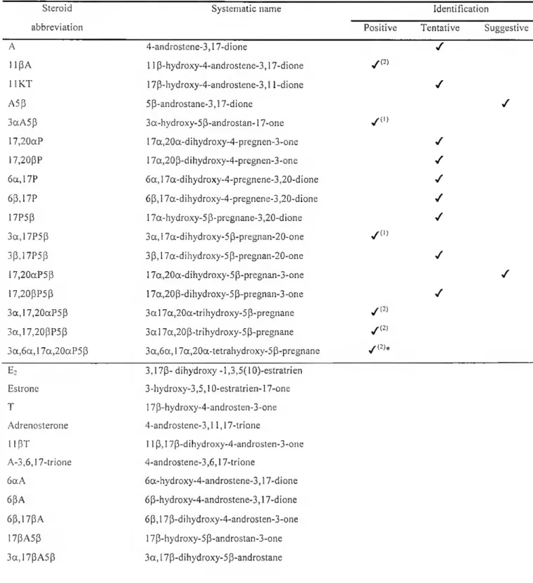
3a.1 lBA5B-17one 3a. 11 B-dihvdroxv-5 B-androstan-17-one 3a,17aA5p-l Ione 3 a, 17p-dihydroxy-5P-androstan-11 -one 3p,l lpA5a-17one 3 p, 11 p-dihydroxy-5a-androstan-17-one 3a,17,20aP5p-l Ione 3a, 17a,20a-trihydroxy-5P-pregnan-11 one 3a,1 ip,17,20aP5p 3 a, 11 p, 17a,20a-tetrahydroxy-5p-pregnane 3a,17,20p,21P5p 3a, 17a,20p,21 -tetrahydroxy-5p-pregnane 3a,11 p,17,21P5p 3a, 11 p, 17a,21 -tetrahydroxy-5p-pregnane 1 ip.l7aP-3,20dione 11P, 17a-dihydroxy-4-pregnene-3,20-dione
ethE2 17a-ethynilestradiol
estriol 1,3,5(10)-estratriene-3,16a, 17p-triol
A dourada, Sparus aurata, é um peixe hermafrodita protândrico, i.e, atinge a primeira maturação sexual como macho podendo inverter o seu sexo para fêmea nas épocas de reprodução subsequentes. O fenómeno, designado como inversão sexual, sendo bastante comum nos teleósteos em geral, é ainda mal conhecido, sobretudo do ponto de vista genético e fisiológico (desconhecem-se, nomeadamente, quais e como se relacionam as moléculas indutoras e mediadoras do processo). É bastante difícil compreender que os mesmos factores genéticos possam regular ciclicamente o aparecimento dos dois sexos na gónada e conceber um modelo onde os mesmos factores possam interactuar com factores sociais ou ambientais que se sabem poder ter influência na expressão do sexo. Deste modo o hermafroditismo tem sido mais considerado como um processo de (re)diferenciação sexual do que de determinação sexual como, por exemplo, nos mamíferos. De um modo geral, o cérebro é considerado como o centro de controle de todos os fenómenos ligados à reprodução e capaz de integrar toda uma série de estímulos, quer de natureza interna (p. ex. acção retroactiva de esteróides sexuais) quer externa (p. ex. fotoperíodo, temperatura, feromonas etc). A resposta surge depois na fornia de diversos tipos de moléculas estimuladoras ou inibidoras da produção de gonadotrofinas por parte da pituitária que, por sua vez, controlam a esteroidogénese e, consequentemente, a gametogénese. No entanto, nos peixes, o perfil de esteróides sexuais varia consideravelmente de espécie para espécie o que dificulta o conhecimento do papel fisiológico da maioria desses esteróides na diferenciação/inversão sexual. Deste modo o objectivo geral desta tese é procurar relacionar o perfil de esteróides sexuais da dourada com o processo de inversão e /ou diferenciação sexual para gerar informação que contribua para um modelo geral para a compreensão do hermafroditismo na reprodução dos peixes.
O processo de inversão sexual foi seguido em peixes feminizados artificialmente por administração de estrógenio na ração e também em peixes a completar 2 anos de idade e potencialmente em inversão sexual natural. Em ambas as experiências se verificou que a feminização da gónada (completa no primeiro caso e parcial no segundo) se processou à custa da inibição de desenvolvimento da parte testicular sem que se tenha verificado um aumento real do ovário. Na inversão natural foi mesmo
espermatogónias foram substituídas por massas de tecido conjuntivo de aparência amarelo-castanho não relatadas anteriormente nesta espécie. O nível de estrógenio no plasma sanguíneo foi sempre muito baixo embora na inversão sexual induzida a capacidade de produzir estradiol-17p (E2), tal como a testosterona, a partir da androstenediona (medida por radioimunoensaio) se verificasse inversamente proporcional à proporção de tecido testicular. A capacidade esteroidogénica das gónadas feminizadas (utilizando 17a-hidroxiprogesterona radioactiva como precursor) revelou um paralelismo significativo com as mudanças morfológicas. Ou seja, a atrofia do testículo foi sobretudo mais acompanhada pela diminuição de alguns fluxos metabólicos, nomeadamente na produção de 11 p-hidroxiandrostenediona (llpA) e etiocolanolona (3aA5p), do que pelo aparecimento de novas vias enzimáticas. A presença de elevados níveis de 11 PA foi confirmada in vivo assim como a de esteróides 5p-reduzidos (incluindo um de propriedades cromatográficas semelhantes a 3aA5P) o que sugere um possível papel para estes esteróides na espermatogénese desta espécie. Foi detectada a actividade de 6(a/p)-hidroxilase que são enzimas esteroidogénicas muito pouco comuns em peixes, embora a sua acção não estivesse relacionada de um modo evidente com a inversão sexual.
Ao nível ontogénico verificou-se que a gonadogénese ocorre por volta dos três meses de idade (87 dias pos-eclosão) e não é afectada pela exposição ao estrógénio exógeno. A gametogénese verificou-se ter início cerca de 1 mês mais tarde do que em anteriores descrições, por volta dos 5 meses, através da formação das ovogónias junto à cavidade central e como consequência da formação de ninhos de células germinais primordiais. A exposição faseada das larvas ao estrogénio demonstrou a sua insensibilidade a esta hormona durante os primeiros 100 dias de vida apesar da técnica utilizada de incorporação de E2 nas presas vivas (rotíferos e artémias) ter sido satisfatória. A causa para este facto pode dever-se à metabolização do E2 noutros esteróides biologicamente inactivos ou mais provavelmente à inexistência da forma a do receptor de estrogénio da dourada, que se pensa estar envolvido na inversão sexual, nas larvas destas idades.
endógenos no plasma sanguíneo durante as primeiras fases de inversão sexual (antes da iniciação da vitelogénese) é uma hormona largamente associada a inversão sexual na dourada pois é susceptível de induzir a mesma e também porque se verifica maior capacidade para a sua síntese nas gónadas em feminização. No entanto, o efeito da administração de estrógénio parece ser dependente do estado de desenvolvimento da gónada porquanto a gónadas muito indiferenciadas não invertem o seu sexo. O conjunto de resultados dá também ênfase à ideia de que a teoria da primazia de diferenciação sexual feminina geralmente aceite nos mamíferos possa também ser aplicada aos peixes apesar da existência de casos de hermafroditismo protândrico. Apesar das douradas se diferenciarem como machos para a primeira estação de reprodução existe previamente a formação de um ovário efémero que depois degenera. Para alem desta evidência morfológica, também do ponto de vista fisiológico, ao nível da capacidade esteroidogénica das gónadas, parece haver uma via metabólica feminina de base que só é ultrapassada se algum mecanismo masculinizante ocorrer e que retorna a condição feminina quando esse mecanismo activo cessar.
I General Introduction
1.1 Characteristics of the species
The gilt-head sea bream, Sparus aurata, is a marine teleost with a silvery grey ovoid body and a large dark pateh loealised at the anterior end of the lateral line. One of most distinctive features of this species is a golden curved bar across the forehead, bordered by two dark zones, which gives the species its common name (Fig. 1.1). Body length rarely exceeds 70cm but it usually reaches 30-35cm length. Generally, this fish can be found, solitary or in small groups, in littoral waters with a sandy bottom and lagoons. It is mainly carnivorous (molluscs, crustaceans and fishes), although sometimes feeds on plants. This species distribution spreads throughout the Mediterranean, where it is rather common, and the Atlantic, from Great Britain to Cape Verde and the Canary islands (UNESCO, 1986).
1.2 Taxonomic Classification
The gilt-head sea bream taxonomic classification is as follows (Nelson, 1994):
Superclass- Osteichtyes Class- Actinopíerygii Subclass- Teleosíei Superorder- Acanthoptherygii Order- Pcrciformes Suborder- Percoidei Supcrfamily- Percoidea Family- Sparidae Genus- Sparus
Species- Sparus aurata (Linnaeus, 1758)
1.3 Economical and Scientific Importance
Sea bream aquaculture techniques were developed during the late seventies and early eighties and, nowadays, it is possible to control ali stages of the sea bream life cycle, which is fundamental for profitable fish farm production. This production has been continuously increasing over the last ten years. Total European catches raised from 12280 tons in 1994 to 33226 tons in 1997 (FEAP, 1997) and the main origin of production are Mediterranean countries such as Greece, Italy, Spain and Portugal. Therefore, sea bream is not only becoming economically important but is also readily available, providing two good practical reasons to study this species.
Moreover, in contrast to other vertebrates, teleost fishes exhibit a wide variety of different types of ambisexuality, including protogynic and protandric hermaphroditism (as is the case of the sea bream), making them a group of great interest for investigating sex development in vertebrates (Reinboth, 1982). The genetic basis of hermaphrodites as the sea bream is unknown. In fact, hermaphroditism has generally been considered to be largely a process of sex differentiation (and/or inversion) rather than sex determination as the involvement of possible genetic factors in this process is poorly understood (Price, 1984). ll is difficult to understand how the same genetic factors can regulate cyclically the appearance of both sexes in one fish and also to conceive a model where genetic factors can interact with other factors, as sexual steroids, known to be related with sexual differentiation (Borg, 1994). The conflicting data on the effects of steroids in ambisexual fish add to the uncertainty as to whether or not sex hormones have a causative influence on the process of sex inversion (and differentiation) (Reinboth, 1988). Further studies on the sex steroid profile of ambisexual species are needed to improve the current knowledge on the physiological role of sex steroids on sex differentiation and reversal.
*¥m. èé&m ps '■m m •5;V: í* fw- ííl^í WÍ ■ w. *y:' A-V <• Wi ' Ov m «•. ^ii;^ V ''-lí^írM ES%í^: mm mm ?
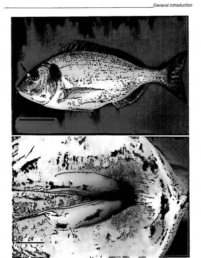
Figure 1.1 (upper photo) - Lateral view of 300g adult sea bream, Sparus aurata, exhibiting the most distinctive feature of this species: the golden curved bar across forehead (arrow). Bar corresponds to 5cm. Figure 1.2 (lower photo)— Sea bream ambisexual gonad, united caudally, located below the dorsal wall of the peritoneal cavity. Note the simultaneous presence of testes (Te) and ovary (Ov). Bar corresponds to Icm.
1.4 Reproductive biology
The sea bream is a protandrous hennaphrodite, i.e., individuais spawn as males during the first breeding season but may undergo a sex change to female in one of the subsequenl spawning seasons (D'Ancona, 1941; Pasquali, 1941). Although there is either testicular (ílrst) or ovarian (later) predoininance in sea bream gonad at spawning, both germinal tissues are present during the whole gonadal cycle. Sea bream gonads are paired elongated organs placed along the dorsal wall of the peritoneal cavity just bellow the swim bladder. The gonads unite caudally lo form a common genital duct that exits the body between the anus and the urinary pore (Fig. 1.2). According to Reinboth (1982) the fact that the teleost genital apparalus is not connected in either sex with the excretory system, permits, from a morphological point of view, the protandric and protogynic sex reversals observed in these fish. The spawning season in southern Portugal is from October/November to January/February although it can be advanced or delayed several months by temperature and photoperiod manipulations in artificial environments.
1.4.1 Sex differentiation
Gonadogenesis is thought to occur around 90 days post hatch (Power, D.M. unpublished data; see chapter V) although gametogenesis takes place much later. Zohar eí al (1978) followed the development of the sea bream gonad during the first two years of life. The first germ cells to differentiate are the oogonia, which arise from primordial germ cell (PGC) nests that, unevenly, border the vicinity of the central cavity (see chapter V) at the age of 4 months. One month later, a topographic differentiation becomes evident and the gonad divides into a dorsal region containing the central cavity which will form the ovary and a ventral region, separated by connective tissue, forming the future testis. Until the age of 8 months the oogonia proliferate forming an ephemeral ovary that starts to degenerate simultaneously with a process of active spennatogenesis, leading the gonad to function as a testis in the first (Zohar et al, 1978) or second breeding season (Bruslé-Sicard and Fourcault, 1997). The sea bream testis is of a lobular type, i.e, germ cells are scattered along lobules and the cysts, which remain roughly in the same place during
spermatogenesis, and release the spennatozoa into a central lúmen {vas eferens) that comunicares with the sperm duct {vas deferens) (Billard et ai, 1982). The subsequent period is characterized by the start of sex reversal that affects up to 80% of the males (Zohar et ai, 1978) but that can be delayed for later seasons (Bruslé-Sicard and Fourcault, 1997). As sex inversion proceeds the lobular organization is disrupted and degenerative processes including phagocytic activity result in testicular regression. In contrasl, the ovary recovers its oogenic capacity and develops the ovigerous lamellae increasing the ovarian component of the gonad. However, caution must be taken when diagnosing histologically sex reversal, because protandry involves coexistence of both sexes during the whole life cycle (Yeung and Chan, 1987). Also, sea bream gonads are very plastic and unexpected abrupt sex changes have been reported (Kadmon et ai, 1985), emphasizing the need of more reliable histológica! criteria for sex reversal recognition (Bruslé-Sicard and Fourcault,
1997). Sea bream is a cyclical breeder and therefore its ovary, like most teleosts, contains oocytes at ali stages of development, i.e, it is an asynchronous ovary (Nagahama, 1983). Simultaneously with the progressive dominance of the ovary in the gonad, oocytes enter a growth phase, vitellogenesis, before the resumption of meiosis. Oocyte maturation is the following event and consists of germinal vesicle breakdown, chromosome condensation and assembly of the first polar body, ali of which are prerequisites for ovulation and successful fertilization (Nagahama, 1997).
1.5 The role of steroids in the Brain-Pituitary-Gonad axis
1.5.1 Teleosts in general
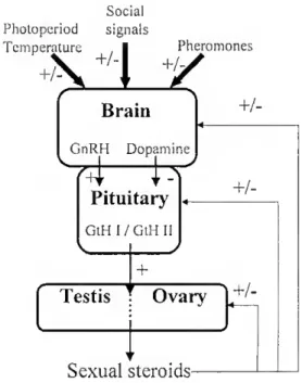
It is now very well established that sexual steroids acting on the gonads are not the only factors regulating fish reproduction and both the brain and the pituitary are involved in this process (Dufour et ai, 1999; Kah et ai, 1999). The brain is one of the main organs controlling reproduction and is capable of perceiving both internai (e.g feedback of steroid hormones) (Francis et al, 1993; Soga et a/., 1998) and externai stimuli such as photoperiod (Amano et al, 1995; Senthilkumaran and Joy, 1995), temperature (Shimizu, 1996), social
signals (Francis eí ai, 1993; Rissman, 1996) or pheromones (Christensen and Sorensen, 1996). For example, in sea bream a significam inhibition of sex inversion was observed when a group of young inales was mixed with larger females (Happe and Zohar, 1988). The brain response to these stimuli can be made by several stimulatory or inhibitory molecules of gonadotropin synthesis but the most important seem to bc gonadotropin-releasing hormones (GnRH, stimulatory), dopamine (DA, inhibitory) and y-aminobutyric acid (GABA, modulatory) (Trudeau and Peter, 1995; Kah et ai, 1999). These substances control gonadotropin release from the pituitary, GtFl 1 and GtFl II (homologous to the folheie stimulating hormone, FSH, and the luteinizing hormone, LH, respectively, in tetrapods) which, in turn, control gametogenesis and steroidogenesis (Fig. 1.3). In teleosts, however, differences in the pubertal profiles of GtFl I and GtH 11 suggests species-specific variations in the regulation of gonadotropin synthesis and their respective roles in the initiation of puberty, gametogenesis and steroidogenesis (Dufour et ai, 1999). As a consequence of the variety of reproductive strategies in teleost fish, the role of steroid hormones as major mediators of sexual differentiation and gametogenesis are complex and also vary considerably. Nevertheless, knowledge on the mechanism of action of some criticai steroids with known functions in several teleosts, e.g. estradiol-17p (£2; ovarian development and oocyte growth), 11-ketotestosterone (11KT; spermatogenesis) and 17a,20(3-dihydroxy-4- pregnen-3-one (17,20(3P; gamete maturation), can provide valuable insight into lhe evolution ofthe reproductive function (Nagahama, 1999).
The synthesis of gonadal steroids through GtH stimulation is generally mediated by specific cells. In the testis, interstitial cehs (homologous to mammalian Leydig cells) occur between testicular lobules and are considered to be the major site of androgen synthesis (Nagahama et ai, 1982) but boundary lobular cells, or Sertoli cells, are also of major importance for spermatogenesis. Germ cells are structurally and functionally supported by Sertoli cells, which are also considered to mediate androgen effeets on germ cells (Shulz et al, 1999). In the preovulatory ovary, the main steroidogenic role is attributed to the follicle layers, i.e., the thecal and granulosa cells. In common with mammals, in teleosts these cells interact in a utwo-cell-type" model to establish a steroid biosynthetic pathway (Kagawa et
al, 1982). Furthermore, £2 and 17,20(3P, two major steroids in oocyte development, are
synthesized by the granulosa layer after their precursors have been produced in the theca layer (Nagahama, 1999). Social Photopcriod signals Temperature +^ rature , 1 1'her KHy Pheromones Brain GnRH Dopamine ív Pituitary GtH I/GtH 11 \ y + Testis Ovary +/- +/-
Figure 1.3 - Simple schematic representation of the cascade of reproductive events acting on the brain-pituitary-gonad axis in teleosts. GnRH, gonadolropin-releasing hormone; GtH,
gonadotropin; +, stimulatory effect; -, inhibitory effect; +/- possible stimulatory and/or inhibitory effects.
Sexual steroids-
Sex steroids can have complementary physiological roles as a consequence of their action at multiple sites (Kah eí al., 1999) or else their physiological role may involve stimulatory or inhibitory effects on other steroids (Antonopoulou et ai, 1999). For example, in the proposed endocrine mechanism of sex reversal in protandrous black porgy, Acanthopagrus shlegeli, E2 is thought to act through a positive feedback to stimulate GnRH and GtH synthesis and release, which in turn stimulates aromatase activity to produce more E2 (Chang et ai, 1997). In the Atlantic salmon, Salmo salar, silastic implants of testosterone (T) prevented the normal testicular decline after the breeding season by
interfering at the levei of GtH, which affects the steroidogenic axis and hence the gonad status (Antonopoulou eí ai, 1999). The mechanism proposed was that exogenous T induced a positive feedback on GtH II which prevented reduction in plasma 17,20pP and, in turn, the suppression of Sertoli cell phagocytic activity upon the spermatozoa, prolonging the spermiation period.
1.5.2 The hermaphrodite Sea Bream
The number and type of GnRHs, may vary among species. In the sea bream, three different forms have been isolated from the brain, e.g. salmon (s)GnRH, chicken (c)GnRH and sea bream (sb)GnRH (Gothilf eí ai, 1996) but only sbGnRH and cGnRH reached the pituitary (Holland et ai, 1998). Furthermore, sbGnRH (and GtH II) pituitary content was 3- 17 fold higher in both male and females with developing gonads, but not in recrudescent bisexual gonads, suggesting that sbGnRH is the form which controlls reproduction in this species (Holland eí ai, 1998). Other substances with GnRH-like activity were, however, detected in lhe sea bream ovary (Nabissi et al, 1997).
An assay for sea bream GtH I has not been developed yet but the genes for p-GtH I and II were found to be differentially expressed in male and female sea bream (Elizur et al, 1996). Mature inales appeared to have higher GtH I mRNA leveis than mature females while the opposite is true for GtH 11 mRNA leveis. Pituitary and plasma GtH II leveis showed, however, different profiles since in mature females leveis of both were high, whereas in mature males only the plasma content was high (Holland et al, 1998). Furthermore, plasma GtH II gradually increased during both the male and female reproductive cycle. The temporal differences in pituitary GtH II content further supported the hypothesis that GtH 1 and II are regulated differently and their roles in male and female sexual development may, therefore, not be equivalem (Holland et al, 1998).
There is no evidence to show that sex steroids are stimulated directly by sea bream gonadotropins aí any of their characteristic sexual stages. Neverlheless, pioneering studies performed by Zohar and his co-workers have shown that low doses of human chorionic gonadotropin (hCG) induced maturation, ovulation and spawning, for at least 4 days, in female sea bream in the last stages of oocyte vitellogenesis (Zohar and Gordin, 1979). Furthermore, hCG treatment in vitro enhanced 11KT in both ovary and testis, 1 Ip-hydroxy- testosterone (1 ipT) and T in the ovary whereas T synthesis was strongly inhibited in the testis (Eckstein et al, 1978). The sea bream sex steroid profile is discussed in detail in chapter IV of this lhesis.
1.4 Objectives
Early studies on fish sex steroids were predominantly carried out on species that first had major commercial importance such as the salmonids and the results obtained were frequently assumed to be typical of ali teleosts. More recent studies, however, gradually indicated that salmonids are atypical teleosts in many respects and that there is a wide diversity in the nature ofthe steroid hormones produced in this group (Kime, 1993). Typical examples are many where hermaphrodite species in addition to the ignorance on the genetic basis of sexuality and the relative discrepancy on the species-specific steroid profiles, it is very difficult to link steroid plasma leveis or metabolism to the sex inversion process since in the few studies carried out comparisons were made of the male and female stages reflecting the sex distinctive characteristics rather than the regulation of sex
inversion itself (Baroiller a/., 1999).
The main objective of this thesis is to correlate the steroid profile of the sea bream with the process of sex inversion and/or differentiation and generate further information to contribute to a general model to hermaphroditism in fish reproduction. To achieve this goal, sex steroid plasma profiles and steroidogenic capacities were followed in either estrogen- induced and naturally sex inverting sea bream. Hormonal sex reversal as been widely used as a lool for a better understanding of the mechanisms of sex differentiation and/or reversal in many teleost families (see review on hormonal induction of sex reversal in fish Pandian and Sheela, 1995). This strategy will allow to control differences in the sex inversion process due to hormonal exogenous interference making it possible to monitor in detail physiological and morphological changes in induced sex inverting specimens that are more difficult to obtain in natural sex inverting fish due to sea bream gonadal sex plasticity (Kadmon et ai, 1985). Very few studies on sex differentiation in hermaphroditic fish have been reported (Nakamura et ai, 1998). Specifically, in sea bream it is unknown when gonadogenesis occur and if the larval early stages are sensitive to hormonal treatments in order to differentiate directly as a female the same way adult sea bream can redirect gonadal sex from male to female (Happe and Zohar, 1988). Therefore time-phased estrogen
larval feeding scheme will be carried out in order to assess larval sensitiveness to estrogen encompassing gonadogenesis and gametogenesis in sea bream.
1.6 References
Amano, M., Hyodo, S., Kitamura, S., Ikuta, K., Suzuki, Y., Urano, A., and Aida, K. (1995). Short photoperiod accelerates preoplic and ventral telencephalic salmon GnRH synthesis and precocious maturation in underyearling male masu salmon, Gen. Comp. Endocrinol. 99, 21-11.
Antonopoulou, E., Mayer, 1., Borg, B., Swanson, P., Murza, I., and Christoforov, O. (1999). Effects of testosterone on gonadotropins, testes, and plasma 17a,20P-dihydroxy-4-pregnene-3-one leveis in postbreeding mature Atlantic salmon, Salmo salar; male parr. J. Exp. Zool. 284, 425-436.
Baroiiler, J.-F., Guiguen, Y., and Fostier, A. (1999). Endocrine and environmental aspects of sex differentiation in fish. Celi Mol. Life Sei. 55, 910-931.
Billard, R., Fostier, A., Weil, C., and Breton, B. (1982). Endocrine control of spermatogenesis in teleost fish. Can. J. Fish. Agua. Sei. 39, 65-79.
Borg, B. (1994). Androgens in teleost fishes. Comp. Biochem. Physiol. 109C, 219-245.
Bruslé-Sicard, S., and Fourcault, B. (1997). Recognition of sex-inverting protandric Sparus aura ta: ultrastrutural aspects. J. Fish Biol. 50, 1094-1 103.
Chang, C. F., Lin, B. Y., Lau, E. L., Lee, M. F., Yueh, W. S., Lee, Y. FL, Chang, C. N., Fluang, J. D., Tacon, P., Lee, F. Y., Du, J. L., and Sun, L. T. (1997). The endocrine mechanism of sex reversal in the protandrous black porgy, Acanthopagrus schlegeli: a review. Chin. J. Physiol. 40, 197-205.
Christensen, T. A., and Sorensen, P. W. (1996). Pheromones as tools for olfactory research. Chem. Senses 21, 241-243.
D'Ancona, U. (1941). Ulteriori osservazioni sull' ermafroditismo e el differenziamento sessuale deli' orata {Sparus auratus L.). Publicazioni delia Stazione Zoologica di Napoli 18, 313-336.
Dufour, S., Huang, Y. S., Rousseau, K., Sbaihi, M., Le Belle, N., Vidal, B., Marchelidon, J., Quérat, B., Burzawa-Gérard, E., Chang, C. F., and Schmitz, M. (1999). Puberty in teleosts; New insights into the role of peripheral signals in the stimulation of pituitary gonadotropins. In "6lh International Symposium
on the Reproductive Physiology of Fish" (B. Norberg, O. S. Kjesbu, G. L, Taranger, E. Andersson and S. O. Stefansson, Eds.), pp. 455-461. John Grieg AS.
Eckstein, B., Abraham, M., and Zohar, Y. (1978). Production of steroid hormones by male and female gonads of Sparus aurala (Teleostei, Sparidae). Comp. Biochem. Physiol. 60B, 93-97.
Elizur, A., Zmora, N., Rosenfeld, FL, Meiri, L, Hassin, S., Gordin, FL, and Zohar, Y. (1996). Gonadotropins P-GtHI and P-GtHII from the gilthead seabream, Sparus aurala. Gen. Comp. Endocrinol. 102, 39-46. FEAP (1997). Summary of fish farm production. Aquaculture Europe 22.
Francis, R. C., Soma, K., and Fernald, R. D. (1993). Social regulation of the brain-pituitary-gonadal axis. Proc NatlAcad Sei U S A 9Q, 7794-7798.
Gothilf, Y., Munozcueto, J. A., Sagrillo, C. A., Selmanoff, M., Chen, T. T., Kah, O., Elizur, A., and Zohar, Y. (1996). Three fornis of gonadotropin-releasing hormone in a perciform fish (Sparus aurata): Complementary deoxyribonucleic acid characterization and brain localization. Biol. Reprod. 55, 636- 645.
Happe, A., and Zohar, Y. (1988). Self-fertilization in the prolandrous hermaphrodite Sparus aurata: Development ofthe technology. In "Reproduction in fish- Basic and applied aspecls in endocrinology and genetics" (Y. B. Zohar, B., Ed., pp. 177-180. Les colloques de TINRA, n0 44. fNRA, Paris.
Holland, M. C. H., Gothilf, Y., Meiri, 1., King, J. A., Okuzawa, K., Elizur, A., and Zohar, Y. (1998). Leveis of the native forms of GnRH in the pituitary of the gilthead seabream, Sparus aurata, at several characteristic stages of the gonadal cycle. Gen. Comp. Endocrinol. 112, 394-405.
Kadmon, G., Yaron, Z., and Gordin, H. (1985). Sequence of gonadal events and oestradiol leveis in Sparus aurata (L.) under two photoperiod regimes. J. Fish Biol. 26, 209-220.
Kagawa, H., Young, G., Adachi, S., and Nagahama, Y. (1982). EstradioI-17(3 production in amago salmon {Oncorhynchus rhodurus) ovarian follicles: Role of thecal and granulosa cells. Gen. Comp. Endocrinol. 47, 440-448.
Kah, O., Madigou, T,, Mazurais, D., and Le Dréan, G. (1999). Aspects of the central regulation of reproduction in teleost fish. In "6lh International Symposium on the Reproductive Physiology of Fish"
(B. Norberg, O. S. Kjesbu, G. L. Taranger, E. Andersson and S. O. Stefansson, Eds.), pp. 27-34. John Grieg AS.
Kime, D. E. (1993). 'Classical' and 'non-classical' reproductive steroids in fish. Reviews in Fish Biology and Fisheries 3, 160-180.
Nabissi, M., Pati, D., Polzonettimagni, A. M., and Habibi, H. R. (1997). Presence and activity of compounds with GnRH-like activity In the ovary of seabream Sparus aurata. Am. J. Physiol. 41, R 111-R 117. Nagahama, Y. (1983). The functional morphology of teleost gonads. In "Fish Physiology" (W. S. Hoar, D. J.
Randall and E. M. Donaldson, Eds.) Vol. IXA, pp. 223-275. Academic Press, New York & London. Nagahama, Y. (1997). 17a,20P-Dihidroxy-4-pregnen-3-one, a maturation-inducing hormone in fish oocytes:
Mechanisms of synthesis and action. Steroids 62, 190-196.
Nagahama, Y. (1999). Gonadal steroid hormones : Major regulators of gonadal sex differentiation and gametogenesis in fish. In "6lh International Symposium on the Reproductive Physiology of Fish" (B.
Norberg, O. S. Kjesbu, G. L. Taranger, E. Andersson and S. O. Stefansson, Eds.), pp. 211-222. John Grieg AS.
Nagahama, Y., Kagawa, H., and Young, G. (1982). Cellular sources of sex steroids in teleost gonads. Can. J. Fish. Aqua. Sei. 39, 56-64.
Nakamura, M., Kobayashi, T., Chang, X.-T., and Nagahama, Y. (1998). Gonadal sex differentiation in teleost fish. J. Exp. Zoo/. 281, 362-372.
Nelson, J. S. (1994). "Fishes of the world". Third ed. John Wiley & Sons, New York.
Pandian, T., J., and Sheela, S. G. (1995). Hormonal induction of sex reversal in fish. Aquacidture 138, 1-22. Pasquali, A. (1941). Contributo alio studio deli' ermafroditismo e dei differenziamento delia gonada nell' orata
{Sparus auratus L.). Publicazioni delia Stazione Zoologica di Napoli 18, 282-312.
Price, D. J. (1984). Genetics of sex determination in fishes - A brief review. In "Fish Reproduction: Strategies and tactics" (G. W. Potts and R. J. Wotton, Eds.), pp. 77-87. Academic Press Inc., London. Reinboth, R. (1982). The problem of sexual bipotentiality as exemplified by teleosts. Reprod. Nutr. Dev. 22,
397-403.
Reinboth, R. (1988). Physiological problems of teleost ambisexuality. Env. Biol. Fish. 22, 249-259.
Rissman, E. F. (1996). Behavioural regulation of gonadotropin-releasing hormone [Review], Biol. Reprod. 54,413-419.
Senthilkumaran, B., and Joy, K. P. (1995). Changes in hypothalamic catecholamines, dopamine-p- hydroxylase, and phenylethanolamine-N-methyltransferase in the catfish Heteropneustes fossilis in relation to season, raised photoperiod and temperature, ovariectorny, and estradioI-17P replacement. Gen. Comp. Endocrinol. 97, 121-134.
Shimizu, A. (1996). Long-term effects of a luteinizing hormone-releasing hormone analogue and/or a dopamine antagonist, pimozide, on gonadal activity in an autumn-spawning bitterling, Acheilognalhus Rhombea, during various phases of the annual reproductive cycle. J. Exp. Zool. 276, 279-286.
Shulz, R. W., Bogerd, J., and Goos, H. J. T. (1999). Spermatogenesis and its endocrine regulation. In "6th
International Symposium on the Reproductive Physiology of Fish" (B. Norberg, O. S. Kjesbu, G. L. Taranger, E. Andersson and S. O. Stefansson, Eds.), pp. 225-232. John Grieg AS.
Soga, T., Sakuma, Y., and Parhar, I. S. (1998). Testosterone differentially regulates expression of GnRH messenger RNAs in lhe terminal nerve, preoptic and midbrain of male tilapia. Molecular Brain research 60, 13-20.
Frudeau, V. L., and Peter, R. E. (1995). Functional interactions between neuroendocrine systems regulating GTH-II release. In "Proceedings of the fifth International Symposium on Reproductive Physiology of Fish" (P. Thomas and F. Goetz, Eds.), pp. 44-48. Fish Symposium 95, Austin.
UNESCO (1986). "Fishes of the north-eastern Atlantic and the Mediterranean". UNESCO.
Yeung, W. S. B., and Chan, S. T. H. (1987). The gonadal anatomy and sexual pattern of the protandrous sex- reversing fish, Rhabdosargus sarba (Teleostei: Sparidae). Journal ofZoology, Lond. 212, 521-532. Zohar, Y., Abraham, M., and Gordin, H. (1978). The gonadal cycle of captivity-reared hermaphroditic teleost
Sparus aurata (L.) during lhe first two years of life. Ann. Biol. anim., Biochim., Biophys 18, 877-882. Zohar, Y., and Gordin, H. (1979). Spawning kinetics in the gilthead seabream, Sparus aurata L., after low
doses of human chorionic gonadotropin. Journal of Fish Biology. 15, 665-670.
THE EFFECT OF ESTROGEN ON THE GONADS
AND ON IN VITRO CONVERSION OF
ANDROSTENEDIONE TO TESTOSTERONE,
11-KETOTESTOSTERONE AND ESTRADIOL-17p
IN Sparus aurata (TELEOSTEI, SPARIDAE)
Co-author Adelino V.M. Canario
II The effect of estrogen on the gonads and on in vitro conversion of androstenedione to testosterone, 11-ketotestosterone and estradiol-17p in Sparus aurata (Teleostei, Sparidae)
II. 1 Abstract
I he effects of estrogen on gonad morphology and steroidogenesis of sea bream, Sparus aurata, a protandrous hermaphrodite teleost, were investigated. Fish were treated in winter/spring for different periods with 17a-ethynilestradiol (ethE2; experiment 1) and in summer with two doses of E2 (experiment 2). Estrogen was more effeetive in summer. Its main effect on the gonad was inhibition of testicular growth and of male germ cell development beyond the spermatogonia stage, including mitosis. The effect of estrogen on ovarian development was slight and only apparent at the end of experiment 2 in the higher dose group. Gonadal fragments were incubated at different times during treatment with androstenedione and the output of T, E2 and 11KT were measured by radioimmunoassay. T and E2 production were inversely correlated with the proportion of testicular tissue (and positively with ovarian tissue) in the gonad in experiment 2. However, the production of 11KT was not correlated with any type of tissue, possibly because of further metabolism. Inhibition of testicular development by estrogen was also associated with higher output of steroid conjugates.
KEY WORDS: sex reversal, feminisation, gonads, estradiol-17p, testosterone, 11-ketotestosterone
II.2 Introduction
Several endogenous and exogenous factors may trigger or mediate sexual inversion. Exogenous factors are mainly of social origin (Fishelson, 1970; Robertson, 1972; Fricke and Fricke, 1977; Reinboth, 1980; Cole and Robertson, 1988; Warner and Swearer, 1991; Sunobe and Nakazono, 1993). Sex ratios and hierarchical changes are perceived by the brain initiating a cascade that appears to involve neuropeptide Y (NPY, Kramer and Imbriano, 1997), GnRH (Grober and Bass, 1991; Kramer eí ai, 1993) and GtFI (Koulish and Kramer, 1989; Yeung et ai, 1993). Actions of GtH (and possibly GnRH) on sex reversal most likely occur through changes in steroidogenesis and steroid receptors in the gonads.
Steroids given at the appropriate time can influence sex differentiation both in gonochoristic (Yamamoto, 1958; Yamamoto and Kajishima, 1968; Badura and Friedman, 1988) and hermaphrodite species (Happe and Zohar, 1988; Chang et ai, 1995). Generally androgens induce masculinisation and estrogens feminisation. However, little is known of the short-term histological and steroidogenic changes that occur during natural and induced sex reversal. There are close relationships between developing male tissue and 11KT in blood plasma or its gonadal biosynthesis in vitro (Yeung and Chan, 1985; Nakamura et al, 1989; Guiguen et al, 1993; Guiguen et al, 1995). Chang et al (1995) found in black porgy, Acanthopagrus schlegeli, that, depending on the dose of E2 used to induce sex change, testicular or ovarian tissue would develop and only 11KT, not T, correlated with testicular development. In the protandrous Sparidentex hasta, 11 KT has also been suggested lo be a reliable indicator for sex reversal or differentiation (Kime eí al, 1991). In the anemonefish, Amphiprion melanopus, 11KT was higher in males than females, while the opposite was true for E2. During feminisation there is a reduction in 11KT and an increase in E2 (Godwin and Thomas, 1993). Among other protandrous hermaphrodites, an unknown sterol is present in higher concentrations during the transition phase of sex reversal in sea bass, Lates calcarifer (Guiguen et al, 1995). In protogynous species such as the grouper, Epinephelus tauvina, a shift toward the production of 1 Ip-hydroxytestosterone (1 ipT) and 11KT occurs during masculinisation (Lee et al, 1995).
The objective of the present work was to detail lhe histological and steroidogenic changes that result from estrogen-induced feminisation of the protandrous hermaphrodite sea bream, Sparus aurata. Previous studies have shown that sea bream can be readily feminised by E2 (Reinboth, 1983; Happe and Zohar, 1988) and that 11- oxo androgens are produced by testicular tissue (Eckstein et ai, 1978). Plasma leveis of E2 were very low (108 + 11 pg.mP1) in fish with ovaries containing mainly pre-
vitellogenic oocytes, with highest leveis (1669 + 312 pg.ml'1) found during the early
vitellogenic phase (Kadmon et ai, 1985). In contrast, in fish with mainly testicular tissue highest leveis were found when spermatogonia were mainly present (745 + 142 pg.mr1, Kadmon et ai, 1985). Ovulatory females produce large quantities of 20p-
reduced pregnanes (Canario et al, 1995). No data on sea bream plasma androgens are available. In the present study sea bream were fed estrogen and changes in the gonadal status was assessed from their histology and from steroidogenic capacities. To overcome the problem presented by low endogenous steroids production, small fragments of gonads were incubated in vitro with androstenedione as precursor and the output of T,
11KT and E2 measured.
11.3 Materials and Methods
Reagents - Hormones were purchased either from Sigma-Aldrich Co. (Poole, Dorset, UK) or Steraloids (Newport, RI, USA). In vitro culture reagents were purchased from Sigma-Aldrich Co. Hormone treatments were prepared by dissolving the steroid in ethanol, spraying the solution evenly onto the food pellets followed by evaporation in air.
Fish - Fish used in the experiments carne from the same spawning stock and were raised in 3m3 tanks at the CIMSul experimental fish station (Instituto de Investigação
das Pescas e do Mar, Olhão, Portugal) where the experiments were also carried out. Fish were placed in InT round fibreglass tanks a week prior to starting the experiments and fed daily with 3% dry feed weight/wet fish weight of a commercial diet (Ewos Ltd,
Scotland). The tanks were supplied with well-aerated running seawater under natural photoperiod and temperatura conditions.
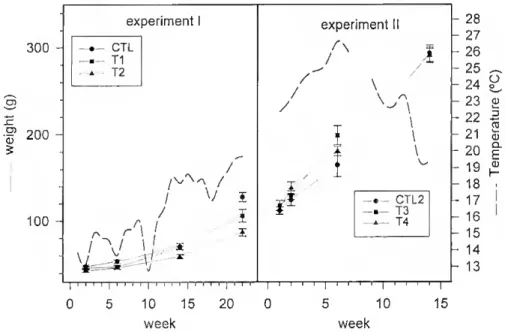
Experiment I (effecí of 17a -ethynilestradiol) - In a preliminary experiment, carried out between 23 June and 26 Seplember 1991 (temperatura range 22-280C), 25
mg.kg'1 diet of 17a-ethynilestradiol (ethE2), a powerful estrogen agonist, was
administered on alternate weeks to 150 g average weight sea bream. Treatment caused feminisation of the gonads (81.4 + 3.3% ovarian tissue, compared with 14.3 +4.1% in controls, n=ll) but it also caused a reduction in growth and food intake. In the actual experiment, to reduce the effect of ethE2 on appetite, fish were treated with ethE2 at a lower dosage and for different lengths of time. Forty fish, 40.7 + 0.30g (mean + SEM) were randomly placed in six tanks assigned to three duplicate experimental groups. Two of the groups were given a diet containing 15 mg.kg"1 ethE2 for 37 days (Tl) or 112 days
(T2). The third group (control; CTL1) was fed the same diet sprayed only with ethanol. Initially fish were treated daily with hormone but owing to their reduced appetite compared with controls, treatment with hormone was given every other day from day 24. Average weekly temperatures ranged between 12.80C in December 1992, when the
experiment started, to 19.70C when it finished 22 weeks later (May 1993; Fig. 2.1).
300 - I 200 - 100 - experiment CTL Tl T2 / / V í rs r \ \ i / \/ 1 í W « \ 1 / / experiment II J \ CTL2 T3 T4 | i i i i | i i i i | i i i i ; i ' i ? [ ? i 0 5 10 15 20 0 week 10 15 week 28 27 26 25 24 23 p 22 21 20 19 18 17 16 15 14 13 O D ro a3 o. E Q) H
Figure 2.1 - Changes in weight and temperature in control and estrogen-treated fish in the two experiments. In experiment I there were no slatistical differences in weight among groups until the 6'h
significant differences between lhe three groups. In experiment 2, there was significam growth from weeks 2 to 14, but no difference between groups (two-way analysis of variance at p<0.05 levei)
To foi lo w lhe changes induced by treatment, samples of general ly 12 fish per treatment were collected at weeks 2, 6, 14 and 22. Fish were anaesthetised with 2-phenoxyethanol, measured, weighed, bled, killed by decapitation and lhe gonads taken for histology and in vitro steroidogenesis.
Experiment 11 (effect of estradiol-17p) - To overcome lhe lack of appetite shown by fish treated with ethE2 in lhe preliminary and in experiment I, in experiment II il was decided instead to treat the fish with E2. To further improve appetite, cod liver oil (10 ml.kg"1) was mixed in the diet of ali groups. Twenty-seven fish from the same stock as
the fish used in experiment I, mean weight 92.7 + 0.98g, were randomly placed in six tanks assigned to three duplicate experimental groups. Two of the groups were treated with, respectively, 2 mg.kg'1 (T3) and 15 mg.kg"1 (T4) of E2 in the diet. The third group
acted as control and received the same diet sprayed only with ethanol (CTL2). The experiment lasted 14 weeks starting in June 1993 and finishing in October 1993. During this period average weekly temperatures ranged between 26.6 and 19.40C (Fig. 2.1).
The effect of treatment was followed by sampling 6 fish per treatment at 1, 2, 6 and 14 weeks (13 fish), as given above.
In vitro steroidogenesis - Fractions (5-10 mg) of gonads were incubated in vitro in Gostar 12 well plates (Gostar Corporation, Cambridge, USA) containing Iml trout balanced salt solution (Jalabert and Fostier, 1984), 5 LU. human chorionic gonadotrophin (hCG) to stimulate steroidogenesis and androstenedione (2 pg.mf1 in
experiment I and 0.5 pg.ml'1 in experiment II). No cofactors were used. Incubations
were carried out for 48h at room temperature (21 ± 20C) in a humid, oxygenated
atmosphere and under constant gentle agitation. Incubates were stored at -20oC for later
extraction and radioimmunoassay.
Steroid assays - Free, glucuronidated and sulphated fractions from blood plasma (100 pl) and in vitro incubates (1 ml) for radioimmunoassay were obtained using methodologies described by Scott and Canario (1992) and Canario and Scott (1989). Specificity tables for the E2, T and 11KT radioimmunoassays (RIAs) used in the present
study have been previously published (Scott et ai, 1984). The E2 RIA cross-reacted less than 0.1% with ethE2. The limit of detection of the three radioimmunoassays was 200 pg/ml (blood plasma) and 40 pg.well"1 (incubates). Intra-assay and inter-assay precision
(coefficient of variation) were, respectively, 7.5% and 12.4% for T, 8.2% and 11.6% for 11KT and 9.1% and 9.2% for E2.
Histology - Gonads were fixed in BouhVs fixative for 36h, dehydrated and embedded in paraffin wax. Thick transverse sections (6 pm) were stained with EhrliclVs hematoxylin and eosin. Clear camera drawing and a polar planimeter were used to quantify areas occupied by testicular and ovarian tissues. The gonads were further classified on the basis of stage of maturity of germinal cells and the area occupied by each maturity stage (see Table 2.1). The gonadosomatic (GSI) index was calculated according to the formula:
GSI (%) = gonadweight (§) xioo
fish total weight (g)
To verify if there were differences in the proportion of testicular or ovarian tissue in different parts of the gonads, cross-sections from the anterior quarter, middle and posterior quarter of three gonads of each group from experiment II (week 14) were analysed by repeated measures one-way analysis of variance. Since the regions were statistically similar (p=0.189 for testicular tissue and p=0.532 for ovarian tissue), a cross-section posterior to the middle region was used to calculate testicular and ovarian Índices as the product of GSI by the proportion of testicular or ovarian tissue. Parallel sections of tissue were used for the steroidogenic studies.
Statistics - Results are presented as mean ± SEM. The effect of treatment on growth, gonadal morphology and steroidogenesis was tested by two-way Analysis of Variance (ANOVA) followed by Tukey's Honestly Significant Difference test. Data was first log (weights and concentrations) or inverse sine (percentages) transformed before ANOVA. Plots in Figures are based on untransformed data. Statistical significance was considered at the 5% levei.
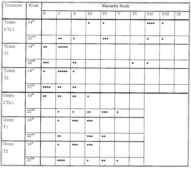
Table 2.1 - Maturity scale used for staging testicular and ovarian regions of sea bream Ireated with estrogen.
Stage | Testicle Ovary
0 Absence of, or a few scatlered, male germ cells Absence of, or a few scattered, female germ cells
I Spermatogonia are the only germ cells present (<30% testicular area)
Exclusive, or almost exclusive, presence of primary germ cell nests
11 Spermatogonia are the only germ cells present (30-60% testicular area)
Primary germ cell nests predominate in relation to oogonia and exclusion of any other female cell
III Spermatogonia are the only germ cells present (>60% testicular area)
Oogonia predominate in relation to oogonia mother cells nests and exclusion of any other female cell
IV Presence of spermatocytes 1 Pre-vitellogenic oocytes cover <50% ovarian area
V Presence of spermatocytes 11 Pre-vitellogenic oocytes cover 50-75% ovarian area
VI Presence of spermatids Pre-vitellogenic oocytes cover >75% ovarian area
VII Presence of spermatozoa
VIII Spermatozoan cysts present (>10% testicular area) but no sperm duct formed (vos deferem) IX Spermatozoan cysts of large dimensions and
sperm duct present.
II. 4 Rcsults
II. 4.1 Effect of hormone treatment on gonadal morphology
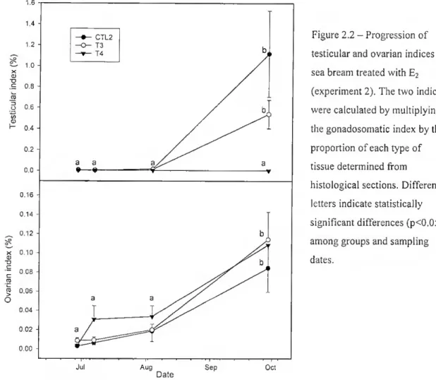
Growth and gonadosomalic index - During experiment I, ethE2-fed groups (Tl and T2) had reduced appetites and reduced growth (p<0.001) compared with control (CTL1; Fig. 2.1). When ethE2 was replaced by E2 (experiment II) a reduced appetite was not observed and growth was very similar among groups (Fig. 2.1). In experiment I the gonads were too small to obtain accurate GSIs. In experiment II, the initial GSI ranged
from 0.022 + 7.78x10° to 0.030 + 6.16x10'^ and it grew continuously throughout the experiment. However, the largest increase was in the control and T3 groups (p<0.05) aíter the 6lh week (1.01 + 0.019 and 0.77 + 0.013). The ovarian and testicular portions of
the gonad considered separately (Fig. 2.2), revealed that the testicular index was very significantly different between T4 and the other two groups at week 14 (pO.Ol). In T4
1.6 1.4 - 1,2 - 10 - CTL2 13 14 <u "O .E O.i d 0.6 - co d) ^ 04 - 0.2 - 0.0 - a a ^—*- 0.16 - 0 14 0.12 - x 0.10 0) - 0.08 - ? 0.06 - 0,04 0.02 - 0.00 -
Aug Sep Oct
Date
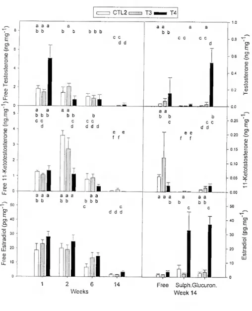
Figure 2.2 - Progression of testicular and ovarian Índices in sea bream treated with E2 (experiment 2). The two Índices were calculated by multiplying the gonadosomatic index by the proportion of each type of tissue determined from histological sections. Different letters indicate statistically significam differences (p<0.05) among groups and sampling dates.
it was essentially unchanged during the experiment. From the ó1'1 week there was an
increase in lhe testicular index in T3 and CTL2 reaching 0.54 + 0.14% and 1.12 + 0.41%, respectively, by the 14lh week. In contrast, the ovarian index increased steadily
in ali groups until week 6 but there was a sharp increase beyond this period. At week 14, treated groups had an ovarian index slightly higher (0.11% in T3 and T4) than CTL2 (0.08%).
Effect of 17cc-ethynilestrcidiol - Up to the 6th week the gonads in ali groups
contained essentially undifferentiated tissue (Fig. 2.3). By the 14th week control fish had
treated fish (p<0.05). Although there was a tendency for a slight decrease in testicular tissue in the control group between weeks 14 and 22, it was not significam (p>0.05). The proportion of testicular tissue in the treated groups was also virtually unchanged during this period as was lhe proportion of undifferentiated tissue in ali groups (p=0.93). Treated fish had a higher proportion of undifferentiated tissue than control fish at both weeks 14 and 22 (p=0.003). No differences between groups in the proportion of female tissue were found (p=0.384) but there was an average increase of 32% for ovarian tissue in control and treated groups between weeks 14 and 22 (p=0.009), a period when hormone treatment had already ceased. No differences in the proportion of any type of tissue were ever found between the two leveis of oestrogen treatment.
At week 14, 70% of control fish had spermatozoa but this proportion had decreased sharply by week 22. At this time most fish had only spermatogonia and spermatocytes I (Table 2.2). Greater development of testicular tissue was observed in the control group when compared with both treated groups at week 14 (p=0.0004). A significam difference with T2 was still evident at week 22 (p=0.035). Changes in testicular tissue were not so clear in Tl and T2. At week 14 most fish in Tl and T2 had a few spermatogonia covering less than 30% of testicular area. At week 22 less than 30% of the fish in Tl progressed to produce spermatids and spermatozoa, the remaining fish contained scarce spermatogonia or no visible male germ cells. In T2 none of the gonads progressed beyond spermatogonia.
In contrast to testicular tissue, ovarian tissue developed in a similar manner in ali groups of fish during the whole experimental period. At week 14 the ovary consisted mainly of primordial germ cell nests or oogonia scattered throughout the ovary. At the end of the experiment 40% of ali fish contained pre-vitellogenic oocytes in their ovarian tissue and no differences between groups were found (p=0.57). Only a thin layer of squamous follicular cells was present in pre-vitellogenic oocytes indicating that the folheies were not fully differentiated.
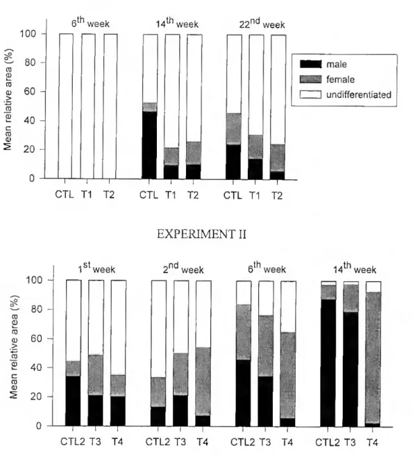
EXPERIMENT I 100 -1 — 80 ro ro d) 60 40 - 20 6th week CTL T1 T2 14th week 22ndweek ■■ mele i -y-i female ZH undifferentiated CTL T1 T2 CTL T1 T2 EXPERIMENT II 100
1st week 2nd week 6th week 14th week
80 60
20
CTL2 T3 T4 CTL2 T3 T4 CTL2 T3 T4 CTL2 T3 T4
Figure 2.3 - Changes in the proportion of testicular, ovarian and undifferentiated tissues in the gonads of sea bream. Top: experimenl 1 - fish were given a diet containing no estrogen (CTL1), 15 mg.kg'1 ethE2 for
37 (Tl) or 112 days (T2). Control had a higher % of testicular and smaller undifferentiated area than treated groups only at week 14 (p<0. 05). In contrast, treated groups had more undifferentiated tissue than control at weeks 14 and 22 (P=0. 003). Bottom: experiment II - fish were given a diet containing no estrogen (CTL2), 2 mg.kg"1 E2 (T3) or 15 mg.kg'1 E2 (T4). There were no statistical differences in area
occupied by ovarian or testicular tissue in CTL2 and T3 at any time during the experiment. T4 had a statistically significant lower proportion of testicular tissue than the other groups from week 6 and a higher proportion of ovarian tissue at week 14. See text for further details
Table 2.2 - Results from scoring maturity stages of testicular and ovarian tissue (for cri teria see Table 2.1) in sea bream treated (Tl and T2) and not treated (CTL1) with ethE2. Each dot (•) represents one fish.
Treatment Week Maturity Scale
0 I 11 111 IV V VI VII VIII IX Testes CTL1 14lh • • • ••• • 22'^ •• • • •• • • Testes Tl 14th •• 2 2 ^ ••• •• • • Testes T2 14th • • 2 2 "d •••• •• •• Ovary CTL1 14th •• •• •• • 22'»d • • •• • •• • Ovary Tl u» • ••• ••• •• ••• • • Ovary T2 14lh • ••• ••• 22^^ •••• • • • •
Effect of Estradiol-17p -At the start of the experiment most of the gonads were filled with largely undifferentiated tissue (>50%), the remainder being divided almost equally between testicular and ovarian tissue (Fig. 2.3). At the end of the experiment the gonads of fish receiving the highest hormone dosage (T4) consisted mainly of ovarian tissue. In contrast, the gonads of the control group (CTL2) contained mainly testicular tissue (Fig. 2.4). There were no statistically significam differences in area occupied by ovarian or testicular tissue in the control and the lower dose E2 groups. Significam differences were detected, however, in the proportion of testicular tissue between T4 and the other groups from week 6 of the experiment, and at week 14 for the proportion of
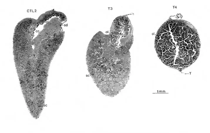
CTL2 o> 6 cc :>•: - S' 'mm ti ?. *r m "o T3 T4 ■(K :í'""4" * f: ^ m w e- ■v • ;; r " ■ - '' JrV 1 mm
Figure 2.4 - Histológica! cross sections (H & E ófim) of gonads ffom experiment II at week 14. CTL2; gonad from control fish showing a largely dominant mature testis filled with mainly spermatozoan cysts (sc) and a clearly formed sperm duct (sd). A small ovary (O) with ovarian lamella (ol) and central cavity (cc), is located dorsally. T3: gonad from a fish treated with 2 mg.kg'1 E2 showing less prominent testis
and ovary. T4: gonad of fish treated with 15 mg.kg 1 E2 showing ovary and regressed testis.
ovarian tissue. There were no statistically significant differences in the proportion of undifferentiated tissue between the three groups (p=0.202). After the 2nd week and until
the end of the experiment there was a progressive and significant reduction in undifferentiated tissue in ali groups (p<0.05). Cytological ohservations also confirmed significant inhibition in testicular development in T4 six weeks after the beginning of E2 treatment (p=0.004) and until the end of the experiment (p<0.001) (Table 2.3). Only one T4 fish had spermatocytes 1 at the first week and no stages beyond spermatogonia were observed after this period. In contrast, fish in CTL2 and T3 developed ali types of male germ cells and at the end of the experiment they had functional testes filled with spermatozoa. The ovarian region in ali groups showed a progressive change from a high proportion of oogonia and few pre-vitellogenic oocytes to a stage where pre-vitellogenic
Fable 2.3- Results from scoring maturity stages of testicular and ovarian tissue (for criteria see Table 2.1) in sea bream treated (T3 and T4) and not treated (CTL2) with E2. Each dot (•) represents one fish.
Treatment Week Maturity Scale
0 I
11
III IV V VI VII VIII
IX Tesles CTL2 1 •• • • • 2 •• •• 6 • • • • 14 Testes T3 1 • •• • • 2 •• • • 6 • • • • • • 14 • • ••• Testes T4 1 • • • • 2 ••• 6 •• •••• 14 •••••• •• Ovary CTL2 1 • •• • • 2 • • • • 6 • •• • 14 •••••• • •••• Ovary T3 1 • • • • • 2 • • • • 6 • • •• • • 14 ••• • • 1 • •• • Ovary 2 • • • T4 6 • • • • •• 14 • • •• • ••
oocytes covered ca 50% of the ovary (CTL2 and T2) and in some físh up to 75%. At week 14, 9 out of 13 fishes from T4 had more than 75% of ovarian section fílled with pre-vitellogenic oocytes. This was greater than that observed in T3 and CTL2 (P=0.004) (14; Table 2.3). There were no vitellogenic oocytes in any of the groups.
II.4.2 Steroid output by gonadal tissue
Effect of ethEi - Blood plasma steroid leveis were very low and in a large number ot samples below the detection limit of the radioimmunoassays (200 pg.ml'1). For this
reason they are not discussed.
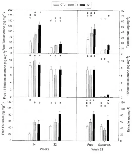
In viíro incubates of transverse sections of gonads with androstenedione at week 14 and 22, contained mainly free T and 11KT (Fig. 2.5). However, at week 22 significant leveis of glucuronides were also detected. No sulphates were detected in samples from either week 14 or 22.
1 he effect of ethE2 treatment on 11KT production was only statistically significant in the glucuronide fraction at week 22. Glucuronide in T2 incubates was significantly higher than either Tl or CTL1, which contained similar leveis. Production of free E2 was significantly higher in Tl and T2 incubates than in control incubates at week 14. T2 contained significantly more free T than control incubates at week 14. No significam differences between any of the steroids were present at week 22. Indeed by week 22 the relative abundance of free and glucuronide of T or E2 did not appear to be related to treatment, although, overall, more T and E2 were present in T2 incubates than CTL1 or Tl incubates (Fig. 2.5). In ali the fish studied more free E2 was produced (p<0.05) than glucuronide. The proportion of the E2 glucuronide fraction (45%) was higher than the glucuronides of androgens. Positive Pearson correlations (r>0.60, n=22) of total production (free + conjugated) between the three steroids were highly significam (P<0.002). None of the steroids produced was correlated to type of tissue.
Effect of E2 - Blood plasma steroid leveis were very low and were not considered to be meaningful. The production of ali three steroids in the incubates decreased during the experiment (PO.OOl; Fig. 2.6) and conjugates were very low with the exception of
CTL1 [=) Tl hh T2 200 v CJ) 175 fc Ó) 150 c 125 c 0 100 0 /5 to íl) h- 50 (1) £ 25 U CO fc O) 10 c tu c 0 tu CO 0 6 cu 0 4 ? * ~ tu u. 120 CO E 100 O) 80 0 "D ro 60 (O LU 40 CU tu LL 20 - 0 - a a b b c c c d d d I a a a a a a b b b b b 14 22 Weeks a a a b b b c c i b b a a a a b b b b c c c Free Glucuron. Week 22 80 70 60 50 40 30 20 10 0 10 o 120 100 E O) 80 % a. 60 "õ -o *0 £ LU 20
Figure 2.5 - Steroid output by gonadal tissue íragmenls incubated with androstenedione and measured by RIA in fish treated with ethE2. Different letters above bars represent statistically significam differences (p<0.05) among groups and sampling dates on the left column and statistically significam differences among groups and fonn of steroid on lhe right column. Results are expressed per wet weight of tissue. Note differences of scale.
samples from week 14, which was carried out in October, at the onset of the period of natural spawning. Until the 6th week, no consistent significam differences between
v; 3 o QJ i CTL2 T3 T4 a a a a b b b b b b b c c d d I1 1 ílfii a a a a b b b b b c c c d d b c c d d d I e e f f I a a a aaa aa b b b b b b b c d d d i r^i ri I 1 2 6 14 Weeks a a b b A a a b b c c c b d d e e f f -n õL aaa a a a b b b b c ( A h Free Sulph.Glucuron, Week 14 0,25 O) -o.io ^ o "S 0 05 id -20 2
Figure 2.6 - Steroid output by gonadal tissue fragments incubated with androstenedione and measured by RIA in fish treated with £2- Different letters above bars represent statistically significant differences (p<0.05) among groups and sampling dates on the lefl column and statistically significam differences among groups and forni of steroid on the right column. Results are expressed per wet weight of tissue. Note differences of scale.
At lhe end of the experiment more total T was produced by T4 than by T3 or CTL2 (p<0.05), the difference between the latter groups was not significant. Sulphated T was only a small fraction compared to both free and glucuronidated T (p<0.05). I here was a tendency for higher doses of E2 treatment to cause increased production of glucuronides. Similar amounts of T sulphate and glucuronide were produced by CTL2, the main product being free steroid. T sulphate was present at the lowest concentration
in samples trom T3 and there were no significant differences between the concentration ot glucuronide and free steroid. There were statistically significant differences in production ot T, its sulphate and glucuronide in T4. Glucuronide was present in the highest concentration followed by free and sulphate. Omission of the precursor (androstenedione) from gonadal incubates resulted in systematically low leveis of T (9.2 ± 1.7 pg.mg"1 ofin CTL2; 14.2 + 3.1 pg.mg 1 in T3 and 22.4 ± 4.9 pg.mg1 in T4).
The concentration of E2 administered to fish influenced the production of 11KT and its metabolites. Overall, more 11KT and its metabolites were produced by T3, with no significam differences between CTL2 and T4. More free 11KT was produced followed by glucuronide and sulphate with significam differences between the three forms. T3 produced more free 11KT than T4 and more sulphated hormone than CTL2. No significant differences in production of the two forms of 11KT were found between T4 and CTL2. Glucuronide production was significantly higher in T4 than in CTL2. In the absence of precursor, total 11KT production was very low (CTL2= 6.07 + 1.57 pg.mg'1, T3= 12.2 ± 4.59 pg.mg'1, T4=4.38 ± 0.98 pg/mg).
E2 production followed a similar pattern to T production, although at leveis 100 fold lower (Fig. 2.6). At week 14, more E2 was produced by T4 (p<0.05) and mainly as sulphates and glucuronides. Without precursor (androstenedione), production of total E2 was 1 fold lower (5.55 ±1.51 pg.mg"1 in CTE2, 2.65 ±1.16 pg.mg'1 in T3 and 8.08 ±
1.29 pg.mg"1 in T4). This contrasts with what was observed in androgen output, where
omission of precursor caused a 2-3 fold reduction.
At the end of lhe experiment highly significant negative correlations were found between the proportion of testicular tissue in the incubation and T (r<-0.65, pO.OOl, n=23) and E2 (r<-0.83, pO.OOl n=23). The same steroids also showed highly significant positive correlations (albeit slightly lower) with the proportion of ovarian tissue. 11KT output was not correlated to any type of tissue (r=0.04, p=0.843 with ovary and r=-0.36, p:=:0.871 with testis, n=23 for both correlations).
11.5 Discussion
The main effect of estrogen on sea bream gonads was inhibition of testicular growth and inhibition ot development of germ cells beyond spermatogonia, including mitosis. This effect was most likely, a result of direct action of estrogens on the testicular tissues. When the first experiment started, and up until at least 6 weeks, gonadal tissue was undifferentiated. By the 14111 week there was a signiflcantly lower
proportion of testicular tissue present in ali the treated groups, even that which received only 5 weeks treatment. While control fish had testes with germ cells at ali stages of spermatogenesis, in the treated groups spermatogenesis did not progress beyond spermatogonia. At the end of the experiment (6 weeks after T2 stopped receiving ethEa) there was a gradation in the proportion of testicular tissue which was inversely related to the amount of estrogen treatment. A clear inhibition of spermatogenesis by estrogen was still noticeable with only 2 individuais in Tl and none in T2 having progressed beyond spermatogonia. In the second experiment, which started with about half of the gonadal volume already differentiated, estrogen administration caused a clear reduction in testicular tissue, which was noticeable after 6 weeks in the high dose group (T4). Although at the start of the experiment spermatogonia filled most of the testis, as the experiment progressed they were progressively reduced until few remained. In contrast a progressivo development to spermiation was observed in control and low E2 dose groups.
The effect of estrogen on ovarian development was very slight and was apparent only after 14 weeks in the second experiment. This was noticeable histologically by the predominance of pre-vitellogenic oocytes in the ovary. Thus, in the present study, the effect of estrogen is to increase the proportion of ovarian tissue at the expense of testicular tissue and the result is referred to as feminisation. This definition contrasts with that proposed by other authors who have considered feminisation the presence of a functional ovary (producing gametes) or clear inhibition of spermiation (Reinboth, 1962; Reinboth, 1983; Happe and Zohar, 1988; Chang and Lin, 1998). Measurements of E2 and histological observations in the anemonefísh Amphiprion melanopus, also