(Linné, 1758) and astaxanthin quantity in each stage
Sühnel, S.
a*, Lagreze, F.
a, Bercht, M.
a, Ferreira,
JF.
a,
Carneiro-Schaefer, AL.
b, Magalhães, ARM.
band Maraschin, M.
caLaboratório de Moluscos Marinhos, Universidade Federal de Santa Catarina – UFSC, Servidão dos Coroas, s/n, Barra da Lagoa, CEP 88061-600, Florianópolis, SC, Brazil bNúcleo de Estudos em Patologia em Aquicultura, Universidade Federal de Santa Catarina – UFSC,
Rodovia Edmar Gonzaga, 1346, Itacorubi, CEP 88040-900, Florianópolis, SC, Brazil cLaboratório de Morfogênese e Bioquímica Vegetal, Universidade Federal de Santa Catarina – UFSC,
Rodovia Admar Gonzaga, 1346, Itacorubi, CEP 88040-900, Florianópolis, SC, Brazil, *e-mail [email protected]
Received July 25, 2008 – Accepted October 28, 2009 – Distributed August 31, 2010 (With 3 figures)
Abstract
This work describes the gametogenic cycle of the scallop Nodipecten nodosus kept in a culture system. To this end, during one year, samples were taken from the broodstocks every 30 days to be submitted to macroscopic and microscopic analyses and to measure the amount of astaxanthin. To perform the microscopic evaluation, 5 µ slices from the median portion of the female part of the gonad were submitted to the pattern methodology for histological analyses with paraffin and HE coloration. The remaining portion of the female gonad was lyophilised to extract and quantify the levels of astaxanthin using HPLC. The microscopic analyses revealed four well defined stages for the reproductive cycle. Analyses of data taken throughout the year indicated preferential spawning periods from December to January and from July to September. The astaxanthin analyses showed higher amounts of this carotenoid during the advanced pre-spawning and the initial spawning periods than during gametogenesis, initial pre-spawning, advanced spawning, and the spent stages. According to these results, it was possible to establish a descriptive table of the sexual stages of the female portion of the gonad and the amount of astaxanthin in the sexual stage of the scallop Nodipecten nodosus.
Keywords: reproductive cycle, astaxanthin, Nodipecten nodosus, scallop gonadic maturation.
Estágios sexuais da porção feminina da gônada da vieira
Nodipecten nodosus
(Linné, 1758) e a quantidade de astaxantina em cada estágio
Resumo
Este trabalho descreve o ciclo gametogênico da vieira Nodipecten nodosus mantida em ambiente de cultivo. Para isto, durante um ano, amostras de indivíduos reprodutores foram coletadas a cada 30 dias e submetidas à avaliação macroscópica e microscópica e à quantificação de astaxantina. Para a avaliação microscópica, secções de 5 µ da porção mediana feminina da gônada foram submetidas à metodologia de análise histológica padrão em parafina e coloração HE. O restante da porção feminina da gônada foi liofilizado para extração e quantificação de astaxantina em HPLC. A avaliação microscópica permitiu a descrição de quatro estágios bem definidos para o ciclo reprodutivo. Na análise ao longo do ano, foram observados períodos preferenciais de desova em dezembro e janeiro e de julho a setembro. A análise da quantidade de astaxantina, mostrou, nos estádios de pré-desova avançada e de desova inicial, uma maior quantidade desse carotenoide em comparação aos estádios de gametogênese, pré-desova inicial, desova avançada e repouso. Em função desses resultados, foi possível estabelecer um quadro descritivo dos estágios sexuais da porção feminina da gônada e quantidade de astaxantina em cada estágio sexual da vieira Nodipecten nodosus.
1. Introduction
The world production of pectens, a bivalve mollusks, in 2007, was 12,616,738 t, generating 11,740,962 dollars (FAO, 2009).
Along the Brazilian coast, the Pectinidae family is represented by 4 genera and 14 species (Rios, 2009). Of these 14 species, Nodipecten nodosus (Linné, 1758) stands out as a species of economic value in Brazil. According to Manzoni and Rupp (1993), N. nodoss has significant potential for culture in the natural environment due to fast growth and large adult size (Rupp et al., 2004; Rupp and Parsons, 2006). The high market price is another factor that is of interest for culture. Scallops have been offered not only in sophisticated Brazilian restaurants but also all around the world, including in haute cuisine. Almost all scallops consumed in Brazil come from abroad; this is the main reason why the study of its biology, reproductive cycle, culture and other factors are of great importance for its production.
In 2007, there were about 24 thousand units of scallops produced in Santa Catarina (EPAGRI, 2008). It thus represents a new source of income for the fishers who before, depended only on fishing. The cultivation of the native species of scallops, Nodipecten nodosus, represents an advantage for cuisine and tourism, for being very tasty.
The knowledge of the reproductive cycle of Nodipecten nodosus is fundamental for the production of scallop seeds in the laboratory.
The bivalve mollusc N. nodosus is a functional (simultaneous) hermaphrodite (Rupp, 1994; Lodeiros et al., 1997; Freitas, 2001). Adult animals in their natural environment spawn throughout the year, with periods of higher intensity concentrated in spring and summer around Arvoredo Island, Southern Brazil (Manzoni, 1994; Manzoni et al., 1996).
Research on N. nodosus spats settlements, using artificial collectors in the natural environment, has demonstrated that this species has a low rate of settlement, due to the low density and high dispersion of the populations (Ostini and Poli, 1990; Manzoni and Rupp, 1993; Manzoni et al., 1996). These data, combined with asynchronous spawning, suggest that the seed supply of this species for commercial production would be viable through hatchery production rather than extraction from its natural habitat.
One of the factors that affects seed production is the maturation stage of the female portion of the gonad to be used for spawning. This differentiation is therefore very relevant for commercial hatchery production. Lubet (1959), identified several different phases of the reproductive cycle of bivalve molluscs through the observation of histological sections of the gonad. Repeating this analysis for N. nodosus, we also identified different phases of gonad maturation, described later in this paper.
N. nodosus is characterised by one well defined gonad surrounded by tegumentum. The tegumentum is composed of an external epithelium, a basal lamina and connective tissue (sub-epithelial perigonadic tissue). The
gonad contains gonoducts, blood vessels, muscle fibre and part of the intestine. The white male portion of the gonad is proximate and the orange female portion is distal. The follicles have a bulb-like shape and are delineated by connective interstitial tissue that is thin when the follicles are filled with gametes and more evident and enlarged when the follicles are empty. The tubular gonoducts link the follicle to the external side and are formed by a simple prismatic epithelium. The gametes leave the paleal cavity through the urogenital pore (Román et al., 2001).
Sühnel et al. (2009) observed that the N. nodosus pectinid contains an unusually large amount of carotenoids in the female portion of the gonad. The present study quantifies the accumulation of astaxanthin in the female portion of the gonad of N. nodosus broodstock in each sexual stage, and provides a histological description of these different stages based on observation of individuals produced in the hatchery over the period of one year. The relation between astaxanthin levels and the reproductive stages is also evaluated.
2. Material and Methods
Nodipecten nodosus (Linné, 1758) scallop broodstock were collected from the EPAGRI (Empresa de Pesquisa Agropecuária e Extensão Rural/Rural Extension and Agronomic Research Company) experimental culture area, at Canto Grande beach near Bombinhas (27° 12’ 5.39” S and 48° 30’ 47.7” N) in the State of Santa Catarina, Southern Brazil. High Performance Liquid Chromatography (HPLC) for carotenoids analyses were carried out at the Laboratory of Plant Morphogenesis and Biochemistry/ UFSC (Universidade Federal de Santa Catarina), and histological analyses were carried out at the Aquaculture Pathology Studies Nucleus/UFSC.
2.1. Experimental design
For the description of the female gametogenic cycle 45 animals of 70 ± 5 mm in height, 1.5 years of age, were collected from the EPAGRI research area. The amount of astaxanthin was quantified in histological sections of the female part of the gonad.
To describe the reproductive cycle over the year, approximately 70 individuals of 70 ± 5 mm in height and 1.5 years old, from the same spawning group (30 adults), were kept in a lantern net in a long line at the experimental area of EPAGRI, and 4 individuals were collected each month for determination of maturation stage.
a central longitudinal cut and one section was used for the histological analysis. The other half, used for the astaxanthin analysis, was stored at –18 °C for 24 hours and then lyophilised (Terroni Favel LT 1000/8).
2.2. Histology
The sectioned gonadal tissues were fixed in a Davidson’s solution and stored in 70% Ethanol. After 6 Ethanol (from 70-100%) and Xhylol baths, they were embedded with paraffin.
The blocks containing the gonad samples were cut into 5 µ-thick slices and coloured with Harris’s Hematoxylin and Eosin.
The identification of the reproductive stages involved two independent evaluations with the aid of a microscope, carried out by two different persons, without previous identification of the slide, enlarged to 200 and 400×.
To define the reproductive stage, a score system was developed for each stage, with values from 1 to 6 (Table 1), which was compared using statistical analyses. The different reproductive stages were defined according to their histological characteristics.
The sea-water temperature was recorded between October 2006 and November 2007 in the long line where
the animals were kept using a TidBit (Optic StowAway-TEMP) installed at the culture area.
2.3. Astaxanthin analysis
Carotenoid extraction followed the methodology described in Sühnel et al. (2009). The lyophylised sample was placed in a ceramic container with liquid nitrogen, mixed and 50 mg were removed. To this sample, we added 5 mL of acetone extracting solution (Ac = O, Nuclear, P.A.) and n-hexane (Hex, TediaBrazil, P.A.) in a proportion of 1:3 respectively, and agitated it in a magnetic agitator Fisatom. Thus treated, the samples were kept at room temperature (23 °C) away from light during extraction. After extraction, the samples were filtered through filter paper, their final volume was adjusted to 3 mL, and they were then stored in amber flasks in an N2 atmosphere. The samples were stored in a freezer (–18 °C) for subsequent determination of astaxanthin levels.
The astaxanthin was measured using HPLC. The methodology used for the analysis is described in Sühnel et al. (2009), where 300 µL of the carotenoid extracts were filtered (0.22 µ filter), and 10 µL of each sample was injected in a liquid chromatograph (Shimzadu LC - 10A), equipped with a C18 column of reverse phase Vydac (BioRad, 25 cm × 4.6 mm
Table 1. Histological description of the female portion of the gonad stages in the reproductive cycle of the scallop Nodipecten nodosus.
Stage Stage
No.
No. of points
Microscopic description
Macroscopic description
Gametogenesis 1 1 Few oocytes; Different sizes of oocytes; Heterogeneous aspect; Presence of inter-follicle tissue; Non-juxtaposed inter-follicle walls; Thicker follicle wall (presence of internal oogonia); With empty inter-follicle and intra-follicle spaces.
Deflated gonad; Pale Orange coloration; Visible granules.
Initial pre-spawning
2A 2 Many follicles; Follicles full of gametes; Elongated oocytes;
Clear but recognisable follicle walls; Empty tube.
Swollen gonad; Bright orange coloration;
No granules present. Advanced
pre-spawning
2B 3 More oocytes per follicle than in 2A; Extremely long and elongated oocytes; Very juxtaposed and not clearly visible follicle walls; Many follicles; Empty tube; Slight stimulus or cut releases gametes; No empty inter-follicle or intra-follicle spaces. Initial
spawning
3A 4 Initial phase of gamete elimination; Visible and not completely juxtaposed follicle walls; Possible presence of gametes in the genital tubes; Intra-follicle and inter-follicle empty spaces.
Swollen gonad; Bright orange coloration; Granules present.
Advanced spawning
3B 5 Empty follicles present; Irregularly formed follicle due to recent spawning; Completely or partially empty follicle; Few remaining gametes.
Spent 4 6 Few follicles; Remaining follicles have small diameter; Abundant inter-follicle connective tissue.
∅) and UV-visible detector, operating with a reading of 470 ηm. A solution of acetonitrile and methanol (at a 90:10 proportion, TediaBrazil, HPLC degree) with a 0.8 mL/min flow was used as a mobile phase.
Astaxanthin was identified based on the period of retention obtained from the analysis of the standard sample (0.461 mg.10 mL–1 Hex, Sigma – MO, USA) under the same experimental conditions, and was measured by calculating the whole area of the corresponding peaks and applied to the external standard curve (y = 7044.96×, r2 = 0.994).
2.4. Statistical analyses
The descriptive analysis of the astaxanthin levels in the female portion of the gonad and the reproductive cycle (using the points in Table 1, for each reproductive stage) was performed. As samples were not homogeneous, the evaluation of normality of error and homogeneity of variance (Barttlet) was applied using a non-parametric Kruskal-Wallis analysis followed by the Mann-Whitney U-test separation of two-by-two averages. The reference consulted for the statistical analyses was Zar (1974).
3. Results
3.1. Description of reproductive stages
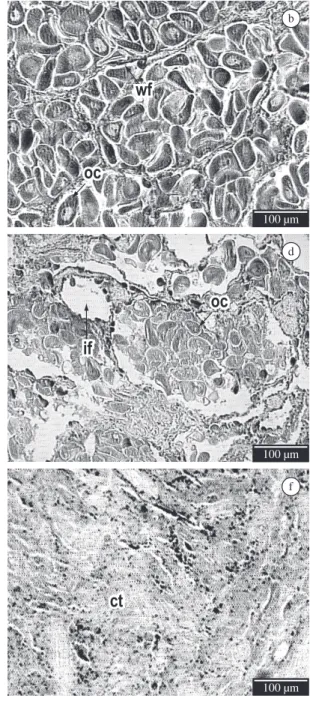
The description of morphological aspects of the female gonadal portion was based on the observation of: (a) the macroscopic aspects: coloration, granulation and appearance of the gonad; and (b) the microscopic aspects: the aspects of oocytes, follicle walls, inter and intra follicular spaces, tube (gonoduct), connective tissue and disintegration of the gametes. The reproductive stages were classified into four stages (Figure 1, Table 1): (i) Stage 1 - gametogenesis; (ii) Stage 2A - initial pre-spawning and Stage 2B - advanced pre-spawning; (iii) Stage 3A - initial spawning and Stage 3B - advanced spawning; and (iv) Stage 4 - spent.
Gametogenesis stage (Stage 1) is characterised by the presence of visible follicles with thick non-juxtaposed walls, containing few oocytes of different size and heterogeneous appearance. It is also possible to observe the oogonias in the luminal side of the follicle, the presence of inter-follicle connective tissue, and inter-follicle and intra-follicle empty spaces.
The initial pre-spawning stage (Stage 2A) is characterised by numerous follicles, which are filled with gametes, elongated oocytes, and clearly visible and recognisable follicle walls. The gonoducts have no oocytes (empty tube) and no inter-follicle and intra-follicle spaces are observed.
The stage of advanced pre-spawning (Stage 2B) is characterised by a much larger number of oocytes per follicle than in Stage 2A, extremely elongated oocytes, follicle walls that are very juxtaposed and hard to see, with many follicles. Gonoducts have no oocytes (empty tubes), and no empty intra-follicle and inter-follicle spaces are observed. As the gonad is filled with gametes, the medial cut applied for histological preparation stimulates their liberation.
The initial spawning stage (Stage 3A) is characterised as the initial phase of gamete liberation. The follicle walls are not completely juxtaposed, but still recognisable; there may be gametes in the genital tubes and intra-follicle and inter-follicle empty spaces can be observed.
The advanced spawning stage (Stage 3B) is characterised by empty and irregularly-shaped follicles, as a result of the recent spawning. At this stage, the follicles may be completely or partially empty, with few visible remaining oocytes.
The spent stage (Stage 4) is characterised by just a few remaining follicles, which are small in diameter and have abundant inter-follicle connective tissue.
3.2. Variation of the reproductive stages in the culture tank
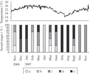
The histological analysis of gonads from animals collected between November 2006 and November 2007 indicated two major spawning periods from December 2006 to January 2007, and from July to September 2007 in Canto Grande (South of Brazil). During the remaining months, the analysed animals were predominantly in the pre-spawning stage (Figure 2).
Seawater temperature between December 2006 and January 2007 varied from 22.64 ± 0.77 °C to 24.75 ± 0.97 °C, respectively; and in July, August and September 2007 average temperatures were 15.84 ± 1.40 °C, 16.47 ± 1.09 °C and 19.65 ± 1.37 °C, respectively (Figure 2).
3.3. Astaxanthin quantity at each reproductive stage To evaluate the carotenoid quantity in each reproductive stage (Figure3) the homogeneity of the samples was verified. A Mann-Whitney U-Test comparing pairs of samples was then carried out to identify which stages differed significantly from others. Results indicated that astaxanthin levels in the advanced pre-spawning Stages 2B (45.06 ± 6.35 µg.mL–1) and initial spawning 3A (38.01 ± 7.16 µg.mL–1) have no significant differences, but when gametogenesis 1 (8.31 ± 0.41 µg.mL–1), initial pre-spawning 2A (17.71 ± 4.87 µg.mL–1), advanced spawning 3B (17.12 ± 0.85 µg.mL–1), and spent 4 (6.76 ± 0.91 µg.mL–1) are compared a significant difference is observed (p < 0.05). The gametogenesis 1 and spent 4 stages are significantly different (p < 0.05) both from each other and from the other stages (p < 0.05). The stages of initial pre-spawning 2A and advanced spawning 3B showed no significant differences.
4. Discussion
Figure 1. Histological sections of Nodipecten nodosus female gonad showing the different stages of the reproductive cycle: (a) gametogenesis; (b) initial pre-spawning; (c) advanced pre-spawning; (d) initial spawning; (e) advanced spawning; (f) spent. ct: connective tissue; oo: oogonia; wf: wall follicles; oc: oocytes; if: intra-follicle space; it: inter-follicular space (few); rg: remaining gametes. The bar represents 100 micra.
Notwithstanding these methods, the histological method (microscopic evaluation) is the most accurate; it is a method that unequivocally defines the reproductive phase of the animal (Barber and Blake, 2006).
Sastry (1963) described four reproductive stages for Argopecten ventricosus: (1) immature; (2) partially mature; (3) mature; and (4) spent. Villalejo-Fuerte and Ochoa (1993) and Benninger and LePennec (1991) described the following stages for this same species: (1) initial
gametogenesis; (2) advanced gametogenesis; (3) mature; (4) spawning; and (5) spent.
Figure 2. Temperature of the sea water during the experiment and the maturation stages of the animals (N. nodosus)
determined monthly (n = 4). Stages: (a) gametogenesis; (b) initial pre-spawning; 09(c) advanced pre-spawning; (d) initial spawning; (e) advanced spawning.
Figure 3. Astaxanthin quantity in the female portion of the scallop N.nodosus gonad in the four stages of the
reproductive cycle: (a) gametogenesis; (b) initial pre-spawning; (c) advanced pre-pre-spawning; (d) initial pre-spawning; (e) advanced spawning; (f) spent.
(2) differentiation; (3) recovery; (4) swelling; (5) initial maturation; (6) advanced maturation; and (7) partial or total spawning.
According to Penchaszadeh et al. (2000), the pectinid Amusium laurenti has four sexual stages, these being: (1) follicle development; (2) full follicle; (3) partial spawning; and (4) total spawning. This author does not describe the stage of spent for this species.
According to Freitas (2001) the reproductive cycle of the N. nodosus scallop has three distinct phases with the following characteristics: (1) spent, at this stage it is not possible to distinguish the male or female portions of the gonad; (2) follicle organisation and sex cell multiplication;
(3) reproductive period, which includes the gonad maturation, gamete elimination and gonad restoration.
Individual N. nodosus scallops with 50 mm in height (approximately 6-7 months after hatching) are already sexually mature and able to reproduce (Freitas, 2001). For this phase, (phase 3 - reproductive period) our work identified four distinct reproductive stages, where the 2nd and 3rd stages were subdivided into A and B.
The four reproductive stages presented in this work were adopted because they are easy to recognise, even when we consider the dynamic aspects of the reproductive cycle of Nodipecten nodosus.
Differentiation between the four stages described in this study was based on microscopic evaluation of each stage. This division into stages is particularly useful in the process of captive breeding in nurseries, as it facilitates the identification of the favourable reproductive stages of broodstock to be used for seed production. In captivity, after N. nodosus are collected from the sea, the animals go through a period of maturation when they are kept in tanks and fed daily. The quality of this feed is important for the success of maturation and later production of viable larvae.
In our macroscopic evaluation and description of N. nodosus, gonads that are not significantly swollen, a pale orange colour, and visible granules characterise the stage of gametogenesis. No macroscopic differentiation was observed for the stages of initial pre-spawning (Stage 2A) and advanced pre-spawning (Stage 2B); both stages presented swollen gonads, bright orange coloration and no granules. Differences between these two sub-stages could only be detected under the microscope. The stages of initial spawning (Stage 3A) and advanced spawning (Stage 3B) also showed no macroscopic difference, both showed apparently swollen gonads, bright orange coloration and granules. For the spent stage (Stage 4) the gonad was not swollen, and presented an opaque, very pale orange colour without granules.
The spent stage (Stage 4) described in this study was also noted by Manzoni et al.(1996) for N. nodosus over the winter; however, it was not commented on by Freitas (2001) in a study of the same species. Luna-González (1997) and Luna-González et al. (2000) observed that the spent stage for Argopecten ventricosus in Mexico occurred during the month of July.
In some bivalves, during the spent stage the connective tissue surrounds the follicles, and spreads and expands according to the level and degree of gamete maturation (Lunetta, 1969; Calvo, et al. 1998). This is why connective tissue fills the empty inter-follicle spaces in the spent stage when there are only a few leftover gametes.
during the winter (12.0 to 12.7 °C) and spring, when the temperature reaches 16 °C.
According to Manzoni et al. (1996), N. nodosus of the Arvoredo Island show partial and asynchronous spawning throughout the year. However, this does not automatically imply that the animals are spawning viable gametes, rather that they may be simply expelling gametes due to environmental stress factors, such as temperature. The same authors observed that the period between November and December presented the highest gonadosomatic index, and this is associated with high water temperatures.
In the present study, the histological evaluation of the maturation stage throughout the year in a cultivation environment, two distinct spawning seasons were observed, one in the summer (December and January) and another at the end of winter and the beginning of spring (July to September). These winter and spring spawning peaks with lower seawater temperatures were also observed by Manzoni and Banwart (2000).
To evaluate the relation of carotenoid quantity with reproductive stage, the levels of the carotenoid astaxanthin were measured in the female part of the gonad. This analysis indicated that during the advanced pre-spawning and initial spawning stages there are higher levels of astaxanthin in the female portion of the gonad than in gametogenesis, initial pre-spawning, advanced spawning, and spent the stages.
The highest carotenoids concentration at the advanced pre-spawning (Stage 2B) suggests that carotenoids are accumulated in the oocytes and not in the interfollicular tissue. This is because the spent stage (Stage 4), which has a large amount of connective tissue (inter-follicle tissue) and few remaining oocytes, is the smallest amount of carotenoids in the stages of the reproductive cycle for this species.
According to Farías-Molina (2001), carotenoids are required for gonad development and gamete maturation in invertebrates.For other species of aquatic animals, like shrimp (Lorenz, 1998; Howell and Matthews, 1991; Cuzon et al., 2004) and fish (Meyers, 1994), the carotenoids showed a positive effect on maturation and reproduction.
The higher astaxanthin quantity during the advanced pre-spawning and initial spawning stages suggests that carotenoid accumulation is correlated to oocyte maturation. However, further studies are needed to evaluate the effect of carotenoids on reproduction and survival of molluscs larvae.
As the bivalve molluscs are unable to synthesise carotenoids (Johnson and Schroeder, 1995; Farías-Molina, 2001), our study suggests that their absorption through a diet rich in phytoplankton carotenoids is important for good reproductive results.
The reserves for the early embryonic development of the bivalve molluscs are found in the oocyte cytoplasm. The eggs of molluscs are small (about 50 µm, according to Mackie, 1984), with little calf, allowing a total cleavage, type holoblastic spiral. However, the calf needs to ensure the development of the embryo, without an external food
source, to form a larvae that swims and feeds. Carotenoids also compose this vitelline reserve of molluscs eggs.
In mussels Perna perna (Linné, 1758), according to Lunetta (1969), a salmon or orange-yellow colour in the oocytes is related to a lipid inclusion. On the other hand, in N. nodosus this colour can be related to both the lipid influence and presence of carotenoids in the oocytes, other evidence of this relationship is the fat-soluble carotenoids property.
As demonstrated by the results of our work, it is possible to establish a very close relation between the stages of the reproductive cycle and the astaxanthin content in the female portion of the gonad during the maturation and spawning processes of N. nodosus. This study also observed, for the captive environment, similar periods for reproductive peaks as those observed in the natural environment.
Acknowledgements — We are thankful to FINEP, FAPESC and CNPq for financial support and to CAPES/UFSC for a PhD scholarship.
References
BARBER, BJ. and BLAKE NJ., 1991. Reproductive physiology. In SHUMWAY, SE. (Ed.). Scallop: biology, ecology and aquaculture.
Amsterdam: Elsevier.
BARBER, BJ. and BLAKE, NJ., 2006. Reproductive physiology. In SHUMWAY, SE. and PARSONS, GJ. (Eds.). Scallops: biology,
ecology and aquaculture. 2 ed. Amsterdam: Elsevier. p. 357-415. (vol. 35)
BENNINGER, PG. and LEPENNEC, M., 1991. Functional anatomy of scallops. In SHUMWAY, SE. (Ed.). Scallop: biology, ecology
and aquaculture. Amsterdam: Elsevier. p. 133-223.
CALVO, J., MORRICONI, E. and ORLER, PM., 1998. Estratégias reproductivas de moluscos bibalves y equinoideos. El Mar Argentino y sus Recursos Pesqueros, vol. 2, no. 2, p. 195-231.
CUZON, G., LAWRENCE, A., GAXIOLA, G., ROSAS, C. and GUILLAUME, J., 2004. Nutrition of Litopenaeus vannamei
reared in tanks or in ponds. Aquaculture, vol. 235, no. 1-4, p. 513-551.
Empresa de Pesquisa Agropecuária e Extensão Rural de Santa Catarina – EPAGRI, 2008. Cultivo da vieira Nodipecten nodosus em Santa Catarina: influência da profundidade, densidade e frequência de limpeza. Florianópolis. Boletim Técnico, 135. Available from: <http://www.epagri.rct-sc.br/>. Access in: 08/10/2009.
Food and Agriculture Organization – FAO, 2009. Fishery and aquaculture statistics. Rome. Available from: <http://www.gfcm.
org/figis/servlet/SQServlet?file=/usr/local/tomcat/FI/5.5.23/figis/ webapps/figis/temp/hqp_3713.xml&outtype=html>. Access in: 08/10/2009.
FARÍAS-MOLINA, A., 2001. Nutrición en moluscos Pectínidos. In MAEDA-MARTINZ, AN. (Ed.). Los Moluscos Pectínidos de Iberoamérica: ciencia y acuicultura. México: Editora Limusa.
p. 89-104.
MANZONI, GC. and BANWART, JPF., 2000. Aspectos da biología reprodutiva da vieira Nodipecten nodosus, cultivada na enseada da
Armação do Itapocoroy (26046’ S – 480 37’ W) (Penha – SC). In
Anais da XIII Semana Nacional da Oceanografia. Itajaí: Editora da Univalli. p. 537-539.
MEYERS, PS., 1994. Developments in World aquaculture, feed formulation and role of carotenoids. Pure and Applies Chemistry,
vol. 66, no. 5, p. 1069-1076.
OSTINI, S. and POLI, CR., 1990. A situação do cultivo de moluscos no Brasil. In HERNANDEZ, RA. (Ed.). Cultivo de moluscos en América Latina. Bogotá: Ed. Guadalupe. p. 311-325.
PAZOS, AJ., RÓMAN, G., ACOSTA, CP., ABAD, M. and SÁNCHEZ, JL., 1996. Stereological studies on hte gametogenic cycle of the scallop, Pecten maximus, in suspended culture in Ria de Arousa (Galicia, NW Spain). Aquaculture, vol. 142, no. 1-2,
p. 119-135.
PENCHASZADEH, PE., PAREDES, C. and SALAYA, JJ., 2000. Reproductive cycle of the south American scallop Amusium laurenti
(Gmelin, 1791) (Bivalvia, Pectinidae). Aquaculture International,
vol. 8, no. 2-3, p. 227-235.
RIOS, EC., 2009. Compendium of brazilian sea shells. Rio
Grande: Evangraf. 668 p.
ROMÁN, G., MARTÍNEZ, G., GARCÍA, O. and FREITES, L., 2001. Reproducción. In MAEDA-MARTINZ, AN. (Ed.). Los Moluscos Pectinídeos de Iberoamérica: ciência y acuicultura.
México: Editora Limusa. p. 27-59.
RUPP, GS., 1994. Obtenção de reprodutores, indução à desova e cultivo larval e pós-larval de Nodipecten nodosus (Linnaeus, 1758) (Bivalvia: Pectinidae). Florianópolis: Universidade Federal de Santa Catarina. 125 p. [Dissertação de Mestrado]
RUPP, GS., THOMPSON, RJ., PARSONS GJ. and BEM, MM., 2004. Influence of food supply on postmetamorphic growth and survival of hatchery-produced lion’s paw scallop, Nodipecten nodosus (Linnaeus, 1758). Journal of Shellfish Research, vol.
23, no. 1, p. 5-13.
RUPP, GS. and PARSONS, GJ., 2006. Scallop aquaculture and fisheries in Brazil. In SHUMWAY, SE. and PARSONS, GJ. (Eds.). Scallops: biology, ecology and aquaculture. Amsterdam:
Elsevier.
SASTRY, AN., 1975. Physiology and ecology of reproduction in marine invertebrates. In VERMBERG, FJ. (Ed.). Physiological ecology of estuarine organisms. Columbia: University of South Carolina Press. p. 279-299.
SÜHNEL, S., LAGREZE, F., FERREIRA, JF., CAMPESTRINI, LH. and MARASCHIN, M., 2009. Carotenoid extraction from the gonad of the scallop Nodipecten nodosus (Bivalvia: Pectinidae).
Brazilian Journal Biology, vol. 69, no. 1, p. 209-215.
VILLALEJO-FUERTE, M. and OCHOA, RI., 1993. The reproductive cycle of the scallop Argopecten circularis (Sowerby, 1835) in
relation to temperature and photoperiod, in Bahia Concepcion, B.C.S. Mexico. Ciencias Marinas, vol. 19, no. 2, p. 181-202.
ZAR, JH., 1974. Biostatistical analyses. Englewood Cliffs: Prentice-Hall. 357 p.
JOHNSON, EA. and SCHROEDER, WA., 1995. Microbial carotenoids. In FIECHTER, A. (Ed.). Advances Biochemical Engineering and Biotechnology. Berlin: Springer-Verlag.
p. 119-178.
HOWELL, BK. and MATTHEWS, AD., 1991. The carotenoids of wild and blue disease affected farmed tiger shrimp (Penaeus monodon fabricus). Comparative Biochemistry and Physiology,
vol. 98B, no. 2-3, p. 375-379.
LODEIROS, CJ., RENGEL, JJ., FREITES, L., MORALES, F. and HIMMELMAN, JH., 1997. Growth and survival of the tropical scallop Lyropecten (Nodipecten) nodosus maintained
in suspended culture at three depths. Aquaculture, vol. 165, no. 1-2, p. 41-50.
LORENZ, TA., 1998. Review of the Carotenoid, Astaxanthin, as a Pigment and Vitamin Source for Cultured Penaeus Prawn.
Kailua-Kona (Hawaii): NatuRose. Available from: <www. cyanotech.com>. Access in: 01/05/2008.
LUBET, P., 1959. Recherches sur le cycle sexuel et l’émission des gamètes chez les mytilides et les pectinides (Mollusques Bivalves). Revue des travaux de l’Institut des peches maritimes, vol. 23, no. 4, p. 387-548.
LUNA-GONZALEZ, A., 1997. Ciclo reproductivo de la almeja catarina Argopecten ventricosus (Sowerby II, 1842), cultivada en la rada del puerto de Pichilingue, B.C.S. u su relación con el medio. México: Universidad Autónoma de Baja California Sur.
74 p. [Dissertação de Mestrado]
LUNA-GONZALES, A., CÁCERES-MARTÍNEZ, C., ZÚÑIGA-PACHECO, C., LÓPEZ-LÓPEZ and CEBALLO-VÁZQUEZ, P., 2000. Reproductive cycle of Argopecten ventricosus (Sowerby,
1842) (Bivalvia: Pectinidae) in the Rada del Puerto de Pichiligüe, B.C.S., México and its relation to temperature, salinity and food.
Journal of Shellfish Research, vol. 19, no. 1, p. 107-112. LUNETTA, JE., 1969. Fisiologia da reprodução de mexilhões
(Mytilus pernaL. Mollusca Lamellibranchia). Boletim de Zoologia e Biologia Marinha, vol. 26, n. 324, p. 33-111.
MACKIE, GL., 1984. Bivalves. In TOMPA, AS., VERDONK, NH. and BIGGELAAR, JAM. (Eds.). The Mollusca. Orlando: Academic Press. p. 351-418. (vol. 7).
MANZONI, GC. and RUPP, GS., 1993. Estudo da biologia reprodutiva e viabilidade de cultivo de Lyropecten nodosus (Linnaeus, 1758) (Mollusca: Pectinidae) na Ilha do Arvoredo – SC. Florianópolis: Universidade Federal de Santa Catarina. 35 p
(Relatório final).
MANZONI, GC., 1994. Aspectos da biologia de Nodipecten nodosus, (Linnaeus, 1758) (Mollusca: Bivalvia), nos arredores da ilha do Arvoredo (Santa Catarina – Brasil), com vistas a sua utilização na aqüicultura. Florianópolis: Universidade Federal de Santa Catarina. 98 p. [Dissertação de Mestrado]
MANZONI, GC., POLI, CR. and RUPP, GS., 1996. Período reproductivo del pectínido Nodipecten nodosus (Mollusca:
Bivalvia) en los alrededores de la Isla Arvoredo (27o 17’S – 48o