2019
UNIVERSIDADE DE LISBOA
FACULDADE DE CIÊNCIAS
DEPARTAMENTO DE BIOLOGIA VEGETAL
Development of novel bacterial enzymes for medical diagnosis
through directed evolution
André Filipe Teixeira Taborda
Mestrado em Microbiologia Aplicada
Dissertação orientada por:
Prof. Lígia O. Martins
This Dissertation was fully performed at Instituto de
Tecnologia Química e Biológica – ITQB Universidade Nova
de Lisboa under the direct supervision of Professor Lígia O.
Martins
Professor Ana Tenreiro was the internal supervisor designated in the scope of the Master
in Applied Microbiology of the Faculty of Sciences of the University of Lisbon
I
Acknowledgments
Na última etapa deste ciclo, que culmina com a entrega desta dissertação de tese de mestrado, queria nomear e sobretudo agradecer a todos os que de forma direta ou indireta contribuíram para a sua realização.
Em primeiro lugar gostaria de agradecer ao laboratório Microbial and Enzyme technology (ITQB) a disponibilidade em me terem aceite assim como os recursos que me forneceram para que pudesse realizar todo o trabalho laboratorial e ainda a possibilidade de estar presente nalguns seminários e conferências que ajudaram a complementar a minha formação.
A todos os investigadores e alunos do laboratório assim como do gabinete em que me encontrava o meu obrigado pela troca de ideias e desabafos sempre que necessário.
Em particular, o meu grande agradecimento à professora Lígia Martins, orientadora deste trabalho, por ser das pessoas mais focadas, dinâmicas, dedicadas e trabalhadoras que já conheci, tentando estar presente em todas as decisões experimentais e motivando sempre que os resultados não foram tão animadores. É, indubitavelmente, um exemplo a seguir. Pode ter a certeza que contribuiu para o meu percurso como investigador/cientista.
À Doutora Vânia Brissos, por todos os conselhos fornecidos durante o decorrer do trabalho experimental que foram essenciais para o seu sucesso.
À professora Teresa Catarino pela disponibilidade e conhecimento que foram importantes na obtenção das cinéticas por Stopped-flow.
À Mariana Lima, aluna de licenciatura que frequentou o laboratório no âmbito do seu projeto de curso ajudando na otimização da produção em larga escala assim como na obtenção de alguns dados de Stopped-flow.
Às técnicas do ITQB Teresa Silva e Isabel Pacheco pela disponibilidade, ajuda e formação na execução de alguns procedimentos experimentais.
E finalmente a toda a minha família e amigos, evidenciando especialmente os meus pais, pela ajuda que demonstraram ao longo de toda a minha formação, apoiando e motivando sempre.
II
Abstract
Pyranose 2-oxidases (P2Oxs) have a great potential to replace the typical glucose oxidases (GOxs) and glucose dehydrogenases (GDHs), specific for β-D-glucose anomer, in glycemia monitoring biosensors. P2Oxs are flavoenzymes mostly identified in Fungi that catalyzes the regioselective oxidation of C2 alcohol moiety of several aldopyranoses originating the correspondent keto-sugar with the concomitant reduction of O2 to H2O2. The use of O2 as cheap and clean oxidant and in particular the lack of D-glucose
anomer preference represent very attractive points for the biotechnological application of P2Oxs. In this work, directed evolution (DE) methodologies were applied to improve the first identified bacterial P2Ox, from Pseudoarthrobacter siccitolerans, AsP2Ox, in its specificity and activity for D-glucose. This strategy was supported by transient-state analysis in combination with oxygen consumption steady-state kinetics showing that the reductive half-reaction (oxidation of D-glucose) is the limiting step of the catalytic mechanism. We first optimized and validated colorimetric screening enzymatic assays based on ‘activity-on-plate’ and 96-well plates using cell crude extracts. These screenings allowed analyze in a high-throughput mode, thousands of variants generated by error-prone PCR. The hit variant from the first-generation 1A1 harbors only one mutation, G366S, located close to the substrate binding site. Biochemical and kinetic analysis using purified enzyme showed that 1A1 has a 2-fold increased kcat
(turnover number) and 2-fold higher protein production yields as compared with the wild type enzyme. In a second round of directed evolution a new hit variant 5D5 was selected, carrying four additional mutations (S22S, A75T, A206T, Q295H). The pH profile of 5D5 revealed an optimum pH shifted 1 unit towards the alkaline range, a 6-fold higher kcat and 3-fold production yields than the wild-type enzyme.
The analysis of mutations using site-directed mutagenesis showed that both G366S and Q295H are key mutations, which under an epistatic effect contributed to the higher catalytic efficiency exhibited by 5D5 hit variant. The comparison of kinetic parameters of 5D5 variant with GOxs and GDHs show that its Km remains too high and the kcat too low in order to replace the traditional enzymes in biosensors.
Therefore, the evolution of AsP2Ox must continue using 5D5 as the parent in new rounds of DE to achieve an improved variant exhibiting the properties that fit the desired application.
key-words: Biosensors, Flavoproteins, Pyranose 2-oxidase, Laboratory enzyme evolution,
III
Resumo
Piranoses 2-oxidases (P2Oxs) são enzimas que oxidam açúcares, nomeadamente D-glucose, no carbono C2, o que representa uma vantagem na substituição das atuais enzimas usadas em biossensores para monitorização da glicemia, as glucose oxidases (GOxs) e glucose desidrogenases (GDHs), que apenas oxidam no carbono C1, o anómero β-D-glucose. Como consequência, o uso das P2Oxs em biossensores poderá conduzir a uma medição mais sensível dos níveis de D-glucose no sangue.
As P2Oxs são flavoenzimas que foram identificadas maioritariamente em fungos com funções relacionadas com a degradação da biomassa vegetal. Estas enzimas catalisam a oxidação seletiva do grupo álcool do carbono C2 de diversas aldopiranoses originando as respetivas ceto-piranoses. Como aceitador de eletrões, estas enzimas podem utilizar um vasto repertório de moléculas (como quinonas e radicais) mas a sua grande vantagem reside na utilização de oxigénio molecular, que é um oxidante barato e “limpo”, que é reduzido a peróxido de hidrogénio durante o seu ciclo catalítico. Este divide-se em duas semirreações, uma redutiva, relacionada com a redução do cofator FAD a FADH2 em
consequência da oxidação do carbono C2 dos açúcares libertando como produto, o respetivo ceto-açúcar, e uma oxidativa, associada à reoxidação do FADH2 a FAD, na presença de um aceitador de
eletrões, como o dioxigénio.
Atualmente, para além das P2Oxs fúngicas, três P2Oxs de origem bacteriana foram caracterizadas entre as quais se destaca a AsP2Ox, da bactéria Pseudoarthrobacter siccitolerans por ter sido a primeira e cuja identificação foi realizada no laboratório de Tecnologia Microbiana e Enzimática do ITQB NOVA. A caracterização desta enzima revelou, no entanto, uma baixa afinidade para a D-glucose. Assim, este trabalho de dissertação teve como principal objetivo a aplicação de técnicas de evolução dirigida (DE) para melhorar a enzima AsP2Ox de modo a aumentar a sua especificidade e atividade para o uso da D-glucose como substrato de modo a que possa ser empregue em biossensores de monitorização da glicemia. Esta estratégia de engenharia foi eleita, uma vez que a ausência de uma estrutura cristalina da enzima ou de um modelo estrutural fidedigno, não é possível fazer previsões de melhoramento racional baseado em relações entre a estrutura e a função.
A engenharia de enzimas por Evolução Dirigida requer a otimização de um número de parâmetros desde o crescimento das estirpes recombinantes, métodos de rutura celular até ao rastreio de atividade enzimática. Neste trabalho desenvolveu-se, otimizou-se e validou-se duas metodologias para rastreios em larga escala (high-throughput screenings) que possibilitaram a análise de alguns milhares de variantes gerados por error-prone PCR nas duas gerações de evolução dirigidas efetuadas. Uma das metodologias utilizou como alvo colónias de células em placa de Petri (‘activity-on-plate’) e outra, extratos celulares brutos, após crescimento das colónias em meio líquido em placas com 96 poços. Uma das otimizações realizadas foi no método de deteção da atividade enzimática da AsP2Ox. Sendo o oxigénio molecular o aceitador final de eletrões pretendido, a atividade da enzima é medida através de uma reação acoplada utilizando uma peroxidase (e.g. Horseradish peroxidase, HRP), que consume o peróxido de hidrogénio formado pela AsP2Ox e oxida um substrato que origina um produto corado, cuja formação possa ser facilmente monitorizada num espectrofotómetro. Provou-se que o ABTS, substrato da HRP mais comumente utilizado neste tipo de reações acopladas, levava a medições subestimadas de atividade da AsP2Ox. Por conseguinte, testou-se um novo sistema de substratos baseado em dois compostos (AAP e DCHBS) que, por atividade da HRP e na presença de H2O2 origina
um composto cor-de-rosa, doseado espetrofotometricamente a 515 nm que se revelou um método mais adequado para seguir a atividade da AsP2Ox.
As biblioteca de variantes geradas por “error-prone PCR” foram rastreadas num primeiro passo através da metodologia ‘activity-on-plate’, que sendo qualitativa, permitiu identificar clones com atividade e diminuir o número de variantes a serem avaliados com maior rigor. O segundo screening, em placas de
IV 96 poços, com carácter quantitativo, necessitou de ser exaustivamente otimizado de modo a diminuir o risco de se selecionar falsos positivos. Assim, observou-se que o uso da estirpe KRX de E. coli como estirpe de expressão da proteína de interesse, em placas de 96 poços de poços fundos e consequente disrupção dos pellets celulares recorrendo a lisozima era a combinação que conduzia a um coeficiente de variação (CV) para a atividade total da AsP2Ox menor, sendo por isso, aplicado na avaliação dos variantes selecionados por ‘Activity-on-plate’.
Na primeira geração de DE, dos 7300 variantes analisados em ‘Activity-on-plate’, cerca de 100 foram inoculadas em placas de 96 poços de poços fundos e sujeitos a screenings, o que permitiu a identificação de um variante, 1A1, que continha uma mutação na posição 366 (glicina para serina), a cerca de 10 Å do N5 do cofator FAD e cuja atividade relativa ao wild-type era de 3.7 ± 0.9 (em extratos celulares brutos). Posteriormente, numa segunda geração de DE, dos cerca de 900 variantes pré-selecionados para análise em placa de 96 poços, um, denominado de 5D5, apresentou atividade enzimática melhorada obtendo uma atividade relativa ao parente (1A1) de 5.0 ± 0.9. Os resultados da sequenciação do variante 5D5 revelaram a existência de 4 mutações adicionais, sendo uma delas sinónima (S22S), duas localizadas na superfície da enzima (A75T e A206T) e uma (Q295H) dentro da suposta cavidade de ligação ao substrato distando cerca de 11 Å do N5 da molécula de FAD.
A análise dos dois variantes obtidos durante a DE, após crescimento em escala grande e purificação, revelou que 1A1 apresenta um aumento nos níveis de produção da enzima (cerca de duas vezes) assim como do parâmetro kcat de duas vezes quando comparado com o wild-type. Já o mutante da segunda
geração, 5D5, mostrou um desvio do pH ótimo de 7.5 para 8.5, um ligeiro aumento na produção de proteína funcional, e uma melhoria notória do kcat 6 vezes superior em relação ao wild-type,
apresentando uma eficiência catalítica de 3.22 ± 0.39 M-1 s-1.
De modo a contribuir para o conhecimento sobre o mecanismo catalítico da AsP2Ox utilizou-se um aparelho de stopped-flow para determinar as constantes de segunda ordem das semirreações redutiva e oxidativa da AsP2Ox. Os resultados indicaram que a semirreação redutiva apresenta uma velocidade 6 ordens de magnitude mais lenta do que a semirreação oxidativa, fazendo com que o passo limitante do ciclo catalítico seja a oxidação da D-glucose justificando que o seu melhoramento seja um dos principais objetivos deste trabalho. A técnica de stopped-flow (seguindo a semirreação redutiva) e ainda cinéticas medindo diretamente o consumo de oxigénio num Oxygraph® permitiram ainda suportar os dados de
estado estacionário garantindo que a evolução da proteína desde o wild-type até ao variante 5D5 foi bem sucedida.
Finalmente, de modo a tentar entender o papel das mutações que foram introduzidas durante o processo evolutivo, recorreu-se à técnica de mutação dirigida para originar os mutantes simples (A75T, A206T e Q295H) e o mutante duplo (Q295H/G366S) que permitiu concluir que as mutações Q295H e G366S, que distam ~7 Å entre si, estão sob um efeito epistático envolvido na melhoria da atividade obtida no variante 5D5, uma vez que o efeito das duas mutações conjuntas é superior ao efeito individual de cada uma das mutações. Adicionalmente, concluiu-se que a mutação Q295H era a responsável pelo desvio de pH ótimo verificado no variante da ultima geração e que as mutações A75T e A206T, embora não desempenhem um papel ativo na melhoria cinética, podem estar relacionadas com os melhores rendimentos de produção proteica obtidos no ultimo variante.
Uma comparação dos parâmetros cinéticos (estado estacionário) do último variante obtido, 5D5, com os das GOXs e GDHs, sugere que o Km da AsP2Ox permanece alto e o kcat baixo o que invalida, por
enquanto, a sua aplicação em biossensores de rastreio da glicemia, motivando assim o grupo de investigação a continuar a evolução desta proteína, partindo do variante 5D5 como parente para novas rondas de evolução dirigida, de modo a encontrar uma enzima que cumpra os requisitos para a desejada aplicação.
Palavras-chave: Biosensores, Flavoproteinas, Piranose 2-oxidase, Evolução dirigida, Análise
V
List of contents
Acknowledgments ... I Abstract ... II Resumo ... III List of contents ... V Figures index ... VII Tables index ... IX Abbreviations list ... X1. Introduction ... 1
1.1 Overview on biosensors ... 1
1.2 Enzyme-based biosensors to measure D-glucose concentration ... 1
1.3 Pyranose 2-oxidase: General properties, Catalytic mechanism, and activity assays ... 3
1.4 Fungal Pyranoses 2-oxidases ... 4
1.5 Bacterial Pyranoses 2-oxidases ... 6
1.6 Other biotechnological applications of P2Oxs ... 7
1.7 Protein engineer: How to make better enzymes? ... 8
1.7.1 Rational design in protein improvement ... 8
1.7.2 Directed evolution: using Darwinian evolution principles to improve biocatalysts ... 9
1.7.3 Semi-rational design in protein engineering ... 11
1.7.4 The New Era in protein engineering: bioinformatic and machine-learning approaches .. 12
1.8 Context of the project ... 12
2. Material and methods ... 14
2.1 General materials and procedures ... 14
2.1.1 Bacterial strains, plasmids and cultivation medium ... 14
2.1.2 Preparation of electrocompetent E. coli cells ... 14
2.1.3 Transformation of E. coli cells ... 15
2.2 AsP2Ox large-scale production and purification ... 15
2.2.1 Optimization of large-scale production ... 15
2.2.2 Optimized large-scale production and purification of recombinant AsP2Ox variants .... 15
2.2.3 Determination of protein concentration ... 16
2.3 Wild-type AsP2Ox steady-state characterization using an HRP coupling assay ... 16
2.3.1 Selection of the suitable substrate system for HRP coupling assay ... 16
2.3.2 pH profile of AsP2Ox wild-type ... 17
2.3.3 Steady-state characterization of Wild-type AsP2Ox for different substrates using HRP-AAP/DCHBS coupling assay ... 18
2.4 Directed evolution of AsP2Ox ... 18
2.4.1 Optimization of directed evolution using 96-wells plate high throughput screening ... 18
VI
2.4.3 ‘Activity-on-plate’ screening ... 20
2.4.4 Optimized 96-wells plates growth and screening ... 21
2.5 Directed evolution AsP2Ox hits characterization ... 21
2.5.1 pH profile and steady-state kinetics using HRP-AAP/DCHBS coupling assay ... 21
2.5.2 Transient-state kinetics ... 22
2.5.3 Steady-state kinetics following the O2 consumption ... 23
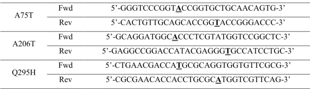
2.6 Site-directed mutagenesis and characterization of variants ... 23
3. Results and Discussion ... 25
3.1 Development of useful tools ... 25
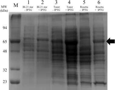
3.1.1 Selection of E. coli strain for large-scale production ... 25
3.1.2 Optimization of the detection method of AsP2Ox enzymatic activity ... 27
3.1.3 Re-characterization of wild-type AsP2Ox using the HRP-AAP/DCHBS method ... 29
3.1.4 Characterization of AsP2Ox variants obtained previously in the course of DE ... 30
3.2 Directed evolution of AsP2Ox ... 31
3.2.1 Screenings optimization ... 31
3.2.2 First generation of directed evolution ... 35
3.2.3 Second generation of directed evolution ... 37
3.3 Kinetic characterization of DE’s hit variants ... 39
3.3.1 Protein production and pH profiles ... 39
3.3.2 Transient state kinetic analysis of wild-type and variants ... 40
3.3.3 Steady-state kinetic parameters for D-glucose ... 42
3.3.4 Steady-state kinetic parameters for dioxygen ... 43
3.4 Effect mutations introduced during DE ... 43
4. Conclusions ... 46
5. References ... 47
6. Supplementary material ... 52
6.1 Selection of E. coli strain for large-scale production ... 52
6.2 Preliminary test of AsP2Ox with B-PER detergent ... 53
6.3 Directed evolution hit variants characterization for O2 ... 53
VII
Figures index
Figure 1.1 Schematic representation of three generations D-glucose biosensors ... 2
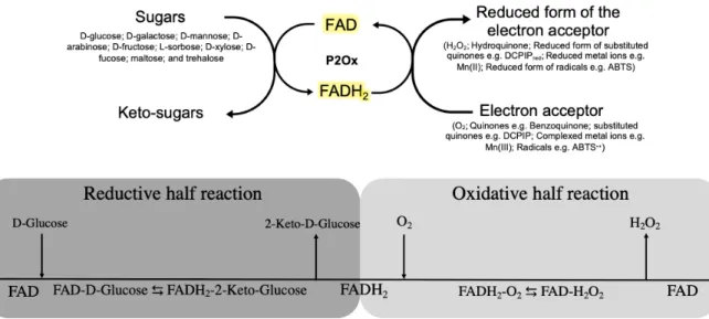
Figure 1.2. Overall redox reaction of Pyranoses 2-oxidases. ... 4
Figure 1.3. Recent proposal on the mechanistic steps of reductive half-reaction of TmP2Ox. ... 6
Figure 1.4. Comparison of the main steps of rational design and direction evolution. ... 11
Figure 3.1 SDS-PAGE of cell crude extracts of different E. coli strains after growing at small-scale (50 mL). ... 25
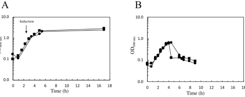
Figure 3.2 Growth curves of two distinct E. coli strains carrying the wild-type gene of AsP2Ox in 2.5 L-scale. ... 26
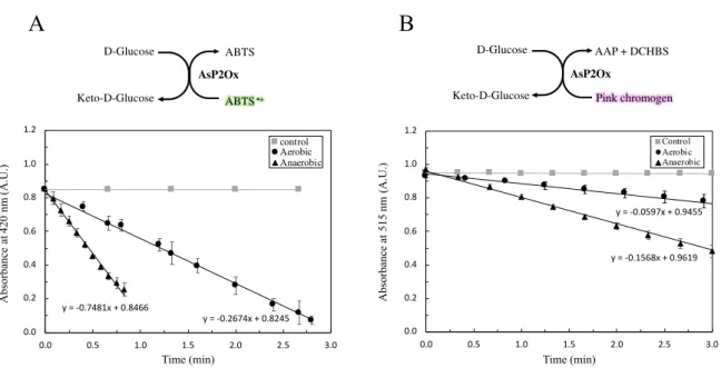
Figure 3.3 Overall scheme of the catalytic cycle of AsP2Ox and of coupled reaction assay to measure (indirectly) its activity. ... 27
Figure 3.4 Preliminary spectrophotometric assay to test the bleaching of oxidized chromogens formed during wild-type AsP2Ox reaction using D-glucose as electron donor. ... 28
Figure 3.5 pH profile of wild-type AsP2Ox. ... 29
Figure 3.6 Comparison of steady-state kinetics obtained using the two substrate systems of HRP. ... 29
Figure 3.7 Evolution tree of AsP2Ox with variants that were performed previously on the MET Lab.31 Figure 3.8 Directed evolution main steps and high throughput ‘Activity-on-plate’ screening procedure. ... 32
Figure 3.9 Example of an ‘Activity-on-plate’ screening performed during the evolution of AsP2Ox . 35 Figure 3.10 Activity of 95 variants from the first generation relative to wild-type after the first 96-wells plate liquid screening. ... 35
Figure 3.11 Activity relative to wild-type of each AsP2Ox variant picked to 96-wells plates during the first generation of directed evolution. ... 36
Figure 3.12 ‘Activity-on-plate’ rescreening to compare activity of wild-type with 2G10 and 1A1 AsP2Ox variants. ... 36
Figure 3.13 Summary of the first generation of directed evolution. ... 37
Figure 3.14 Activity relative to 1A1 obtained for each variant in 96-wells plate screening during the second generation of directed evolution. ... 37
Figure 3.15 Final screening that support the selection of 5D5 as the hit variant of second generation. 38 Figure 3.16 Summary of the second generation of directed evolution. ... 39
Figure 3.17 pH profile of wild-type, 1A1 and 5D5 AsP2Ox variants. ... 40
Figure 3.18 Time-course spectra set for one shoot in stopped flow apparatus. ... 40
Figure 3.19 Transient-state analysis of the different variants of AsP2Ox (Traces). ... 41
Figure 3.20 Transient steady-state analysis of AsP2Ox enzyme variants. ... 41
VIII Figure 3.22 pH profile of AsP2Ox single mutants A75T, A206T and Q295H and for double mutant Q295H/G366S. ... 44 Figure S6.1 Growing curve performed for four distinct growth of BL21 star carrying the wild-type AsP2Ox in large-scale production (1L). ... 52 Figure S6.2 Growing curve performed for three distinct E. coli strains carrying the wild-type gene of AsP2Ox in small-scale production (50 mL). ... 52 Figure S6.3 Assay to test the B-PER detergent as substrate of AsP2Ox ... 53 Figure S6.4 Steady-state curves obtained during the Oxygraph assays for the AsP2Ox variants ... 53 Figure S6.5 Steady-state kinetic curves for the AsP2Ox SDM variants (single and double mutants) .. 54
IX
Tables index
Table 2.1 - Primers used in the site-directed mutagenesis for the construction of different mutants. ... 24 Table 3.1 Apparent steady-state kinetic parameters of wild-type AsP2Ox for different sugar substrates (D-glucose, D-xylose, D-ribose, L-arabinose and D-galactose), using O2 as electron acceptor ... 30
Table 3.2 Apparent steady-state kinetic parameters of wild-type, 2C9* and CM3 AsP2Ox variants using D-glucose as electron donor and O2 as electron acceptor ... 30
Table 3.3 Optimization of the method of cell disruption using liquid screening in 96-wells plates. .... 33 Table 3.4 Optimization of E. coli expression strain for directed evolution liquid screening in 96-wells plate. ... 34 Table 3.5 Final validation of optimized directed evolution procedure for liquid media screenings in 96-wells plates. ... 34 Table 3.6 AsP2Ox protein production yields and optimal pH for the directed evolution hit variants. . 39 Table 3.7 Apparent steady-state kinetic parameters of wild-type , 1A1 and 5D5 AsP2Ox variants using D-glucose as electron donor and O2 as electron acceptor. ... 42
Table 3.8 Apparent steady-state kinetic parameters obtained by following the O2 consumption for
wild-type, 1A1 and 5D5 AsP2Ox variants (using the Oxygraph®) ... 43
Table 3.9 Apparent steady-state kinetic parameters (for D-glucose and O2), protein yields production
X
Abbreviations list
1,5-AG 1,5-anhydro-D-glucitol
AAP 4-Aminophenazone
Abs Absorbance
ABTS 2,2’-azino-bis (3- ethylbenzothiazoline-6-sulfonic acid) AsP2Ox Arthrobacter siccitolerans pyranose 2-oxidase
B-PER Bacterial Protein Extraction Reagent
CV Coefficient of variance
DE Directed evolution
DCHBS 3,5-Dichloro-2-hydroxybenzene-sulfonic acid
DCPIP 2,6-Dichlorophenolindophenol
dNTPs Deoxyribonucleotide triphosphate
epPCR Error prone polymerase chain reaction
FACS Fluorescence-activated cell sorting
FAD Flavin adenine dinucleotide
GDH Glucose dehydrogenase
GMC Glucose-methanol-choline
GOx Glucose oxidase
HPLC High Performance Liquid chromatography
HRP Horseradish peroxidase
IPTG Isopropyl β-D-1-thiogalactopyranoside
IVC in vitro compartmentalization
LB Luria-Bertani
NMR Nuclear magnetic resonance spectroscopy
OD600nm Optical density at 600 nm
P2Ox Pyranose 2-oxidase
PCR Polymerase chain reaction
Pink chromogen N-(4-antipyryl)-3-chloro-5-sulfonate-p-benzoquinone-monoimine
RT Room temperature
SDS-PAGE Sodium dodecyl sulfate polyacrylamide gel electrophoresis
SOB Super Optimal Broth
SDM Site-directed mutagenesis
SSM Site-saturation mutagenesis
TB Terrific Broth
Tris Tris (hydroxymethyl)aminomethane
UV-Vis Ultraviolet-Visible
1
1. Introduction
1.1 Overview on biosensors
Biosensorics, the science of biosensors, represents a branch of biotechnology arising in the 20th century as a result of symbiotic contributions from biology, biophysics, chemistry, physics, electronics, and informatics [1].
Biosensors are analytic systems constructed to detect an analyte whereas the analyzer is from a biological source [1]. On the backstage of an analyzer, a more or less developed electronic system transduce the biosensor activity in electric current to produce a quantitative signal [2]. Biosensors are developed from biological material such as antibodies or nucleic acids but the most extensively studied are the enzyme-based biosensors, in some cases, the use of whole cells as biosensors as a base of the immobilized biocatalysts proved to be advantageous originating the cell-based biosensors in which the microbial biosensors are common [2].
The microbial biosensors are mostly used in the environmental biotechnology branch and their advantages are due to the simplicity of microbial cultivation, high analytical prospects, and reliability. In these biosensors the microorganism can be seen as a complex “bag of intracellular enzymes” where the analyte crosses microbial cells, are metabolized by intracellular enzymes (where the co-substrates are consumed) and reaction products are generated (frequently they are electrochemically active). However, the major challenges for the use of this type of biosensors relies on the selection of a microorganism that allow for high substrates specificity (high selectivity) and enough sensitivity of products detection [1].
Enzyme-based biosensors show a broad range of applications and are considered a key focus of research in the area. The advantages related with their utilization are due to the usual high specificity of enzyme-substrate interactions and the high turnover rates of biocatalysts without the interference of intracellular metabolism (as is the case in microbial-based biosensors). The basics in enzyme-based biosensors is the ability to detect the presence of certain analytes by measuring changes (e.g. proton concentration) that occurred during the substrate consumption (or product formation) by the enzyme. The challenging steps in the development of enzyme biosensors are in the improvement of the biocatalyst sensitivity to produce a signal at lower concentrations of the analyte, and robustness, that lead to the optimization of immobilization processes [2].
1.2 Enzyme-based biosensors to measure D-glucose concentration
One of the most popular applications of enzyme-based biosensors is the measurement of carbohydrates in samples, in particular the monitoring of glycemic levels in blood [3].Glucose sensors were evolved to adapt to the desired functions and properties and currently they are categorized in three different generations distinguishable by the final electron acceptors used. In first-generation, the electron acceptor is dioxygen that is reduced to H2O2 that can further transfer the electrons to the electrode. In the
second-generation some mediators are used, replacing the O2 utilization. In the third-generation the mediators
are eliminated and the enzyme can transfer directly the electrons to the electrode avoiding error introduced, by e.g. the oxygen concentration in samples (Figure 1.1) [3]. These biosensors are mainly based in the action of Glucose oxidases (GOxs) and Glucose dehydrogenases (GDHs), responsible for the C1 oxidation of D-glucose but can be distinguished by the capability of using (GOXs) or not (GDHs) dioxygen as electron acceptor. Both GOxs and GDHs show advantages and disadvantages, for example,
2 GOxs showed are in general more specificity for β-D-glucose while GDHs show higher activity. These properties can be typically improved by protein engineering [3].
Recently, others carbohydrate-using enzymes, namely, Pyranose-2-oxidases (P2Oxs), are being considered as biosensors for determine glycemic levels in blood. Unlikely GOxs or GDHs, that catalyses the oxidation of D-glucose in C1 which is susceptible of anomers distinction using the β-D-glucose, P2Ox shows a major advantage of acting on the C2 alcohol moiety which makes both, α- and β-D-glucose anomers, susceptible of enzymatic oxidation leading in improved sensitivity during the response time [4], [5].
The substrate specificity of P2Ox is broader when compared with GOXs and GDHs, and P2Oxs were reported to use 1,5-anhydro-D-glucitol (1,5-AG) as substrate allowing the determination of the concentration of this natural D-glucose analogue in human cerebrospinal fluid and serum [6], [7]. 1,5- AG is in the bloodstream, being provided from diet but also from liver. When the levels of blood glucose are normal 1,5-AG is filtered and reabsorbed by kidneys but in hyperglycemia episodes, glycosuria blocks the reabsorption of 1,5-AG which leads to decreased levels of this metabolite in blood. This makes 1,5-AG a marker to access the glycemic control in diabetes mellitus [6], and Glycomark®, that
allows the measurement of 1,5-AG levels is already available in the market. In this device, P2Ox oxidizes 1,5-AG releasing H2O2 that is quantified through a coupled horseradish peroxidase (HRP) assay
that gives indirectly assess to the levels of 1,5-AG on the samples. As P2Ox can also react with D-glucose, a pre-treatment of the sample with a Glucokinase is applied to phosphorylate D-glucose that will no longer be a P2Ox substrate [8].
Figure 1.1 Schematic representation of three generations D-glucose biosensors. In the first generation, the electrons from
enzymes are transferred to dioxygen that become reduced and further transfer the electrons to the electrode, in the second generation, the electron acceptors are external mediators and in the third generation, the electrons are transferred directly to electrode. Figure taken from [3].
3
1.3 Pyranose 2-oxidase: General properties, Catalytic mechanism, and activity
assays
Pyranose oxidases (P2Ox, pyranose:oxygen oxidoreductase; EC 1.1.3.10; synonym, glucose 2-oxidase) are members of the glucose-methanol-choline (GMC) oxidoreductase superfamily of enzymes [9]. This superfamily includes enzymes such as glucose oxidase, choline oxidase, cholesterol oxidase, cellobiose dehydrogenase, aryl-alcohol oxidase and pyridoxine 4-oxidase that oxidizes an alcohol moiety to the corresponding aldehyde [10]. P2Oxs contain a flavin adenine dinucleotide (FAD) molecule as prosthetic group and therefore the typical UV-Vis absorption spectra of oxidized holoenzymes show two characteristic bands at ~ 390 nm and ~ 460 nm. The 460 nm maximum of absorption is responsible for the yellow color of oxidized form of the enzyme. In accordance, the reduced holoenzymes loses this band and the protein turns colorless [11], [12]. The P2Oxs were found in Eubacteria and Fungi with some different properties.
The catalytic cycle of P2Ox, as all oxidoreductases, requires a molecule with sufficiently negative redox potential to act as an electron donor and at the end of the catalytic cycle, the presence of a molecule with a redox potential to act as an electron acceptor. As a result, the reaction can be divided into two distinct half-reactions. In the first reductive half-reaction , the electron donor (sugar) transfer two electrons to the cofactor FAD that becomes reduced (FADH2) and in the second oxidative half-reaction, the FADH2
transfers two electrons to an acceptor (e.g. dioxygen) and the enzyme turn-over ends [10] (Figure 1.2). Several aldopyranoses and some disaccharides are reported to act as electron donors of P2Oxs including D-glucose, D-galactose, D-mannose, D-arabinose, D-fructose, L-sorbose, D-xylose, D-fucose, maltose, and trehalose but in all described non-engineered P2Ox the D-glucose is the preferred electron donor substrate [4], [10], [13], [14]. The oxidation of these pyranoses occurs regioselectively in the C2 position releasing a sugar in the 2-keto-aldopyranose form [4], [14], [15]. In the literature, it was reported that some sugars can be also oxidized to a smaller extent at carbon C3 [16] and, in other cases, the C3 oxidation by P2Ox occur as a side reaction in substrates whereas the C2 hydroxyl group is compromised (e.g. when 2-deoxy-D-glucose was used as substrate) [17], [18].
In the oxidative half-reaction, these enzymes use O2 as final electron acceptor which releases H2O2 as
co-product of the reaction but some quinones (as para-benzoquinone), substituted quinones (e.g. DCPIP), complexed metal ions (e.g. ferrocenium and manganese complexes) and radicals (e.g. radical cation ABTS•+) were reported to also act as final electron acceptors of the P2Ox redox cycle [19].
Interestingly, in the oxidative half-reaction when O2 is used as substrate, it was observed, for the first
time, the formation of a C-4a-hydroperoxyflavin intermediate [20] which is an intermediate barely detected in other oxidases due to the spatial constraints that bypass this intermediary step [21]. Considering the redox cycle described, the methodologies that have been used to follow the reaction progress of these enzymes include UV-Vis absorbance, HPLC and NMR that measure the reduced or oxidized sugar forms in a time-course manner [22]–[24]. UV-Vis is the most common technique and has been used to follow the reduced/oxidized final acceptor (e.g. at 290 nm to follow hydroquinone formation, the reduced form of para-benzoquinone ) [19] or when dioxygen is used as a final electron acceptor, a reaction coupled assay is typically set-up with horseradish peroxidase (HRP) and ABTS that culminates in the formation of a dark green compound (ABTS•+) that can be monitored at 420 nm [25].
The dioxygen consumption can also be measured in an Oxygraph® device. Finally, the stopped-flow
apparatus has been widely used to find the mechanistic properties of these enzymes and it allows to follow independently the two half-reactions of the catalytic cycle by monitoring the absorbance changes at ~ 460 nm [26]. Additionally, this apparatus allows studying the kinetic mechanism and the identification of the Michaelis complex (D-glucose-Enzyme complex) of the reaction (where
4 disassociation occurs before the interaction of the enzyme with the final electron acceptor), compatible with a bi-bi ping pong kinetic mechanism (the typical mechanism of oxidoreductases) [26].
1.4 Fungal Pyranoses 2-oxidases
P2Oxs were originally studied from Fungi and the first enzyme was isolated from the basidiomycete fungi Polyporus obtusus in 1975 [27]. Currently, ten fungal P2Oxs mainly from white and brown-rot wood degrading basidiomycetes have been investigated [4], [28]. The fungal enzymes are homotetrameric, many times glycosylated, and the FAD co-factor is typically covalently linked to the apoprotein by a histidyl linkage [4]. Fungal P2Ox are hypothetically expressed with an N-terminal prepropeptide which can target the enzyme to hyphal periplasmic space from where they can be secreted in later stages of the fungal life cycle to act as exoenzymes [29], [30]. Their physiological role has been associated to its oxidase activity which supplies hydrogen peroxide during the catalytic cycle that can be putatively used as co-substrate for lignin-degrading peroxidases (e.g. dye-decolorizing peroxidases) [31], the reason why these enzymes are classified as members of the auxiliary activity family 3 of redox enzymes by Carbohydrate Active Enzymes (CAZy) (www.cazy.orf) [32]. The hydrogen peroxide released during the redox cycle of P2Ox can also have some antifungal properties against phytopathological fungus [33]. Moreover, it has been considered that the dehydrogenase activity of these enzymes shows a relevant role in the maintenance of quinone/hydroquinone redox cycle equilibrium in decomposing wood process as well as in the regeneration of reducing metals for radical based lignin depolymerization reactions [34].
Metabolically, the C2 oxidation of D-glucose by P2Oxs releases 2-keto-D-glucose that can act as an intermediate metabolite of a secondary pathway which leads the formation of the antibiotic cortalcerone, hypothetically responsible for protection against bacterial attack and/or advantage in nutrients competition with bacteria [35], [36].
There are only three known crystal structures of P2Oxs of fungal origin: Trametes multicolor (synonym
Trametes ochracea) [9], Peniophora sp. [37] and Phanerochaete chrysosporium [38]. P2Ox from T.
Figure 1.2. Overall redox reaction of Pyranoses 2-oxidases. On the top it is displayed the general catalytic cycle of P2Oxs
that can use several sugars as electron donors releasing the correspondent keto-sugars. In the presence of electron acceptors such O2, quinones, substituted quinones, complexed metal ions or radicals, the FADH2 transfer the electrons releasing the reduced form of the electron acceptor and regenerating the FAD cofactor. On the bottom it is shown the Cleland notation of the proposed bi-bi ping pong mechanism of P2Ox whereas it is possible to divide the overall reaction into two half-reactions. During the reductive half-reaction, the FAD is reduced, and the oxidized sugar is release before the starting of the oxidative half-reaction in which the enzyme cofactor is re-oxidized regenerating the cofactor for a new enzyme turnover.
5
multicolor, TmP2Ox, has been the one most studied P2Ox and the association between structure and
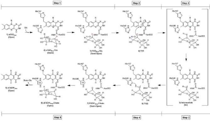
function in this protein was an important step to elucidate the mechanistic action of P2Oxs. For example, using TmP2Ox it was demonstrated that the structure of each subunit of the homotetramer has a peanut-shaped with three distinct regions. An oligomerization arm responsible for the association of the protein monomers, an head domain with a putative function associated with immobilization of the protein on the fungal cell-wall polysaccharides and a protein core domain [9]. The core domain is subdivided in a FAD-binding subdomain (F subdomain) that features a Rossmann fold motif in which FAD cofactor is covalently linked to a histidine residue (His167), part of the 165STHW169 conserved motif [9], [15]. The substrate-binding subdomain (S subdomain), bigger than the F subdomain contains the active-site cavity, in the interior of the protein, three peripherical a-helices and a six-stranded b sheet in the central part [9].In each subunit, the sugar substrate has to cross through an internal cavity that contains water molecules before reaching the enzyme active site. The residues His548, Asn593, Gln448, His450, and Val546 showed an important role in the sugar stabilization inside the substrate pocket and the alignment of the available sequences revealed that these residues are conserved among fungal P2Ox. The histidine 548 was originally hypothesized to act as a base, allowing the deprotonation of the sugar substrate in the alcohol moiety of C2, followed by hydride transfer to the N5 atom of the FAD cofactor and the Asn593 is proposed to participate in the interaction enzyme-sugar complex by the formation of a hydrogen bond with the deprotonated C2-OH moiety [9].
Very recently, and based in TmP2Ox studies, it was proposed a new mechanism for the reductive half-reaction performed in which P2Oxs show five mechanistic steps before the complete FAD reduction (Figure 1.3) [39]. In this new study, it was proposed for the first time in flavoproteins that the oxidation of a C-H bond can occur through a hydride transfer between the C2 of the sugar to the N5 of FAD, generating a protonated ketone sugar intermediate (stabilized by the key residues Thr169, His548, Asn593, and Phe474). The reaction mechanism follows with a proton transfer from the protonated keto-sugar to a conserved His548 residue culminating in the release of 2-keto-glucose and in the reduction of the protein cofactor [39].
The activity of TmP2Ox is proposed to be controlled by a highly conserved loop (residues 452 to 461) that is determinant for substrate binding and catalysis in each subunit. In the models performed with different ligands, this loop displayed two different forms, a closed and an open conformation, depending on the stage of the P2Ox redox cycle [9]. The transition between both conformations was associated with the loop motif 454FSYG457 that becomes closer to the active site in the oxidative half-reaction blocking the entrance of sugar substrates to the pocket but still allowing the interaction with small molecules such as dioxygen [9].
6
1.5 Bacterial Pyranoses 2-oxidases
The first bacterial P2Ox named AsP2Ox was characterized from the dry desiccation resistant actinobacteria Arthrobacter siccitolerans (renamed for Pseudoarthrobacter siccitolerans [40]) in 2016, based in sequence analysis and alignment with different GMC family members and also fungal P2Ox enzymes [24]. The AsP2Ox show highly conserved residues in the GMC family (13 out of 15 residues) and additionally, contains three major conserved regions in this family: the FAD-binding domain, the attachment loop, and the substrate-binding domain. Additionally, multiple alignment analysis with four pyranoses dehydrogenases (members of GMC family) and with three fungal P2Ox revealed an identity of 26 %, around the double obtained in the comparison with pyranoses dehydrogenases suggesting that AsP2Ox is more related with fungal P2Ox than with other members of GMC enzyme family [24]. Within the AsP2Ox sequence, the flavin binding domain was shown to be conserved when compared with fungal P2Oxs but the STHW motif that would allow the covalent ligation of FAD to apoprotein have a replacement of serine and threonine for two alanines (AAHW). These aminoacidic changes can putatively interfere in FAD ligation leading to a non-covalently bond to the apoprotein with consequences in protein stability and lower redox potential [12], [24]. Besides, the tertiary structure model suggests that in AsP2Ox His440 and Asn484 are conserved and putatively exert the same role as His548 and Asn593 of TmP2Ox.
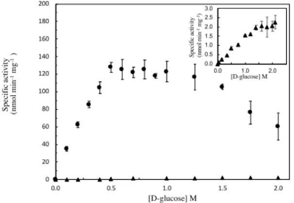
The characterization of the recombinant enzyme shows that the optimal temperature of AsP2Ox is 37ºC. The optimal pH activity and also the kinetic parameters were re-evaluated in this thesis and will be discussed in the Results and Discussion section. The role of the AsP2Ox in A. siccitolerans is not well understood but the high Km values for D-Glucose (see below) of this enzyme suggests this is not the
intracellular substrate, although within the sugar substrates tested it is still the preferred one.
Figure 1.3. Recent proposal on the mechanistic steps of reductive half-reaction of TmP2Ox. The figure shows the steps
proposed by Wongnate et al[39] for the reaction of P2Oxs with glucose. In Step 1 the formation of the active enzyme substrate complex (FADEnz:Glu) takes place. In step 2, the hydride transfer from the carbon position 2 of glucose to the N5 of the flavin
occurs. Step 3 relates to the formation of the protonated ketone intermediate (Int), while in step 4 the proton transfer of the protonated keto to His548 takes place. In step 5, 2-keto-glucose is produced and the reduced flavin generated. Figure and descriptive subtitle taken from [39].
7 Two more P2Oxs from bacterial origin were later identified from soil and environmental bacteria. PaP2Ox, is from Pantoea ananatis, a g-proteobacteria with a cosmopolite distribution that acts as endophytic lignocellulolytic bacterium [41], while KaP2Ox is from Kitasatospora aureofaciens (formerly Streptomyces aureofaciens) that belong to actinobacteria and is a microorganism that in its genome contains genes that codifies for lignocellulosic enzymes which suggest with a role in plant biomass degradation [28].
The PaP2Ox enzyme shares a sequence similarity of 27 % with TmP2Ox and 26 % with AsP2Ox. This protein shows two conserved domains: a FAD-binding domain, and a steroid-binding domain with unknown function but that can hypothetically act as a substrate-binding domain. Like the fungal counterparts, PaP2Ox is an homotetramer where the FAD-binding domain does not exhibit the characteristic fungal motif STHW mentioned before resulting in a FAD non-covalently bonded to each subunit, although this protein has an optimal temperature of 50ºC which is higher than the one obtained for AsP2Ox but still less thermostable as compared with fungal P2Oxs [41]. PaP2Ox showed that in conjunction with bacterial laccases can originate diverse compounds from lignin which makes it an interesting enzyme for biotechnological applications [41].
KaP2Ox, the most recently identified bacterial P2Ox showed an identity of 39 % with TmP2Ox matching 545 out of 623 of its residues. The structure in solution of KaP2Ox, unlike the previous P2Ox, is a homodimer, and the FAD is covalently bounded to apoprotein. The sequence of KaP2Ox show one Histidine (His464) that can be homologous to His548 in TmP2Ox [28].
A phylogenetic analysis based on sequences maximum of likelihood grouped KaP2Ox with other putative bacterial P2Ox in one clade distinct from fungal P2Ox but interestingly AsP2Ox was placed in another different clade far from PaP2Ox and KaP2Ox [28].
1.6 Other biotechnological applications of P2Oxs
In the late nineteenth century and early twentieth century, significant advances were made in the extraction, characterization and commercial exploitation of enzymes [42].
The potential of P2Ox in C2 regioselectivity oxidation is very promissory for the carbohydrate chemistry industry allowing the development of efficient ways to convert bulk carbohydrates in valued products [43]. Using D-glucose as substrate, P2Ox can provide 2-keto-D-glucose which can act as intermediate of other sugar precursors for the production of rare sugars, fine chemicals, and drugs [22]. For example, the antibiotic cortalcerone can be produced synthetically from the 2-keto-D-glucose supplied by P2Ox activity followed by dehydration using another enzyme [44], [45].
In the same field, an engineered P2Ox was recently used to perform the first reaction of a cascade in the one-pot bioconversion of L-arabinose to L-ribulose leading to higher production yields and replacing a process that resulted in almost 90 % of substrate waste [46]. In this cascade, the fungal P2Ox is responsible to convert L-arabinose in 2-keto-arabinose that is further converted in L-ribulose by a xylose reductase. Interestingly the conversion of L-arabinose to L-ribulose represents an increasing value from 0.1 US$ g-1 to 995 US$ g-1 which makes the investment on this enzyme as catalyst rewarding [46]. It
was reported that P2Ox can also produce 2-keto-D-galactose a precursor for the production of the rare, low-caloric and noncariogenic sugar D-tagatose [43], [47] or 2-deoxy-3-keto-D-glucose that can be an intermediate for vitamin B1 and B6 production [43].
Lastly, the oxidase activity of P2Oxs can be employed as a dioxygen scavenger providing anoxic conditions that are usually hard to achieve in analytical chemistry procedures [48]. P2Ox was used in an oxygen removal system in a small open volume of a biofuel cell and the results suggested that these enzymes can be applied without interference on the biosensor detection characteristics neither in the pH
8 of solution which represent an advantage comparing for example, with GOx that requires a strong ionic strength buffer to maintain the pH [48].
Fungal P2Ox had been engineered for improved characteristics such as the reactivity for O2 [49],
increased thermostability [50], pH stability [50], substrate specificity and/or catalytic efficiency [46], [51], [52]. These improvements were performed using all the three methods of protein engineering (rational, semi-rational or directed evolution) with different final purposes (such as for biofuel cells [49] or biocatalysts for industry [46]).
1.7 Protein engineer: How to make better enzymes?
Enzymes are biomolecules that occur in nature associated with living organisms but they can also be synthesized de novo mimicking native enzymes [53]. Nowadays the de novo synthesis approach starts to have some impact because of the challenging of engineer native enzymes although the bulk of enzymes are still discovered and characterized from nature [53].
The major problem with organisms’ native enzymes is that they are synthesized intracellularly with a defined metabolically role at relatively low levels, in amounts that are not cost-effective for commercial and industrial applications. Moreover, in most cases they show poor stability, low activity for alternative substrates, (product) inhibition and limited conversion yields [54].
Protein engineer is a revolutionary branch of science that works with different techniques to allow improving enzymes properties within short time frames [55].
Typically, protein engineering uses recombinant proteins that are usually expressed in E. coli systems and amino acid substitutions can be accessed by two main approaches rational design and directed evolution that can be applied individually or in conjugation to achieve the best-improved variant hits [56]. In some cases, another method a semi-rational design approaches that shares characteristics of both can also be applied.
1.7.1 Rational design in protein improvement
When an enzyme has the structural and biochemical data available and the relationship between structure and function is relatively well known, the rational design can be considered a good approach to improve enzymes [57]. This method relies on computational and biochemical studies that allow identifying residues that if mutated for others can increase the fitness of the enzyme. The methods to introduce mutations in a gene (pre-cloned onto an expression vector) in rational design is based in site-directed mutagenesis (SDM) which use a pair of designed oligonucleotides primers carrying a nucleotide that will mismatch with the template sequence allowing the change of nucleotide sequence after a PCR reaction with an high-fidelity DNA polymerase [58]. There are more than one strategy to design primers for SDM (the mutation can be in both primers or in only one primer and the other will pair fully with template, for example) but all of them results in a linear PCR product, that is digested (e.g. with DpnI) to eliminate template sequences, and then used to transform E. coli strains. In these bacteria self-mechanisms, turn the PCR product, in a circular plasmid that allow for expression of the gene of interest [58].
In this technique, the correct introduction of the mutations is confirmed by DNA sequencing followed by enzyme production, purification and biochemical characterization [59], [60] (Figure 1.4). If the variant is not as good as expected, all computational analysis could be performed again, and the experimental procedure has to be repeated. Due to the fact of this approach relies on specific operator-defined mutations it is a good technique to test a pre-elaborated hypothesis on the structural and mechanistic role of specific residues [58].
9
1.7.2 Directed evolution: using Darwinian evolution principles to improve
biocatalysts
When the crystal structure or the model structure of an enzyme is not available the application of rational design does not apply. If an enzyme has N amino acids and considering 20 amino acid possibilities for each position, there is a space of possible enzyme variants given by 20N [61]. This imaginary space of
hypothetically mutant enzymes hides variants with so far undetected characteristics although the time limitations just allow us to explore a tiny part of them.
The directed evolution (DE) technique, also known as laboratory evolution, appears in the 1980’s to bypass that gap [60].
In DE, the concept is to simulate in vitro, speeded-up, evolutionary trends that occurs in nature which leads organisms with improved fitness when compared with ancestors. The well-adapted enzyme mutants, during protein engineer through DE, are selected as parent for further rounds to improve some characteristic(s), this process is repeated iteratively simulating a process that, in nature, could take hundreds or thousands of years [62].
The major advantage of DE is that mutations are introduced randomly in the protein gene without the need of previously computational analysis [60].
The usage of DE requires four main prerequisites as follows, (I) the availability of the gene that codifies for the protein that will be engineered, (II) a suitable expression system, (III) a method to generate diversity onto parent gene (library) and (IV) a method to screen and select the improved variants within the library [63]. For the first and second pre-requisites, the most common approach is to have a gene of the desired protein cloned into a plasmid vector that could be heterologously expressed in E. coli cells.
1.7.2.1 Generation of diversity on the parent gene
The creation of diversity on a gene can be performed using two main distinct techniques: random mutagenesis or recombination [60], [64], [65].
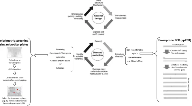
In random mutagenesis the introduction of mutations to construct a variant library can be performed using, for example, chemical and/or physical mutagenesis, mutator strains, but the most used approach is the error-prone PCR (epPCR) (Figure 1.4) [64].
In epPCR the gene that codifies for the protein of interest is amplified in low fidelity conditions using a polymerase that usually can introduce mutation mistakes during elongation of DNA strand (e.g. Taq polymerase). Additionally, several factors can be optimized to tune mutation rates, increasing the concentration of MgCl2, use of mutagenic dNTPs analogs, increase the DNA polymerase concentration,
increase the number of cycles or inclusion in the PCR mix compounds that interfere in Taq polymerase elongation (e.g. MnCl2) [66], [67]. These conditions should be optimized to achieve a relatively low
mutation rate (2-3 mutations in a gene) which amplify the probability to achieve beneficial mutations and prevent the introduction of exceeding mutation numbers that culminate in non-functional proteins [60]. During the PCR the Taq polymerase has a high tendency to replace A to G and T to C which can lead to a higher GC content of product sequence and also limited diversity of mutations achieved. Sometimes to counteract this tendency the concentration of dNTPs used in the PCR mix can be unbalanced [66].
Gene recombination is also used to create enzyme gene variants randomly. The most common recombination technique is named DNA shuffling. In this approach, chimera enzymes that result from the association of diverse homologous genes are constructed [64]. The genes used in DNA shuffling
10 could derive from variants of the same protein or from genes from different origins that codify for structurally similar protein, showing high homology [54], [68]. A common protocol of DNA shuffling includes a controlled fragmentation of the gene (50 to 150 bp) with DNase I and then all fragments are joined randomly in a PCR without primers because the fragments by themselves can align and cross-prime to each other [56]. Next, a standard PCR with appropriate flanking cross-primers is performed to amplify the low number of segments that are full-length, generating a gene library with different chimeric genes. The size of a recombinant library is typically smaller than a library generated by random mutagenesis [56], [60], [69]. However, the great advantage of DNA shuffling is that in later stages of random mutagenesis evolution it can be applied to accumulate the beneficial mutations and eliminate the deleterious or neutral mutations [70].
1.7.2.2 Screening and selection in directed evolution
Finally, in DE, the development of a suitable method to screen and/or select the enzyme variants of a library is mandatory and is considered the most challenging step [64].
Screening methodologies are developed in order to discover phenotypes associated genotypes and to allow to identify improved enzyme variants for the desired characteristic [64]. The screening methods are wide diverse, but they can be grouped into three major types that englobes screenings of spatially separated variants, high-throughput screenings in flow cytometry and screening in artificial cell-like compartments [64].
In spatially separated variant screenings the library size is small (~104 variant members per screening
round) [64]. The main advantage of this approach is its compatibility with several assay techniques, including screening in solid media by, for example, following the zones of degradation of a substrate in agar plates surrounding the colonies, or in liquid medium, that are usually performed in multi-well plates using a coupled assay by, for example, fluorescence spectrophotometry (Figure 1.4). Both solid and liquid media screenings are typically based in single E. coli colonies that contain one gene variant and the respectively expressed variant enzyme. The main disadvantage of these screenings is the high time-consumption which limits the throughput capacity [64], [65].
Flow cytometry screenings are performed in a fluorescence-activated cell sorting (FACS) apparatus and the size of the library can be higher than in the methods described before (> 108 members can be
analyzed in less than 24 h) [65]. In this approach, the aim is to analyze individually each cell that could emit fluorescence (or luminescence), for example, by monitoring the level of expression of reporter proteins such as green fluorescent protein (GFP) that were coupled to the target enzymes [71]. The FACS stringency can be adjusted which allows to perform different rounds with an increment of this parameter using the gate of cell sorted in the round before. Cell surface display can as well be performed with the help of FACS to sort positive improved mutants. In this approach, the aim is to develop a system that allow the display of the target protein in cell surface (for example via anchoring) followed by treatment with modified antibody’s (coupled with fluorophores) that will allow identifying the desired enzyme variants [64], [65].
The screening in artificial cell-like compartments are used in cases of hard establishment of a cell system that allow the implementation of a fluorescent reporter for a given gene and phenotype [65]. Then, in
vitro compartmentalization (IVC) can provide an alternative method to high throughput using the FACS
apparatus. The main advantage of this system is the inexistence of the diverse metabolic network of the cells eliminating the possibility that improved phenotypes come from mutations non-related with target gene [64], [65]. In IVC approach aqueous droplets are used in water-in-oil emulsions (or also droplets in water-in-oil-in-water emulsions) to compartmentalize individual genes and gene products with alternative fluorogenic substrates.
11 As an alternative to the screening methods that require the individual evaluation of phenotypes, selection methods link the desired activity to a physical separation of the encoding DNA or survival of the organism producing active library members [65]. "Rejective to the unwanted" is the feature of selection methods that make them pure high throughput [65]. DE selection methods include several techniques such as cell surface display and cell compartmentalization that are very elaborated techniques with several variations [65]. Other technique that is used for selection in DE of enzymes with active role in cell metabolism is organismal growth complementation (or organismal survival) in which the desired enzyme property is coupled with the fitness of the host cell allowing that only the cells containing the protein with the desired property can survive under selective pressure [65].
1.7.3 Semi-rational design in protein engineering
The semi-rational design approach for enzyme engineering uses the site-saturation mutagenesis (SSM) technique. This method is a conjugation between rational and non-rational. In a first step, computational analysis based on protein structure, identify amino acids that could interfere directly on the improvement of the desired property. In a second step, the replacement of that amino acid position will be performed for the other nineteen amino acids available, creating a small library of enzymes (called smart library) that will be screened for activity (as a DE library) [68].
In SSM the mutations are introduced by mutagenic primers that are synthetic oligonucleotides with randomized codon flanked by parent sequence. These primers are degenerated, and their sequence is identical with the parent template except in the specific position where the mutation will be introduced by the randomized codons. After a standard PCR reaction with mutagenic primers, the library is constructed [54]. This approach can be applied to randomize several positions simultaneously creating a library with variants for more than one position [54].
Colorimetric screening using microtiter plates
Cell culture in 96-wells plate Cell disruption Collect the cell crude extracts after centrifugation
Select the improved variants (e.g. by increase absorbance) Parent of new round of DE
Non-recombinative epPCR Recombinative e.g. DNA shuffling
Mutations randomly distributed on the enzyme gene PCR with Mn2+using Taq polymerase Error-prone PCR (epPCR) Enzyme gene Mu ta nt s lib ra ry Screening Chromogenic/fluorogenic substrates Coupled enzyme assays
IVC Selection
Figure 1.4. Comparison of the main steps of rational design and direction evolution. In the scheme four main steps are
displayed related with protein engineering by rational design or directed evolution. Within directed evolution, the main procedures are exemplified, how epPCR is performed and also exemplified is a high through put screening using microtiters and a chromogenic assay that allow to identify the best hit variant to be used as parent of a new round of directed evolution. The directed evolution technique is iterative, i.e. when a new parent is achieved all steps (generation of diversity, expressing in host system and screenings) are repeated. The figure was adapted from [60].
12
1.7.4 The New Era in protein engineering: bioinformatic and machine-learning
approaches
Computational protein design is an in silico technique that relies on semi-rational design [56]. In computational protein design, the aim is to apply a combination of force field and algorithms to provide different tridimensional configurations of the same protein in which all amino acids can be substituted by other nineteen resulting in proteins with different energies. The ones with lower energy are chosen to test experimentally [56].
Machine-learning-guided directed evolution for protein engineer is a new tool that is nowadays being an intensive target of research. The concept is to create DE principles in computing-based programs without the need for experimental screens and selection. This approach allows to bypass the time-consuming and expensive DE approach and also to overcome problems related with constrains in evolution imposed in DE [72]. However, it is still not possible to predict how much time can be saved using this process. Instead of DE that uses only the improved variants, in machine-learning-guide processes, information of unimproved sequences are used to expedite evolution and expand the number of properties that can be optimized [72]. The most challenging part of this method is to build a sequence-function model that can be applied to choose sequences to screen. In the end, machine-learning-guided DE uses all information of the sequence-function pairs to construct a landscape to achieve improved sequences. [72].
1.8 Context of the project
In this master thesis the aim was to apply directed evolution methodologies, one of the main focus of Microbial and Enzyme Technology Lab at ITQB, to identify improved variants of AsP2Ox, a bacterial pyranose 2-oxidase recently characterized in this laboratory.
The goal was to achieve variants exhibiting higher catalytic efficiency and lower Km for D-Glucose and
O2 allowing its application as first generation biosensors for monitoring the D-glucose levels replacing
the typical GOx or GDHs.
When I arrived at the Microbial and Enzyme technology (ITQB), the directed evolution of AsP2Ox was already being performed by a fellowship researcher. After a round of DE and rational design a variant, CM3, had been identified, showing a catalytic efficiency for D-glucose and O2 around 10-fold higher
than the wild-type. When I started the studies two initial tasks were planned based on identified issues. The first concerned the cell cultivation at large scale (5L-Erlenmeyers containing 1L of LB medium) where a lack reproducibility was identified; many times, cell cultures die before the induction of gene expression with IPTG. This led me to investigate alternative E. coli strains. The second, related to literature that reported that the oxidized form of ABTS (ABTS•+), produced upon activity of HRP, in
the coupled assay to measure AsP2Oxs activity, could act as an electron acceptor of AsP2Ox, competing with O2 and resulting in non-reliable activity measurement. This aspect needed investigation since if
this is confirmed all previously optimized DE procedures need re-validation and re-optimization as well as the characterization of wild-type, intermediates and hit variant.
The work performed resulted in the optimization of the cultivation at large-scale of recombinant E.coli strains producing AsP2Ox, optimization of the coupled enzymatic assay to measure activity of wild-type and AsP2Ox variants, re-investigation of kinetic parameters and successful implementation of a DE protocol to improve the first P2Ox from bacterial origin, in which the diversity of the gene was promoted by epPCR and the screening method was implemented based on a reaction coupled assay with
13 HRP monitored by UV-Vis absorbance. Hit variants were identified and the proteins were purified and biochemically characterized showing improved remarkable kinetic properties.
14
2. Material and methods
2.1 General materials and procedures
2.1.1 Bacterial strains, plasmids and cultivation medium
During this study the Tuner (DE3, Novagen), KRX (Promega), Rosetta (DE3, Novagen) and BL21 star (DE3, Novagen) E. coli strains were used to heterologously express the different AsP2Ox variant genes previously cloned onto pET-15b vector (Novagen). In BL21 star, Tuner and Rosetta the target genes are controlled by T7 promoter, induced by isopropyl β-D-1-thiogalactopyranoside (IPTG) and in the KRX strain the genes are under control of rhaPBAD promoter, induced by rhamnose. The pET-15b vector has some important characteristics, namely a region that codifies for a β-lactamase that confers the resistance to ampicillin and a region that codifies for a 6xHis tail localized close to the cloning site that allow the protein produced to be purified using an His-tag affinity column. Luria-Bertani (LB) and Terrific Broth media (TB) were used as routine liquid media to grow the different E. coli strains. The cultivation media was supplemented with 100 μg/mL of ampicillin (NZYTech) for all strains that were transformed with the plasmid pET-15b carrying the asp2ox gene plus 20 μg/mL of chloramphenicol (NZYTech) when the host strain used was Rosetta. LB medium contains (per liter): 1 % of tryptone, 0.5 % of yeast extract and 170 mM of NaCl. TB medium contains the following components (per liter): 1.2 % of tryptone, 2.4 % of yeast extract, 4 mL of glycerol (86%), 17 mM of KH2PO4 and 72 mM of K2HPO4. Super Optimal
Growth medium (SOB) was used for the culture of electrocompetent cells. SOB medium contains (per liter): 2 % of tryptone, 0.5 % of yeast extract, 10 mM of NaCl and 2.5 mM of KCl, 10 mM of MgCl2
and 10 mM MgSO4. Luria-Bertani Agar (LA) was used as the solid media and has the same composition
than LB with addition of 1.5 % of bacteriological agar. All culture media was sterilized in an autoclave at 121ºC and stored at room temperature until use.
2.1.2 Preparation of electrocompetent E. coli cells
The frozen stock of E. coli strains from the Lab culture collection was used to stretch a LA plate that was incubated overnight at 37ºC. In the following day, one single colony was picked to inoculate 20 mL of SOB medium (pre-inocula) and the cultures had grown overnight at 37ºC, 180 rpm on Innova 44 incubator shaker (New Brunswick Scientific. This incubator was used for all growths except when 96-wells plates were used). Growth in 250 mL of pre-warmed SOB medium started with OD600nm ≃ 0.01
using the pre-inocula. Culture were incubated at 37ºC, 180 rpm. After 3 h (OD600nm ≃ 0.8), the
Erlenmeyers containing the cultures were placed in an ice-cold bath for 20 min (after this step all procedures were performed at 4ºC). Cultures were transferred to ice-cold centrifuge bottles and spun down (4420 × g, 10 min at 4ºC). The supernatants were discarded, and the cell pellets were washed with 250 mL of a sterile ice-cold 10% glycerol solution. The cells were centrifuged again in the same conditions. The washing step was performed twice. The final cell pellets were resuspended in 10 % glycerol solution that remained in the centrifuge bottles. Aliquots of 150 μL were frozen at - 80ºC and stored until use.