Bruno Pacheco Fernandes
Preparation of particles for
cosmetic applications in human hair
Br uno P ac heco F er nandes Outubro de 2012 UMinho | 201 2 Pr eparation of par ticles f or cosme
tic applications in human hair
Universidade do Minho
Outubro de 2012
Tese de Mestrado
Micro/Nano Tecnologias
Trabalho efectuado sob a orientação do
Professor Doutor Artur Manuel Cavaco-Paulo
e coorientação da
Doutora Raquel de Jesus Marques da Silva
Bruno Pacheco Fernandes
Preparation of particles for
cosmetic applications in human hair
Universidade do Minho
“The scientist is not a person who gives the right answers, he is one who asks the right questions.”
vii
A
CKNOWLEDGMENTSNow that this stage of my education is coming to an end, it is time to express my gratitude to all those who, in this last year, contributed for the success of this journey.
I thank to Professor Artur Cavaco-Paulo for the opportunity to work on his research group, for trusting in me to accomplish this project, for his support and guidance as well as for his availability. I am also grateful to Dra. Raquel Silva, my co-supervisor, for the dedication and manifested patience, for the knowledge transmitted and for all the constructive comments and suggestions that helped me in the challenges of this work and improved the writing of my thesis.
To my colleagues in the Bioprocess Research Group, I express my appreciation for the help, for never refusing to take my doubts and for the excellent work environment they provided to me. A special thanks to Artur Ribeiro for the valuable assistance in the last part of this work.
For the support, for always being there for me and for emotionally raise me up, thank you to all my friends, especially to those who are closest to me: Ademar, Carina, Félix, Laura and Nuno. For helping me with the fluorescence microscope, I also thank to Rita.
I also want to show gratitude to my amazing family because of the endless support I received from them since ever. To my cousin Carla, a special thanks for the laptop you lent me when mine broke down, allowing me to write the thesis on time.
And last but not least, my most heartfelt recognition goes to my parents for their unconditional love and care, for teaching me to be who I am, for always support my choices and without whom this thesis would not exist.
Preparation of particles for cosmetic applications in human hair
ix
A
BSTRACTNanoparticles (NPs) have a huge interest for transdermal applications, demonstrating their ability to be trapped in the hair follicles (HFs) where they release the entrapped compounds. For hair cosmetics, this discovery isparticularly important as it can improve the treatment of several hair follicle related disorders/diseases. Therefore, the aim of this work was to obtain NPs of Poly (Lactic Acid) (PLA-NPs) for topical delivery of drugs at the level of the HFs. Further, these particles may be used for cosmetic applications in human hair.
The effect of several parameters was examined, in order to establish an improved nanoprecipitation protocol for the preparation of suitable PLA carriers for follicular targeting. Thus, the application of mechanical stirring or ultrasound, the use of Acetone/Ethanol (50/50, v/v) as the solvent phase and the addition of 0.6% (w/w) of Pluronic F68 to the formulation showed the best compromise between the desired properties (monodispersed populations of particles with a mean size of ≈150 nm were obtained, the ζ-potential was less than -18 mV and particles exhibited a spherical shape with smooth surface) and the yield of nanoparticles (≈90% was showed for particles produced with agitation and ≈70% when ultrasound was employed).
After the encapsulation of model compounds, no significant changes were found in the properties of particles and the entrapment efficiency was above 80%. Nevertheless, the use of sonication promoted higher loading efficiencies. In turn, the release kinetics of PLA nanoparticles indicated an anomalous dye transport mechanism (diffusion and polymer degradation) for Nile Red (lipophilic) and a Fickian diffusion of first order for FITC (hydrophilic). Furthermore, the release from particles produced with agitation was slightly faster than from particles produced with sonication.
Finally, fluorescence microscopy on porcine skin cryosections showed that the produced PLA-NPs can effectively transport lipophilic and hydrophilic compounds into the HFs, with fluorochromes reaching a maximal depth corresponding to the full follicles length after 24h.
In conclusion, the modified nanoprecipitation protocol presented in this study allows the preparation of PLA nanocarriers with potential for hair follicle therapy and the yields obtained are acceptable for industrial purposes.
Preparação de partículas para aplicações cosméticas em cabelo humano
xi
R
ESUMOAs nanopartículas (NPs) são de grande interesse para aplicações transdérmicas, acumulando-se nos folículos capilares (FC) onde libertam os compostos encapsulados. Esta descoberta é particularmente importante para a cosmética capilar, podendo melhorar o tratamento de várias doenças/distúrbios associados aos FC. Consequentemente, este trabalho visou a obtenção de NPs de Poli (Ácido Lático) (PAL-NPs) para entrega de agentes terapêuticos ao nível dos FC. No futuro, estas partículas poderão ser utilizadas para aplicações cosméticas em cabelo humano.
Inicialmente, o efeito de algumas variáveis experimentais foi testado, de modo a melhorar o protocolo de nanoprecipitação para a produção de PAL-NPs aptas para entrega de princípios ativos ao nível dos FC. Assim, a melhor relação entre as propriedades desejadas e o rendimento em nanopartículas foi obtida com a aplicação de agitação mecânica ou ultrassons, o uso de Acetona/Etanol (50/50, v/v) como fase solvente e a adição de 0.6% (w/w) de Pluronic F68 à formulação. Com estas condições obtiveram-se populações monodispersas de partículas esféricas com uma superfície lisa, tamanhos médios de 150 nm, carga superficial inferior a -18 mV e rendimentos de ≈90% e ≈70% para partículas produzidas com agitação e sonicação, respetivamente.
Após um encapsulamento eficiente (> 80%) do Nile Red e do FITC, não foram registadas alterações significativas nas propriedades das partículas. Por sua vez, o mecanismo de libertação dos compostos mostrou-se dependente da sua natureza: para o Nile Red (lipofílico) ocorreu transporte anómalo (difusão e degradação do polímero) e a libertação do FITC (hidrofílico) foi controlada por difusão fickiana de primeira ordem. É também de salientar que a libertação dos fluorocromos foi ligeiramente mais rápida para partículas produzidas com agitação.
Por último, usando microscopia de fluorescência em secções de pele de porco, provou-se que as NPs produzidas são eficazes no transporte de compostos lipofílicos e hidrofílicos para o interior dos FC. Mais ainda, ao fim de 24h foi possível detetar a presença dos corantes ao longo de toda a extensão destas estruturas.
Em suma, as alterações introduzidas ao protocolo de nanoprecipitação permitiram a preparação de nanopartículas de PAL com potencial para terapia folicular sendo que, os rendimentos obtidos são aceitáveis para aplicação industrial.
xiii
T
ABLE OFC
ONTENTSAcknowledgments... vii
Abstract ... ix
Resumo... xi
Table of Contents ... xiii
List of Abbreviations...xvii
List of Figures ...xxi
List of Tables ...xxv
List of Equations...xxvii
1. Motivation and Aims of the Study... 1
2. Review of the Literature ... 5
2.1. Nanoparticles as Carriers for Drug Delivery... 7
2.2. Drug Administration through the Skin... 9
2.2.1. Approaches to Overcoming the Dermal Barrier... 10
2.2.2. Nanotechnology for Conquering the Skin Barrier... 11
2.3. The Dermal Barrier... 12
2.3.1. The Structure of the Skin... 12
2.3.1.1. Organization of the Stratum Corneum... 14
2.3.1.1.1. Transport Routes across the Stratum Corneum... 16
2.4. The Follicular Pathway... 18
2.4.1. Anatomy of the Hair Follicles... 19
2.4.1.1. Target Structures for Follicular Therapy... 21
2.5. Nanoparticles for Hair Follicle Therapy... 22
xiv
2.5.1.1. Preparation of Drug-loaded Polymeric Nanoparticles... 25
2.5.1.2. Drug Release from Polymeric Nanoparticles... 30
2.6. Safety Aspects of Nanoparticles... 32
3. Materials and Methods... 35
3.1. Materials and Equipment... 37
3.1.1. Chemicals and Solvents... 37
3.1.2. Porcine Skin Tissue... 37
3.1.3. Equipment... 37
3.2. Methods... 39
3.2.1. Preparation of PLA Nanoparticles... 39
3.2.1.1. Addition of a Non-Solvent to the Solvent Phase... 40
3.2.1.2. Effect of the Concentration of Pluronic F68... 40
3.2.2. Yield of Nanoparticles... 40
3.2.2.1. Effect of Ethanol in the Yield of Nanoparticles... 41
3.2.3. Characterization of the Particles... 42
3.2.3.1. Particle Size and Size Distribution Measurements... 42
3.2.3.2. Analysis of the Zeta-Potential... 42
3.2.3.3. Morphology of Nanoparticles... 43
3.2.4. Preparation of Dye-loaded Nanoparticles... 43
3.2.4.1. Determination of Entrapment Efficiency... 44
3.2.4.2. In vitro Release Profile and Dye Release Kinetics... 45
3.2.5. In vitro Follicular Penetration Studies... 47
3.4.5.1. Preparation of the Skin... 47
xv
3.4.5.3. Cryosections and Fluorescence Microscopy... 49
4. Results and Discussion ... 51
4.1. Preparation of PLA Nanoparticles... 53
4.1.1. Comparison of Preparation Techniques... 53
4.1.2. Influence of the Solvent Phase... 57
4.1.3. Effect of the Surfactant Concentration... 60
4.2. Yield of Nanoparticles... 62
4.3. Morphology of Nanoparticles... 66
4.4. Encapsulation of Model Compounds... 67
4.4.1. Entrapment Efficiency... 69
4.4.2. In vitro Release Profile... 71
4.5. Follicular Penetration of PLA Nanoparticles... 75
5. Conclusions and Future Perspectives... 79
xvii
L
IST OFA
BBREVIATIONS AACS approx.
American Chemical Society Approximately C °C Celsius D Da DDS DGV DMSO Dalton
Drug Delivery Systems Direção Geral de Veterinária Dimethyl sulfoxide
E
e.g. For example
F
FDA FITC
Food and Drug Administration Fluorescein 5(6)-isothiocyanate G g Gram H HF h HPLC Hair Follicle(s) Hours
High Performance Liquid Chromatography
I
xviii K kV kHz Kilovolts Kilohertz L
LDA Laser Doppler Anemometry
M µL µm m MEC min mL mm MTC mV Microliters Micrometers Meters
Minimum Effective Concentration Minutes
Milliliters Millimeters
Minimum Toxic Concentration Millivolts N nm NP(s) NR Nanometers Nanoparticle(s) Nile Red O o/w •OH Oil in water Hydroxyl radical P PBS PCL PCS PDI
Phosphate Buffered Saline Solution Poly-ε-caprolactone
Photon Correlation Spectroscopy
xix
PLA PLA-NPs PLGA Pluronic F68
Poly (Lactic Acid)/Poly (D, L-Lactic Acid) Nanoparticles of Poly (D, L-Lactic Acid) Poly Lactic-co-glycolic Acid
Pluronic acid F68
R
rpm Rotations per minute
S SC SESD SLN s STEM SD Stratum Corneum
Spontaneous Emulsion Solvent Diffusion Solid Lipid Nanoparticles
Second
Scanning Transmission Electron Microscopy Standard Deviation U UV UV-Vis Ultraviolet Ultraviolet-Visible V v/v Volume/volume W w/o w/o/w w/v w/w Water in oil
Water in oil in water
Weight/volume
Weight/weight
Z
xxi
L
IST OFF
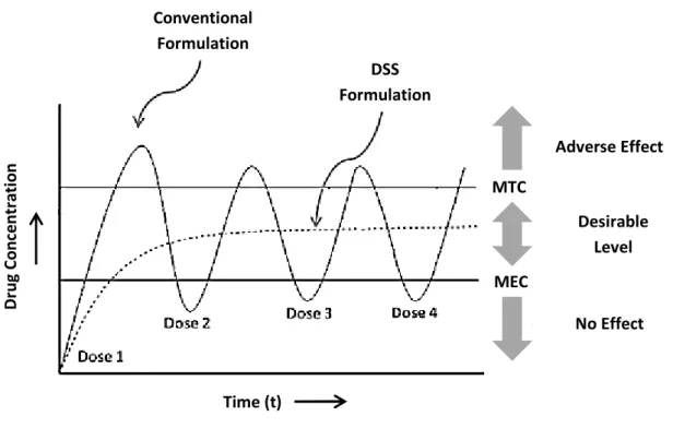
IGURESFigure 2-1: Release profile of conventional formulation and drug delivery
systems (DDS), over time (Adapted from Das S. et al., 2011 [19])...8
Figure 2-2: Anatomy of the skin (Online image, available at
http://www.meb.uni-bonn.de/Cancernet/Media/CDR0000579036. jpg; accessed on September 10, 2012)...13
Figure 2-3: Schematic representation of the “brick and mortar” model of the
stratum corneum (Adapted from Moghimi H.R. et al, 1996 [39])...15
Figure 2-4: Routes for the penetration of substances through the stratum
corneum (Adapted from Prausnitz M.R. et al., 2004 [42]). ...17
Figure 2-5: Cross-section diagram of a human hair follicle (Adapted from
Meidan V.M. et al., 2005 [46])...20
Figure 2-6: Chain structure of PLA (Online image, available at
http://www.medicinescomplete.com/mc/excipients/current/image s/Ecpoly_dl_lactic_acidC001_default.png; accessed on September 10, 2012). ...24
Figure 2-7: Mechanisms of drug encapsulation on the nanoparticles (Adapted
from Guterres S.S. et al., 2007 [22]). ...25
Figure 2-8: Production of polymeric particles with single emulsion method
(Adapted from Gomes V.M.A., 2009 [15])...26
Figure 2-9: Mechanism of nanoparticles formation by the SESD method
(Adapted from Murakami H. et al., 1999 [57])...27
Figure 2-10: Method for the preparation of nanoparticles based on a
xxii
Figure 2-11: Schematic illustration of the nanoprecipitation method (Adapted
from Gomes V.M.A., 2009 [15])...29
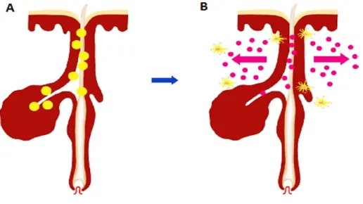
Figure 2-12: Release of the drug and targeting of follicular structures. A)
Accumulation of polymeric drug-loaded nanoparticles within the hair follicle canal, at the level of the target structure. B) Release of the drug from the particles and its penetration through the hair follicle barrier. (Adapted from Papakostas D. et al., 2011 [11])...31
Figure 3-1: Sketch of a typical Franz Diffusion Cell (Online image, available at
http://www.permegear.com/fc01.gif; accessed on October 11, 2012). ...48
Figure 3-2: Depth of penetration of PLA nanoparticles into the hair follicles,
after topical application (Adapted from Rancan F. et al., 2009 [45])...49
Figure 4-1: Mean size and size distribution (PDI) of PLA nanoparticles, obtained
after a sonication treatment of 15 and 18 minutes. ...54
Figure 4-2: Effect of Pluronic F68 on the size and PDI of nanoparticles produced
by nanoprecipitation, using agitation and sonication. ...60
Figure 4-3: Effect of the volume fraction of ethanol on the yield of
nanoparticles, prepared by nanoprecipitation...63
Figure 4-4: Effect of volume fraction of ethanol on the properties of PLA
nanoparticles, prepared with agitation and sonication. ...65
Figure 4-5: S-TEM photographs (x50000 magnification) of PLA nanoparticles
obtained with A) agitation and B) sonication, using a binary mixture of Acetone/Ethanol (50/50, v/v) and Pluronic F68 concentration of 0.6% (w/w). ...67
xxiii
Figure 4-6: Z-Average (nm), PDI and Zeta-Potential (mV) of PLA nanoparticles,
prepared with agitation and sonication, after the entrapment of Nile Red and FITC. ...68
Figure 4-7: Entrapment efficiency of Nile Red and FITC into PLA nanoparticles,
produced with agitation and sonication...69
Figure 4-8: In vitro release profile of A) Nile Red and B) FITC from PLA-NPs,
produced by nanoprecipitation with agitation and sonication...72
Figure 4-9: Fluorescence microscopy images (x5 magnification) of porcine skin
xxv
L
IST OFT
ABLESTable 3-1: List of equipment, used in the course of this experimental work...38
Table 4-1: Effect of the employed techniques on the properties of PLA
nanoparticles, obtained by nanoprecipitation. ...55
Table 4-2: Effect of the solvent phase on the properties of PLA nanoparticles,
prepared with agitation and sonication. ...57
Table 4-3: Loading efficiency of Nile Red and FITC into nanoparticles produced
by nanoprecipitation, using agitation and sonication. ...71
Table 4-4: Dye release kinetic data obtained from fitting experimental release
data to Ritger-Peppas Equation, where “n” is the diffusion exponent and R2is the correlation coefficient. ...75
xxvii
L
IST OFE
QUATIONSEquation 3-1: Determination of efficiency of nanoparticles formation...41
Equation 3-2: Equation of Stokes-Einstein used to determinate the diameter of
the particles...42
Equation 3-3: Equation of Henry used to determinate the electrophoretic
mobility of the particles. ...43
Equation 3-4: Entrapment efficiency of compounds in the PLA nanoparticles...45
Equation 3-5: Loading efficiency of the dyes in the PLA nanoparticles. ...45
Equation 3-6: Ritger-Peppas equation...46
Equation 3-7: Ritger-Peppas modified equation. ...47
Equation 4-1: Theoretical determination of dielectric constant of the mixtures
1. MOTIVATION AND AIMS OF
THE STUDY
Motivation and Aims of the Study
3
Nanotechnology is an interdisciplinary area that deals with the development, manipulation and control of materials at nanometric scale, in order to take advantage of their singular physical, chemical and biological properties [1, 2]. Currently, due to the many new options that this scale can offer, nanotechnology is a key field of research, providing means for achieving otherwise unreachable goals [3, 4].
The cosmetic industry was among the first to use nanotechnology in the development of products. From 1994 to 2005, L’Oreal SA (France) was worldwide ranked in 5th placed on the total number of nanotechnology-based products and in
2009, more than 13% of nanostructured products available in the market were classified for cosmetic use [4]. This growing interest in the development of nanotechnological cosmetics occurred because nanomaterials have the potential to radically change the way cosmetics deliver their benefits, leading to product innovation, which consequently may stimulate the economy of one of the most important worldwide industries [5, 6].
Among nanomaterials for cosmetic use, nanoparticles (NPs) are of particular interest because they improve the stability of various cosmetic ingredients like unsaturated fatty acids, enhance the penetration of certain agents as vitamin and other oxidants, increase the efficacy and tolerance of UV filters on the skin surface and make the product more aesthetically pleasing [7]. Moreover, NPs are also important for cosmetic applications because they can encapsulate a wide range of ingredients as vitamins, fragrances, and drugs with cosmetic or dermatological purposes and act as Drug Delivery Systems (DDS) [5].
Regarding to the route of administration, for cosmetic applications but not only, the transport of DDS through the skin has been widely studied because it offers numerous advantages over other routes [8]. After topical application, although nanoparticles may accumulate at different skin sites depending on their properties, they tend to be trapped inside the hair follicles (HFs), reaching high concentrations at these sites [9]. This localized accumulation of nanoparticles is very important for hair cosmetics because, using NPs it will be possible to achieve adequate concentrations of active ingredients into these structures, in order to treat many hair follicle related disorders/diseases without damages to the hair or skin lesions [10].
Preparation of Particles for Cosmetic Applications in Human Hair
4
The present study aimed to obtain nanoparticles that could be used as carriers for topical delivery of drugs at the level of the HFs. Thus, these particles may be used for cosmetic applications in human hair. Initially, to achieve the ultimate goal proposed, the influence of several experimental factors was tested in order to obtain an adequate formulation for the production of Poly (D,L-Lactic Acid) nanoparticles (PLA-NPs) by nanoprecipitation methodology. The yield of nanoparticles was further determined and the entrapment efficiency and release profile of hydrophilic and lipophilic compounds from PLA-NPs were achieved. Finally, the ability of these particles to penetrate the skin and accumulate in the HFs was assessed in vitro.
Review of the Literature
7
2.1. Nanoparticles as Carriers for Drug Delivery
In the last decades, significant advances have been made in the production of nanoparticles (NPs) as carriers to deliver therapeutic agents into the body [11]. Thus, the application of nanotechnology on drug delivery enabled the creation of entirely new therapeutic agents [12].
The main advantage of NPs as drug delivery systems (DDS) is their ability to target specific cell populations in the human body and to help in the intracellular uptake of the drugs [13]. The efficiency of drug delivery to various parts of the body is directly affected by particle size but it can be modulated through the modification of particle surface (e.g., binding to specific ligands as monoclonal antibodies to target a selected cell population) [11]. By providing a high local concentration just at the desired site of action, NPs enhance the bioavailability of drugs, minimizing the amount of active substances needed. The application of a low dose of drugs reduces the risk of side-effects in other tissues and it makes possible to use certain drugs that were previously impractical because of their toxicities [11, 13, 14]. Another important feature of NPs is their capacity to promote a sustained drug release over a prolonged period of time [11]. Controlling the drug carrier architecture (e.g., porosity), the release of the drug can be tuned to achieve a desired kinetic profile, fixating the concentration of the encapsulated substance between the Minimum Effective Concentration (MEC) and the Minimum Toxic Concentration (MTC), for an adequate period of time (Figure 2-1); achieving a constant drug level in tissues, the therapeutic index (ratio between the efficacy of the drug and its undesirable side effects) is maximized [11, 13, 15]. In conventional systems, many applications are need to maintain the drug level above MEC (otherwise there is no beneficial effect) and bellow MTC (above this limit, the active substance becomes toxic) because, following a relatively short period at the therapeutic level, drug concentration drops off until re-administration [15, 16]. Thus, NPs can reduce the frequency of drug re-administration, increasing the consumer’s compliance and acceptance [11]. Finally, nanoparticles can also provide protection to the encapsulated drugs, making them more stables, resulting in the maintenance of their bioactivity until they reach the target [17, 18].
Preparation of Particles for Cosmetic Applications in Human Hair
8
Figure 2-1: Release profile of conventional formulation and drug delivery systems
(DDS), over time (Adapted from Das S. et al., 2011 [19]).
A broad range of materials like lipids, proteins, metals and polymers have been studied for the production of nanoparticles [11]. The choice of the material must to take into account their biodegradability and biocompatibility because in vivo degradation must be fast, originating products that can be metabolized and eliminated by the organism [15]. A careful choice of material is also important once that it will affect the properties of target and controlled release of nanoparticles [18]. To synthetize the particles, numerous protocols are already described based on the type of material, type of drug used or the desired delivery route and several structures like liposomes, micelles, solid lipid nanoparticles (SLN), dendrimers and polymeric nanoparticles were already obtained and reported as efficient systems for delivery purposes [11, 20].Liposomes are hollow particles, composed of one or several closed phospholipid bilayers offering a hydrophobic shell (lipid layer) as well as a hydrophilic core (inner volume of the liposome); lipophilic and hydrophilic substances can be integrated into the shell or the core, respectively [3, 5, 11, 21]. Micelles are structures smaller than liposomes, which has either a fatty core separated from an aqueous
Conventional Formulation DSS Formulation Adverse Effect No Effect Desirable Level Time (t) D ru g Co nc en tr at io n MTC MEC
Review of the Literature
9
solvent by the polar heads (normal micelle) or else an aqueous core separated by the polar heads from a fatty solvent (inverse micelle) [3]. SLNs are formed by a matrix of lipids which are biodegradable raw materials that are physiological well tolerated [22]. Dendrimers, a unique class of polymers, are highly branched macromolecules whose size and shape can be precisely controlled [13]. Finally, polymeric nanoparticles have a core surrounded by a polymeric shell and generally, they exhibit greater stability [23]. While there is plenty of research in the production of nanoparticles for drug delivery, only a few of them have reached the market because they are still several limitations associated with the design and characterization of these materials (ability to reproducibly fabricate specific nanoparticle shapes and sizes, to achieve optimal drug loading of carrier, to control drug release and delivery and to design stable materials, which do not release harmful degradation products) [14, 24]. The initial cost of materials and the expensive processes also has limited the commercial distribution of these systems [15]. However, the remarkable results achieved with nanoparticulate DSS in animal models are promising for their extensive commercialization in the coming years [11].
The administration of nanoparticulate DSS to humans can be made through injection or across oral, pulmonary, ocular, transmucosal and dermal routes [13]. Lately, special attention has been given to the transport of drugs through the skin (topical application), because this route offers a lot of vantages when compared, for example, to intravenous injection or oral administration [8]. The administration of
nanoparticles through the skin is particularly important in the field of cosmetic, once those particles had proved to be valuable for optimizing current galenical formulations for topical dermatotherapy [11].
2.2. Drug Administration through the Skin
The main function of the skin is to provide a protective barrier against external agents; however, its passive diffusion mechanism can also allow the penetration of substances, making it a potential route for the delivery of drugs into the
Preparation of Particles for Cosmetic Applications in Human Hair
10
body [25, 26]. According to the purpose, the delivery through the skin can be divided into topical and transdermal delivery; topical delivery involves drug administration for local therapeutic effects, whereas transdermal delivery uses the skin as a route for the transport of drugs into the circulatory system [27].
The skin offers many advantages over other routes of drug administration because it is a large and readily accessible surface area for absorption, the application is a noninvasive procedure (allowing continuous intervention) and it is possible to cease the absorption preventing overdose or undesirable effects [28]. When compared with the traditional oral route of drug administration, additional advantages are shown: it avoids the gastrointestinal tract (and consequently, drug degradation under the extreme acidity of the stomach), it avoids hepatic first pass metabolism (increasing the bioavailability of drug) and it reduces the fluctuations on the drugs level in the plasma, minimizing the risk of systemic side effects [25, 28]. Despite all the advantages of drug administration through the skin, this route also has some disadvantages such as, higher molecular weight candidates fail to penetrate without modifying the nature of skin, drugs with very low or high partition coefficient fail to reach systemic circulation and high melting drugs have difficult to permeate due to their low solubility in the skin [25]. Therefore, to make these compounds suitable for topical administration is necessary to increase skin permeability or increase solubility of drugs in the skin [28].
2.2.1. Approaches to Overcoming the Dermal Barrier
Chemical and physical methods have been aimed to disrupt or weaken the skin in an attempt to enhance drug transport across it [29]. Physical enhancement methods usually involve the use of an energy source to overcome the barrier properties of the skin and include iontophoresis (driving charged molecules into the skin by a small direct current), electroporation (application of short micro- to milli-second electrical pulses to create transient pores in skin) and sonophoresis (cavitation caused by low frequency ultrasound energy, which increases skin fluidity) [28, 30, 31].
Review of the Literature
11
Alternative physical approaches to increase skin permeability, without the use of an energy source, involves the use of microneedles (which can be inserted into the skin producing a channel for drug transport), jet-propelled particles (high-velocity jet of compressed gas carrying drug particles) and ablation of the stratum corneum [30, 31]. For chemical enhancing of percutaneous absorption, water is the safest and most widely used penetration enhancer; the increasing of hydration leads to a diminished resistance of the skin [25]. Other chemical enhancers, which promote changes in the structure of skin, have also been reported [32]. These chemicals can disrupt the lipid organization of skin (e.g., azone, terpenes, fatty acids, dimethyl sulfoxide (DMSO) and alcohols) or alter its protein organization (DMSO or urea) [28]. Although these methods have proved to be effective for increase skin permeability to drugs, they also induce irritation and they cause damages to the skin, reducing its barrier function [32].
2.2.2. Nanotechnology for Conquering the Skin Barrier
Since an ideal penetration enhancer should reversibly reduce the barrier resistance of skin without damaging it, nanoparticulate DSS are the most promising alternative to the physical and chemical penetration enhancer systems [14, 32, 33]. Nanoparticles can modify the physicochemical properties of the encapsulated molecules and offer a means to facilitate the percutaneous delivery of substances, increasing the permeability and transport of therapeutic agents into/across the skin without damaging it [14, 26, 32, 33]. NPs can also protect drugs from be metabolized by mircoflora or enzymes on the surface of skin and prevent allergic responses in skin induced by the encapsulated drugs [25].
Despite the advances in recent years, the transport of a wide range of drugs through the skin is still limited due to the special composition and structure of skin, which provides a formidable barrier to penetration [25, 31, 34]. Thus, explore and understanding the anatomy, physiology and chemical composition of the skin is important to improve the nanoparticles design and increase the ability to deliver these materials with wide systemic and topical application [14, 28]. An understanding of the
Preparation of Particles for Cosmetic Applications in Human Hair
12
interactions between these novel drug delivery systems and the skin, as well as the transport pathways within the skin, is also required to establish the feasibility of using nanoparticles to optimize the transport process [33].
2.3. The Dermal Barrier
The skin, or cutis, is considered the largest human organ; being normally less than 2 mm thin, it accounts more than 10% of the body mass and have an average surface of approximately 2 m2. This organ enables the body to interact dynamically
with the environment and plays many functions (protective, homeostatic and sensorial), essential for the survival of the human beings [35]. Specifically, the excellent protective/biological barrier function of skin protects the body against external mechanical, chemical, microbial and physical influences [3]. To maintain its characteristics, this organ is in a continual renewing process [28].
Because of large surface area and easy accessibility, skin delivery has potential application in drug delivery [36]. However, the functional properties that enable skin to act as an excellent barrier also serve to limit the access of drugs into and across it [28]. Whereas an initial consideration of the skin structure might suggest a simple barrier, a closer examination reveals a complex combination of a range of cell types [31]. Thus, to optimize the delivery of drugs into or across the skin is important to learn its structure, in order to learn how to overcoming its barrier function [37].
2.3.1. The Structure of the Skin
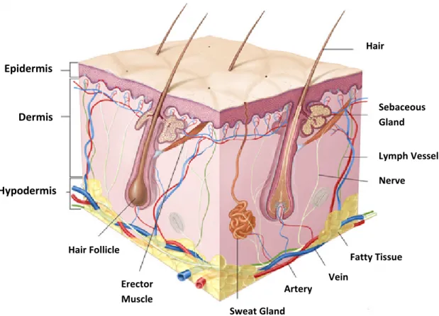
The Figure 2-2 shows the structure of the skin. Anatomically, it is composed of epidermis, dermis and hypodermis (also called subcutaneous tissue),being a complex barrier as a consequence of its anatomical organization and special chemical composition [22, 27]. In addition to these main layers, several pilosebaceous units (hair follicles and associated sebaceous glands) and apocrine or eccrine sweat glands are
Review of the Literature
13
dispersed throughout the skin [14, 36]. Owing to its unique biochemical and anatomical properties, each individual skin layer is mechanically different [3].
Figure 2-2: Anatomy of the skin (Online image, available at
http://www.meb.uni-bonn.de/Cancernet/Media/CDR0000579036.jpg; accessed on
September 10, 2012).
The innermost layer, the hypodermis, is composed of loose textured, white, fibrous connective tissue in which fat and elastic fibers are intermingled [36]. This layer acts as insulator, shock absorber, and reserve depot of calories and supplier of nutrients for the more superficial skin layers [37]. On its domain, it can be found the base of the hair follicles, the secretory portion of the sweat glands, the cutaneous nerves and the networks of lymph and blood vessels [28].
Above the hypodermis, there is dermis which normally measure up to 2 mm in thickness [3]. The dermis is a fibrous layer that supports and strengthens the epidermis and it consists of a matrix of loose connective tissue composed by collagen (a fibrous
Epidermis Dermis Hypodermis Hair Follicle Erector Muscle Sweat Gland Artery Vein Fatty Tissue Nerve Lymph Vessel Sebaceous Gland Hair
Preparation of Particles for Cosmetic Applications in Human Hair
14
protein) embedded in a semigel matrix, which contains water, ions and mucopolysaccharides; this matrix allows the oxygen and nutrients to diffuse to the epidermal cells [37]. The dermis shelters a network of blood capillaries, lymphatic vessels, nerve endings and nearly all elastic fibers giving mechanical stability to the skin [36]. Finally, this region of skin contains only few cells, predominantly fibroblasts (responsible for the connective tissue synthesis), mast cells (involved in the immune and inflammatory responses) and melanocytes (involved in the production of melanin) [37].
The top layer of the skin (epidermis) is typically 50-150 μm thin and it contains four histologically distinct strata of keratinocytes, which varying in the differentiation level [3, 37]. The innermost Stratum Basale is composed of two keratinocyte types, one that acts as stem cells having a proliferation capacity, and the second one which serves as anchor to the basement membrane; in this strata is also possible to find Merkel cells (sensory perception), Langerhans cells (immunological function) and melanocytes (melanin production) [14, 36]. During the differentiation process to form the higher epidermal layers, keratinocytes undergo a process of keratinization, in which the cell differentiates and moves upward from Stratum Basale, through the
Stratum Spinosum and Stratum Granulosum, to the outermost layer, the Stratum Corneum (SC), also called horny layer or non-viable epidermis [14]. Therefore, the SC represents the final stage of epidermal cell differentiationand it has an approximately thickness of 5 - 20 µm but a number of factors, including the degree of hydration and skin location, can influence it [29, 37]. This layer is the main responsible for the resilient absorption properties of skin, providing protection to the body against the entry of external materials but it is also the main barrier for diffusion of water out of the skin [14, 27, 36].
2.3.1.1. Organization of the Stratum Corneum
The structure of SC has been described by “brick and mortar” model (Figure 2-3) and it was first presented by Michaels et al. [38].
Review of the Literature
15
Figure 2-3: Schematic representation of the “brick and mortar” model of the
stratum corneum (Adapted from Moghimi H.R. et al, 1996 [39]).
The “bricks” of the model correspond to hydrophilic corneocytes (also called horny cells) which are dead, anuclear, flattened and hexagonal cells [14, 27, 28]. They are composed mainly of insoluble bundled keratins (70%) and lipids (approx. 20%); their approximate diameter and thickness is 40 µm and 0.5 µm, respectively [28, 37]. In SC, corneocytes are vertically stacked into columns (perpendicular to the skin surface) and each column contains between 10 and 15 cellular layers of keratinized cells. The “bricks” are joined together by desmosomes, which help to form a tough outer layer by maintaining cellular shape and regular packing [14, 28]. The inter-cellular space in SC is usually less than 100 nm thick except at cell clusters junctions, in the 2 to 3 partly detached cellular layers close to skin surface (where corneocytes are less densely packed or even partially detached) and at the 2 to 3 layers at the bottom of SC where the terminal cells differentiation has only just begun [3].
In the model, the “mortar” represents the hydrophobic continuous matrix of lipids in which the corneocytes are embedded; this lipid matrix provides the only continuous diffusion pathway from the skin surface to the base of the SC [27, 37]. The
Preparation of Particles for Cosmetic Applications in Human Hair
16
main lipids in the intercellular spaces of SC are ceramides, fatty acids, cholesterol,
cholesterol sulphate and sterol/wax esters; unlike almost all other membranes in the body, the phospholipids in SCare largely absent. Ceramides are the largest group of lipids and its compact stacking is the primary determinant of the extremely stable physical properties of the SC [27]. According to Landmann model, the intercellular lipid matrix is generated by keratinocytes in the mid to upper part of the Stratum Granulosum, discharging their lamellar contents into the intercellular space [40]. In the initial layers of the SC, this extruded material rearranges to form broad intercellular lipid lamellae which then associate intomultiplelipid bilayers, parallel to the surface of SC [31]. The hydrocarbon chains became arranged into regions of crystalline, lamellar gel and lamellar liquid crystal phases, thereby creating various domains within the lipid bilayers; it has been proposed that the distribution of crystalline and gel phases of the membrane lipids influences the diffusion of small molecules across the SC and into the dermis. [33, 38] Between the lipid bilayers, there are layers of water [28]. Water is an essential component, because it acts as a plasticizer to prevent cracking of the SC;
some of extra water in maximally hydrated SC is sequestered into these layers, so-called “lacunae” [3, 31].
At the surface of SC, there is also an irregular and discontinuous layer (0.4–10 µm) consisting of sebum secreted by the sebaceous glands, along with sweat, bacteria and dead skin cells. However, this layer is considered to have a negligible effect as an additional barrier to permeation through the SC [14].
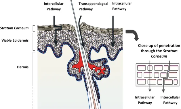
2.3.1.1.1. Transport Routes across the Stratum Corneum
Despite the composition and structure of the SC offers a main obstacle and a real challenge on drug delivery into and through the skin, its “brick and mortar” organization also provides some possible pathways to the passive diffusion of substances into the skin, as schematized in Figure 2-4. These pathways are through the keratinocytes (intracellular pathway), through the lipid matrix occupying the spaces between the keratinocytes (intercellular pathway) and across hair follicles, sebaceous
Review of the Literature
17
glands and sweat glands (transappendageal pathway) [14, 36]. However,the transport pathway is highly influenced by the structure, size and solubility of the penetrant [27].
Thus, understanding the physicochemical properties of the penetrant is essential to determine the predominant route of drug permeation and thereby optimize its delivery [31].
Figure 2-4: Routes for the penetration of substances through the stratum
corneum (Adapted from Prausnitz M.R. et al., 2004 [42]).
The major route of penetration for lipophilic molecules is through the lipid matrix [21].Although this diffusional pathway (500 μm) is much longer than the simple thickness of the SC, it contains fluid lipids and channel-like structures, increasing the molecules insertion and migration through the intercellular lipid layers, providing higher diffusivity to them [3, 21, 41]. However, the intercellular space also contains hydrophilic domains which restrict the invasion of too lipophilic compounds. As a consequence, only substances with partition coefficients (logP) between 1 and 3 are well suited for skin absorption [21]. Finally, the size of penetrating species also
Stratum Corneum
Viable Epidermis
Dermis
Close up of penetration through the Stratum
Corneum Transappendageal Pathway Intracellular Pathway Intercellular Pathway Intercellular Pathway Intracellular Pathway
Preparation of Particles for Cosmetic Applications in Human Hair
18
influences the absorption through this pathway, because penetrants need to fit into the intercellular space in order to move along it [21].
The hydrophilic entities have therefore, the propensity to cross normal skin
within the aqueous regions near the outer surface of corneocytes or through corneocytes,along the most curved cell plasma membranes - intracellular pathway [3].
However, in this route, not only the drug must partition into and diffuse through the corneocyte but also, in order to move to the next corneocyte, the permeant must partioning into and diffuse through the lipid lamellae between corneocytes [31].
Until recently, the transappendageal pathway was barely considered as a penetration pathway because it covers only 0.1% of the skin surface area. Nevertheless, this route became to receive considerable attention once it provides an efficient penetration pathway and more important, it was realized that the appendages can act as reservoirs of topically applied substances [43, 44]. Among the appendages, the hair follicles (HFs) are of particular interest because they seem to be the preferential targets for nanoparticles [21, 43]. This favored penetration of nanoparticles through the HFs and their reservoir function for topically applied substances has the potential to revolutionize the treatment of hair follicle associated diseases (e.g., acne and some skin cancers) and the treatment of many diseases/disorders of human hair (e.g.hirsutism, graying, alopecia) that havedramatic
effects on the appearance, socio-cultural status and self-esteem of the affected individuals[10, 45, 46].
2.4. The Follicular Pathway
Even though the HFs are increasingly recognized as a potentially route for the entrance of topical applied substances into the skin,the invasion of a substance into these structures is not an absorption process itself because, by definition, the compounds inside these appendages are still on the outside of the body [31]. However, since HFs are invaginations of the epidermis extending into the dermis, they provide a much greater actual area for absorption bellow the skin surface [47]. Also, as
Review of the Literature
19
it extend into the hair follicles, the surrounding SC layer is progressively reduced, which may lead to a faster and more efficient diffusion of solutes from out of the follicles to the inside of the skin; the dense network of blood capillaries at the lower levels of the HFs also facilitates the uptake of topically applied substances [14, 31]. Another feature of the HFs that makes them interesting for topical delivery is their reservoir function [41]. While the reservoir of the SC is located in the uppermost cell layers of the horny layer (approximately 5 μm), the reservoir of the HFs is usually extended deep in the tissue up to 2000 μm [48].
Even if the HFs are potential routes for the absorption of topically applied substances, not all hair follicles are available for penetration and they have to be distinguished between “open” and “closed” [49]. The hair follicles “open” to penetration exhibit hair growth and/or sebum production which act as a pumping system, pushing topically applied substancesmechanically into the hair follicles when the hairs are in motion [50].This was kind of surprising as the direction of hair growth and sebum flow are opposed to the invasion of materials into the follicles, suggesting an “active” uptake mechanism transporting them deep into the follicle [48]. In the “open” hair follicles, the release of sebum also influences the absorption and, since sebum consist mostly neutral, non-polar lipids, it provides a lipoidal pathway that it will favor the penetration of more lipophilic permeants[26, 47].Depending on the skin samples, approximately 50-70% of hair follicles are open for penetration [36].
2.4.1. Anatomy of the Hair Follicles
In order to develop appropriated studies of follicular delivery, an understanding of the structure of the HFs is useful. The hair follicle, represented in Figure 2-5, is composed of a hair bulb and a hair shaft [46, 47]. The hair bulb, localized at the base of the hair follicle, encloses a small papilla of dermis, which it is a structure of actively growing cells that produce the long fine cylinder of a hair [51]. The dermal papilla is connected to the blood vessels, an important feature when the HFs are used as route for transport of drugs into the systemic circulation [47, 52]. The hair bulb and
Preparation of Particles for Cosmetic Applications in Human Hair
20
shaft are enveloped in an inner root sheath and an outer root sheath [52]. The outer root sheath is a stratified epithelium that is continuous with the epidermis and this continuity is responsible for the increased surface area of absorption beneath the surface of the skin, making of this layer the most important with regard to drug delivery [47]. On the contrary to the outer root sheath, the inner root sheath ends about halfway up the follicle [46].
Figure 2-5: Cross-section diagram of a human hair follicle (Adapted from Meidan
V.M. et al., 2005 [46]).
As it can be seen in the figure above, the hair follicle is associated through ducts (in the upper part of the follicle canal) with sebaceous glands that are responsible for the production of sebum and their release into the follicular canal [46, 47]. Connected to the hair follicle is also an adjoining arrector pili muscle. The whole structure, comprising the hair follicle, sebaceous glands and adjoining arrector pili
Hair Bulb Suprabulbar Area Isthmus
Infundibulum Hair Shaft
External Root Sheath Internal Root Sheath Dermal Papilla Epidermis Dermis Sebaceous Gland Arrector Pili Muscle Bulge Area Cuticle Cortex Medulla
Review of the Literature
21
muscle, is called pilosebaceous unit; usually, the terms hair follicle and pilosebaceous unit are used interchangeably [52].
The hair follicle (or pilosebaceous unit) can be divided into four parts: the infundibulum (zone between the skin surface and the point where the sebaceous gland duct opening into the follicle canal), the isthmus (between the sebaceous gland duct opening and the bulge region), the suprabulbar zone (between the bulge area and the hair bulb) and the hair bulb. The lower part of the infundibulum, called infrainfundibulum, may experience a continuous loss of epidermal differentiation occurring towards the isthmus and creating the major entry point for applied substances [52]. As the hair follicles are composed of many structures and have several zones with different populations of cells, they offer a lot of therapeutic targets for the treatment of disorders of human hair and hair follicle associated diseases [53].
2.4.1.1. Target Structures for Follicular Therapy
At the HFs, the major target areas for drug delivery are the sebaceous glands, the bulge region and the hair bulb (Figure 2-5) [54]. The sebaceous glands represent an important therapeutic site because they are implicated in the aetiology of androgenetic alopecia (with the clinical consequence of increased hair loss) and acne, as well as other sebaceous gland dysfunctions [10, 49]. Along with the sebaceous glands, the bulge region is the most important target site within the hair follicle. Since the bulge region is the host of epithelial stem cells with high proliferative capacity and multipotency (which make them capable to repopulate hair follicles, sebaceous glands and epidermis), it opened new directions for their utilization in hair and skin regenerative medicine [10]. The gene delivery to stem cells of the bulge region can facilitate long-term gene correction of congenital hair disorders (hair loss or hair overgrowth) or genetic skin disorders; however, for this purpose, profound knowledge of genes and signaling pathways involved in hair diseases are necessary, in order to allow specific modulation using, for example, RNA interference [10, 49, 52]. The localization of melanocytes stem cells at this region also offers the opportunity to treat
Preparation of Particles for Cosmetic Applications in Human Hair
22
pigmentation disorders [47]. Another desirable target in hair follicle is the hair bulb, where matrix cells and melanocytes can be found [11]. Such as epithelial and melanocyte stem cells of hair bulge, the hair matrix cells and melanocytes (involved in the production of melanin through melanogenesis) of hair bulb are also responsible for follicle reconstitution (playing an important role in the control of hair growth) and pigmentation, respectively [10, 52].
2.5. Nanoparticles for Hair Follicle Therapy
As mentioned before, when applied topically, nanoparticles have tendency to accumulate preferentially in the hair follicle orifices (see section about “Transport Routes across the Stratum Corneum”); on the other hand, free drugs can enter the skin through the stratum corneum and the hair follicles, indistinctly. When the hair therapy is intended, the loading of therapeutic agents into nanoparticles is important because it reduces the penetration of drugs through the trans-epidermal pathway, increasing their concentration in the hair follicle [31]. The use of nanoparticles for hair therapy is also favorable because, it is known that drugs penetrate better into hair follicles when they are inside the particles [49]. However, the penetration of nanoparticles within the follicular duct is dependent of their diameter, with smaller particles showing increased penetration depths [11]. This particularity is also significant for hair therapy, since the targets for drug delivery are found at different depths of the follicle (Figure 2-5). Thus, it is possible to reach a specific site of action (located at a certain depth) by controlling the size of the applied particles [52]. Once inside the follicles, the storage time of nanoparticles is longer, when compared to short-time storage in SC. This happens because the depletion of stored nanoparticles from follicular reservoir is dependent of slow processes as penetration of particles into deeper tissue layers or by their release with the outflow of sebum [36, 55]. The long time storage of nanoparticles within the follicle canal ensures a prolonged drug release, which enables the reduction of the applied dose as well as the frequency of application [31].
Review of the Literature
23
Since the follicular pathway favors the penetration of lipophilic rather than hydrophilic drug carriers (see section about “The Follicular Pathway”), many kinds of lipophilic particles have been investigated for follicular target and drug delivery upon topical application [45, 52]. Among them, lipophilic polymeric nanoparticles are the most promising technology and they have shown many advantages over other systems. Polymeric particles helps to mask the intrinsic properties of encapsulated drugs, which can facilitate the entrance of therapeutic agents that otherwise have low solubility on the sebum (e.g., hydrophilic substances) [22, 37].
2.5.1. Polymeric Nanoparticles
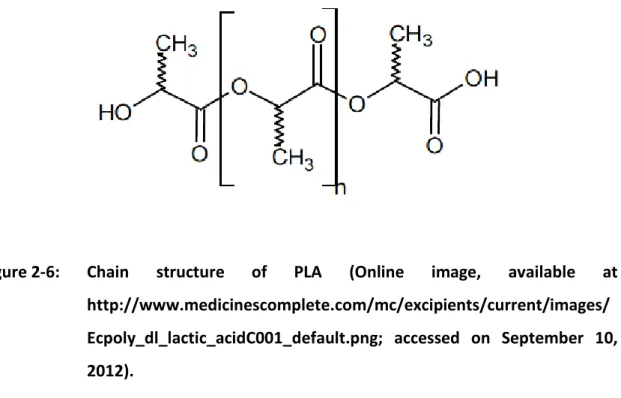
Polymeric nanoparticles can be synthetized from synthetic and natural polymers, varying in polarity from hydrophilic to hydrophobic [56]. However, natural polymers as proteins and polysaccharidesare not extensively used because they vary in purity and often require preparation processes that can lead to drug degradation [37]. Concerning to the synthetic ones,the accumulation and the potential cytotoxicity of non-biodegradable polymers is a major problem and limits their use in humans. Therefore, just a few types of biocompatible and biodegradable polyesters like poly (lactic acid) (PLA), poly lactic-co-glycolic acid (PLGA) and poly-ε-caprolactone (PCL) have been widely studied [11, 57]. Among these polyesters, in the preparation of polymeric drug delivery systems for long-term sustained-release of various drugs,PLA has numerous advantages over the others because, it is produced from renewable resources, it is recyclable and compostable, its physical and mechanical properties can be easily altered though the manipulation of its architecture and it is approved by Food and Drug Administration (FDA) for clinic use [58, 59].
PLA (Figure 2-6) is a linear, lipophilic, thermoplastic, strength and high-modulus polymer, soluble in organic solvents but insoluble in common alcohols and water [11, 18, 60]. Its constituting monomer is lactic acid, obtained annually from renewable resources such as corn starch or sugarcane, by carbohydrate fermentation [11, 60]. Lactic acid exists in two optically active configurations, L- lactic acid and
D-Preparation of Particles for Cosmetic Applications in Human Hair
24
lactic acid and, polymers synthesized from one of them are partially crystalline while the racemic poly (D, L-lactic acid) is amorphous [18].
Figure 2-6: Chain structure of PLA (Online image, available at
http://www.medicinescomplete.com/mc/excipients/current/images/ Ecpoly_dl_lactic_acidC001_default.png; accessed on September 10, 2012).
The synthesis of PLA can follow two different routes of polymerization: condensation/coupling and ring-open polymerization [60]. The first route leads to the formation of a polymer by the linking of monomers with the release of water or similar simple substances, allowing the production of low molecular weight PLAs (< 3000 g/mol). On the other hand, ring-opening polymerization produces polymers with higher molecular weights and the method is based in addition polymerization where the terminal end of a polymer acts as a reactive center and cyclic monomers join to form a larger polymer chain [18, 59].
The presence of ester linkages in the polymer backbone allows its hydrolytic degradation in aqueous environment. The degradation rate is dependent on several parameters such as crystallinity (crystalline PLA degrades slower), molecular weight (low molecular weight PLAs degrade faster) and the environment conditions (pH, ionic strength, temperature) [18]. Regarding to the production of drug delivery systems, these dependence between the properties of polymer and its degradation rate is an important advantage of PLA; since the degradation rate is related to the release of
Review of the Literature
25
drugs, altering the polymeric material, it is possible to manufacture systems with a rate delivery that can be modulated depending on duration of the desired effect [61]. Following the hydrolysis of the polymer, the only degradation product is lactic acid, which it is biocompatible, metabolized through citric acid cycle and then removed from the body [18].
2.5.1.1. Preparation of Drug-loaded Polymeric Nanoparticles
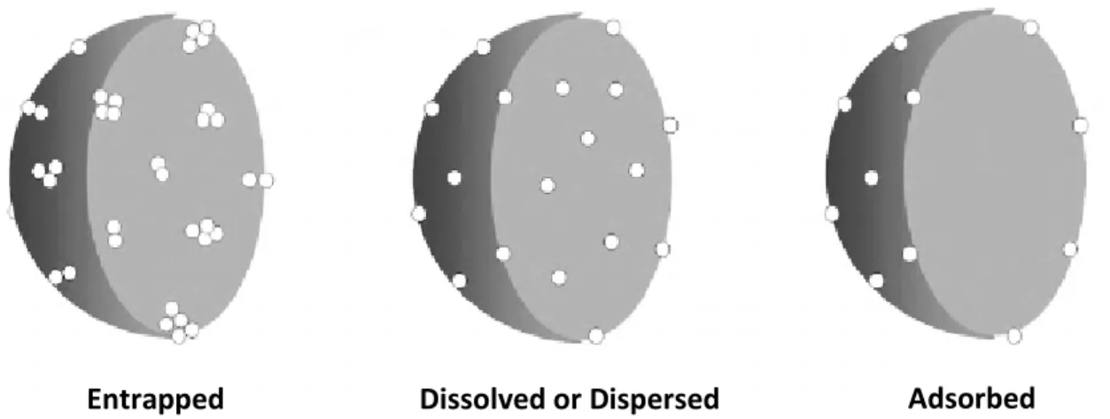
Numerous methods such as single emulsion [62], spontaneous emulsion solvent diffusion (SESD)[63], double emulsion [23], salting-out [64], spray drying [65] and nanoprecipitation [66] have been proposed for the preparation of polymeric drug-loaded nanoparticles.All of these methodsareable to produce NPs with high yield and high encapsulation efficiency and, in general, the process is critical in determining nanoparticle size [67, 68].Considering the encapsulation mechanism, the drugs can be
entrapped, dispersed, dissolved within or simply adsorbed on the matrix of nanoparticles (Figure 2-7), depending upon the method of preparation and the
physico-chemical properties of the therapeutic agent [22].
Figure 2-7: Mechanisms of drug encapsulation on the nanoparticles (Adapted
from Guterres S.S. et al., 2007 [22]).
Preparation of Particles for Cosmetic Applications in Human Hair
26
The great challenge in choosing a method for the preparation of particles consists on an adequate formulation, establishing the ideal amounts of drug, polymer, solvent and non-solvent, which it will allow the production of stable nanoparticles, efficient entrapment of drugs and their release according to the desirable profile [15, 67].
In the production of polymeric particles with single emulsion method (also known as solvent evaporation method), Figure 2-8, the polymer is dissolved in an organic solvent along with the drug and this mixture is emulsified into an aqueous solution, using a surfactant/emulsifying agent. After formation of stable oil in water (o/w) emulsion, the organic solvent is evaporated from dispersed oil droplets (containing both polymer and drug) [62].
Figure 2-8: Production of polymeric particles with single emulsion method
(Adapted from Gomes V.M.A., 2009 [15]).
The spontaneous emulsion solvent diffusion technique is a modified version of the solvent evaporation method, schematized in Figure 2-8 [63]. In this method, the organic solution used to solubilize the polymer and the drug is a binary mixture of a water-miscible organic solvent and a water-immiscible solvent. The polymeric solution is then poured, with mechanical stirring, into an aqueous phase containing a stabilizer
Organic Solution Containing the Polymer and the Drug
Aqueous Solution Containing the Surfactant/Emulsifying agent Dispersed Oil Droplets Evaporation of the organic solvent o/w emulsion
Review of the Literature
27
and emulsion droplets are formed; droplets can also be induced by sonication and homogenization [57, 69]. Due to the quickly spontaneous diffusion of water-soluble solvent out from each emulsion, an interfacial turbulence between the two phases is created and a drastically reduction in the size of particles is obtained (Figure 2-9) [15, 62]. The consequent removal of the organic solvent from the system makes the droplets solidify to finally form polymeric nanoparticles varying in size from 100 to 400 nm. However, a major concern about this method is the large-scale production that sometimes causes a severe aggregation in the particle formation when the polymeric concentration is increased to an acceptable range for industrial purposes [57].
Figure 2-9: Mechanism of nanoparticles formation by the SESD method (Adapted
from Murakami H. et al., 1999 [57]).
The SESD method and solvent evaporation previous described are widely used for the encapsulation of hydrophobic drugs but they have poor results incorporating bioactive agents of a hydrophilic nature. Hydrophilic drugs have a high tendency to move from the organic phase to the outer aqueous phase, not into the polymeric nanoparticles [67, 69]. Thus, another emulsification method - the double emulsion technique, represented in Figure 2-10 - has become the favored protocol for the encapsulation of this kind of compounds [23]. First, the drug for encapsulation is dissolved in water and this phase is dispersed in an organic solvent, which it contains
Formation of emulsion droplets (After dispersion of polymer solution into aqueous phase)
Diffusion of water-soluble solvent out from each emulsion
Removal of the organic solvent
Preparation of Particles for Cosmetic Applications in Human Hair
28
the degradable polymer - the first emulsion, water in oil (w/o), is formed. Then, the w/o emulsion is dispersed into an aqueous medium (containing a stabilizer), forming the final w/o/w emulsion. Once again, as the solvent evaporates, the polymer hardens encapsulating the drug [16].
Figure 2-10: Method for the preparation of nanoparticles based on a
double-emulsion(Adapted from Gomes V.M.A., 2009 [15]).
Another method to prepare polymeric nanoparticles is the salting-out process [64]. Using this technique, a w/o emulsion is formed containing the polymer, acetone, magnesium acetate tetrahydrate, stabilizer, and the active compound. Subsequently, water is added until the volume is sufficient to allow the diffusion of the acetone into the water, which results in the formation of nanoparticles. This suspension is purified by cross-flow filtration and lyophilization. One disadvantage of this procedure is that it uses salts that are incompatible with many bioactive compounds [69].
Organic Solution (containing the polymer)
Aqueous Solution (containing the drug)
Aqueous Solution (containing a stabilizer) w/o emulsion is formed w/o/w emulsion is formed Nanoparticles in suspension Addition
Review of the Literature
29
A preparation technique that it is suitable for industrial scalable processing is spray drying because it is very rapid and convenient, also having very few processing parameters. In this process, drug-loaded particles are prepared by spraying a solid-in-oil dispersion or water-in-solid-in-oil emulsion in a stream of heated air. The type of drug (hydrophobic or hydrophilic) decides the choice of solvent to be used in the process [65].
The nanoprecipitation, first introduced by Fessi et al., is the most commonly used technique to formulate polymeric nanoparticles, intended for cutaneous applications [22, 66]. It is a simple, fast, efficient, economic and reproducible method for the preparation of small particles, based on precipitation and subsequent solidification of the polymer at the interface of a solvent and a non-solvent (Figure 2-11). Thus, the process is often called solvent displacement or interfacial deposition. [18, 22, 68] The choice of the polymer, solvent and non-solvent system is critical for the success of the method and the nature of the polymer-solvent interactions has been reported to affect the properties of the nanoparticle preparation. However, no clear guidelines about the influence of each of the three components of the system have yet emerged [68].
Figure 2-11: Schematic illustration of the nanoprecipitation method(Adapted from
Gomes V.M.A., 2009 [15]). Polymer in solution Non-solvent of the polymer Nanoparticles Evaporation of the solvent Precipitation of the polymer
Preparation of Particles for Cosmetic Applications in Human Hair
30
To achieve nanoprecipitation, the polymer is dissolved in an organic solvent (or mixture of solvents) and added to a non-solvent of the polymer wherewith the organic solvent is miscible; depending on its solubility, the drug to be encapsulated must be dispersed in the aqueous solution or dissolved in the organic solvent before the fusion of the phases [18, 68]. The nanoparticles form instantaneously by precipitation of the polymer and Marangoni effect is considered to explain the process: solvent flow, diffusion and surface tension at the interface of the organic solvent and the aqueous phase cause turbulences, which form small droplets containing the polymer; subsequently, as the solvent diffuses out from the droplets, the polymer precipitates. To finish, the organic solvent is typically removed by evaporation [18, 68]. Although the presence of surfactants/stabilizers is not indispensable for the formation of the particles, they can be included in the process to modify the size and the surface properties or to ensure the stability of the nanoparticle dispersion (especially during the early stages of the precipitation). Thus, in this method, no emulsification step (which is usually part of a nanoparticle preparation process), laborious processing conditions or special laboratory ware is needed and the nanoparticles prepared by nanoprecipitation are typically smaller than 500 nm [18]. Nevertheless, this technique suffers the drawback of a poor encapsulation efficacy of water-soluble drugs due to the rapid migration, and therefore loss of drug, into the aqueous phase [70].
The methods previously mentioned can be successfully employed for the encapsulation of many drugs as hinokitinol [71], minoxidil [72], latanoprost [73], finasteride [74], cyclosporine A [75] and tamoxifen [76]. All of these therapeutic agents have been studied and developed for the safe and effective treatment of several hair follicle disorders/diseases.
2.5.1.2. Drug Release from Polymeric Nanoparticles
After the penetration of nanoparticles inside the hair follicle canal, the encapsulated drug is released and, independently, it penetrates through the intact
Review of the Literature
31
barrier of the hair follicle to reach the target structure (Figure 2-12) [50]. The rate of release is dependent upon desorption of the surface-bound/adsorbed drug, diffusion of drug through the polymer wall, nanoparticle matrix degradation or a combined degradation/diffusion process [62]. In nanoparticulate DSS,the ideal release profile of a therapeutic agent is a near zero-order profile, which establishes a more constant flow of the bioactive agent out of the carrier, maintaining more appropriate steady level of the drug at the site of delivery [13]. In the development of a drug carrier, in order to achieve this steady release rate, it is important to know the properties of the delivery system, the characteristics of the drug and the degree of polymer-drug interaction because they can influence the release of the bioactive compound [23].
Figure 2-12: Release of the drug and targeting of follicular structures. A) Accumulation of polymeric drug-loaded nanoparticles within the hair follicle canal, at the level of the target structure. B) Release of the drug from the particles and its penetration through the hair follicle barrier. (Adapted from Papakostas D. et al., 2011 [11]).
Regarding to the properties of the carrier, a decrease in particle size (and increase in the surface area) results in high release. Higher porosity, inducing a larger inner surface, can increase the influx of the release medium into the particles and, thereby, also facilitate the drug diffusion rate. In addition, specific properties of the
![Figure 2-5: Cross-section diagram of a human hair follicle (Adapted from Meidan V.M. et al., 2005 [46]).](https://thumb-eu.123doks.com/thumbv2/123dok_br/17837150.845223/48.892.144.756.366.848/figure-cross-section-diagram-human-follicle-adapted-meidan.webp)
![Figure 2-8: Production of polymeric particles with single emulsion method (Adapted from Gomes V.M.A., 2009 [15]).](https://thumb-eu.123doks.com/thumbv2/123dok_br/17837150.845223/54.892.138.759.525.830/figure-production-polymeric-particles-single-emulsion-method-adapted.webp)
![Figure 2-10: Method for the preparation of nanoparticles based on a double- double-emulsion (Adapted from Gomes V.M.A., 2009 [15]).](https://thumb-eu.123doks.com/thumbv2/123dok_br/17837150.845223/56.892.172.716.287.750/figure-method-preparation-nanoparticles-double-double-emulsion-adapted.webp)
![Figure 2-11: Schematic illustration of the nanoprecipitation method (Adapted from Gomes V.M.A., 2009 [15]).Polymer insolutionNon-solvent ofthe polymer Nanoparticles Evaporation ofthe solventPrecipitation ofthe polymer](https://thumb-eu.123doks.com/thumbv2/123dok_br/17837150.845223/57.892.138.758.729.1025/schematic-illustration-nanoprecipitation-adapted-insolutionnon-nanoparticles-evaporation-solventprecipitation.webp)