Universidade do Minho
Escola de Ciências
Inês Soares Ferreira
outubro de 2015
Lipid based nanocarriers for the delivery of the
bioactive compound resveratrol
Inês Soar
es F
err
eir
a
Lipid based nanocarrier
s for t
he deliver
y of t
he bioactive compound resveratrol
UMinho|20
Universidade do Minho
Escola de Ciências
Inês Soares Ferreira
outubro de 2015
Lipid based nanocarriers for the delivery of the
bioactive compound resveratrol
Trabalho efetuado sob a orientação da
Professora Doutora Maria Elisabete Cunha Dias
Real Oliveira
e do
Professor Doutor Hernâni Varanda Gerós
Dissertação de Mestrado
iii
Acknowledgments
This dissertation benefited from the insights and direction of several people. First, I would like to express my gratitude to Professor Elisabete Oliveira, as head of the research group and my supervisor, for accepting me in her group and for giving me the necessary conditions for my work to be carried out and for all the knowledge she passed on to me. I would also like to express my deepest appreciation and gratitude to my co-supervisor, Professor Hernâni Gerós, for receiving me so well in his research group and for always being available. I will always be grateful for the education, guidance, mentorship and friendship he has provided me.
I would like to give my sincere gratitude to Doctor Marlene Lúcio for having accompanied me throughout my investigation. Her friendship, support, availability and the knowledge she shared with me, along with her useful suggestions, greatly improved this dissertation. I also want to show my thankfulness to Professor Manuela Côrte-Real for teaching me about the flow cytometry technique and to Cristina Ribeiro, for helping me during the flow cytometry experiments.
I want to acknowledge the contributions of my colleague Justine Demaître for the kindness in providing her results regarding the coefficient partition and the HSA binding assays. I also would like to thank my colleagues Jorge Rodrigues and Ana Garcia, which have accompanied me in the biology experiments, for their friendship and for helping me in any way they could. I also wish to extend my gratitude to all my laboratory colleagues for their help and advices whenever I needed. Besides, I want to thank them for their friendship and sympathy and for the relaxing moments, which made my work much more satisfying and amusing.
I want to show my gratefulness to all my caring friends and family. To my brother Pedro for his friendship and good mood which has helped me in the more stressful times. To my boyfriend for all the love and patience he has giving me throughout this year.
Last, but above all, to my loving parents for giving me the freedom to choose my path while teaching in me that freedom entails sense of responsibility. For all the opportunities they have offered me and for always encouraging me to follow my dreams. For their constant love, care and affection. In them I see my role model and I would be happy if my children were one day half as proud of me as I am of them. For all this and more, they will have my eternal gratitude.
v
Lipid based nanocarriers for the delivery of the bioactive compound resveratrol
Abstract
Resveratrol is a phenolic compound produced naturally by 72 different plant species, particularly grapevines, pines and legumes1. This compound has powerful anti-oxidant, anti-inflammatory and anti-cancer effects2,3. However, its fast metabolization and reduced solubility in biological fluids impairs its bioavailability. Therefore, it is essential to obtain a suitable carrier to achieve an effective therapy. Liposomes are great candidates as delivery systems since they present high biocompatibility, protection and controlled release of the drug.
In the present study, plain and resveratrol loaded DODAB:MO liposomes (1:2) were prepared and characterized over time for size, surface charge and polydispersity index to obtain information about the liposomes shelf stability. A thorough characterization of the system was carried out, namely regarding resveratrol biophysical effects in lipid membranes, encapsulation efficiency, controlled release and HSA binding assays. Furthermore, the effect of free and encapsulated resveratrol in the growth of a yeast culture was determined, along with its protective effect against hydrogen peroxide (H2O2) induced oxidative stress. Cell viability and reactive oxygen species production were also evaluated, as well as liposome internalization by yeast cells.
Results showed that resveratrol loaded liposomes produced by incubation were adequate for drug delivery purposes. Resveratrol proved to have a lipophilic character and being unevenly distributed in the lipid formulation, and its partition in the system proved to be spontaneous. Results evidenced the necessity to adapt the formulation to avoid release of the drug in storage conditions, as well as to avoid binding to HSA. Regarding the assays performed in yeast cells, resveratrol did not affect the growth of the yeast and protected the cells against the oxidative stress induced by H2O2. Moreover, neither free nor encapsulated resveratrol affected cell viability and both formulations promoted a decrease of the intracellular reactive oxygen species levels. Resveratrol loaded liposomes are successfully internalized by yeast cells.
vii
Nanotransportadores lipídicos para entrega do composto bioactivo resveratrol
Resumo
O resveratrol é um composto fenólico produzido naturalmente por 72 espécies de plantas, particularmente videiras, pinheiros e legumes1. Este composto apresenta
efeitos antioxidantes, anti-inflamatórios e anticancerígenos2,3. No entanto, a sua rápida
metabolização e a sua reduzida solubilidade em fluidos biológicos diminui a sua biodisponibilidade. Assim, a obtenção de um transportador adequado é essencial, de forma a alcançar uma terapia eficaz. Os lipossomas são ótimos candidatos como sistemas de entrega de fármacos, uma vez que apresentam alta biocompatibilidade, proteção e libertação controlada do fármaco.
No presente estudo, lipossomas compostos por DODAB:MO (1:2) com e sem resveratrol foram preparados e caracterizados ao longo do tempo em relação ao tamanho, carga de superfície e índice de polidispersividade, de forma a obter informação acerca da estabilidade dos lipossomas em condições de armazenamento. Uma caracterização minuciosa do sistema foi conduzida, nomeadamente em relação aos efeitos biofísicos do resveratrol em membranas lipídicas, bem como ensaios de eficiência de encapsulamento, libertação controlada e ligação à HSA. Além disso, foi determinado o efeito do resveratrol livre e encapsulado em culturas de leveduras, juntamente com o seu efeito protetor contra o stresse oxidativo induzido pelo peróxido de hidrogénio (H2O2). Foram também avaliadas a viabilidade celular e a produção de
espécies reativas de oxigénio, bem como a internalização de lipossomas pelas células de levedura.
Os resultados obtidos mostram que lipossomas contendo resveratrol produzidos pelo método de incubação são adequados para fins de entrega de fármacos. O resveratrol provou ter um carácter lipofílico e estar distribuído de forma desigual na formulação lipídica, e a sua partição no sistema mostrou ser espontânea. Os resultados evidenciaram a necessidade de adaptar a formulação de forma a evitar a libertação de fármaco em condições de armazenamento, bem como a evitar a sua ligação à HSA. Relativamente aos ensaios conduzidos em células de levedura, o resveratrol não afetou o crescimento da cultura e protegeu as células contra o stresse oxidativo induzido pelo H2O2. Além disso, o resveratrol, quer livre quer encapsulado, não apresentou qualquer
efeito na viabilidade celular e promoveu a diminuição dos níveis intracelulares de espécies reativas de oxigénio. Os lipossomas com resveratrol são internalizados com sucesso pelas células de levedura.
ix
Table of Contents
Acknowledgments iii Abstract v Resumo vii Table of Contents ix Abbreviations xiii Figure Index xvTable Index xxi
Equation Index xxiii
Dissertation’s Overall Plan and Objectives xxv
CHAPTER 1
STATE OF THE ART 1
1.1. Phenolic compounds in chemoprevention 3 1.1.1. Cancer – a disease of the developed world 3 1.1.2. Phenolics in chemoprevention – the particular case of resveratrol 4 1.1.2.1. Chemical structure, sources and biosynthesis of resveratrol 6 1.1.2.2. Pharmacokinetics of resveratrol: absorption, metabolism, distribution and
excretion 8
1.1.2.3. Toxicity of resveratrol 11
1.1.2.4. Anticarcinogenic activity of resveratrol 11 1.2. Liposomes as anti-cancer drug delivery systems 14
1.2.1. Lipid polymorphism 14
1.2.2. Liposome preparation and classification 17 1.2.3. Liposomes as drug-delivery systems 19
1.2.4. DODAB:MO (1:2) liposomes 23
CHAPTER 2
MATERIALS AND METHODS 27
2.1. Preparation of plain liposomes 29
2.2. Preparation of resveratrol loaded liposomes 30
x
2.2.2. Hydration method 31
2.2.3. Direct mixing method 31
2.3. Size and polydispersity index determination by dynamic light scattering (DLS) 32 2.4. Zeta potential determination by electrophoretic light scattering (ELS) 36
2.5. Shelf stability studies 38
2.6. Quantitative determination of resveratrol by spectroscopy 39 2.6.1. Construction of resveratrol calibration curves by UV/Visible Absorbance
Spectroscopy 42
2.7. Encapsulation efficiency assays 43
2.8. Resveratrol biophysical effects in lipid membranes 45 2.8.1. Influence of resveratrol in the microviscosity and cooperativity of the liposomal
formulation 45
2.8.2. Resveratrol partition coefficient (Kp) assays using a membrane-water system
and derivative spectroscopy 48
2.8.2.1. Determination of the resveratrol partition coefficient (Kp) in liposomal
systems in water 51
2.8.2.2. Determination of the resveratrol thermodynamic parameters of its
membrane partition 51
2.8.2.3. Determination of the resveratrol partition coefficient (Kp) in liposomal systems at biologically relevant pH values 52
2.9. Controlled release assays 52
2.9.1. In storage conditions 52
2.9.2. In physiological conditions 53
2.10. HSA binding assays using dynamic light scattering (DLS) techniques 54 2.11. Preliminary assays with free and encapsulated resveratrol in a yeast model 56
2.11.1. Resveratrol effect in yeast cell growth under fermentative and respiratory
conditions 56
2.11.2. Resveratrol effect in yeast growth under respiratory conditions in the
presence of hydrogen peroxide 58
2.11.3. Effect of hydrogen peroxide in yeast cell death in respiratory conditions 59 2.11.4. Liposome internalization by yeast cells assessed by fluorescence microscopy
60 2.11.5. Cell viability and reactive oxygen species (ROS) quantification assays by flow
xi CHAPTER 3
RESULTS AND DISCUSSION 65
3.1. Liposomes present good characteristics for drug delivery purposes 67 3.2. Liposomes are stable for at least 4 weeks 70 3.3. Resveratrol can be quantified by UV/Vis absorbance spectroscopy 73 3.3.1. In ultrapure water (pH ≈ 5.5) 74
3.3.2. In HEPES buffer (pH = 7.4) 75
3.3.3. In acetate buffer (pH = 5.0) 76
3.4. Incubation is the most efficient method to encapsulate resveratrol 77 3.5. Resveratrol promotes disorganization of the liposomal formulation and diminishes
its microviscosity 81
3.6. Resveratrol has a lipophilic character and it is encapsulated in the liposomal
formulation 83
3.7. Resveratrol partition in the liposomal formulation is spontaneous 89 3.8. Resveratrol is released from liposomes in water 92 3.9. Resveratrol loaded liposomes need PEGylation to avoid binding to HSA 95 3.10. Resveratrol does not affect the growth of yeast cells 98 3.11. Hydrogen peroxide inhibits yeast growth in a dose-dependent manner and
resveratrol slightly counteracts this effect 100 3.12. Resveratrol loaded liposomes are efficiently internalized by yeast cells 104 3.13. Free and encapsulated resveratrol have no effect in cell viability 105 3.14. Free and encapsulated resveratrol decrease endogenous ROS levels 107
CHAPTER 4
CONCLUSIONS AND FUTURE PERSPECTIVES 111
xiii
Abbreviations
µmax Maximum Specific Growth Rate
ADME Absorption, Distribution, Metabolism and Excretion AGEs Advanced Glycation End Products
B Phase Transition Cooperativity COX Cyclooxygenase
DDS Drug Delivery Systems DHE Dihydroethidium
DLS Dynamic Light Scattering DNA Deoxyribonucleic Acid
DODAB Dioctadecyldimethylammonium Bromide DPH 1,6-diphenyl-1,3,5-hexatriene
EE Encapsulation efficiency
ELS Electrophoretic Light Scattering EPR Enhanced permeability and retention FDA Fluorescein Diacetate
GLCRSV resveratrol-3-O-β-glucoside GLURSV resveratrol-3-O-glucuronide
H Hexagonal Phase
HEPES 4-(2-hydroxyethyl)-1-piperazineethanesulfonic acid HSA Human Serum Albumin
IAPs Inhibitor of Apoptosis Proteins family IR Infrared
ki Exponential Inhibition Constant Kp Partition Coefficient
LDV Laser Doppler Velocimetry LUVs Large Unilamellar Vesicles
L Lamellar Phase
Lα Liquid-Crystalline Lamellar Phase Lβ Solid-Crystalline or Gel Lamellar Phase
M Micellar Phase
xiv MO Monoolein (1-oleoyl-rac-glycerol)
mRNA Messenger Ribonucleic Acid MVVs Multivesicular Vesicles
ODC Ornithine Decarboxylase
PCS Photon Correlation Spectroscopy PdI Polydispersity Index
PEG Polyethylene Glycol
pKa Acid Dissociation Constant Q Cubic Phase
RAGE Receptors for Advanced Glycation End Products ROS Reactive Oxygen Species
RR Ribonucleic Reductase RSV Resveratrol
STS Stilbene Synthase SULRSV resveratrol-3-sulfate
SUVs Small Unilamellar Vesicles
Tm Main Phase Transition Temperature UV Ultraviolet
Xmin Minimum Inhibitory Concentration YPD Yeast Extract Peptone Dextrose ζ-Potential Zeta Potential
xv
Figure Index
Figure 1. Worldwide cancer incidence in both sexes (this statistic excludes all
non-melanoma skin cancers)10. 3
Figure 2. Chemical structure of resveratrol (trans-3-5-4’-trihydroxystilbene). 7
Figure 3. Schematic representation of trans-resveratrol biosynthesis by stilbene
synthase51. 7
Figure 4. Schematic representation of the pathways of resveratrol absorption,
distribution, metabolism and excretion. GLCRSV, resveratrol-3-O-β-glucoside (piceid); SULRSV, resveratrol-3-sulfate; GLURSV, resveratrol-3-O-β-glucuronide
(adapted from 18). 10
Figure 5. Impact of the packing parameter (γ) on lipid assemblies formed in
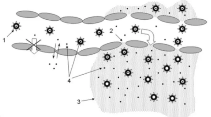
aqueous solutions (adapted from 66). 16 Figure 6. Enhanced permeability and retention (EPR) effect. Long-circulating drug
carriers (1) penetrate through the leaky pathological vasculature (2) into the tumor interstitium (3) and degrade there, releasing a free drug (4) and creating its
high local concentration86. 22
Figure 7. Chemical structure (on the top) and molecular model (on the bottom) of
the lipid dioctadecyldimethylammonium bromide (DODAB). 23
Figure 8. Chemical structure (on the top) and molecular model (on the bottom) of
the lipid monoolein (MO). 24
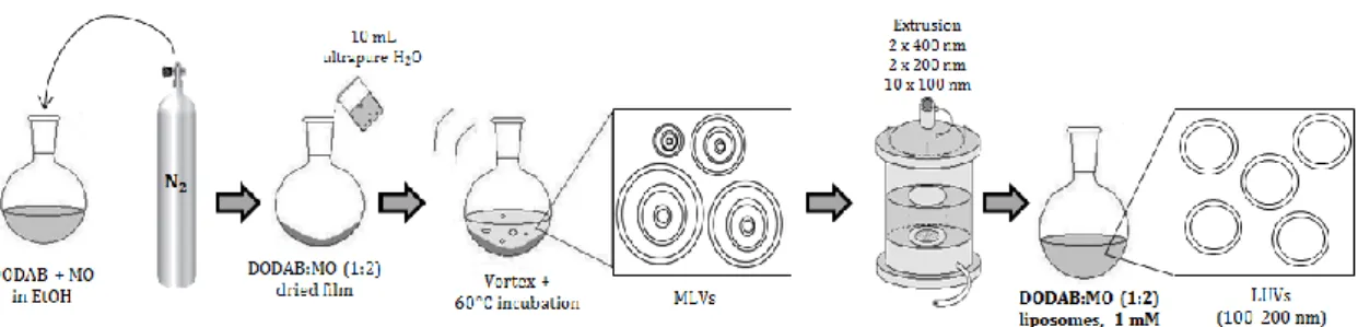
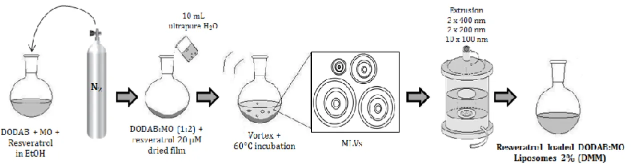
Figure 9. Schematic representation of the preparation of plain DODAB:MO (1:2)
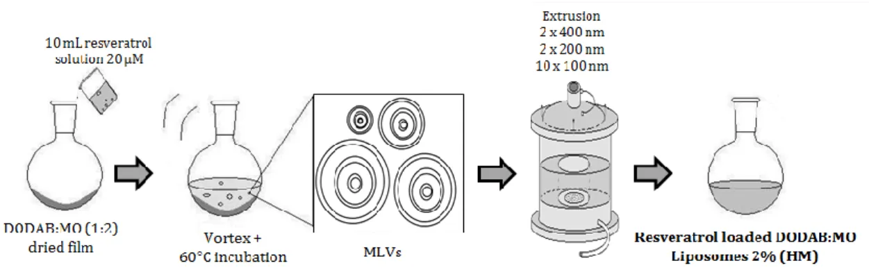
liposomes by the lipid film hydration plus extrusion technique (adapted from 96). 29 Figure 10. Schematic representation of the preparation of resveratrol loaded
DODAB:MO (1:2) liposomes (2%) produced by the incubation method of encapsulation from previously formed liposomes by the lipid film hydration plus extrusion technique (adapted from 96). 30 Figure 11. Schematic representation of the preparation of resveratrol loaded
DODAB:MO (1:2) liposomes (2%) produced by the hydration of the lipid film method of encapsulation (adapted from 96). 31 Figure 12. Schematic representation of the preparation of resveratrol loaded
DODAB:MO (1:2) liposomes (2%) produced by the direct mixing method of
encapsulation (adapted from 99). 32
Figure 13. Schematic representation of a modern dynamic light scattering
apparatus possessing both classic (90°) and backscatter (173°) configuration for detection of scattered light intensity65. 33
xvi
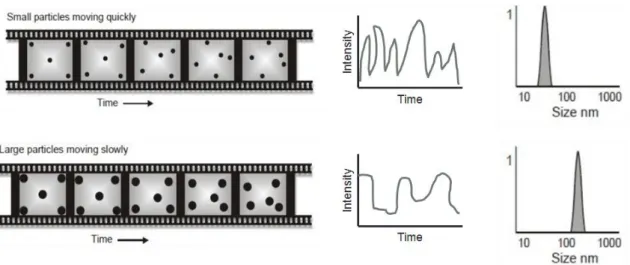
Figure 14. Schematic representation of particles moving randomly in a liquid.
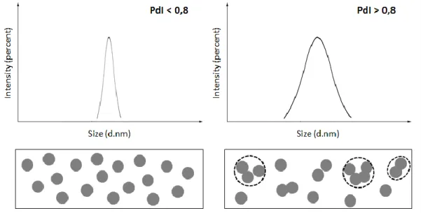
Their motion speed results in different intensity fluctuations which are used to determine particle size (adapted from 101). 34 Figure 15. Schematic representation of the intensity versus the size distribution of
two samples. The sample on the left presents particles with identical hydrodynamic radius, thus being a monodisperse population and having a small PdI, while the sample on the right shows a heterogeneous population with the presence of aggregates which results in a higher PdI, being this a polydisperse
population (adapted from 99). 35
Figure 16. Schematic representation of the electrical double layer surrounding a
particle in suspension101. 37
Figure 17. Schematic representation of a dip cell (left) and the LDV technique
(right)101. 38
Figure 18. Schematic representation of the components of a double beam
spectrophotometer106. 41
Figure 19. Example of a sigmoid profile curve representing the phase transition
after the nonlinear fitting to equation 12, where the refined parameters are Tm
and B. 47
Figure 20. Typical yeast population growth curve in a population grown in a
culture flask. 57
Figure 21. Schematic representation of growth experiments to evaluate the effect
of resveratrol on yeast growth in the presence of H2O2. 58 Figure 22. Schematic representation of the dilution process carried out during the
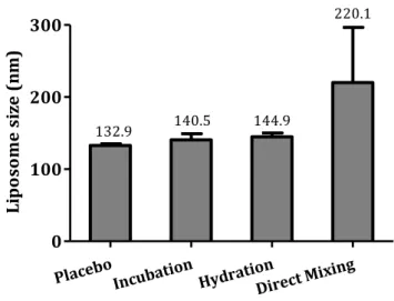
experiments to evaluate the effect of H2O2 in yeast cell death. 60 Figure 23. Mean liposome size and its standard deviation for placebo liposomes
and 2% resveratrol loaded liposomes produced by incubation, hydration and direct mixing methods of encapsulation. 67
Figure 24. Mean PdI and its standard deviation for placebo liposomes and 2%
resveratrol loaded liposomes produced by incubation, hydration and direct mixing
methods of encapsulation. 68
Figure 25. Mean ζ-potential and its standard deviation for placebo liposomes and
2% resveratrol loaded liposomes produced by incubation, hydration and direct
mixing methods of encapsulation. 69
Figure 26. Mean liposome size and its standard deviation for placebo liposomes
and 2% resveratrol loaded liposomes produced by incubation, hydration and direct mixing methods of encapsulation during the three months following
liposome production. 70
Figure 27. Mean PdI and its standard deviation for placebo liposomes and 2%
xvii methods of encapsulation during the three months following liposome production.
71
Figure 28. Mean ζ-potential and its standard deviation for placebo liposomes and
2% resveratrol loaded liposomes produced by incubation, hydration and direct mixing methods of encapsulation during the three months following liposome
production. 72
Figure 29. pH values and its standard deviation for placebo liposomes and 2%
resveratrol loaded liposomes produced by incubation, hydration and direct mixing methods of encapsulation during the three months following liposome production.
72
Figure 30. Absorbance spectra of resveratrol with growing concentrations
solubilized in ultrapure water, HEPES buffer and acetate buffer. 74
Figure 31. Absorbance spectra of resveratrol standard solutions with increasing
concentrations solubilized in ultrapure water. 74
Figure 32. Resveratrol calibration curve in ultrapure water at maximum
wavelength of 305 nm. 75
Figure 33. Absorbance spectra of resveratrol standard solutions with increasing
concentrations solubilized in HEPES buffer (pH = 7.4). 75
Figure 34. Resveratrol calibration curve in HEPES buffer (pH = 7.4) at maximum
wavelength of 305 nm. 76
Figure 35. Absorbance spectra of resveratrol standard solutions with increasing
concentrations solubilized in acetate buffer (pH = 5.0). 76
Figure 36. Resveratrol calibration curve in acetate buffer (pH = 5.0) at maximum
wavelength of 305 nm. 77
Figure 37. First derivative of the absorbance spectra of twelve resveratrol
standard solutions with growing concentrations ranging from 0.5 to 100 µM
solubilized in ultrapure water. 77
Figure 38. Resveratrol calibration curve in ultrapure water using the first
derivative of the absorbance at maximum wavelength of 341 nm. 78
Figure 39. First derivative of the absorbance spectra of the placebo liposomes that
remained in the filter after amicon ultracentrifugation (light red line) and of the resveratrol loaded liposomes produced by incubation that remained in the filter after amicon centrifugation (dark red line). 78
Figure 40. Graphic representation of the resveratrol (2%) encapsulation
efficiency in resveratrol loaded DODAB:MO (1:2) liposomes produced by incubation, hydration and direct mixing methods. 79
Figure 41. Drug loading efficiency of increasing concentrations of resveratrol (0.5,
xviii
Figure 42. Representation of the mean count rate in percentage of DODAB:MO
(1:2) liposomes with (white circles) and without (black circles) resveratrol versus temperature. Each point corresponds to the mean value of three measurements and the correspondent standard deviation is represented. The lines (black for plain liposomes and grey for resveratrol loaded liposomes) represent the best non-linear fitting according to the equation 12, where the refined parameters were Tm
and B. 82
Figure 43. Absorbance spectra of three samples of resveratrol (43 µM)
represented by RSV 1, RSV 2 and RSV 3, and of the samples of resveratrol loaded liposomes with increasing lipid concentrations and a fixed resveratrol concentration (S1-S9). The correspondent references prepared in the same manner as the samples but without the incorporation of the drug are presented in
grey. 84
Figure 44. Representation of the first (A), second (B) and third (C) derivatives of
three samples of resveratrol (43 µM) represented by RSV 1, RSV 2 and RSV 3, of the samples of resveratrol loaded liposomes with increasing lipid concentrations and a fixed resveratrol concentration (S1-S9), and of the reference samples presented in grey. The peaks used to calculate the resveratrol Kp are identified in
the spectra. 85
Figure 45. Representation of the absorbance values of the first derivative at λ =
341 nm (A), of the second derivative at λ = 351 nm, and of the third derivative at λ = 343 nm (C) and at λ = 359 nm (D), and respective nonlinear regressions fitted to equation 14. Below each graphic representation is presented the respective partition and correlation coefficients. The Log Kp value is presented in mol.L-1. 86 Figure 46. Representation of the second derivative of the absorbance values of
three samples of resveratrol (43 µM), of the samples of resveratrol loaded liposomes with increasing lipid concentrations and a fixed resveratrol concentration, and of the reference samples. The bathochromic shift is also
represented in the graphic. 88
Figure 47. Fiting of the third derivative spectrophotometric data collected at λ =
311 nm for resveratrol loaded DODAB:MO (1:2) liposomes at different temperatures calculated with derivative spectroscopy at different temperatures: 30°C (black), 37°C (red), 50°C (green), 55°C (dark blue), 60°C (light blue). 90
Figure 48. Van’t Hoff regression for the resveratrol partition in DODAB:MO (1:2)
LUVs. The pink square represents the Lα phase and the blue square represents the
Lβ phase. 90
Figure 49. Cumulative resveratrol release from DODAB:MO (1:2) liposomes in
storage conditions (ultrapure water, pH ≈ 5.5) and in acetate buffer (pH = 5). 93
Figure 50. Cumulative resveratrol release from DODAB:MO (1:2) liposomes in
HEPES buffer (pH = 7.4) and in acetate buffer (pH = 5). 94
Figure 51. Size and ζ-potential changes upon binding to HSA of increasing
xix
Figure 52. Mean sizes (columns) and PdI (dots) of resveratrol loaded liposomes
in the presence (blue) and in the absence (red) of HSA, when the lipid
concentration is increasing. 96
Figure 53. ζ-potential variation of plain and resveratrol loaded liposomes with
increasing lipid concentrations in the presence of HSA. 97
Figure 54. Growth of S. cerevisiae W303 in YPD medium (fermentative conditions)
(A) and in lactate/ethanol medium (respiratory conditions) (B) in the presence and absence of 100 µM resveratrol. Resveratrol was prepared in ethanol 100% (v/v) before addition to the culture medium, so the final concentration of ethanol in the growth experiment was 2.2% (v/v). Control experiments with ethanol alone (final concentration: 2.2% (v/v)) and neither resveratrol nor ethanol are also
shown. 98
Figure 55. Growth of S. cerevisiae W303 in lactate/ethanol medium (respiratory
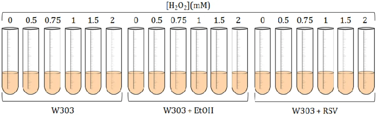
conditions) in the absence (A) and in the presence of 200 µM resveratrol (+ EtOH 2.2% (v/v)) (B) and with ethanol 2.2% (v/v) (C) before and after the addition of H2O2 with growing concentrations (0, 0.5, 0.75, 1, 1.5 and 2 mM) to the culture
media. 100
Figure 56. Protective effect of resveratrol on the inhibition of yeast cell growth
mediated by 0.5 mM H2O2. Growth of S. cerevisiae W303 occurred in
lactate/ethanol medium (respiratory conditions) and 0.5 mM H2O2 in the absence
and in the presence of 200 µM resveratrol (+ EtOH 2.2% (v/v)) or ethanol alone. 101
Figure 57. Dependence of the specific growth rates of S. cerevisiae W303 grown in
lactate/ethanol medium (respiratory conditions) on the extracellular H2O2
concentration in the absence (A) and in the presence of 200 µM resveratrol (+ EtOH 2.2% (v/v)) (B) or ethanol alone (C), and the respective Xmin. 102
Figure 58. Spot test analysis representing the growth of S. cerevisiae on YPD agar
medium when cells were incubated with up to 5 mM H2O2. 103 Figure 59. Bright-field (A), fluorescence (B) and overlay of both (C) micrographs
of S. cerevisiae W303 yeast cells incubated with (1) 3 µM DPH free fluorescent probe to study cell capacity to internalize it (control experiment) and with (2) resveratrol loaded DODAB:MO (1:2) liposomes labeled with 3 µM DPH fluorescent
probe. Scale bar = 7.5 µm. 105
Figure 60. Flow cytometry analysis of S. cerevisiae W303 yeast cell populations to
study cell viability with the FDA probe. Scattergram of a population of yeast cells in the absence of resveratrol (A); overlay histogram of autofluorescence (black line) and FDA induced fluorescence (green line) in the absence of resveratrol (B) and upon treatment of yeast cells with 200 µM of free resveratrol (C) and
resveratrol loaded liposomes (D). 106
Figure 61. Effect of free and encapsulated resveratrol on cell viability in S.
cerevisiae W303 grown in lactate/ethanol medium. 107
Figure 62. Flow cytometry analysis of S. cerevisiae W303 yeast cell populations to
study the effect of resveratrol against endogenous ROS with the DHE probe. Scattergram of a population of yeast cells in the absence of resveratrol (A); overlay
xx histogram of autofluorescence (black line) and DHE induced fluorescence (orange
line) in the absence of resveratrol (B) and upon treatment of yeast cells with 200 µM of free resveratrol (C) and resveratrol loaded liposomes (D). 108
Figure 63. Effect of free and encapsulated resveratrol on the percentage of cells
producing intracellular ROS in yeast cells S. cerevisiae W303 grown in
xxi
Table Index
Table 1. Biophysical parameters (B and Tm) of DODAB:MO (1:2) liposomes in the
absence and presence of resveratrol. 83
Table 2. Coefficient partition (Kp) values of resveratrol in a
LUV((DODAB:MO)(1:2))/H2O system and the respective logarithms (Log Kp) and
coefficient partition values of resveratrol in octanol:water systems (Log P). 87
Table 3. Coefficient partition (Kp) values of resveratrol at physiological relevant
pH’s in a LUV((DODAB:MO)(1:2))/H2O system and the respective logarithms (Log
Kp). 88
Table 4. Coefficient partition (Kp) values of resveratrol in a
LUV((DODAB:MO)(1:2))/H2O system and the respective logarithms (Log Kp) at
different temperatures. 89
Table 5. Variation of the enthalpy (ΔH)±SD, entropy (ΔS)±SD and Gibbs free
energy (ΔG) obtained for the resveratrol partition between the aqueous phase and DODAB:MO (1:2) liposomes at different temperatures. 91
Table 6. Maximum specific growth rate (µmax) of S. cerevisiae W303 liquid cultures.
99
Table 7. Effect of H2O2 on the growth of S. cerevisiae. Cells were cultivated in
lactate/ethanol medium (respiratory conditions) in the absence and in the presence of 200 µM resveratrol (+ EtOH 2.2% (v/v)) and with ethanol alone, in the presence of different H2O2 concentrations. 103
xxiii
Equation Index
Equation 1. Determination of the critical packing parameter (γ). 15
Equation 2. Stokes-Einstein equation for the diffusion coefficient (D) of the particle. 33
Equation 3. Henry’s equation for electrophoretic mobility (µE) of a colloidal particle. 37
Equation 4. Determination of the intensity of transmitted light (Iλ). 40
Equation 5. Determination of the transmittance (Tλ). 40
Equation 6. Determination of the absorbance (Aλ). 40
Equation 7. Determination of the absorbance coefficient (αλ). 40
Equation 8. Beer-Lambert equation. 40
Equation 9. Equation of a straight line. 43
Equation 10. Determination of the encapsulation efficiency (EE) in percentage. 44
Equation 11. Determination of the drug loading efficiency in percentage. 45
Equation 12. Modified Boltzmann equation. 47
Equation 13. Correlation between the absorbance and the partition coefficient (Kp). 49
Equation 14. Derivative of the partition coefficient (Kp). 50
Equation 15. Van’t Hoff equation. 50
Equation 16. Gibbs free energy equation. 50
Equation 17. Determination of the dissociation constant (Kd). 54
Equation 18. Variation of the Langmuir isotherm. 55
Equation 19. Gibbs free energy (∆G) equation of the binding. 55
Equation 20. Determination of the culture’s specific growth rate (µmax). 57
xxv
Dissertation’s Overall Plan and Objectives
In the past recent years, intensive research has been done in order to improve the therapeutic efficacy against several diseases, including cancer. It is known that conventional drug formulations present numerous limitations and thus new strategies have been developed to improve treatment performance. Liposomes are one of the most successful drug delivery systems that apply nanotechnology to potentiate the therapeutic effectiveness and reduce toxicities of conventional medicines. Numerous studies have reported the promising properties of resveratrol, including anti-inflammatory, antioxidant and anti-cancer roles.
With this in mind, this work aimed to develop and characterize a resveratrol loaded liposomal formulation with the purpose of trying to improve the therapeutic efficiency of this natural occurring polyphenol. After a thorough biophysical characterization of the resveratrol loaded liposomal formulation, some preliminary work was performed with the purpose of exploring the properties of free and encapsulated resveratrol in the yeast model Saccharomyces cerevisiae, one of the most intensively studied model organism in molecular and cell biology. Basic cellular mechanics of replication, recombination, cell division and metabolism are well conserved between yeast and larger eukaryotes, including mammals. Moreover, the complete sequence of its genome has proved to be extremely useful as a reference towards the sequences of human and other higher eukaryotic genes4,5. Furthermore, this budding yeast grows well in culture, is stable as either a diploid or haploid cell type, and is amenable to both classical genetic as well as molecular genetic manipulations6.
xxvi
This masters’ dissertation is divided in four chapters, which are summarized below:
Chapter 1 – State of the Art
Presents an overview of the literature and of the previous research concerning this topic. This section includes an outline regarding the use of phenolic compounds, particularly resveratrol, in chemoprevention, as well as a review concerning liposomes and why these should be used as anti-cancer drug delivery systems.
Chapter 2 – Materials and Methods
Describes the experimental work carried out during this study. In each subchapter, the techniques employed, their theoretical basis, the instruments used and the protocols are described.
Chapter 3 – Results and Discussion
Presents the experimental results that arose from the laboratory work, as well as a discussion regarding these findings.
Chapter 4 – Conclusions and Future Perspectives
Depicts the main conclusions to be drawn from the experimental work, along with the suggestion of some new experiments to be performed in the near future in order to further comprehend the applications of the formulation at study.
1
C
HAPTER
1
S
TATE OF THE
A
RT
Contents
1.1. Phenolic compounds in chemoprevention
1.1.1. Cancer – a disease of the developed world
1.1.2. Phenolics in chemoprevention – the particular case of resveratrol
1.1.2.1. Chemical structure, sources and biosynthesis of resveratrol
1.1.2.2. Pharmacokinetics of resveratrol: absorption, metabolism,
distribution and excretion
1.1.2.3. Toxicity of resveratrol
1.1.2.4. Anticarcinogenic activity of resveratrol
1.2. Liposomes as anti-cancer drug delivery systems
1.2.1. Lipid polymorphism
1.2.2. Liposome preparation and classification
1.2.3. Liposomes as drug delivery systems
3
1.1. Phenolic compounds in chemoprevention
1.1.1. Cancer – a disease of the developed world
Cancer is one of the leading causes of morbidity and mortality worldwide (figure 1), with approximately 14 million new cases and 8.2 million cancer related deaths in 2012, numbers which are expected to rise by about 70% within the next two decades. Many different types of cancer are known, although breast, lung, liver, stomach, colorectal, oesophageal and prostate cancers account for over half of all new cases and are the most common causes of cancer death7. This complex biological disorder results from integrated effects of environmental, physical, metabolic and genetic factors3. Determining the real causes of cancer is a complex subject, but the well-known risk factors are alcohol and tobacco abuse, infections, radiation, obesity and lack of physical activity. Ageing is another important factor for the development of cancer since the overall risk accumulation is combined with the tendency for cellular repair mechanisms to be less effective as a person grows older2.
Figure 1. Worldwide cancer incidence in both sexes (this statistic excludes all
non-melanoma skin cancers)10.
Cancer, also known as malignant neoplasia, refers to an extensive group of diseases that are associated with a disturbance in the control of cell growth and
4
metabolism. Indeed, the unbalanced control of cellular proliferation is one of the main characteristics of cancer cells11. Since tumor formation is a multistep process, normal cells evolve progressively to the neoplastic stage. Along their way, these cells acquire particular abilities that enable them to become tumorigenic. These distinct hallmark capacities were proposed in 2000 by Hanahan and Weinberg11 and are: (1) sustaining proliferative signaling through uncontrolled activation of oncogenes; (2) evading growth suppressors; (3) enabling replicative immortality through increased telomerase activity; (4) activating invasion and metastasis through the over activation of invasion-related proteases; (5) inducing angiogenesis, which consists in the building of an extensive network of blood vessels to maintain continuous supply of nutrients, and be able to sustain it; and (6) resisting cell death by evading apoptosis through the inhibition of proapoptotic signaling and through the stimulation of survival factor pathways. All these changes make cancer cells unresponsive to antigrowth signals, resulting in the loss of tumor suppressor gene activity8. Over the last decade, noteworthy progress was made in the field of cancer research which led to an improved understanding of these hallmark capabilities, but also led to modifications and, ultimately, expansions of the original concept12.
Considering these concerns and knowing that chemoprevention aims to decrease the occurrence of cancer by the administration of natural or synthetic compounds13, the ideal chemopreventive agent would be one which could inhibit or reverse these processes in neoplastic cells while protecting normal cells9. In fact, the current treatments available are limited because they do not differentiate between normal and cancer cells, which causes side effects and early termination of therapy, ultimately harming the patient14.
1.1.2. Phenolics in chemoprevention – the particular case of resveratrol
Phenolic compounds, also known as phenols, represent the major group of phytochemicals found in plants, particularly in fruits, seeds and leaves15, and other types of foods and beverages such as chocolate, tea and wine16. These are a class of phenylalanine-derived chemical compounds that consist of a reactive hydroxyl group (-OH) bonded directly to an aromatic hydrocarbon ring, and they can be classified as simple phenols or polyphenols, based on the number of phenol units in
5
the molecule17,18. Polyphenols comprise a large class of antioxidants, which are normally produced by plants for their antibiotic and antifungal properties19, and include flavonoids, anthocyanins, phenolic acids, lignans and stilbenes.
Dietary polyphenols have received tremendous attention among nutritionists due to their benefits on human health, since a high intake of fruits, vegetables and whole grains, which are rich in polyphenols, has been associated with lowered risks of various diseases, including cancer, chronic inflammation, cardiovascular and neurodegenerative diseases20. Polyphenols reveal these health benefits through complementing and adding to the functions of antioxidant vitamins and enzymes as defense against oxidative stress, mainly caused by excess of reactive oxygen species (ROS)15. Polyphenols can decrease the oxidation rate either by preventing the free radicals formation or by deactivating the active species and the precursors of the free radicals, usually by donating an electron or a hydrogen atom16. More often, they act as chain breakers, this is, they act as direct radical scavengers of the lipid peroxidation chain reactions by donating an electron to the free radical, which neutralizes the radical and makes polyphenols to become less reactive radicals themselves, therefore stopping the chain reactions21–23. In addition to this radical scavenging, polyphenols also act as transition metal chelators, which can prevent the oxidation caused by highly reactive hydroxyl radicals22,24. So, while polyphenols are undeniably strong antioxidant molecules, most of the evidence of their antioxidant activity is based on in vitro studies, which are limited in terms of similarity to the mechanisms of antioxidant actions in a biological model. Nevertheless, these methods may portray well how polyphenols function as antioxidants, thus shedding light on the actual role of polyphenols in human health. However, caution must be taken since increasing evidence indicates that they may act in ways beyond the antioxidant functions in vivo16. For instance, once polyphenols have donated an electron or hydrogen atom, they become free radicals themselves, which can theoretically lead to pro-oxidant activities. Still, this is a question which needs to be further researched25.
In recent times, natural substances isolated from food and developed as medicines have attracted substantial interest in the field of cancer, mainly because these compounds are part of the daily diet and can be consumed within a reasonable wide range of concentrations without major side effects14. With this in mind, the
6
connection between the potential health benefits of polyphenols and the biological routes associated with cancer has been widely investigated. The action of polyphenols has been studied in various cancers including breast, lung, skin, oral cavity, ovarian, esophagus, stomach, liver, pancreas, endometrial, thyroid, testicular bladder, small intestine, colon, urinary tract and prostate26,27. It has been proved that polyphenols exhibit anti-cancer properties by interfering with molecular events involved in initiation, promotion, and progression stages28.
Over the past 20 years, case-control studies have shown that a high intake of fruit and vegetables, and specific polyphenols found within these, helps to prevent the onset and progression of various types of cancer29,30. At the cellular level, there is noteworthy evidence that some polyphenols influence carcinogenesis and tumor development31 by interacting with reactive intermediates32 and activated carcinogenic and mutagenic agents33, by modulating the activity of key proteins involved in controlling cell cycle progression34, by modulating cancer cell signaling13,35 and enzymatic activities36, by promoting apoptosis37–39 and by influencing the expression of many cancer associated genes40.
Another distinctive property of tumors is the increase in glucose uptake and the high rate of glycolysis which leads to the non-enzymatic glycation of proteins and the generation of advanced glycation end products (AGEs). The amount of some of these AGEs in several human tumors has been related to their involvement in cancer progression41. Interestingly, some polyphenols have been suggested to counteract AGEs formation both in vivo and in vitro, which indicates that these phenols may limit their impact on the carcinogenesis process42–45. Additionally, receptors for AGEs, such as RAGE, play a significant role in regulating cancer cell invasion and metastasis46,47 and some polyphenols can possibly inhibit cancer cell proliferation by blocking RAGE related signaling48.
1.1.2.1. Chemical structure, sources and biosynthesis of resveratrol
Within the phenolic compounds subclass of stilbenes, resveratrol (RSV) (figure 2) is the common term for 3-5-4’-trihydroxystilbene which exists in both trans and cis isomeric forms. However, the trans isomer is widely studied and undoubtedly more abundant in plants18. This natural polyphenol and phytoalexin is
7
produced naturally by 72 different plant species, particularly grapevines, pines and legumes1.
Figure 2. Chemical structure of resveratrol (trans-3-5-4’-trihydroxystilbene).
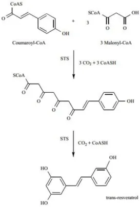
Grapes are the most abundant source of resveratrol for humans, particularly Vitis vinifera, V. labrusca and V. muscadine grapes, which are used in the production of several wines. This compound can be found in roots, seeds and stems of vines, but the concentration is higher in grape’s skin, which contains from 50 to 100μg/g49. The production of resveratrol is induced in response to multiple stress conditions, such as injury, chemical signals from pathogen fungi attack, exposure to ozone, sunlight, heavy metals50, among others, and this process is carried out by the stilbene synthase (STS) enzyme, as shown in figure 3.
Figure 3. Schematic representation of trans-resveratrol biosynthesis by stilbene
8
STS catalyzes three condensation reactions between coumaroyl-coenzyme A and three molecules of malonyl-coenzyme A via cleavage of three carbon dioxide molecules. Moreover, STS also catalyzes the loss of the terminal carboxyl group, leading to the production of the C14 molecule resveratrol51.
1.1.2.2. Pharmacokinetics of resveratrol: absorption, metabolism, distribution and excretion
To understand the potential beneficial properties of a given compound, it is necessary to study its absorption, distribution, metabolism and excretion (ADME). Several studies concerning the resveratrol bioavailability have been conducted both in rodents and humans.
Resveratrol consumed orally tends to be oxidized in the human digestive tract, and this process can be avoided when these molecules undergo glycosylation. Therefore, glycosylated resveratrol will not be oxidized, which will preserve its biological activity and increase its stability and bioavailability. However, several studies show that resveratrol is absorbed in the small intestine, particularly at the jejunum and the ileum, and the intestinal epithelial cells (enterocytes) are unable to absorb glycosylated resveratrol, and so this absorption process requires glycosidases. Once in the enterocytes, resveratrol is extensively metabolized resulting mainly in sulfatide and glucuronide conjugates18, suggesting that resveratrol is released from the intestinal epithelial cells in these conjugated soluble forms into the blood stream52. This biochemical changes that occur almost immediately after ingestion can also occur in the liver51. Although modifications such as glucuronidation and sulphation typically reduce the resveratrol cell permeability and aid in its excretion, which will diminish its bioavailability, resveratrol administered in vivo showed high efficiency. This combined with the fact that in vivo concentrations of individual metabolites can be more than ten times higher than those of the native compound, has led to speculation that resveratrol metabolites could themselves be active in promoting many of the health benefits attributed to resveratrol53. However, several studies show less pharmaceutical impact of these metabolites51.
9
Numerous studies performed in humans show that about 70% of orally administered resveratrol (25 mg) is absorbed and metabolized in less than 30 minutes with a peak plasma level of approximately 2 µM of resveratrol metabolites and a half-life of 9 to 10 hours. However, the resveratrol absorption and metabolization can vary between individuals depending on factors such as the hepatic function and the metabolic and enzymatic activity of the local intestinal microflora51, and also depending on the relative amounts of resveratrol and its conjugates present in the food source or dietary complement18.
One of the factors that impair resveratrol bioavailability is its poor water solubility, which hinders its ability to be solubilized in the blood. However, resveratrol is able to bind itself to plasma proteins and thus assure its body distribution and bioavailability51. Resveratrol can be bound to serum proteins such as hemoglobin and human serum albumin (HSA) by taking advantage of the electrostatic interactions which result, among other factors, from positively charged residues that are close to the binding compound. Both complexes formed are spontaneous and exothermic54. The conversion of resveratrol into hydrophilic conjugates also facilitates its entry into the blood stream and its diffusion throughout the body. Although resveratrol can be found in the colon shortly after oral intake, its distribution in tissues requires a few hours. The liver and the gallbladder filter resveratrol and its metabolites from the circulation and transport them back into the intestine through the bile for a delayed absorption and this process is called recirculation. Figure 4 shows a schematic representation of the pathways of resveratrol ADME. Studies regarding the uptake and metabolism of resveratrol by human liver have shown that human hepatocytes exhibit an initial increasing rate of uptake (minutes), followed by a stable rate in the next few hours55. Even though treatment with high doses of resveratrol results in great accumulation of the drug in the liver, no toxicity or hepatocyte lysis is observed, which suggests that resveratrol has an important role in the prevention of liver diseases51.
10
Figure 4. Schematic representation of the pathways of resveratrol absorption, distribution,
metabolism and excretion. GLCRSV, resveratrol-3-O-β-glucoside (piceid); SULRSV, resveratrol-3-sulfate; GLURSV, resveratrol-3-O-β-glucuronide (adapted from 18).
In the last step of pharmacokinetics, resveratrol metabolites are eliminated from the organism and excretion is almost equally distributed between urine and feces51. However, the excretion time depends strongly on the resveratrol concentration present in plasma – small amounts of resveratrol are rapidly metabolized and eliminated whereas a higher dose of intake results in retention and accumulation of the compound in tissues, thus becoming available for cellular uptake and intracellular signaling18. Resveratrol and its metabolites are almost completely eliminated from tissues 72 hours after a single dose. In humans, the two major metabolites identified in urine were glucuronide- and sulfate- -conjugates of resveratrol and of dihydro-resveratrol. The total recovery of glucuronic and sufate conjugations in urine and feces was about 71-98% after oral doses and 54-91% after intravenous doses, while the native form of resveratrol presented a near to zero retrieval, which suggests that the circulating form of resveratrol is primarily the modified metabolites rather than its native form51.
11 1.1.2.3. Toxicity of resveratrol
Toxicity assessments are an important part of new drug safety profiling, since bioactive drugs may have adverse effects on the organism and on its metabolism.
Resveratrol, when administered to rodents and dogs for 13 weeks at doses up to 1000 and 1200 mg/kg/day respectively, resulted in dose-related increases in plasma levels of free and conjugated resveratrol. However, clinical observations failed to recognize any proof of resveratrol toxicity56. Moreover, human clinical studies have been performed with single doses of 5 g of resveratrol and no adverse side effects were observed51. These observations indicate that 450 mg/day of resveratrol represent a safe dose for a 70 kg individual. Therefore, in humans, resveratrol seems to be well tolerated and to have weak toxicity, indicating that this bioactive compound can be used as a pharmacological drug in human medicine57.
1.1.2.4. Anticarcinogenic activity of resveratrol
Antitumor agents are compounds that inhibit cancer development by blocking tumor cell transformation and proliferation, and by inducing tumor cell death18. Thus, chemoprevention and chemotherapy consist in using natural, synthetic or biologic substances to reverse, suppress or prevent the development of cancer. Amongst the food-derived molecules that can be used as antitumor agents, resveratrol is particularly interesting since it has been shown to modulate a wide range of different intracellular mediators involved in multi-stage carcinogenesis, inflammation, cell cycle and apoptosis. Evidence also supports its association with antioxidant and anti-inflammatory activities51.
Antioxidant effects
Electron acceptors react easily with free radicals, originating ROS. These chemically reactive molecules are continuously generated in cells exposed to aerobic environments, and have been associated with the initiation and progression of cancer58, through directly damaging deoxyribonucleic acid (DNA) and other macromolecules53. Resveratrol has an intrinsic antioxidant capacity that is related
12
to its chemopreventive effects, since it is an excellent radical scavenger that can protect cell membranes against lipid peroxidation and avoid DNA damage caused by the generation of ROS. These protective effects result from the activation of antioxidant enzymes, such as superoxide dismutase, catalase, glutathione reductase, glutathione peroxidase, glutathione transferase and oxidoreductases51. In vivo, resveratrol has been shown to increase plasma antioxidant capacity and decrease lipid peroxidation53.
Anti-promotion effects
Cyclooxygenases (COX) produce prostaglandins from arachidonic acid and these compounds are able to stimulate tumor growth by acting on cell proliferation, angiogenesis and immunosuppression. Therefore, COX inhibitors are considered valuable therapeutic agents against several cancers59. Resveratrol decreases the total COX activity of tumors and normal tissues in vivo through selective inhibition of COX-1 activity and/or reduction of COX-2 at the messenger RNA (mRNA) level. In vitro studies have shown that the transcriptional inhibition of COX-2, as well as another important player in carcinogenesis, ornithine decarboxylase (ODC), could be accomplished through inhibition of protein kinase C. Resveratrol does not directly inhibit ODC activity, but reduces its expression in vivo and prevents its induction by carcinogens53.
Moreover, inflammation mediators such as COX-2, inducible nitric oxide synthase, interferon-γ, pro-inflammatory cytokines and tumor necrosis factor-α, are also involved in carcinogenesis, particularly in the promotion and progression stages. Resveratrol is able to block the expression of these various components of pro-inflammatory signaling through the suppression of the nuclear factor-κB and of the activator protein-1. Also, resveratrol promotes a reduction of the intracellular levels of Ca2+ which act as a secondary messenger during cell inflammatory activation. Resveratrol also downregulates the Akt/CREB activation, a pathway that responds to various signals that drive the cell proliferation, differentiation and adaptive responses51. All functions mentioned above imply that resveratrol could slow tumor development through multiple complementary mechanisms53.
13
Inhibition of angiogenesis
Angiogenesis is the physiological process through which new blood vessels form from pre-existing vessels and it is necessary to maintain the growth of most solid tumors with a diameter beyond 3 mm. Resveratrol, when delivered systemically at a 2.5-100 mg/kg dose, has shown to prevent tumor-induced neovascularization and to promote wound healing. Moreover the suppression of COX and ODC by resveratrol could have a role in the inhibition of angiogenesis since these enzymes promote vascularization and tumor growth.
Alterations in cell cycle and apoptosis
Resveratrol can also combat tumor formation and development by inducing cell cycle arrest and apoptosis. In vivo tumor models indicate that resveratrol has anti-proliferative and pro-apoptotic effects by downregulating cell cycle proteins and increasing apoptosis53. Resveratrol is an effective inhibitor of ribonucleotide reductase (RR), and this inhibition leads to the arrest of the cell cycle in the G1 phase. Resveratrol also inhibits the oncogenic and oxidative stress activated tyrosine kinase Src and therefore blocks the activation of the signal transducer and activator of transcription Stat3 in malignant cells, also resulting in cell cycle arrest and loss of viability. Furthermore, resveratrol leads to accumulation, phosphorylation and acetylation of p53, a tumor suppressor protein that activates the cyclin inhibitor p21 and results in the G1/S arrest. Moreover, resveratrol also downregulates the cyclin D1 enzyme which is overexpressed in cancers and is required for cell cycle G1/S transition51.
The capacity of resveratrol to induce cell cycle arrest leads to subsequent cell apoptosis59. Resveratrol has also been suggested to downregulate surviving, which belongs to the inhibitor of apoptosis proteins family (IAPs). Moreover, in acute lymphoblastic leukemia cells, resveratrol has been shown to induce mitochondria- -mediated apoptosis through the depolarization of mitochondrial membranes by inhibiting the F1 complex of the F0/F1 ATPase proton pump51. Interestingly, resveratrol exerts its pro-apoptotic effect on tumor cells alone, while normal cells remain unharmed59. For instance, resveratrol has been shown to sensitize several
14
tumor lines, but not normal human fibroblasts, to TRAIL (tumor necrosis factor-related apoptosis-inducing ligand)-induced apoptosis53.
1.2. Liposomes as anti-cancer drug delivery systems
1.2.1. Lipid polymorphismAmphiphilic lipids are those consisting of molecules with a polar water- -soluble headgroup covalently linked to a water-insoluble hydrocarbon chain, also referred to as tails60. Thus, amphiphilic lipids tend to lower the contact surface tension between the two media, acting as surfactants or tensioactive agents61.
Lipids have the ability to self-assemble in dynamic macrostructures in water, which is driven by its amphiphilic nature. Amphiphilic lipids tend to aggregate so that its hydrophobic portions are well apart from the water and its hydrophilic portions are in contact with the solvent, and the aggregation process is held by the hydrophobic effect61. Lipid molecules assemblies tend to form polymorfic structures or phases62 upon hydration. These different phases result from an optimization of the hydrophobic effect with a variety of intra- and intermolecular interactions, in combination with a number of geometric packing constraints. The lipid arrangement of major importance in cell biology is the lipid bilayer that constitutes the biological membranes. Such an aggregate possesses lipids in lamellar fluid phase (Lα) and is comprised of a periodic arrangement of lipid bilayers alternating with water layers to define a one-dimensional liquid crystal. Other common lipid arrangements are the micelles. Lipid molecules that are dispersed in an aqueous solution tend to form aggregates with the hydrophilic portions in contact with the surrounding solvent, being the hydrophobic portions entrapped in the micelle center. These are considered normal micellar phases (MI). When the lipid molecules are exposed to a non-polar solvent, the hydrophilic groups are entrapped in the micelle center and the hydrophobic groups are exposed to the surrounding solvent, resulting in inverted micellar phases (MII). Micellar phases can polymerize in two types of hexagonal phases corresponding to two-dimensional arrays of hexagonally coordinated cylinders in which the lipid acyl chains are oriented inside (HI) and outside (HII) the cylinders63. Cubic phases (Q) are also lipid arrangements that show
15
a particular interest. These phases have interesting thermodynamically stable structures that consist of curved bicontinuous lipid bilayers in three dimensions, separating two congruent networks of water channels. It is suggested that the cubic phase is an intermediate of a phase transition between the HII and the Lα phases, and that it is stabilized at a particular temperature64.
All the lipid phases described above are considered disordered (liquid-crystalline) lipid phases. However, there are other lipid phases that are known as ordered (gel) lipid phases, which are an evidence of the structural characteristics and dynamics of lipid membranes. Amphiphilic lipids possess a specific temperature at which they undergo a transition from a two-dimensional constrained plane (gel phase) to a more freely dispersed state (liquid-crystalline phase) referred to as main phase transition temperature (Tm). The gel-to-liquid-crystalline phase transition increases the fluidity of the lipid membranes and has important consequences in lipids behavior65.
The abovementioned lipid phases are also known as lyotropic phase structures, because the structural arrangement of lipid aggregates depends on several factors such as the temperature, the amount of water, the system’s pH, the concentration of the amphiphilic molecule and the critical packing parameter (γ). The critical packing parameter is the relation between the size of the polar headgroups and the size of the nonpolar tails and is determined by equation 1, as follows:
𝛾 = 𝑣
𝛼0. 𝑙𝑐 (1)
where γ is the critical packing parameter, v is the volume of the hydrophobic tail, lc is the effective length of the hydrophobic tail and α0 is the mean surface area occupied by the hydrophilic head. The values of the packing parameter are associated with different lipid aggregates that are entropic driven and favored by the geometry of the monomers (figure 5).
16
Figure 5. Impact of the packing parameter (γ) on lipid assemblies formed in aqueous
solutions (adapted from 66).
When γ < 1 3⁄ , the molecules preferentially adopt a conic shape, since they possess a large polar headgroup area and a single nonpolar tail, and tend to form
17
spherical micelles (M). When 1 3⁄ < 𝛾 < 1 2⁄ , the area of the polar headgroup diminishes until 0.33 when compared with the nonpolar tail, and the molecules adopt a geometry resembling a truncated cone, forming non-spherical micelles which can polymerize originating type one hexagonal phases (HI). When 1 2⁄ < 𝛾 ≤ 1, the molecules adopt a nearly cylindrical shape and only planar bilayers are formed that bend to form closed vesicles that are entropically able to exist (L). When 𝛾 = 1, the molecules adopt the form of a cylinder and only planar bilayers are formed. Finally, when 𝛾 > 1, the area of the nonpolar tails is larger than the area of the polar headgroups, leading to the formation of inverted structures with negative spontaneous curvature, such as inverted micelles (MI) which can polymerize originating type two hexagonal phases (HII)66,67.
1.2.2. Liposome preparation and classification
Currently, liposomes are the most clinically established nanocarrier systems for drug delivery68. Because liposomes are composed by amphiphilic lipids, in aqueous media, their thermodynamic phase properties and self-assembling characteristics influence entropically focused confiscation of their hydrophobic sections into spherical bilayers69. Therefore, liposomes are artificial and spherical lipid bilayer vesicles that are formed by the self-assembly of amphiphilic lipids in aqueous solutions, resulting in one or several concentric lipid bilayers, with an aqueous phase in their lumen and in-between their bilayers70. Liposome size can vary from 50 nm to several micrometers68.
There are different subclasses of liposomes that can be produced by a wide variety of techniques. Small Unilamellar Vesicles (SUVs) are liposomes with a diameter comprised between 25 and 100 nm and can be produced by techniques such as sonication, French press and through several extrusion cycles. Large Unilamellar Vesicles (LUVs) are composed by a single lipid bilayer and have diameters between 100 and 500 nm. These vesicles can be prepared via dilution from organic solvents, detergent dialysis or extrusion of Multilamellar Vesicles (MLVs). MLVs are liposomes that can have diameters ranging from 500 up to 1000 nm and are composed of concentric layers. These can be produced by techniques involving organic solvents, freeze-thaw procedures, lipid film hydration and ethanol