Vol.57, n.4: pp. 578-586, July-August 2014 http://dx.doi.org/10.1590/S1516-8913201402174
ISSN 1516-8913 Printed in Brazil
BRAZILIAN ARCHIVES OF BIOLOGY AND TECHNOLOGY
A N I N T E R N A T I O N A L J O U R N A L
Immobilization of Lipases Produced by the Endophytic
Fungus
Cercospora kikuchii
on Chitosan Microparticles
Lara Aparecida Buffoni Campos Carneiro
1, Tales Alexandre Costa-Silva
1, Cláudia Regina
Fernandes Souza
1,
Luciano Bachmann
2, Wanderley Pereira Oliveira
1and Suraia Said
1*1Departamento de Ciências Farmacêuticas; Faculdade de Ciências Farmacêuticas de Ribeirão Preto; Universidade
de São Paulo;Ribeirão Preto - SP - Brasil. ²Departamento de Física; Faculdade de Filosofia, Ciências e Letras de Ribeirão Preto; Universidade de São Paulo; Ribeirão Preto - SP - Brasil
ABSTRACT
This work studied the immobilization of Cercospora kikuchii lipases on chitosan microparticles by chemical attachment on chitosan acetate microparticles activated by glutaraldehyde (CAM) added before or after the enzyme and physical adsorption on highly deacetylated chitosan hydrochloride microparticles (CHM). Lipases covalently immobilized on pre-activated CAM showed better performance retaining 88.4% of the enzymatic activity, with 68.2% of immobilization efficiency (IE). The immobilized enzyme retained an activity of about 53.5 % after five reuses, using p-NPP as substrate. Physical adsorption of lipase onto highly deacetylated CHM showed 46.2 % of enzymatic activity and 28.6% of IE. This immobilized derivative did not lose activity up to 80 days of storage at 4°C, while lipases immobilized on pre-activated CAM maintained its activity up to 180 days at same conditions. Taken together the results indicate that chitosan microparticles provide an optimal microenvironment for the immobilized enzyme to maintain good activity and stability.
Key words: enzyme immobilization, chitosan, Cercospora kikuchii, lipase
*Author for correspondence: [email protected]
INTRODUCTION
Lipases (triacylglycerol acylhydrolases
E.C.3.1.1.3) are enzymes that catalyze the total or partial hydrolysis of triacylglycerols, releasing diacylglycerols, monoacylglycerols, glycerol and free fatty acids. The wide industrial application of lipases extend from the food sector such as dairy,
beverages, oil processing, meat and fish
processing, up to the chemical sector in ester synthesis, production of detergents, and in cosmetics. The lipases also have potential use in biodiesel transesterification (Castro et al. 2004). Lipases from microorganisms are widely used in these industrial processes. They are easily isolated from the fermentation broths and have higher
stability and more diversified properties than lipases from other sources (Borgston and Brockman 1984; Castro and Anderson 1995). However, its inability of reuse and high cost of single use limit its use in industrial production. Besides, the activity of soluble enzymes is controlled by several parameters such as aggregation, autolysis, or proteolysis by proteases during catalysis. In recent years, enzyme immobilization technology has provided an effective method to circumvent these issues, due to the physical stability, which is needed to maintain
the intrinsic enzyme structure. Enzyme
continuously automatic operation in industrial production (Polshettiwar and Asefa 2013; Zhang et al. 2013).
Many microorganisms have been reported as lipase producers, but because of the recent interest in the application of this enzyme in new industrial processes such as resolution of chiral drugs, fat modification and for synthesis of personal care products and flavor enhancers, new strains from different sources and with specific characteristics are being isolated (Deive et al. 2009; Dadavate et al. 2009; Bussamara et al. 2010). Endophytic fungi have also drawn attention as potential producers of many products, including industrially interesting enzymes. Endophytic fungi exhibit a complex web of interactions with the host plants and have been extensively studied over the last several years as prolific sources of new bioactive natural products, including new enzymes with interesting properties for industrial application (Verza et al. 2009; Borges et al. 2009; Fernandes et al. 2009). However, very few endophytic fungi have been reported as producers of lipase.
Different supports and methods have been used to immobilize fungal lipases with results of activity retention varying between 30 and 61% (Kim et al. 2006; Simões et al. 2011; Liu et al. 2011; Zhu and
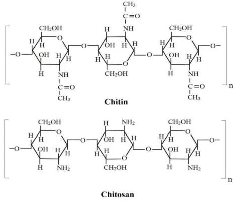
Sun 2012). Chitosan is a biopolymer derived from crustacean skeleton chitin and commonly used as support (Biró et al. 2008). The features of chitin and chitosan include its abundance as a by-product of the fishing and fermentation industries, its low cost over other supports for enzymes, its lack of toxicity emphasized by the fact that chitin is present in the foods of large consumption such as shellfish and sake, its chemical reactivity allowing easy fixation of enzymes. The structure of chitin
relates a polymer in which N-acetyl-D-glucosamine
units prevail over the glucosamine units, while the
chitosan is a polymer where D-glucosamine is the
prevailing recurring unit in the chain (Muzzarelli 1980). The residues of these linear polymers are joined by (β1→4) linkages (Fig. 1). The ratio between these units along the polymeric chain
depends on the conditions used in the
deacetylation process (Muzzarelli and Peter 1997). Chitosan amino groups are functional groups commonly used for the anchoring a large variety of molecules, including enzymes with or without crosslinker.
The present study aimed to explore different strategies to immobilize lipases from the
endophytic fungus Cercospora kikuchii onto
chitosan microparticles.
Figure 1 - Segments of chitin and chitosan polymers. Chitin is represented by N-acetyl-D-glucosamine
MATERIALS AND METHODS
Microorganism and Lipase Production
Cercospora kikuchii was grown and maintained on
potato dextrose agar (PDA) medium as previously reported by Costa-Silva et al. (2011). For lipase production, the fungus was cultivated on PDA
medium at 30oC for seven days. Agar pieces
(1 cm2) containing mycelium were inoculated on
Vogel’s minimum medium (Vogel 1956)
supplemented with 2% soybean oil and incubated at 30oC in a rotatory shaker at 120 rpm for six days. The culture broth containing extracellular lipases was filtered, dialyzed and concentrated and used in the immobilization process (Costa-Silva et al. 2011).
Lipase Immobilization
Lipases from C. kikuchii were immobilized on
chitosan microparticles by chemical attachment on
glutaraldehyde-treated chitosan acetate
microparticles (CAM) and physical adsorption to
highly deacetylated chitosan hydrochloride
microparticles (CHM).
Chemical Attachment of Lipase on CAM
Two methods were used. Method 1: A suspension (400 mL) of 2% (w/v) chitosan (Polymer, Amherst, MA) in 1.0% (w/v) acetic acid was prepared by stirring at 7930 xg for 30 min. After the addition of the enzyme solution to a final concentration of 500 U/g chitosan, the mixture was stirred to complete the homogenization. The procedure was completed by the addition of 13.2 mL of 2.5% (w/v) glutaraldehyde (Sigma Chemical Company, St. Louis, MO) and by stirring for 15 min more. The suspension (acetate microparticles with immobilized lipase) was dried at 160oC and atomized (Costa-Silva et al. 2010). Method 2: Lipases were immobilized according to Rorrer and Hsien (1993) with some modifications. Briefly, a suspension of powdered chitosan (4.0 g) in 5% acetic acid (96 mL) was gently dropped into 1M NaOH solution and stirred at 25°C for 24h. The acetylated product was washed with an excess of distilled water until neutral pH (Kimura et al. 1999) and dehydrated by lyophilization. For glutaraldehyde activation, the chitosan beads were added to 50 mm sodium phosphate buffer, pH 6.5 (buffer 1) containing 2.5% (v/v) glutaraldehyde and incubated at 25 ºC for 1h. Excess reagent was removed by washing with distilled water. Dried beads were used for lipase immobilization by
covalent binding. Chitosan (2.5 g), previously activated as described, was mixed with aqueous lipase solution (50 mL, 125 U/g of chitosan) under agitation at 25°C for 1h. The derivative was filtered, thoroughly rinsed with buffer 1 and lyophilized.
Physical Adsorption of Lipase to CHM
Chitosan (1.0 g) was suspended in 0.05 M HCl (150 mL) and the mixture was kept under gentle stirring for 24 h. After the addition of 0.2 M NaCl (150 mL) the suspension was vacuum filtered through a porous membrane (0.20 µm) (Signini and Campana Filho 2001). The filtrate was slowly mixed with absolute ethanol until precipitation and the suspension was centrifuged at 5800 xg for 10 min. Both the supernatant (CHMS) and the precipitated (CHMP) were dried at room temperature. Lipase (7.0 mL) and 100 mg of dried CHMS were mixed and kept under stirring at 2600 xg for 3 h at room temperature. The mixture was centrifuged at 4000 xg for 5 min and the pellet washed three times with 2.0 mL of buffer 1 and again centrifuged as above until no activity was detected in the supernatant. Microparticles without lipases underwent the same washes for infrared analyses. The final pellet obtained was suspended with 10 mL of buffer 1 and lyophilized. Protein content was evaluated in each supernatant.
Lipase Assay
Lipase activity assay was performed using ρ
-nitrophenyl palmitate (pNPP) as substrate
according to Mayordomo et al. (2000) with few modifications. In brief, the reaction mixture consisted of 205 µL of buffer (200 mg of Triton X-100 and 50 mg of gum arabic in 50 mL of 50 mm phosphate buffer, pH 6.5), 45 µL of substrate
(15 mg of pNPP in 10 mL of 2-propanol), and 250
µL of enzyme solution. The mixture was incubated at 40°C for 30 min and then 0.5 mL of 2% trizma base was added. The optical density was measured at 410 nm. One unit (U) of lipase activity was the
amount of enzyme that released 1 µmol of p
-nitrophenol/min under the assay conditions. The operational stability of immobilized derivatives
too was determined using pNPP as the substrate.
The immobilization efficiency, IE (%), was determined by equation 1 (Menoncin et al. 2009), where P0 was the protein content in the lipase
solution (mg) and P1 was the amount of protein
adsorbed on the chitosan microparticles (mg). P1
protein content offered to immobilization process and the protein content washed from the microparticles.
The activity retention (AR %) was calculated following equation 2 (Kaewthong et al. 2005). IE (%) = 100 x (P0/P1) (1)
AR (%) = 100 x [Immobilized enzyme activity (U/mg) / Soluble enzyme activity (U/mg)] (2)
Protein Assay
Protein concentrations were determined by the Bradford method (Bradford 1976) with bovine serum albumin (Sigma Chemical Company, St. Louis, MO) as a standard.
Morphological Characterization of chitosan microparticles
Analysis of the shape and surface characteristics of the microspheres was performed by scanning electron microscopy (SEM; JEOL, JSM 5200 model), with 1000 x of magnification. Powdered samples were mounted for observation on a double-coated, stub adhered, conductive carbon tape.
Infrared Spectroscopy
Fourier Transform Infrared (FTIR) spectroscopy was employed for the analysis of chitosan samples
(CHMS, CHMP, pre-activated CAM, and
commercial chitosan). For this, the samples were covered a diamond crystal in an ATR (Attenuated Total Reflectance) accessory coupled to a FTIR spectrometer (Nicolet 380, Thermo Fisher Scientific Inc, USA). The degree of deacetylation was evaluated using the ratios of band intensities
between 1560 cm-1 and 2920 cm-1. Band intensities
were determined after background removal using adjacent minima or valleys near each band.
RESULTS AND DISCUSSION
One of the properties that generally improve due to immobilization is enzyme stability, under both storage and operational conditions (Mateo et al. 2007). Their physicochemical properties are often
significantly modified compared to the
corresponding enzyme in free solution. A significant advantage of immobilized enzyme is the increase of its thermal stability, allowing their use for longer periods at higher temperatures. Also, interesting and valuable are the changes in the pH-activity curves that often accompany
immobilization. Improvement of selectivity and specificity may be achieved in certain cases too (Mateo et al. 2007).
Chemical Attachment of Lipase onto CAM.
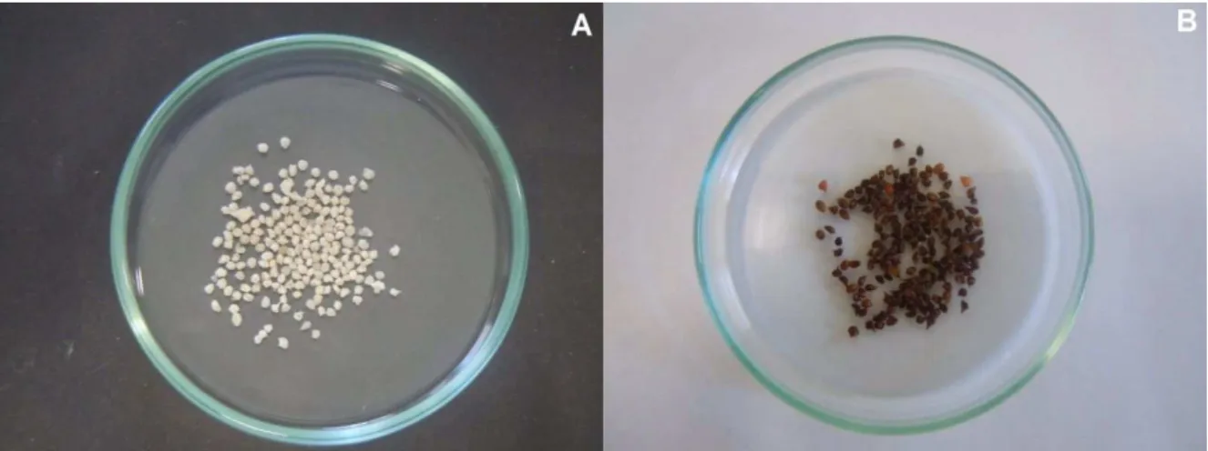
Lipase immobilized on glutaraldehyde pre-activated CAM (method 2) (Fig. 2) showed higher activity retention than the immobilized derivative from the method 1 in which the crosslinker was added after adsorption of the enzyme by the chitosan acetate microparticles. In this condition, only 0.1% of free enzyme activity was recovered on the support (Table 1). Concerning immobilized
lipases, strategies for reactivation should
regenerate not only the catalytic site of the lipase, but also the mechanism of opening and closing of the molecule (Betancor et al. 2006). Hence, low activity retention of lipases immobilized onto chitosan acetate microparticles by method 1 could be due to binding of key amino acids of lipase protein by glutaraldehyde, which hindered the conformational changes required for enzymatic activity. In addition, acid pH might also have a negative impact in enzyme activity.
In contrast, in the pre-activated support the activity retention was about 88% (Table 1), likely due to
glutaraldehyde crosslinkages as
amine-glutaraldehyde-amine,
amine-glutaraldehyde-glutaraldehyde-amine, or
amine-(glutaraldehyde)n,-amine (Fernandez-Lafuente et
al. 1995). It seemed that glutaraldehyde activation originated dimmers that conferred hydrophobicity to the support surface, and thus improved the retention of lipases with affinity for hydrophobic regions (Barbosa et al. 2012). This characteristic may have a positive impact in the immobilization process due to catalytic mechanism of lipases so-called interfacial activation (Verger 1997). Thus, the hydrophobic nature of glutaraldehyde may have permitted the adsorption of lipases via this mechanism, before the covalent attachment could take place (Barbosa et al. 2012).
Table 1 - Comparison between the methods applied for
immobilization of lipases produced by endophytic fungus C. Kikuchii on chitosan microparticles.
Technique Support AR (%) IE (%)
Chemical attachment
CAM –
Method 1 0.10±0 13.5±0.14 CAM –
Method 2 88.40±1.23 68.20±1.86 Physical
Figure 2 - CAM. A) After drying by lyophilization; B) after activation with glutaraldehyde.
The possibility to reuse the biocatalyst is of practical and economic importance in industrial applications. The CAM immobilized derivative obtained by the method 2 retained about 54% of enzyme activity after five reuses using p-NPP as substrate (Fig. 3). The stability of the immobilized derivatives produced by method 2 was determined during the storage period. Shelf-life of the product maintaining activity was six months at 4°C (Fig. 4) with only 3.3% of loss in enzymatic activity, whereas the free form of the enzyme lost 85.8% of its initial activity in the same period (Costa-Silva et al. 2013). These data were indicator of the viability of using the chitosan as support and the covalent binding as a way to protect the enzyme properties and control their stability.
Figure 3 - Operational stability of CAM immobilized
derivative (method 2) after reuse cycles.
Figure 4 - Stability of CAM immobilized derivative
(method 2) under storage at 4 °C.
Scanning Electronic Microscopy (SEM) of particles obtained by the method 2 showed a grooved surface in an undefined format (Fig. 5). Fissures and pores can facilitate the fixation of the enzyme on the support originating an immobilized
derivative with high activity. The efficiency of
Figure 5 - Scanning electron photomicrographs of pre-activated CAM.
Figure 6 - FTIR spectra for free lipase,
glutaraldehyde-pre-activated CAM and immobilized derivative (CAM + lipase).
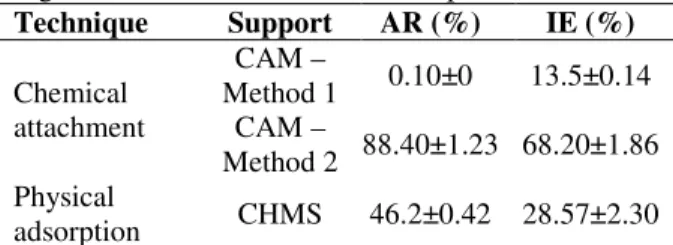
Physical Adsorption of Lipase on CHMS
As described in the methods, both the supernatant (CHMS) and the precipitated (CHMP) from the CHM suspension were dried at room temperature (Fig. 7). The deacetylation of CHMS and CHMP were evaluated by FTIR spectroscopy (Fig. 8), using the ratio A(1560 cm
-1
)/A(2920 cm
-1
) in the IR
spectra to calibrate the chitosan deacetylation
degree. According to Dong et al. (2001), the
intensity of the 1560 cm-1 band of spectrum
decreases with the chitosan deacetylation,
becoming very weak for highly deacetylated specimens because of the increase of the intensity of the 1600 cm−1 band and the ratio A(1560)/A(2920)
from IR spectra is suitable to infer about the
chitosan deacetylation degree. Lipases
immobilized on CHMS retained 46.2% of activity (Table 1). FTIR spectroscopy indicated higher deacetylation of CHMS when compared to CHMP (Table 2), which could have contributed to successful immobilization of lipases on that support. The degree of deacetylation affects the functional and sorption properties of chitosan through exposure of its amino groups available for interactions with the enzyme (Orrego and Valencia 2009), including low-energy interactions such as van der Waals, ionic and hydrogen bonds. The treatments of commercial chitosan that included HCl 0.05 M, NaCl 0.2 M and ethanol might have promoted breakdown of O-glycosidic bonds, thus contributing to the high deacetylation of chitosan in CHMS.
Figure 8 - FTIR spectra of CHMS, CHMP and commercial chitosan.
Table 2 - Absorbances of 1560 cm-1 band relative to
2920 cm-1 band of CHMS, CHMP and commercial chitosan.
CHMS Commercial
chitosan
CHMP
A(1560 cm-1) 0.0175 0.0436 0.0250
A(2920 cm-1) 0.0210 0.0296 0.0076
A(1560 cm -1
) / A(2920 cm-1)
0.833 1,473 3,290
In order to obtain the A(1560 cm-1) and A(2920 cm-1) values, the baseline absorbance value was subtracted from the absorbance at 1560 cm-1 or 2920 cm-1 band.
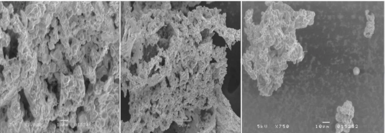
Scanning electron photomicrographs of CHMS showed sizes varying from 8 to 80 µm with a deeply porous surface (Fig. 9), which allowed for a wide surface area available for immobilization, maximizing the amount of protein immobilized. This could have contributed to the large storage stability of lyophilized lipase immobilized on highly deacetylated CHMS that was stored with no loss of activity up to 80 days at 4°C (Fig. 10). The larger surface area as a result of the micro/nanostructure of the materials could be the reason for better substrate-enzyme interaction, owing in part to a higher number of active sites exposed to the media. Indeed, enzyme activity is strongly dependent on the three-dimensional (3D) structure of the enzyme molecules because the active site should be conserved in order for the catalytic reaction between and enzyme and its substrate to occur (Polshettiwar and Asefa 2013). Vertegel et al. (2004) studied whether particle size
contributed to protein 3D structure
preservationand found a greater loss of enzyme activity for lysozyme immobilized onto 100 nm
SiO2 when compared with the same enzyme
immobilized on 4 nm and 20 nm nanoparticles (Polshettiwar and Asefa 2013).
Figure 9 - Scanning electron photomicrographs of CHMS.
Figure 10 - Storage stability of CHMS derivatives at
4ºC.
CONCLUSIONS
This study developed methods of Cercospora kikuchii
chitosan microparticles were potential support in C. kikuchii lipases immobilization technology for a wide range of industrial applications, mainly due the simplicity of the process involved in support production.
ACKNOWLEDGMENTS
The authors gratefully acknowledge the financial support of Fundação de Amparo à Pesquisa do Estado de São Paulo - FAPESP (Process no. 2011/00743-8).
REFERENCES
Barbosa O, Torres R, Ortiz C, Fernandez-Lafuente R. Versatility of glutaraldehyde to immobilize lipases: Effect of the immobilization protocol on the properties of lipase B from Candida antarctica.
Process Biochem. 2012; 47: 1220-1227.
Betancor L, Lopez-Gallego F, Hidalgo A, Alonso-Morales N, Dellamora-Ortiz G, Mateo, C, Fernandez-Lafuente R, Guisan JM. Different mechanisms of protein immobilization on glutaraldehyde activated supports: Effect of support activation and immobilization conditions. Enzyme Microbiol Technol. 2006, 30:877-882.
Biró E, Németh A, Sisak C, Feczkó T, Gyenis J. Preparation of chitosan particles suitable for enzyme immobilization. J Biochem Biophys Methods. 2008; 70:1240-1246.
Borges WS, Borges KB, Bonato PS, Said S, Pupo MT. Endophytic fungi: Natural products, enzymes and biotransformation reactions. Curr Org Chem. 2009; 13: 1137-1163.
Borgston B and Brockman HL. Lipases. 4th ed. Amsterdan: Elsevier; 1984.
Bradford MM. A rapid and sensitive method for the quantitation of microgram quantities of protein utilizing the principle of protein-dye binding. Anal Biochem. 1976; 72:248-254.
Bussamara R, Fuentefria AM, Oliveira ES, Broetto L, Simcikova M, Valente P, et al. Isolation of a lipase-secreting yeast for enzyme production in a pilot-plant scale batch fermentation. Bioresource Technol. 2010; 101:268-275.
Castro HF, Anderson WA. Fine chemicals by biotransformation using lipases. Quim Nova. 1995; 18:544-554.
Castro HF, Mendes AA, Santos JC, Aguiar CL. Modificação de óleos e gorduras por biotransformação. Quim Nova. 2004; 27:146-156.
Costa-Silva TA, Cognette RC, Souza CRF, Said S, Oliveira WP. Spouted Bed Drying as a Method for Enzyme Immobilization. Drying Technol. 2013; 3:1756-1763.
Costa-Silva TA, Nogueira MA, Souza CRF, Oliveira WP, Said S. Lipase production by endophytic fungus
Cercospora kikuchii: Stability of enzymatic activity after spray drying in the presence of carbohydrates.
Drying Technol. 2011; 29:1112-1119.
Costa-Silva TA, Said S, Souza CRF, Oliveira WP. Stabilization of endophytic fungus Cercospora kikuchii lipase by spray drying in the presence of maltodextrin and β-cyclodextrin. Drying Technol. 2010; 28:1245-1254.
Dandavate V, Jinjala J, Keharia H, Madamwar D. Production, partial purification and characterization of organic solvent tolerant lipase from Burkholderia multivorans V2 and its application for ester synthesis.
Bioresour Technol. 2009; 100:3374-3381.
Deive FJ, Carvalho E, Pastrana L, Rua ML, Longo MA, Sanroman MA. Strategies for improving extracellular lipolytic enzyme production by Thermus thermophilus HB27. Bioresource Technol. 2009; 100:3630-3637.
Dong Y, Xu C, Wang J, Wang M, Wu Y, Ruan Y. Determination of degree of substitution for N-acylated chitosan using IR spectra. Sci China, Ser B: Chem. 2001; 44: 216-224.
Fernandez-Lafuente R, Rosell CM, Rodriguez V, Guisan JM. Strategies for enzyme stabilization by intramolecular crosslinking with bifunctional reagents. Enzyme Microbiol Technol. 1995; 17:517-523.
Fernandes MRV, Silva TA, Pfenning LH, Costa-Neto CM, Heinrich TA, Alencar S, et al. Biological activities of the fermentation extract of the endophytic fungus Alternaria alternata isolated from
Coffea arabica L. Braz J Pharm Sci. 2009; 45: 677-685.
Kaewthong W, Sirisansaneeyakul S, Prasertsan P, H-Kittikun A. Continuous production of monoacylglycerols by glycerolysis of palm olein with immobilized lipase. Process Biochem. 2005; 40: 1525-1530.
Kim MI, Kim J, Lee J, Jia H, Na HB, Youn JK, et al. Crosslinked enzyme aggregates in hierarchically-ordered mesoporous silica: A simple and effective method for enzyme stabilization. Biotechnol Bioeng. 2006; 96: 210-218.
Liu Y, Jia S, Wu Q, Ran J, Zhang W, Wu S. Studies of Fe3O4-chitosan nanoparticles prepared by co-precipitation under the magnetic field for lipase immobilization. Catal Commun. 2011; 12:717-720. Mateo C, Palomo JM, Fernandez-Lafuente G, Guisan
JM, Fernandez-Lafuente R. Improvement of enzyme activity, stability and selectivity via immobilization techniques. Enzyme Microbiol Technol. 2007; 40:1451-1463.
Mayordomo I, Randez-Gil F, Pietro JA. Isolation, purification, and characterization of a cold-active lipase from Aspergillus nidulas. J Agric Food Chem. 2000; 48(2):105-109.
Menoncin S, Domingues NM, Freire DMG, Oliveira JV, Di Luccio M, Treichel H, et al. Imobilização de lipases produzidas por fermentação em estado sólido utilizando Penicillium verrucosum em suportes hidrofóbicos. Cienc Tecnol Aliment. 2009; 29:440-443.
Muzzarelli RAA. Immobilization of enzymes on chitin and chitosan. Enzyme Microbiol Technol. 1980; 2:177-184.
Muzzarelli RAA, Peter MG. Chitin Handbook. Grottammare:Atec; 1997.
Orrego CE, Valencia JS. Preparation and characterization of chitosan membranes by using a combined freeze gelation and mild crosslinking method. Bioprocess Biosyst Eng. 2009; 32:197-206. Polshettiwar V, Asefa T. Nanocatalysis: Synthesis and
Applications. 1st ed. New Jersey: John Wiley & Sons, Inc.; 2013.
Rorrer GL, Hsien TY. Synthesis of porous-magnetic chitosan beads for removal of cadmium ions from waste water. Ind Eng Chem Res. 1993; 32: 2170-2178.
Signini R, Campana Filho SP. Características e propriedades de quitosanas purificadas nas formas neutra, acetato e cloridrato. Polim: Cienc Tecnol. 2001; 11:58-64.
Simões AS, Mori RY, Faria R, Castro HF, Mendes AA. Desempenho da matriz híbrida SiO2-quitosana na imobilização da lipase microbiana de Candida rugosa. Quim Nova. 2011; 34:33-38.
Verger R. ‘Interfacial activation’ of lipases: Facts and artifacts. Trends Biotechnol. 1997; 15:32-8.
Vertegel AA, Siegel RW, Dordick JS. Silica nanoparticle size influences the structure and enzymatic activity of adsorbed lysozyme. Langmuir. 2004; 20: 6800-6807.
Verza M., Arakawa NS, Lopes NP, Kato MJ, Pupo MT, Said S, Carvalho I. Biotransformation of a tetrahydrofuran lignan by the endophytic fungus
Phomopsis sp. J Braz Chem Soc. 2009; 20:195–200. Vogel HJ. A convenient growth medium for
Neurospora crassa. Microb Genet Bull. 1956; 13:42-43.
Zhang DH, Yuwen LX, Peng LJ. Parameters Affecting the Performance of Immobilized Enzyme. J Chem. 2013; 2013:1-7.
Zhu J, Sun G. Lipase immobilization on glutaraldehyde-activated nanofibrous membranes for improved enzyme stabilities and activities. React Funct Polym. 2012; 72: 839-845.