Chemical Industry & Chemical Engineering Quarterly www.ache.org.rs/CICEQ
Chem. Ind. Chem. Eng. Q. 20 (4) 549−564 (2014) CI&CEQ
TANJA PETRESKA IVANOVSKA1 LIDIJA PETRUSHEVSKA-TOZI1 ANITA GROZDANOV2 RUMENKA PETKOVSKA1 JASMINA HADJIEVA1 EMIL POPOVSKI3 TRAJCE STAFILOV3 KRISTINA MLADENOVSKA1
1Faculty of Pharmacy, University
“Ss. Cyril and Methodious”, Skopje, Macedonia
2University “Ss Cyril and
Methodius”, Faculty of Technology and Metallurgy, Skopje, Macedonia
3University “Ss Cyril and
Methodius”, Faculty of Natural Sciences and Mathematics, Skopje, Macedonia
SCIENTIFIC PAPER
UDC 615.331:663/664:544:66 DOI 10.2298/CICEQ130218036P
FROM OPTIMIZATION OF SYNBIOTIC
MICROPARTICLES PREPARED BY
SPRAY-DRYING TO DEVELOPMENT OF
NEW FUNCTIONAL CARROT JUICE
Article Highlights
• A spray-drying method was successfully applied to obtain synbiotic microparticles
• Optimal formulation ensures high viability of encapsulated Lactobacillus casei
• Microparticles effectively preserved the cells at the upper intestine and during cold storage
• High viability was maintained during storage of carrot juice enriched with encap-sulated cells
• Synbiotic juice with encapsulated cells may be health drink suitable for all population groups
Abstract
Lactobacillus casei loaded chitosan-Ca-alginate microparticles enriched with the prebiotic fructooligosaccharide were prepared using a spray-drying method associated with polymer complexation and cross-linking with calcium. The con-centrations of the formulation factors of alginate, chitosan and CaCl2 were optimized using 23 full factorial design. Experiments showed that micropar-ticles with favorable physicochemical properties and high probiotic viability during preparation and storage could be obtained when 40 mg/g sodium algi-nate, 5 mg/g chitosan and 50 mg/g CaCl2 is used. Stability of L. casei during microencapsulation was identified by FTIR spectroscopy. The viability of the probiotic in the optimal formulation of synbiotic microparticles remained above the therapeutic minimum during incubation of 24 h in simulated gastrointestinal conditions (7.67±0.4 log cfu/g) as well as after 3 months of cold storage (8.1±0.6 log cfu/g). High viability of L. casei was maintained during 6 weeks of cold storage when carrot juice was enriched with encapsulated cells. The effective preservation of L. casei into synbiotic microparticles provided pro-duction of new non-dairy functional food as an alternative of the population who is at risk of lactose intolerance.
Keywords: Lactobacillus casei, fructooligosaccharide, chitosan-Ca--alginate microparticles, spray-drying, synbiotic carrot juice.
Probiotics are live microorganisms which, when administered in adequate amounts, confer a health benefit on the host [1]. They mainly include species belonging to the genera Lactobacillus, Bifidobact-erium, Streptococcus, Enterococcus, Leuconostoc and Pediococcus, which are normal constituents of
Correspondence: Tanja Petreska Ivanovska, Faculty of Phar-macy, University “Ss. Cyril and Methodious”, Vodnjanska 17, 1000 Skopje, P.O. Box 36, Macedonia.
E-mail: [email protected] Paper received: 18 February, 2013 Paper revised: 9 May, 2013 Paper accepted:22 November, 2013
the human gastrointestinal flora. With respect to safe administration, the most utilized are the strains of Lactobacillus and Bifidobacterium [2].
pro-mote the growth of lactobacilli and bifidobacteria in the colon followed by short chain fatty acid production [5], mainly butyrate which serves as a source of energy for colonocytes, interfering with apoptosis, pro-liferation and differentiation of transformed cells [6].
Because of already perceived health effects of the probiotics, they have been incorporated alone or as synbiotics into a range of food and pharmaceutical products. However, the benefit from the probiotics strongly depends on their ability to survive and multi-ply in the host. There are certain factors that decrease their viability during processing, storage and adminis-tration (e.g., temperature, moisture, oxygen, pressure, gastric acid, bile salts, etc.). In order to enhance the viability in these conditions and provide targeted and controlled delivery of the probiotics in the GIT, micro-encapsulation of probiotics using various techniques (e.g., extrusion, emulsion, spray-drying, spray-coating) may be utilized [7]. There are evidences that spray- -drying method for microencapsulation of the probiotic bacteria results in production of stable microparticles, with low diameters and homogenous size distribution [8-10]. In addition, different biopolymers as coating materials (e.g., alginate, chitosan, gelatin, pectin, car-rageenan) are successfully utilized to provide safe transit of active probiotic cells through the upper GIT [11]. From the functionality point of view, encapsul-ating materials have to provide mild conditions for encapsulation, be biocompatible, non-toxic to the cells and host, impermeable for antibody-sized mole-cules, have sufficient membrane permeability and abi-lity to overcome the acidic and enzymatic environ-ment of the stomach and to increase adherence cap-acity and residence time of the probiotics in certain segments of the GIT as are the terminal ileum and colon. Because one material could not possess all these properties, usually, combinations of biocom-patible materials with different properties are used. Combination of chitosan and alginate for microencap-sulation of probiotics is frequently used due to the possibility for forming semi-permeable membrane between the positively charged chitosan and nega-tively charged alginate, which could not be dissolved in the presence of Ca2+ chelators or antigelling agents [12,13]. Although the coating of alginate beads with polycations and their functionality have been exten-sively studied, the search for optimal composition model for the encapsulating materials and method for microencapsulation continues in order ability of the microparticulate systems to control and target the delivery of the probiotics to be improved and probiotic viability enhanced. Further, microparticulate systems that contain live probiotic cells are convenient for
incorporation in food products, thus improving their functional value. The increased interest of fruit juices as probiotic carriers can be explained by the lack of dairy allergens and cholesterol [14] and their pleasant taste profile and refreshing characteristics. Carrot juice is an interesting medium to prepare a functional product due to the carotenoids and different com-pounds with antioxidative properties, i.e., vitamins and minerals which may have an important role as inhibitors of free radicals production and subsequent oxidative processes related to the onset of cardiovas-cular diseases or cancer. However, survival of probio-tics is a problem due to various factors of the environ-ment including effects of acidic pH of fruit juices in storage conditions. Once ingested, probiotics are additionally exposed to unfavorable acidic conditions in the upper gastrointestinal tract and antimicrobial effects of bile salts solutions. Even some authors have reported that carrot juice containing Bifidobac-terium strains [15] and cashew apple juice enriched with Lactobacillus casei [16] allowed probiotic cells to grow in fruit juices and to maintain the viability for determined time period, it was demonstrated that sen-sory off-flavors are not well accepted [17].
fermentation and storage of the fermented and non- -fermented beverages at 4 °C for 6 weeks.
MATERIALS AND METHODS
Materials
As an encapsulating agent for manufacturing of synbiotic microparticles, sodium alginate (Protanal LF 10/60 LS, fG 35–45%), which was kindly donated by IMCD, FMC BioPolymer (Ayrshire, UK), was used. Chitosan with deacetylation degree ≥85% and low viscosity 342 (viscosity of 10 mg/g solution in acetic acid 20–100 mPa s, Mw 150 kDa (France Chitine, Marseille, France) was used for coating of spray-dried microparticles, while the cross-linking procedure was performed with CaCl2 (Merck, KGaA, Darmstadt, Ger-many) in previously prepared solution of chitosan and CaCl2 in acetic acid (Merck, KGaA, Darmstadt, Ger-many). Freeze-dried probiotic culture of Lactobacillus casei-01 was purchased from Chr. Hansen, Hoers-holm, Denmark. FOS was supplied from Sigma-Ald-rich, St. Louis, MO, USA. Bile salts (Ox bile dried pure) used for preparation of simulated intestinal juice, de Man Rogosa Sharpe (MRS) broth, MRS agar and peptone water were supplied from Merck, KGaA, Darmstadt, Germany. All the reagents were of analyt-ical grade.
Methods
Preparation of synbiotic microparticles
Synbiotic microparticles were prepared by spray-drying method stabilized with subsequent freeze-drying [13]. Shortly, freeze-dried probiotic cul-ture L. casei-01 was inoculated into 5 ml MRS broth and incubated at 37 °C for 24 h under aerobic con-ditions. The cells were harvested by centrifugation at 1500g for 10 min and washed with sterile peptone solution (1 g/L). An aqueous suspension of L. casei with a cell load ca. 12 log cfu/g, FOS (15 mg/g) and alginate was infused into a spray-dryer (Büchi Mini Spray Dryer B-290, Flawil, Switzerland) to obtain mic-roparticles. The conditions of the spray-drying pro-cess were: nozzle diameter 0.7 mm, aspirator pres-sure 90%, atomizer prespres-sure 600 Nl h–1, flow rate 6 ml/min, inlet temperature 120 °C and outlet tempe-rature 60 °C. Spray-dried microparticles were col-lected in sterile containers and then slowly added into previously prepared solution of CaCl2 and chitosan in 10 mg/g acetic acid under continuous stirring using a magnetic stirrer for at least 3 h at room temperature. The hardened microparticles were separated by cen-trifugation at 1500g for 10 min, washed with sterile saline solution and frozen at –20 °C. Afterwards, the
microparticles were freeze-dried at 0.070 mbar and –50 °C for 24 h (FreeZone Freeze Dry System, Lab-conco, Kansas City, MO, USA).
Experimental design
In order to obtain the optimal formulation of syn-biotic microparticles, 23 full factorial design was used to create a set of experiments. Two levels for the fac-tors, upper (“+”) and lower (“–“) level were defined and a zero level (center), in which all variables are fixed at their mean value, was included in order to minimize the risk of missing non-linear relationships. However, the zero level experiment was not included in the calculation of the coefficients. Screening experiments were carried out with three independent variables that affect the experimental responses, sodium alginate in concentration limits of 10 and 40 mg/g (x1), chitosan in concentration limits of 1 and 5 mg/g (x2) and CaCl2 in concentration limits of 5 and 50 mg/g (x3), Table 1. All modeling analyses were performed using experi-mental design software program MODDE 8.0 (soft-ware for design of experiments and optimization, Umetrics, Umea, Sweden). Regression analysis was performed on the basis of experimental results to con-struct mathematical models. The coefficients of the model were obtained using response surface metho-dology to fit the experimental data to polynomial equation and to define the optimal formulation. The applied linear mathematical model for measuring the response of the investigated factors was:
= + + + + +
+ +
0 1 1 2 2 3 3 12 1 2 13 1 3 23 2 3
y b b x b x b x b x x
b x x b x x (1)
where y presents the estimated response, b0 is inter-cept of the linear model showing the average expe-rimental response, coefficients b1, b2 and b3 are the estimated experimental responses of the factors showing the main effects, while the coefficients b12, b13 and b23 are showing two-factor interactions. The response surface plots illustrate the relationship between the factors and the responses by holding constant one of the three independent factors.
Microparticle characterization
micro-particles in 0.1 mM phosphate buffer (pH 6.8) was recorded using Zeta-sizer Nano ZS (Malvern Instru-ments Ltd., Worcestershire, UK). The calcium content of the microparticles was determined by inductively coupled plasma atomic emission spectroscopy, ICP- -AES (Varian, Palo Alto, CA, USA) after degradation of the microparticles with HNO3 (2.5 mg/ml) [13,18].
Stability assay of L. casei during microencapsulation Fourier transform infrared spectroscopy (FTIR) was used to determine the stability of the probiotic L. casei during microencapsulation. FTIR-ATR spectra were recorded at room temperature using a Zinc Selenide crystal and Golden GateTM ATR attachment (Perkin Elmer System 2000 FTIR, ATR-IR Golden Gate, Waltham, MA, USA), in frequency range of 4000-400 cm–1. In order to determine possible mole-cular changes of the probiotic structure during the microencapsulation process, the spectra of non-encapsulated L. casei and released from the micro-particles were compared.
Viability assay of encapsulated L. casei
Viability of the encapsulated L. casei cells was determined using plate-count method. Namely, the microparticles were dispersed in phosphate buffer solution (pH 6.9) until complete release of the pro-biotic cells. Then, L. casei was plated in triplicate on selective MRS agar, incubated at 37°C under aerobic conditions for 72 h and enumerated. The average of the results was expressed as colony-forming units per gram of sample (cfu/g). For viability tests in simulated gastrointestinal juices, method of Mokarram et al. [19]
was used with slight modifications of the simulated gastric juice as Gbassi and Vandamme [20] sug-gested the presence of enzyme in its composition. Thus, the microparticles were incubated in simulated gastric juice (0.08 M HCl with 2 g/L NaCl and 3 g/L pepsin, pH 1.5) using a shaking water bath at 75 strikes/min (Haake, SWB 25, Burlandingen, Germany) for 3 h and in bile salts solution (0.05 M KH2PO4;pH 6.8, with 10 g/L filter sterilized bile salts) for additional 3 h. Washed microparticles were transferred in simul-ated colon medium (0.1 M KH2PO4; pH 7.4) [21] and incubated up to 24 h.
Swelling studies
Swelling properties of the optimal formulation of microparticles were evaluated by measurement of the particle size in simulated gastrointestinal fluids with different pH values. Given amount of microparticles (30–40 mg) was suspended in 5 ml of acetate buffer (pH 1.5) and phosphate buffer solutions with pH 6.8 and 7.4, respectively. An exchange method was used to carry out the swelling test during stirring by mag-netic stirrer at 300 rpm and temperature of 37 °C. At determined time intervals, the samples were removed from the swelling medium and assayed for particle size by Mastersizer (Hydro 2000G, Malvern Instru-ments Ltd., Worcestershire, UK). Percent swelling value was calculated according to:
(
)
− ×
Dt D0 D0 100 (2) Table 1. Experimental matrix for the series of synbiotic microparticles with L. Casei-01 enriched with FOS; independent factors and experimental responses
Experimental matrix Experimental responses Exp.
no.
Independent factors Viability of encapsulated L. casei (log cfu/g) Physicochemical properties
X1
Alginate mg/g
X2
Chitosan mg/g
X3
CaCl2
mg/g
Viability after freeze- -drying
Viability in simulated gastric juice
after 3 h
Viability in simulated intestinal juice
after 6 h
Viability in simulated colon
conditions after 24 h
Particle size
d50
μm
Zeta potential
mV
Ca2+ content mg/10 mg
particles
where Dt and D0 are the mean volume diameters of the microparticles at time t and in the dry state, res-pectively [22].
Synbiotic carrot juices preparation and evaluation of viable cell counts during fermentation and storage
Carrot juice was prepared by extraction of washed and peeled carrots purchased at the local supermarket, with no added water, preservatives or any other nutrient. Then, the juice was pasteurized at 80 °C for 20 min. The initial cell concentration of 7.4±0.1 log cfu/ml was applied to ferment the carrot juice using L. casei and FOS or synbiotic micropar-ticles. This concentration was chosen according to the recommendations of International Dairy Feder-ation for minimum counts of 7.0 log cfu per g or ml of probiotic food to exert beneficial effects [23]. One sample of the carrot juice inoculated with free cells and one sample containing microparticles were firstly submitted to fermentation at 37 °C for 24 h and then stored at 4 °C. The cell population of non-fermented samples with free and encapsulated L. casei was adjusted to 10.5±0.2 log cfu/ml. Another two samples were inoculated with free cells of L. casei or L. casei loaded microparticles, respectively, and then stored in refrigerated conditions for 6 weeks. In the juices con-taining non-encapsulated L. casei, the prebiotic FOS was added simultaneously with the probiotic cells. All samples of synbiotic carrot juices were packed into sterile Erlenmeyers flasks closed with cotton plugs.
The viability of L. casei in all experiments was determined periodically during 24 h fermentation and then on a weekly basis for 6 weeks of storage. When enumeration of bacteria was performed, 1 ml of the sample was mixed with 9.0 ml of peptone water, vor-texes for 15 s, then serially diluted with peptone water and the viable count was determined as it was des-cribed in a previous section. The same procedure was performed for samples with added microparticles, after removing the particles from the juice by filtration [24]. Viability assay for non-fermented samples was performed at the preparation step and then once a week during cold storage. The pH of synbiotic carrot juices were also examined simultaneously with the viability assays (pH meter PB–11 Sartorius, Goet-tingen, Germany).
Chemical analysis of short chain organic acids
The amount of short chain organic acids was determined temporary during fermentation and then once a week in a storage conditions by high-perfor-mance liquid chromatography using a HPLC system apparatus equipped with an ultraviolet detector (Agil-ent Technologies 1200 series, Palo Alto, CA, USA).
The method used by Wang et al. [25] was applied with certain modifications. Shortly, to determine the concentrations of lactic and acetic acid, 2 ml of 0.5 M H2SO4 were added to a 2 ml aliquot of the sample, thoroughly mixed for 30 s and centrifuged (12000g for 15 min). The obtained supernatants were filtered through 0.45 μm membrane (Minisart RC 25, Sar-torius Stedim Biotech, GmbH, Goettingen, Germany). Samples were loaded onto a thermostatically con-trolled reverse phase column (Discovery HS C 18, 250 mm×4.6 mm, 5 μm, Supelco Park, Bellefonte, PA, USA) set at 40 °С and eluted with 0.005 M H2SO4 at flow rate of 1 ml/min. According to the method applied, a detection wavelength was 210 nm, while identification of lactic and acetic acids was done using their respective standards.
RESULTS AND DISCUSSION
Optimization of the formulation of the synbiotic microparticles
produce microparticles with favorable zeta potential, i.e., mucoadhesive properties, minimal concentration of 46.4 mg/g for CaCl2 to protect the probiotic cells in the acidic environment of the gastric juice and inc-reased viability of the probiotic during microencapsul-ation, freeze-drying and in simulated gastric and int-estinal juices with higher alginate concentration. Namely, when chitosan in concentration of 1 mg/g was applied, negative values for the zeta potential of the microparticles were obtained (Table 1), which might lead to repulsive interactions with the negatively charged mucus glycoprotein. Therefore, a compro-mise was imperative and it was assumed that with decreasing chitosan concentration below 4 mg/g, the viability of L. casei in simulated gastric conditions would not be affected, but the surface characteristics of the microparticles would lead to decreased potential for interaction with the mucus glycoprotein. The high CaCl2 concentration as a dominant factor in providing probiotic viability during preparation of syn-biotic microparticles and their exposure to simulated GI conditions is required for efficient cross-linking of microparticles, i.e., mechanical stability of the micro-particles. On the other hand, CaCl2 concentration showed no significant influence on the zeta potential of the microparticles. In addition, positive values for zeta potential were obtained within the experimental range of alginate (10–40 mg/g), pointing to no signi-ficant influence of the alginate concentration on this response. The experimental responses obtained for particle size and Ca-content of the microparticles was also monitored to confirm the favorable physicoche-mical properties of the optimized formulation.
Particle size of the synbiotic microparticles
Particle size is an important factor affecting the survival rate and colonization of encapsulated biotics, sensory properties of the probiotic food pro-ducts and quality and stability of the pharmaceutical products. Depending on the method of microencap-sulation, the size of the produced microparticles
ranges in wide interval, usually from 5 μm to 3 mm. The large particles can negatively affect the textural and sensorial properties of the food products in which they are added [11,24]. Small and controlled size particles are more convenient for incorporation into food products due to the easier managing and higher stability [26]. Furthermore, smaller particles are better for achieving successful adhesion and colonization in the gut, while large particles move faster through the colon and come rapidly in the descending colon prior defecation [27]. In this study, the response for particle size was analyzed in conjunction with the minimal value required to successful encapsulation of the probiotic cells, usually 1 to 5 μm [11], and the recom-mendation for smaller size of particles in order most foods to keep mouth feel characteristics [28]. The method of spray-drying used in our study allowed production of stable microparticles with mean volume diameter ranging from 6.7 to 12.5 μm within all expe-rimental series (Table 1). Considering the values of the regression coefficients (Table 2), it can be con-cluded that the most significant effect on the particle size had alginate concentration (x1), than CaCl2 con-centration (x3) and afterwards, chitosan concon-centration (x2). With increasing alginate concentration, particle size increases and it decreases with increasing CaCl2 concentration. In our experimental system, the par-ticle size was optimized with respect to minimal CaCl2 and chitosan concentrations required to achieve inc-reased probiotic viability and favorable interaction of the particles with the intestinal mucosa, 46.4 and 4 mg/g, respectively. Therefore, the minimal particle size that fulfils the mentioned criteria was the value of 8.19
μm (Table 1).
Zeta potential of the synbiotic microparticles
Another factor that implies adherence property of the particles to the epithelial support of the intestine is the particle charge. There are literature data show-ing that probiotic cells are in general negatively charged [29]. Our measurements of the zeta potential
Table 2. Regression coefficients of the experimental model
Experimental response Regression coefficients
b0 b1 b2 b3 b12 b13 b23
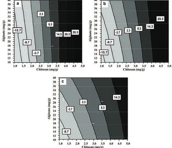
at pH 6.8 pointed that free cells of the probiotic L. casei were also negatively charged (–16.4±0.7 mV). As various polymers able to interact with mucus imp-roving the residence time of the microparticulated delivery systems, we optimized the concentration of positively charged carrier, chitosan, to provide favor-able interaction with the negatively charged glycopro-tein from the mucus, to increase the mucoadhesion capacity and thus, to prolong the residence time of the encapsulated probiotic cells in the lower intestine. With measurements of the zeta potential for the series of microparticles generated with the experi-mental design, values ranging from –21.71 to 25.42 mV were obtained (Table 1). The values of the reg-ression coefficients, b1, b2 and b3 (Table 2) indicate that the surface charge of the synbiotic microparticles is dominantly affected by the chitosan concentration (x2), while the effects of the alginate (x1) and CaCl2 concentration (x3) are significantly lower, with the low-est influence of the concentration of CaCl2. The con-tour diagrams in Figure 1 present the zeta potential values as a function of alginate (x1) and chitosan
concentration (x2), while the CaCl2 concentration (x3) is kept constant at 5 (Figure 1a), 27.5 (Figure 1b) and 50 mg/g (Figure 1c). Although, the best response for zeta potential of 26.3 mV at CaCl2 concentration of 5 mg/g was obtained, high CaCl2 concentration was used due to its effect on the primary response, i.e., viability of the probiotic cells in the acidic gastric con-ditions. At the optimum point for viability in simulated gastric medium (Figure 1c), the zeta potential was predicted to be at least 14.3 mV that satisfies the postulated surface properties of the prepared parti-cles. The positive charge can be attributed to the chi-tosan coating on the surface of the particles. The dominant localization of chitosan in the particle wall was observed by imaging the chitosan-Ca-alginate particles with FITC-labeled chitosan using confocal laser scanning microscopy [18,30].
Calcium content of the synbiotic microparticles
Calcium content of the synbiotic microparticles was used as a measure for mechanical and chemical stability of the obtained particles. Ca-content in the chitosan-Ca-alginate microparticles of different series
ranged from 0.43–1.22 mg Ca2+/10 mg particles (Table 1). In the experimental system, this response was analyzed similarly as the particle size in con-junction with CaCl2 and chitosan concentrations requ-ired to provide high probiotic viability in gastric juice and desirable zeta potential. The value of the regres-sion coefficient b3 points that the Ca-content is mostly affected by the CaCl2 concentration, while the value of the coefficient b1 indicates lower, but also signi-ficant effect of alginate concentration (x1). The con-centration of chitosan (x2) does not affect the Ca-con-tent of the microparticles significantly (Table 2). Inc-rease in CaCl2 concentration in the cross-linking med-ium increased the Ca-content, while with increase in the alginate concentration, the Ca-content decreased. The best response for Ca-content was obtained at chitosan concentration 5 mg/g in the experimental range of alginate 10–20 mg/g and CaCl2 44–50 mg/g. However, the findings that alginate concentration above 25 mg/g is needed to provide sufficient pro-biotic viability in gastric pH and increase in alginate concentration from 10 to 40 mg/g provides higher sur-vival rate during microencapsulation were the main reason for accepting the lower value as an optimum (0.94 mg Ca2+/10 mg particles was obtained vs. the predicted 1.04 mg Ca2+/10 mg particles).
Viability of L. casei after preparation of synbiotic microparticles
Simultaneous microencapsulation of prebiotics and probiotics results in synbiosis that may provide enhanced protection during preparation, storage and exposure to GI conditions. In our previous study [13], higher survival for 4 logs was observed when L. casei was spray-dried in the presence of alginate and FOS in comparison with the survival of the spray-dried L. casei alone. There are other research data points to increased viability of L. casei in a presence of FOSs with different degrees of polymerization [31,32]. Having these in regard, the medium intended for spray-drying as well as the free cells juice was sup-plied with the prebiotic FOS, in addition to the pro-biotic.
In this research, the yield of the spray-drying process was 46.5±9.7, with viability of the probiotic after spray-drying from 7.78 to 11.76 log cfu/g within the different series of the experimental plan. In com-parison with the spray-drying process, the loss of via-bility during freeze-drying was insignificant for all exp-erimental series and viability of the probiotic in the microparticles after freeze-drying was between 7.26 and 11.30 log cfu/g (Table 1). This response was used as a key one for optimization of the formulation
variables during the preparation of the microparticles. According to the values of regression coefficients, b1, b2 and b3 (Table 2), the viability of L. casei in micro-particles obtained after freeze-drying was mostly affected by the alginate (x1) and CaCl2 concentration (x3), while the chitosan concentration had the lowest influence on this response. The best response for the probiotic viability was obtained at chitosan concen-tration of 1 mg/g in the concenconcen-tration range of algi-nate 18.7–40 mg/g and CaCl2 38.5–50 mg/g, however, with no significant effect on the probiotic viability when increased chitosan concentration up to 5 mg/g was used.
Viability of L. casei in simulated gastrointestinal conditions
To estimate the influence of the formulation fac-tors on the stability of the probiotic in gastrointestinal conditions, the viability of the microencapsulated L. casei during incubation in simulated gastric and bile salt solutions was determined. Formulations gener-ated with the experimental design demonstrgener-ated sig-nificantly different values for the viability of the encap-sulated L. casei in a range of 4.32–9.62 log cfu/g during incubation of the microparticles in simulated gastric juice with pH 1.5 (Table 1). The values of the coefficient b1 and especially of b3 (Table 2) demon-strate that the viability of the microencapsulated L. casei in simulated gastric juice is dominantly affected by the alginate (x1) and CaCl2 concentration (x3), with the latest factor as the most significant. The influence of the chitosan concentration was not significant and, in addition, no major interactions were found. We also observed that further increase in CaCl2 concentration improves the probiotic viability in acidic pH, probably as a consequence of the increased degree of ionic cross-linking. The contour diagrams at Figure 2 pre-sent the viability of the microencapsulated L. casei in simulated gastric juice as a function of alginate (x1) and CaCl2 concentration (x3), while the concentration of chitosan (x2) was kept constant at 1 (Figure 2a), 3 (Figure 2b) and 5 mg/g (Figure 2c). One can notice that the probiotic viability of at least 9.1 log cfu/g is predicted when alginate in concentration range of 26.4–40 mg/g and CaCl2 in concentration range of 46.4–50 mg/g were used, with the concentration of chitosan maintained at 5 mg/g (Figure 2c), thus it was considered as an optimal response.
the prepared synbiotic microparticles with respect to the viability of L. casei in intestinal juice was eval-uated during additional 3 h in simulated intestinal juice (pH 6.8) with 1% bile salts. Significantly different values for the viability of encapsulated L. casei in a range of 2.27–8.46 log cfu/g were observed (Table 1). Considering the values of the regression coefficients (Table 2), the probiotic viability in simulated intestinal juice was significantly affected by the concentration of CaCl2 (x3), while the two other factors, x1 and x2, showed almost equal effects. However, the lowest value of the coefficient b2 indicates the lowest influ-ence of the chitosan concentration on the investigated response. The values of interaction terms, b12, b13 and b23 pointed to non-significant interactions between the formulation factors. At the optimum point of concen-trations of the formulation factors (40 mg/g alginate, 5 mg/g chitosan and 50 mg/g CaCl2) high survival rate of L. casei in intestinal juice above 8 log cfu/g was obtained (Table 1).
Effective colonization of the colon with viable and metabolically active probiotic cells is a crucial
prerequisite for providing postulated health effects. Viability and/or release of microencapsulated L. casei in simulated colon conditions were monitored in differ-ent time intervals up to 24 h of the initial incubation. Viability of microencapsulated L. casei in simulated colon conditions (pH 7.4) ranged from non-viable cells to 8.31 log cfu/g (Table 1). All the investigated factors increased the probiotic viability in simulated colon conditions, while the best response for probiotic via-bility of 7.97 log cfu/g was obtained at alginate con-centration (factor with the lowest effect) of 40 mg/g in the concentration range of chitosan, 1–4.6 mg/g and CaCl2, 42–50 mg/g.
Considering above-mentioned, the optimal for-mulation of synbiotic chitosan-Ca-alginate micropar-ticles provides effective protection of the probiotic during exposure in simulated gastric juice (9.62±0.1 log cfu/g) and relatively high survival rate in intestinal juice with pH 6.8 (8.46±0.2 log cfu/g). In addition, the released cell count in colonic pH after 24 h exposure to simulated GI conditions was 7.67±0.4 log cfu/g. We also observed that increasing the microencapsulating Figure 2. A contour diagrams of the viability of microencapsulated L. casei in simulated gastric juice with pH 1.5 as a function of alginate
material concentration increased the survival rate of L. casei and maintained the therapeutic level (at least 6–7 log cfu/g) under storage conditions, thus during 3 months storage of the optimal formulation at 4 °C, viable probiotic cells of 8.1±0.6 log cfu/g were pre-served. In addition, the physicochemical parameters of the optimal formulation, d50 of 8.77±0.4 μm, zeta potential of 21.56±1.1 mV and Ca-content of 0.94± ±0.15 mg/10 mg, point to the great potential for effect-ive colonization of the probiotic in the lower intestine.
Physicochemical properties of the optimal formulation
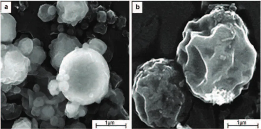
Physicochemical characterization of the optimal formulation that included morphological analysis before and after mechanical treatment of the particles (Figure 3a and b), stability assay of the probiotic during microencapsulation (Figure 4) and swelling properties of the prepared microparticles enabled the potential of the microparticles for successful delivery of the probiotic in the lower intestine and application in carrot juice to be further confirmed.
Scanning electron microscopy of the microparticles
The morphological analysis of the freeze-dried microparticles as we already reported [13] showed spherical shape with wrinkled surface resulted from the encapsulated cells and loss of water during spray- and freeze-drying processes. In this study, when intact microparticles were scanned, the wrinkles were not observed (Figure 3a). In addition, tendency of the particles to agglomerate can be noticed, probably as a result of attractive electrostatic forces between the polymers. The image obtained after mechanical treat-ment of the microparticles using high vacuum SEM clearly shows formation of certain invaginations on the surface of the microparticles with non-significant disruptions of the structure (Figure 3b), thus indicating
that the microparticles may be further processed and/or applied in different food or pharmaceutical pro-ducts.
Stability of L. casei during microencapsulation
In a view of the complex structure of the syn-biotic chitosan-Ca-alginate microparticles, a rough assignment of the corrected FTIR-ATR spectra of L. casei, non-encapsulated and released from the mic-roparticles, has been made (Figure 4). Namely, two distinctive bands at ∼2845 cm–1 and ∼2929 cm–1 due to the asymmetric stretching of methyl and methylene groups, respectively, were detected. These bands are specific to the fatty acids of the wall of probiotic bac-teria [33]. CH3– and CH2– asymmetric and symmetric deformations of proteins (∼1430 and 1372 cm–1, res-pectively) [34] were also detected. A band at ∼1730 cm–1 due to the С=О stretching vibration of the ester groups into the fatty acids and lipids together with Amide I and Amide II bands at ∼1620 and 1530 cm–1 from proteins were observed. In the IR fingerprint region, the symmetric and asymmetric stretching from the phosphodiester component of the nucleic acids at 1030 and 1190 cm–1 were found as well as the C–O–C deformation vibration from the polysaccharides (900– –1200 cm–1) bonded to the glycopeptides and lipopoly-saccharides of the cell wall [34]. Almost identical FTIR spectra of L. casei released from the microparticles and non-encapsulated one suggested preserved sta-bility of the probiotic cells during the microencap-sulation process.
Swelling studies
To confirm the targeted and controlled delivery function of the probiotic loaded chitosan-Ca-alginate microparticles, the swelling behavior of the optimal formulation was investigated in mediums with
rent pH values respective to simulated gastrointes-tinal conditions. An exchange method was used where particles were firstly placed in medium with pH 1.5 for 3 h, then in pH 6.8 for additional 3 h and subsequently in pH 7.4 for 4 h. The results point that synbiotic microparticles have no significant tendency for swelling, especially in mediums with pH 1.5 and 6.8 where increase in d50 of 12.26 and 31.62%, res-pectively, was observed. The protection of probiotic cells encapsulated in chitosan-Ca-alginate microparti-cles was achieved by chemical cross-linking with Ca2+ able to diffuse into the alginate gel network. Increased swelling degree of the microparticles in pH 7.4 was observed, with increase in d50 for 54.89%, probably due to the exchange of Na+ with Ca2+ when pH moves towards more alkaline pH and increased porosity and diffusion of the medium into the vicinity of the par-ticles. Having in regard the morphologic character-istics of the chitosan-Ca-alginate microparticles, i.e., low porosity and non-significant increase in particle size, especially in pH 1.5 and 6.8, one can conclude that the probiotic will be released in the lower inte-stine with the degradation of the microparticles as a dominant release mechanism.
Survival of free and encapsulated L. casei in carrot juices during fermentation and storage
An optimal formulation of the synbiotic micropar-ticles was used to prepare new functional beverages (fermented and non-fermented) that were compared with the properties of the functional juices containing non-encapsulated L. casei and prebiotic FOS. The
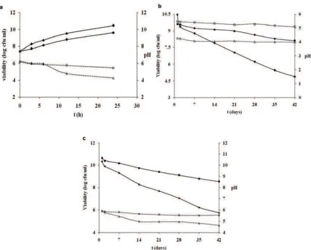
viability of free and encapsulated L. casei in fer-mented samples after 24 h fermentation was 10.46± ±0.4 log cfu/ml and 9.6±0.3 log cfu/ml, respectively, from the starting cell count of 7.4±0.1 log cfu/ml (Fig-ure 5a and b). During the fermentation process, free probiotic cells efficiently utilized carrot juice as an energy source. Kun et al. [15] reported enormous increase in viable bifidobacteria in inoculated carrot juice, but only for the first 12 h of the fermentation when survival rate started to decline upon the 24th hour of fermentation, while rapid and continuous growth of L. casei during 24 h fermentation in cashew apple juice have been documented by Pereira et al. [16]. Although, encapsulated L. casei in our ferment-ation test grew well, however showed slightly lower cell count compared to the juice containing free cells. The selective permeability of the chitosan-alginate membrane [35] may explain the delayed growth of the encapsulated cells.
ment FOS and decrease pH slightly below 6 [36]. Having in regard these findings, one can conclude that FOS as prebiotic is successfully fermented by L. casei-01 and its utilization as a source of energy for the growth of free cells is significantly higher compar-ing to encapsulated cells. In contrast to the observa-tions during the fermentation process, the viable cell counts of microencapsulated L. casei in fermented carrot juice was 8.1±0.13 log cfu/ml after 6 weeks of cold storage at 4 °C, while that of free cells was only 4.89±0.1 log cfu/ml, which confirms the role of the microencapsulation in improving probiotic viability. Moreover, the pH values of the synbiotic carrot juice with free cells were lower than pH values of carrot juice with microencapsulated cells during all inves-tigation period that might negatively affect the sensory properties of the product (Figure 5b).
Survival of free and encapsulated L. casei in non-fermented carrot juices
During 6 weeks storage at 4 °C, the viability of L. casei in non-fermented synbiotic carrot juices with free and microencapsulated cells was also
inves-tigated. Microencapsulated cells survive better than free cells in carrot juice with viable cell counts of 8.52±0.2 and 5.74±0.11 log cfu/ml after storage, res-pectively (Figure 5c). Ding and Shah [37] have also found that encapsulated probiotic cells survived in orange and apple juices throughout the six weeks of storage, while free cells lost their viability within five weeks and confirmed that fruit juices containing microencapsulated probiotic bacteria are more stable than those containing free cells. On the contrary, Champagne and Gardner [38] have been reported viability of L. rhamnosus, L. plantarum, L. reuteri and L. fermentum incorporated in several commercial fruit drinks to be above 6 log cfu/ml during 80 days of storage at 4 °C. In our study, free cells were in ther-apeutically accepted range upon fifth week of storage, whereas encapsulated showed better resistance to the end of the assay. This result confirmed that the chitosan-Ca-alginate matrix could protect probiotic cells exposed to acidic environment as pH value noticed significant reduction in free cells juice (1.56 units) in comparison with the decline of 0.62 units in synbiotic carrot juice containing encapsulated cells. Figure 5. Changes in viability (full lines) of free (♦) and microencapsulated L. casei (•) and pH values (dashed lines) of free cells juice
Short chain organic acid profile of synbiotic carrot juices during fermentation and storage
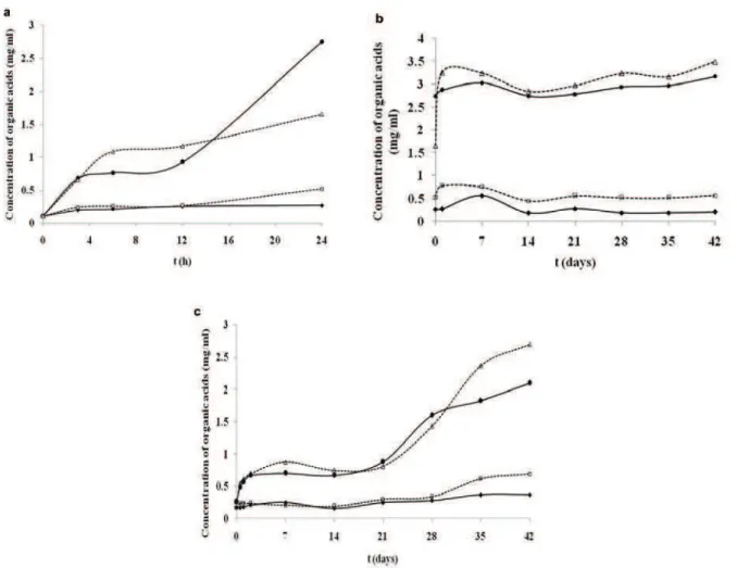
Probiotic bacteria may have utilized carbohyd-rates and produced small amounts of organic acids, thus their metabolic activity can be estimated by the production of lactic and acetic acid. Capability of pro-biotic bacteria to ferment carbohydrates available in the growth medium in general and in this respect in fruit and vegetable juices depends on the bacterial strain itself, but also on the type of the nutrients, growth promoters and inhibitors, osmotic pressure, inoculum size, fermentation period and storage tem-perature [39]. The concentrations of lactic and acetic acid in carrot juice alone were 0.05 and 0.13 mg/ml, respectively. Figure 6a illustrates the lactic and acetic acid concentration in carrot juice containing free and microencapsulated L. casei during fermentation. The production of lactic and acetic acid tends to increase during 24 h fermentation with enhanced production in juice containing encapsulated cells upon 12 h. Further, it is clearly presented that the production of acetic acid in juice containing encapsulated cells was
slightly increased to the end of the fermentation, while the concentration of lactic acid was dramatically inc-reased in free cells juice after 12 h fermentation. At the end of fermentation, the respective values for the concentrations of lactic and acetic acid were 2.75± ±0.03 and 0.27±0.01 mg/ml in free cells juice and 1.66±0.04 and 0.52±0.01 mg/ml for the juice with encapsulated cells, respectively.
Increased production of lactic and acetic acid during fermentation is due to the bacterial growth accompanied with organic acid accumulation. Lactic acid increases nutritional value of fermented products by engendering their taste. In addition, it has signi-ficant role in maintaining proton moving force of bac-terial cells and in absence of carbohydrates, some of bacteria utilize it as an energy source. Moreover, the high content of mineral components in carrot juice may improve the lactic acid fermentation. As the metabolism pathway of lactic acid bacteria lead to the production of lactic acid mainly, the high ratio in the favour of produced lactic acid during the fermentation of carrot juice with L. casei is reasonable. Increased
growth of free L. casei during our fermentation assay (Figure 5a) explains the higher lactic acid production compare to the juice containing encapsulated cells. The significantly lower amount of acetic acid pro-duced is also expected. However, the production of acetic acid in the juice containing microparticles is probably stimulated by carboxylic groups of the alginate that become protonated in the acidic medium [40]. Although, acetic acid is responsible for the sour taste of the juice, its antimicrobial properties improve the functional value of the product.
The amount of acetic acid during 6 weeks of refrigerated storage of fermented samples containing free cells as well encapsulated ones was at constant level albeit certain fluctuations of the concentration were determined. Thus, the acetic acid content dec-reased from 0.27 to 0.21 mg/ml in free cells juice, while in the juice containing encapsulated cells slightly increased from 0.52 to 0.57 mg/ml. In con-trast, the amount of lactic acid tends to increase during the storage period, especially in the juice con-taining encapsulated cells with enhancement of the production of 1.66 to 3.49 mg/ml (Figure 6b). The increase of the amount of lactic acid in carrot juice containing microparticles in storage conditions is pro-bably due to the gradual leakage of the encapsulated cells from the chitosan-Ca-alginate matrix to the medium and slower utilization of the substrate. Although, the level of organic acids in the fermented juice containing free cells remained stable after 6 weeks of cold storage, the progressive cell loss pointed to the microencapsulation as the effective tool to preserve metabolically active cells.
Short chain organic acid profile of non-fermented synbiotic carrot juices during storage
According to the survival rate of L. casei in non-fermented synbiotic carrot juices during 6 weeks of cold storage, the analysis of organic acids showed that higher amount was produced in juice containing synbiotic microparticles, 2.7 and 0.69 mg/ml for lactic and acetic acid, respectively, while in free cells juice the respective quantities were 2.1 and 0.37 mg/ml (Figure 6c). The results indicated that L. casei cells retained the metabolic activity, even in the cold sto-rage conditions without prior fermentation at 37 °C. Increased amounts of lactic and acetic acids in juice containing encapsulated L. casei confirmed the effect-ive preservation of the acteffect-ive probiotic cells by the microencapsulation method used in this study.
CONCLUSIONS
Optimization of the formulation variables of algi-nate, chitosan and CaCl2 have shown that synbiotic chitosan-Ca-alginate microparticles prepared with 40 mg/g alginate, 5 mg/g chitosan and 50 mg/g CaCl2 provide viability high above the therapeutic minimum during preparation and incubation in simulated gastric and intestinal juice with efficient release of viable and metabolically active cells of L. casei able to grow and colonize the lower intestine as well as under invest-igated storage conditions. The viability and metabolic activity of the cells were also confirmed through pro-duction of organic acids when synbiotic microparticles were incorporated into carrot juice. Regarding the sensory characteristics of the carrot juice with micro-particles, non-significant changes of the textural qual-ity due to the low particle size were observed. There-fore, carrot juice containing synbiotic microparticles may be a new functional product while the effect of particles on the consumer acceptance should be further studied. Moreover, in vivo studies are planned to confirm the synergistic effect of the prebiotic FOS on the probiotic viability and the potential of this for-mulation for its effective delivery in the lower intestine as well as beneficiary health effects associated with the consumption of the carrot juice enriched with syn-biotic microparticles.
Acknowledgments
This research was financially supported by the Ministry of Education and Science of the Republic of Macedonia (Project No. 13-3583). The authors would like to thank IMCD (UK) for the donation of sodium alginate (Protanal 10/60 LS, FMC BioPolymer, USA). Gratitude is expressed also to theUniversity of Read-ing, Centre for Advanced Microscopy, United King-dom for providing SEM images of the synbiotic micro-particles.
REFERENCES
[1] FAO/WHO. Guidelines for the evaluation of probiotics in food. Food and Agriculture Organization of United Nations and World Health Organization Working Group report, London, Ontario, 2002
[2] R.D.C.S. Ranadheera, S.K. Baines, M.C. Adams, Food Res. Int. 43 (2010) 1-7
[3] A.C. Ouwehand, K. Tiihonen, H. Mäkivuokko, N. Rauto-nen, in: Functional Dairy Products, M. Saarela Ed., Woodhead Publishing Ltd., Boca Raton, FL, 2007, p. 195 [4] C.B. Fritzen-Freire, E.S. Prudêncio, R.D.M.C. Amboni,
[5] K.P. Scott, S.W. Gratz, P.O. Sheridan, H.J. Flint, S.H. Duncan, Pharmacol. Res. 69(1) (2013) 52-60
[6] X. Pan, F. Chen, T. Wu, H. Tang, Z.Zhao, J. Zhejiang Univ. Sci., B 10(4) (2009) 258-263
[7] M.T. Cook, G. Tzortzis, D. Charalampopoulos, V.V. Khu-toryanskiy, J. Controlled Release 162 (2012) 56-67 [8] N.V. Menshutina, M.G. Gordienko, A.A. Voinovskiy, I.
Zbicinski, Dry. Technol. 28 (2010) 1170–1177
[9] T. Petrovic, V. Nedovic, S. Dimitrijevic-Brankovic, B. Bu-garski, C. Lacroix, Chem. Ind. Chem. Eng. Q. 13(3) (2007) 169–174
[10] A. Picot, C. Lacroix, Int. Dairy J. 14(6) (2004) 505-515 [11] J. Burgain, C Gaiani, M. Linder, J. Scher, J. Food Eng.
104 (2011) 467-483
[12] M. Chávarri, A. Marañón, R. Ares, F.C. Ibáñez, F. Marzo, M. del Carmen Villarán, Int. J. Food Microbiol. 142 (2010) 185–189
[13] T. Petreska Ivanovska, L. Petrusevska-Tozi, M. Dabev-ska KostoDabev-ska, N. Geskovski, A. Grozdanov, C. Stain, T. Stafilov, K. Mladenovska, Maced. J. Chem. Chem. Eng. 31(1) (2012) 115–123
[14] R.C. Ray, P.S. Sivakumar, Int. J. Food Sci. Technol. 44(6) (2009) 1073–1087
[15] S. Kun, J.M. Rezessy-Szabo, Q.D. Nguyen, A. Hoschke, Process Biochem. 43 (2008) 816-821
[16] A.L.F. Pereira, T.C. Maciel, S. Rodrigues, Food Res. Int. 44(5) (2011) 1276–1283
[17] T. Luckow, C. Delahanty, Food Qual. Prefer., A 15 (2004) 751-759
[18] K. Mladenovska, R.S. Raicki, E.I. Janevik, T. Ristoski, M.J. Pavlova, Z. Kavrakovski, M.G. Dodov, K. Gora-cinova, Int. J. Pharm., A 342(1-2) (2007) 124–136 [19] R.R. Mokarram, S.A. Mortazavi, M.B.H. Najafi, F.
Sha-hidi, Food Res. Int 42(8) (2009) 1040–1045
[20] G.K. Gbassi, T. Vandamme, Pharmaceutics 4 (2012) 149–163
[21] S. Mandal, A.K. Puniya, K. Sing, Int. Dairy J. 16 (2006) 1190–1195
[22] I. El-Gibaly, Int. J. Pharm. 249(1-2) (2002) 7-21
[23] A. Homayouni, A. Azizi, M.R. Ehsani, M.S. Yarmand, S.H. Razavi, Food Chem. 111 (2008) 50-55
[24] O. Sandoval-Castilla, C. Lobato-Calleros, H.S. García-Galindo, J. Alvarez-Ramírez, E.J. Vernon-Carter, Food Res. Int. 43 (2010) 111–117
[25] Y.C. Wang, R.C. Yu, H.Y. Yang, C.C. Chou, Food Mic-robiol. 20 (2003) 333-338
[26] J.-H. Cui, J.-S. Goh, S.-Y. Park, P.-H. Kim, B.-J. Lee, Drug Dev. Ind. Pharm. 27(4) (2001) 309-319
[27] N. Washington, C. Washington, C.G. Wilson, Physiolog-ical Pharmaceutics - Barriers to Drug Absorption, Taylor & Francis, London, 2001, p. 143
[28] S. Rokka, P. Rantamäki, Eur. Food. Res. Technol. 231 (2010) 1–12
[29] C. Pelletier, C. Bouley, C. Cayuela, S. Bouttier, P. Bour-lioux, M.-N. Bellon-Fontaine, Appl. Environ. Microbiol. 63(5) (1997) 1725–1731
[30] K. Mladenovska, O. Cruaud, P. Richomme, E. Belamie, R.S. Raicki, M.-C. Venier-Julienne, E. Popovski, J.P. Benoit, K. Goracinova, Int. J. Pharm., B 345 (2007) 59-69 [31] K.J. Aryana, P. McGrew, LWT-Food Sci. Technol. 40
(2007) 1808–1814
[32] S.U. Ping, A. Henriksson, H. Mitchell, Anaerobe 13 (2007) 134–139
[33] J. Schmitt, H.C. Flemming, Int. Biodeter. Biodeg. 41 (1998) 1–11
[34] Z. Filip, S. Hermann, Eur. J. Soil Biol. 37 (2001) 137–143 [35] M.T. Cook, G. Tzortzis, D. Charalampopoulos, V.V.
Khutoryanskiy, Biomacromolecules 12 (2011) 2834-2840 [36] H. Kaplan, R.W. Hutkins, Appl. Environ. Microbiol. 66
(2000) 2682-2684
[37] W.K. Ding, N.P. Shah, Food Res. J. 15(2) (2008) 219-232 [38] C.P. Champagne, N.J. Gardner, Food Res. Int. 41 (2008)
539-543
[39] N.P. Shah, J. Dairy Sci., B 83 (2000) 894-907
TANJA PETRESKA IVANOVSKA1
LIDIJA PETRUSHEVSKA-TOZI1
ANITA GROZDANOV2
RUMENKA PETKOVSKA1
JASMINA HADJIEVA1
EMIL POPOVSKI3
TRAJCE STAFILOV3
KRISTINA MLADENOVSKA1
1
Faculty of Pharmacy, University “Ss. Cyril and Methodious”, Skopje, Macedonia
2University “Ss Cyril and Methodius”,
Faculty of Technology and Metallurgy, Skopje, Macedonia
3
University “Ss Cyril and Methodius”, Faculty of Natural Sciences and Mathematics, Skopje, Macedonia
NAUČNI RAD
OD OPTIMIZACIJE SINBIOTISKIH MAKRO
Č
ESTICA
PRIPREMLJENIH METODOM SUŠENJA
RASPRŠIVANJEM DO RAZVOJA NOVOG
SOKA OD ŠARGAREPE
Korišćenjem metode sušenja raspršivanjem zajedno sa kompleksiranjem polimera i umrežavanjem pomoću kalcijuma, pripremljene su mikročestice hitozan-Ca-alginata, obogaćene prebiotskim fruktooligosaharidima, sa imobilisanim ćelijama Lactobacillus casei. Koncentracije komponenti formulacije alginata, hitozana i CaCl2 su optimizovane korišćenjem 23 punog faktorijelnog plana. Eksperimenti su pokazali da se mikročestice sa povoljnim fizičko-hemijskim svojstvima i visoke probiotske održivosti tokom pripreme i skladištenja mogu dobiti korišćenjem sledećih koncentracija natrijum-alginata, hitozana i CaCl2: 40, 5 i 50 mg/g, redom. Stabilnost L. casei tokom mikroinkapsulacije je utvrđena FTIR spektroskopijom. Viabilnost probiotika u optimalnoj formulaciji sinbiotskih mikroč es-tica ostaje iznad terapeuskog minimuma za vreme inkubacije od 24 h u simuliranim gastrointestinalnim uslovima (7,67 ± 0,4 log cfu/g ), kao i nakon 3 meseca skladištenja u hladnjači (8,1±0,6 log cfu/g). Velika održivost L. casei se postiže tokom 6 nedelja skla-dištenja u hladnjači kada je sok šargarepe obogaćen inkapsuliranim ćelijama. Efikasno
čuvanje L. casei u sinbiotskim mikročesticama omogućuje proizvodnju funkcionalne hrane bez mleka, kao alternative onim ljudima koji su netolerantni na laktozu.