Universidade Nova de Lisboa
Instituto de Higiene e Medicina Tropical
Evaluation of humoral and cellular immune responses to P. berghei-based whole-sporozoite malaria vaccination in rhesus macaques
Andreia Cunha Barreto
DISSERTAÇÃO PARA A OBTENÇÃO DO GRAU DE MESTRE EM CIÊNCIAS BIOMÉDICAS
i
Universidade Nova de Lisboa
Instituto de Higiene e Medicina Tropical
Andreia Cunha Barreto
Evaluation of humoral and cellular immune responses to P. berghei-based whole-sporozoite malaria vaccination in rhesus macaques
Orientador: Helena Nunes Cabaço, PhD, Prudêncio Lab, Instituto de Medicina
Molecular João Lobo Antunes, Faculdade de Medicina da Universidade de Lisboa
Coorientadores: António M. Mendes, PhD, Miguel Prudêncio, PhD, Prudêncio Lab,
Instituto de Medicina Molecular João Lobo Antunes, Faculdade de Medicina da Universidade de Lisboa, Celso Cunha, PhD, Instituto de Higiene e Medicina Tropical, Universidade Nova de Lisboa
Dissertação apresentada para cumprimento dos requisitos necessários à obtenção do grau de Mestre em Ciências Biomédicas.
ii
Acknowledgements/ Agradecimentos
If you can dream it, You can do it!
Em primeiro lugar, quero começar por te agradecer a ti, Helena, pelo voto de confiança, pelo apoio incansável, pela tua paciência, pela tua simpatia e preocupação e por estares sempre disponível para mim. Por teres sempre a palavra certa e por estares sempre com um sorriso. Tenho a certeza que depois de um ano no laboratório, sairei uma pessoa diferente graças principalmente a tudo o que me ensinas-te e me transmitis-te.
Obrigada a ti, Miguel, por me teres aceite no teu grupo e me teres dado a liberdade e permitido aprender tanto neste último ano. És uma fonte de inspiração para todos nós e é um enorme previlégio ter concretizado o sonho de fazer parte do teu laboratório. A ti António, obrigada por toda a tua ajuda e todas as tuas ideias, és uma inspiração pela capacidade incrível que tens de pensar nas coisas de uma maneira ”diferente”.
Não posso deixar de agradecer ao Prof. Dr. Celso Cunha por me ter aceite no mestrado e por se encontrar sempre disponível para nos receber. A todos os professores do Mestrado de Ciências Biomédicas do IHMT por nos terem proporcionado um ano curricular tão intressante e diversificado, e ainda ao IMM,por me ter recebido tão bem e sempre proporcionar um ambiente de excelência científica
Tenho também muito a agradecer a todos os membros do laboratório que me acompanharam ao longo deste ano e me ensinaram tanto. A ti Patricia por me teres ensinado as primeiras coisas no laboratório e me teres dado os meus primeiros trabalhos práticos, és uma pessoa incrivel e desejo-te toda a sorte do mundo. À Fontinha por ser a pessoa mais simpática, acessível e atenciosa e estares sempre disponível para responder às minhas dúvidas. A ti Raquel, obrigada por todas as nossas conversas, eu adoro ouvir as tuas histórias. À Filipa por ser a animação deste laboratório e à Margarida pela tua simpatia e boa disposição. A ti Adriana tenho a dizer que se pudesse levava-te comigo na
iii
mala para Moçambique e que gostei muito de te conhecer.A vocês Isabel e Gonçalo desejo-vos muita sorte para esta próxima etapa da vossa vida.
Teresa, Diana, Denise e Rafael, sem vocês claramente que o meu último ano não teria sido o mesmo. Obrigada por toda a paciência que tiveram, por toda a ajuda, por todas as conversas e gargalhadas e por serem pessoas tão incansáveis. Tenho-vos a dizer que adorei o “grupinho fantástico” e que nunca vos irei esquecer.
Quero também agradecer ao pessoal do Figueiredo Lab por serem uns excelentes companheiros de laboratório, sempre prestáveis e muito acolhedores e ao Mota Lab.
Um enorme obrigado aos meus amigos por estarem sempre presentes em todas as fases da minha vida e por estarem sempre disponíveis apesar da distância.
Obrigada à minha família por ser um apoio tão grande para mim, por estarem sempre presentes em todos os momentos e celebrarem todas as minhas vitórias e por apoiarem as minhas decisões. Obrigada especial aos meus pais e irmão por mesmo longe me fazerem sentir sempre bem e em casa e especialmente por todos os esforços que fizeram para que eu possa seguir os meus sonhos.
Um especial obrigada a ti Rodrigo, por seres incansável e estares comigo nas vitórias e derrotas, nos momentos bons e de mau feitio. Por me levantares o ânimo em todos os dias maus, por me ajudares a ver com mais clareza e por acreditares sempre em mim. Tem sido incrível esta viagem em que temos crescido tanto juntos, obrigada por seres essa pessoa incrivel que tem tanto para dar.
iv
Resumo
A malária é uma doença infecciosa transmitida por mosquitos que continua a ser uma das doenças infecciosas mais impactantes do mundo, matando milhares de pessoas todos os anos. Apesar de todos os esforços para controlar esta doença, como quimioprofilaxia e medidas de controlo de vetores, a falta de conhecimento sobre as respostas imunes desencadeadas pelo parasita Plasmodium dificulta o desenvolvimento de uma vacina eficaz contra a malária, que é necessária com urgência.
A natureza assintomática e altamente imunogénica do estadio hepático da infecção por Plasmodium torna-o num alvo ideal para o desenvolvimento da vacina contra a malária. Prudêncio lab do Instituto de Medicina Molecular (IMM) desenvolveu um novo candidato a vacina contra a malária de esporozoito inteiro que tem como alvo o estágio hepático, a PbVAC. Na qual o parasita roedor P. berghei expressa a proteína circunsporozoítica de superfície do P. falciparum (PfCSP), altamente imunogénica, de modo a promover respostas imunitárias específicas para PfCSP, assim como respostas cruzadas entre espécies, que podem proteger contra uma infecção subsequente por P.
falciparum.
Estudos pré-clínicos em modelos de infecção em murganhos e coelhos mostraram que PbVac é capaz de infectar e desenvolver-se em hepatócitos sem estabelecer uma infecção no estadio sanguíneo. Para imitar o que acontece no fígado humano, os macacos rhesus (Macaca mulatta) foram usados para um teste pré-clínico de P. berghei wild-type (WT) e esporozoítos PbVac geneticamente modificados, como estratégia para induzir imunidade contra P. falciparum. Este estudo tem como objetivo investigar e comparar as respostas imunes humorais e celulares entre animais imunizados e não imunizados.
Primeiro, confirmamos que os esporozoítos do PbWT são capazes de infectar hepatócitos do macaco rhesus in vivo. Posteriormente, os macacos rhesus foram imunizados por picada de mosquito com PbVac ou PbWT e seguidos por 21 semanas, em paralelo com animais não imunizados. Células mononucleares do sangue periférico (PBMCs) e plama foram coletadas periodicamente e células do fígado e esplenócitos foram coletados na eutanásia. Foi realizada uma comparação do plasma pré e pós 3 imunizações para analisar as respostas humorais quantificando IgGs específicas para esporozoítos. As composições dos compartimentos imunes do sangue periférico, fígado e baço foram analisadas por imunofenotipagem e respostas imunes celulares específicas contra esporozoítos PbVAC, PbWT e Pf foram avaliadas em PBMCs e células hepáticas utilizando um ensaio intracelular de citocinas.
As imunizações foram seguras, sem alterações relevantes nos parâmetros de segurança avaliados, e nenhuma infecção relevante foi encontrada. Mostramos que as imunizações contra PbWT e PbVac são capazes de provocar uma resposta humoral contra o antigénio em macacos. É importante ressaltar que a geração de anticorpos anti-esporozoítos Pf observados em animais imunizados com PbVac- mas não com PbWT indica que a PfCS pode desempenhar um papel significativo nas respostas humorais à vacina.
Quanto ao papel da imunidade celular desencadeada por essas imunizações, nem o perfil fenotípico geral, nem a magnitude e especificidade das respostas celulares aos agentes de imunização ou a Pf foram significativamente alteradas após a imunização. Em
v
contraste com o que alguns ensaios em humanos relataram, não encontramos expansão da população de células T γδ após a imunização. No entanto, encontramos alterações fenotípicas nas células linfóides inatas (ILCs), que diminuíram significativamente, e um aumento nas células T CD4+ nos PBMCs.
No geral, os nossos resultados indicam que a imunização com PbVac representa uma plataforma de vacinação segura que gera respostas imunes humorais a Pf. Manipulação adicional da estratégia de imunização com PbVac, como dose ou modo de administração, pode melhorar significativamente as suas respostas imunológicas humorais e celulares, contribuindo assim para o desenvolvimento de uma vacina eficiente contra a malária.
Palavras-chave: PbVac; vacina; reação de espécies cruzada; immunização; imunidade humoral; imunidade celular.
vi
Abstract
Malaria is a mosquito-borne infectious disease that remains one of the most impactful infectious diseases globally, killing thousands of people every year. Despite all efforts to control this disease, such as chemoprophylaxis and vector control measures, the lack of knowledge about the immune responses triggered by the Plasmodium parasite hinders the development of an urgently needed efective malaria vaccine.
The asymptomatic and highly immunogenic nature of the liver stage of Plasmodium infection makes it an ideal target for malaria vaccine development. The Instituto de Medicina Molecular (IMM)’s Prudêncio lab has developed a new pre-erythrocytic whole-sporozoite malaria vaccine candidate, PbVac, in which the rodent P. berghei parasite expresses the highly immunogenic P. falciparum surface circumsporozoite protein (PfCS in order to promote PfCS-specific and cross-species immune responses that may protect against a subsequent P. falciparum infection.
Pre-clinical studies in mouse and rabbit models of infection have shown that PbVac is able to infect and develop in hepatocytes without establishing a blood stage infection. In order to mimic what happens in the human liver, rhesus macaques (Macaca mulatta) were used for a pre-clinical analysis of P. berghei wild-type (WT) and genetically modified PbVac sporozoites, as a strategy to induce immunity to P. falciparum. This study aims to investigate and compare the humoral and cellular-associated immune responses between immunized and non-immunized animals.
First, the ability of PbWT sporozoites to infect rhesus macaque hepatocytes in vivo was confirmed. Subsequently, rhesus macaques were immunized by mosquito bite with
PbVAC or PbWT and followed for 21 weeks, in parallel with non-immunized animals.
Peripheral blood mononuclear cells (PBMCs) and plasma were collected periodically, and liver cells and splenocytes were collected at euthanasia. A comparison between plasma pre- and post- 3 immunizations was performed to analyze humoral responses through quantification of specific IgGs for sporozoites. The compositions of the peripheral blood, liver and spleen immune compartments were analyzed by immunophenotyping, and specific cellular immune responses against PbVAC, PbWT and
Pf sporozoites were assessed in PBMCs and liver cells by an intracellular cytokine
assay.Immunizations were safe, with no relevant changes in the safety parameters evaluated, and no breakthrough infections were found. We show that both PbWT and
PbVac immunizations are capable of eliciting a humoral response against the immunogen
in monkeys. Importantly, the generation of anti-Pf sporozoites antibodies observed in
PbVac- but not PbWT-immunized animals indicates that PfCS may play a significant role
in humoral responses to the vaccine.
As for the role of cellular immunity elicited by these immunizations, neither the overall phenotypic profile nor the magnitude and specificity of cellular responses to the immunization agents or to Pf were significanty altered upon immunization. In contrast to what some human trials have reported, we found no expansion of the γδ T cell population upon immunization. However we found phenotypic changes in innate lymphoid cells ILCs, which decreased significantly, and an increase in CD4+ T cells in PBMCs.
Overall, our results indicate that PbVac immunization represents a safe vaccination platform that generates humoral immune responses to Pf. Additional manipulation of the
vii
PbVac immunization strategy, such as dose or mode of administration, may significantly
enhance its humoral as well as cellular immune responses, thus contributing to the development of an efficient malaria vaccine.
Keywords: PbVac; vaccine; cross-species immunization; humoral immunity; cellular immunity.
i
Table of contents
Acknowledgements/ Agradecimentos ... ii
Resumo ... iv
Abstract ... vi
Index of figures ... iii
Index of tables ...v
Abbreviation ...1
Introduction ...4
1.1. Malaria ...4
1.1.1 Plasmodium life cycle. ...4
1.1.2. Clinical presentation and treatment ...7
1.2. Malaria Immunology ...9
1.2.1. Basic concepts in immunology ...9
1.2.1. Immunology in malaria infection ... 13
1.3. Malaria vaccines ... 16
1.3.1. Overview of malaria vaccines ... 16
1.3.2. Whole organism pre-erythrocytic malaria vaccines ... 17
1.3.3. PbVac vaccine ... 19
1.3.4. Immune response against vaccines ... 21
1.3.5. Rhesus monkeys as malaria vaccine trial models ... 23
2. Aims ... 24
3. Material and methods ... 25
3.1. Animals ... 25
3.2. Mosquito bite... 25
3.3. Study schedule ... 26
ii
3.5. Tissue collection and processing ... 26
3.6. Collection and isolation of mononuclear cells ... 27
3.7. Cell thawing ... 28
3.8. Sporozoites ... 28
3.9. Quantification of antibody titers in the plasma by enzyme-linked immunosorbent assay (ELISA) ... 29
3.10. Quantification of plasma antibody titers by flow cytometry ... 30
3.11. Optimization of flow cytometry panels ... 31
3.12. Phenotypic characterization of immune populations in the blood, liver and spleen of immunized vs. non immunized monkeys ... 34
3.13. Quantification of specific immune responses by ICS ... 35
3.14. Statistical analysis ... 36
4. Results ... 38
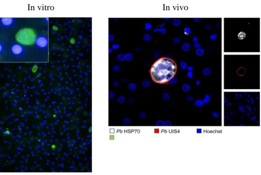
4.1. Invasion of rhesus hepatocytes by PbWT in vivo ... 38
4.2. Safety evaluations ... 38
4.3. Humoral immune responses ... 39
4.4. Phenotypic characterization of immune populations in the blood, liver and spleen of immunized vs. non immunized monkeys ... 41
4.5. Correlations between blood and tissue populations ... 50
4.2. Cellular immune responses ... 52
5. Discussion ... 60
6. Conclusion... 67
7. References ... 68
iii
Index of figures
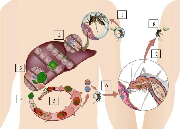
Figure 1- Plasmodium spp. Life cycle...5
Figure 2- Generation of PbVac ... 20
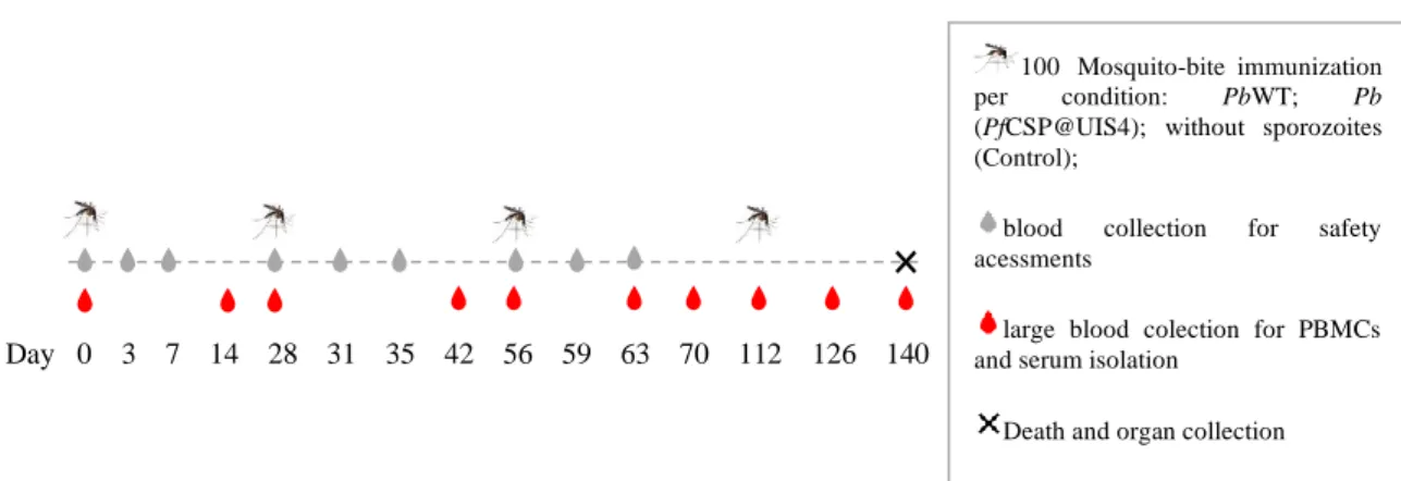
Figure 3- Schedule of parasite administration, sample collection, and death in rhesus monkeys ... 26
Figure 4- Histological liver slices of PbWT infecting rhesus hepatocytes in vivo. ... 38
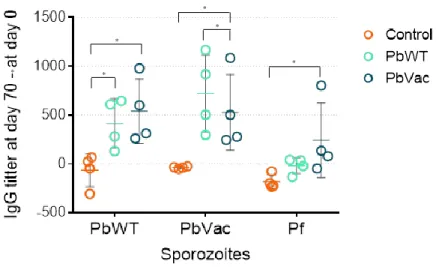
Figure 5- IgG titers were determined in rhesus plasma by ELISA ... 39
Figure 6- IgG titers were determined in rhesus plasma by Flow cytometry ... 40
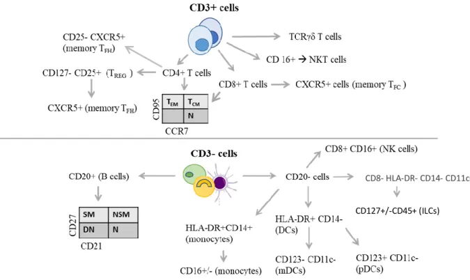
Figure 7- Phenotypic populations to be analysed by flow cytometry. ... 41
Figure 8- Alterations in innate populations with immunization.. Innate populations were analysed within PBMCs at days 0 and 140 ... 42
Figure 9- Frequency of monocytes, DCs, ILCs, NK and NKT cells in blood and tissues on day 140 post-immunization ... 43
Figure 10- Alterations of the T cell compartment and B cells within total PBMCs of immunized and non-immunized monkeys from days 0 to 140... 44
Figure 11- Frequency of T cells, and the 2 major TCR recepors in blood and tissues on day 140 post-immunization ... 46
Figure 12- Frequency of CD4 cell subsets in blood and tissues on day 140 post-immunization ... 47
Figure 13- Frequency of CD8 cell subsets in blood and tissues on day 140 post-immunization ... 48
Figure 14- Frequency of tissue resident memory cells in tissues on day 140 post-immunization ... 49
Figure 15- Frequency of total B cells in blood and tissues on day 140 post-immunization ... 49
Figure 16- Frequency of B cell subsets in blood and tissues on day 140 post-immunization ... 50
Figure 17- Analysis of total lymphocytes specific response in liver cells and PBMCs in liver and PBMCs ... 53
iv
Figure 18- Analysis specific responses of T cells (A) and the major T cell subsets (B) in liver and PBMCs ... 54
Figure 19- Analysis of CD4+ T cells (A) and memory cells (B) specific response in liver and PBMCs ... 55
Figure 20- Analysis of specific responses to CD8+ T cells (A) and CD8+ memory T cells (B) in liver and PBMCs ... 56
Figure 21- Analysis of NKT (A) NK (B) specific response in liver and PBMCs.58 Figure 22- Analysis of total, CD4+ and CD8+ TRM specific response in liver and PBMCs... 59
v
Index of tables
Table 1- Characteristics of the animals used in the study ... 25
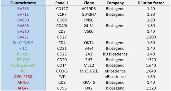
Table 2- Panel 1 to analyse the phenotype of the cells ... 33
Table 3- Panel 2 to analyse the phenotype of the cells ... 33
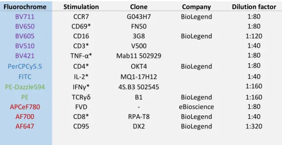
Table 4- Panel to analyse specific responses of the cells against stimulus. ... 34
Table 5- Analysis of cell frequency correlation between blood cells and tissues and between both tissues ... 51
1
Abbreviation
Acid-citrate-dextrose (ACD)
Acute respiratory distress syndrome (ARDS) Alanine transaminase (ALAT)
Allophycocyanin ef780 (apcef780)
Antibody-dependent cellular cytotoxicity (ADCC) Antigen-presenting cells (APC)
Apical membrane antigen 1 (AMA-1) Artemisinin-based combination therapy (ACT) Aspartate transaminase (ASAT)
Biomedical Primate Research Centre (BPRC) Blood-stage vaccines (BSV)
Central memory T cells (T CM)
Cerebral malaria (CM) Chemokine receptor 7 (CCR7) Chemokine receptor type 5 (CXCR5)
Chloroquine chemoprophylaxis sporozoites (CPS) Circumsporozoite protein (CSP)
Controlled human malaria infection (CHMI) Dendritic cells (DCs)
Dimethyl sulfoxide (DMSO) Double negative (DN)
Effector memory cells (T EM)
Enzyme-linked immunosorbent assay (ELISA) Ethylenediaminete traacetic acid (EDTA) Exo-erythrocytic forms (EEFeefs) Fix/Perm (fixation/permeabilization Fixable viability dye (FVD) Fluorescein isothiocyanate (FITC) Follicular cytotoxic T cells (TFC) Follicular helper T cells (Tfh) Forward scatter (FSC)
Gamma glutamyl transpeptidase (γ‐GT) Gene insertion/marker out (GIMO) Genetically attenuated sporozoites (GAS)
2
Heat shock protein 70 (HSP70) Helper T cells (Th)
Heparan sulphate proteoglycans (HSPGs) Hepatitis B surface antigen (hbsag) Horseradish peroxidase (HRP)
Human leukocyte antigen DR (HLA-DR) Immunoglobulins (Ig)
Indoor residual spraying (IRS) Innate lymphoid cells (ilcs) Insecticide treated bed-nets (ITN) Interferon- γ (IFN-γ),
Interleukin- 2 (IL-2)
Intracellular cytokine assay (ICS) Intradermally (id)
Intravenous (iv) Knock out (KO)
Lactate dehydrogenase (LDH) Lipopolysaccharide (LPS)
Major histocompatible complex (MHC)
Malaria Vaccine Implementation Programme (MVIP) Mean corpuscular volume (MVC)
Median fluorescence intensity (MFI) Merozoite surface protein (MSP) Myeloid DCs (mDCs)
Natural killer (NK) Natural killer T (NKT) New Zealand White (NZW) Nitric oxide (NO)
Nonhuman primates (NHP)
P. Berghei (Pb) P. Falciparum (Pf)
Parasitophorous vacuole (PV)
Parasitophorous vacuole membrane (PVM) Pathogen-associated molecular patterns (PAMPs) Pattern recognition receptors (PRRs)
Pfspz (P. Falciparum sporozoite),
3
Phosphate-buffered saline (PBS) Phycoerythrin (PE)
Plasmacytoid dcs (pdcs)
Plasmodium (P.)
Pre-erythrocytic vaccines (PEV) Radiation-attenuated sporozoites (RAS) Rapid diagnostic tests (RDT)
Reactive oxygen species (ROS) Red blood cells (RBC) Regulatory T cells (Treg) Room temperature (RT)
Roswell Park Memorial Institute (RPMI) Side scatter (SSC)
Sporozoite protein essential for cell traversal (SPECT) Subcutaneously (sc)
T cell receptor (TCR)
Tissue-resident memory T cells (TRM)
TMB (Tetramethylbenzidine)
Transmission-blocking vaccines (MSTBV) Tumor necrosis factor (TNF)
Upregulated in infective sporozoites (UIS) Whole sporozoite (Wsp)
World Health Organization (WHO) Α-galactosylceramide (α-galcer)
4
Introduction
1.1. Malaria
Malaria is one of the most impacting infectious diseases worldwide, especially in tropical and subtropical regions of the world. According to estimates from the World Health Organization (WHO), 219 million cases of malaria occurred worldwide in 2017, compared with 216 million in 2016. Of these cases, 92% were registered in the WHO African region. In that same year, 435 000 deaths were estimated from malaria globally, and 61% of them occurred in children under 5 years of age1.
Malaria is a mosquito-borne parasitic disease, caused by a protozoan parasite, phylum Apicomplexa and gender Plasmodium (P.) and transmitted through the bite of a female Anopheles mosquito2. The species capable of causing infection in humans are: P.
falciparum, the major responsible for sub-Saharan Africa cases, complicated malaria and
mortality; P. vivax, the most prevalent in America WHO regions; P. ovale; P. malariae; and P. knowlesi, primarily known as a primate parasite, but recently identified in humans in Southeast Asia3,4. Other Plasmodium species are able to infect different hosts, such as
P. berghei and P.yoelii in rodents and P. knowlesi3 and P. simium in non-human primates5. Their ability to mimic the human infection renders them important for malaria research.
5
The malaria parasite presents a very complex, multistage life cycle involving a mosquito and a mammalian host (Fig. 1). After their entry into the host via mosquito bite, Plasmodium parasites enter the circulation and reach the liver. The liver stage is obligatory, asymptomatic, and induces a strong immune response, thus constituting an ideal target for anti-malarial vaccines and prophylactic drug development6. Parasites released into the bloodstream subsequently infect red blood cells (RBC) and massive replication occurs, initiating the stage responsible for malaria symptoms and transmission.
Figure 1- Plasmodium spp. Life cycle. (1) An infected female Anopheles mosquito injects sporozoites into a mammalian host. This liver-infective parasite forms will travel until reaching the liver. (2) Once in the liver, the parasite transverses several hepatocytes until establishing a productive infective in one, where a PV is formed. (3) Intense asexual reproduction by schizogony occurs to form thousands of merozoites inside vesicles. (4) These merosomes bud off from the hepatocyte and are released into the bloodstream. (5) In the RBC the parasite performs successive cycles of invasion, intracellular growth, proliferation and re-invasion. (6) Some parasites are sexually committed merozoites, that develop into gametocytes that are able to develop within the mosquito when a new blood meal takes place. (7) Inside the vector, the parasite undergoes several transformations, formation of diploid zygotes, division and maturation into motile ookinetes, which are able to penetrate the midgut wall and transform into oocysts. The sporozoite formation initiates, and the accumulation of thousands of mature sporozoites causes the disruption of the oocysts and the release of sporozoites that invade salivary glands. (8) The sporozoites attached to the salivary glands are ready to be injected in the dermis of a new mammalian host.
The Plasmodium life cycle is initiated when an infected mosquito bites the mammalian host to take a blood meal and injects hundreds of sporozoites, the liver-infective forms, into the host’s skin7. Once in the skin, the parasite starts moving
5 2 4 6 1 7 3 8
6
randomly, in a gliding motility through the cells and tissues of the host8, to reach the blood vessels9,10. Most of these Plasmodium parasites actively target the liver (Fig. 1.1), possible due to the recognition and binding of the circumsporozoite protein (CSP), which covers the surface of the parasite, to the negatively charged heparan sulphate proteoglycans (HSPGs) expressed on the surface of the hepatocytes6. Due to its involvement in sporozoite motility as well as hepatocyte invasion and infection, CSP is an important target for drugs and prophylaxis in a pre-erythrocytic phase11,12. Once sporozoites reach the liver, they circulate freely along the sinusoidal epithelium, and transverse endothelial and Kupffer cells and several hepatocytes. Sporozoite protein essential for cell traversal (SPECT) and SPECT2 are two sporozoite proteins of micronemes identified in rodent parasite models that play a crucial role in cell transversal activity and liver infection13.
After migrating through several hepatocytes, the sporozoite establishes a productive infection in one14, where a parasitophorous vacuole (PV) is formed (Fig. 1.2), and asexual reproduction (schizogony) and maturation takes place (Fig. 1.3). Three rodent PV proteins, Upregulated in infective sporozoites (UIS)3, UIS4 and Pb36p, have been identified as being essential for parasite liver stage development within the PV6,12. The end of the exoerythrocytic phase is marked by the formation of thousands of merozoites that are released into the bloodstream inside vesicles, termed merosomes, derived from the host cell to deceive the immune system (Fig. 1.4)15. This exoerythrocytic stage is characteristic of each species and takes a minimum of 6 days in case of infection by P.
falciparum and until a maximum of 16 days for P. malariae 2, and only 2 days for the rodent-infective parasites (P. berghei and P.yoelii)16
Merozoites have the capacity to infect RBCs and perform successive cycles of invasion, intracellular growth, proliferation and re-invasion. The invasion step occurs quickly and involves multiple recognitions and interactions17, from connection of the merozoite to the RBC to its reorientation, which are mediated by the large merozoite surface protein (MSP) and the apical membrane antigen 1 (AMA-1), respectively. Then, merozoite penetration occurs by different parasite transmembrane protein families and at the same time a parasitophorous vacuole membrane (PVM) is formed with its own plasma membrane17,18.
7
The initial form of the erythrocytic cycle is the ring stage, when the parasite begins to feed on the surrounding RBC19, and develops to trophozoite stage, when the synthesis of molecules needed for cell division occurs20,21. During this stage, P. falciparum-infected RBC have the capacity to adhere to the endothelium of capillaries and venules and to non-infected RBC22. This sequestration is suggested to be a parasite defence mechanism to avoid splenic depuration and can lead to serious problems, such as cerebral malaria23. Lastly, a replicative intra-erythrocytic plasmodium stage, termed schizont, leads to the formation of around 16 nuclei that constitute a merozoite. The rupture of the PV and RBC membranes allows the merozoites to be released into the bloodstream and enables them to invade a new RBC and restart the intra-erythrocytic cycle (Fig. 1.5)20. The erythrocyte burst and parasites release is the cause of fever spikes that differ in the number of days according the infective species. For example, P. falciparum peaks occur every third day24. Parasites can also develop into sexual stages, named gametocytes. These are a sexually committed merozoites that invade the RBC following the ring stage and subsequently grow and develop over five stages until becoming sexually differentiated gametocytes25. Only mature sexual stages are able to survive and continue the Plasmodium life cycle in the mosquito host, known as the sporogonic cycle, upon the blood meal of a female anopheles (Fig. 2.6)26. Ingestion of gametocytes stimulates gametogenesis that starts with gametes formation and transformation into fertile female macrogametes and male microgametes. Microgamete exflagellation initiates the fertilization process, which includes fusion of the two gametes27. Fertilization gives rise to a zygote, and meiosis and genetic recombination transform the zygote into a motile ookinete able to penetrate and attach in the midgut wall and develop into oocysts, where occur the CSP formation. Once the oocyst is full of mature sporozoites, it ruptures and release sporozoites into the haemolymph circulation to reach and attach in the salivary glands (Fig. 1.7). At this point, the parasites are infectious and able to be inoculated in the skin of the next mammalian host and reinitiate the cycle (Fig. 1.8)12.
1.1.2. Clinical presentation and treatment
Malaria may have different clinical presentations, since absence of symptoms or very mild symptoms, to severe disease or even death. Therefore, malaria disease is
8
classified as uncomplicated or severe, and its severity may be dependent of the infecting species and on the host’s immune status28.
The gold-standard diagnosis of malaria are rapid diagnostic tests (RDT), which detect parasite antigens in the blood, or microscopy, that allows the quantification and identification of Plasmodium Spp. 29.
The initial symptom of the disease is usually fever due the merozoite bursting during RBC reinfection cycle and can appear 7 days after the infection30. Other symptoms include chills, sweats, headaches, nausea and vomiting, mild jaundice, body aches and general malaise, which can be confused with many other diseases, principally in non-endemic countries30,31. The progress of the symptoms is normally quick and, according to WHO, patients with one or more established clinical or laboratory features are considered to have severe malaria, and therefore treated as such30. Severe malaria has quite an extensive list of manifestations, the most common being cerebral malaria (CM), caused by sequestration of trophozoite-infected RBC into the blood vessels, and severe anaemia, due to the destruction of RBCs. Other clinical manifestations include coma, neurologic abnormalities, haemoglobinuria, acute respiratory distress syndrome (ARDS), blood coagulation abnormalities, low blood pressure, acute kidney injury and hyperparasitemia (more than 5% of RBCs infected by malaria parasites), metabolic acidosis, hypoglycaemia and death31.
The WHO provides some guidelines for malaria control relative to vector control trough the indoor residual spraying (IRS) and sleeping under insecticide treated bed-nets (ITN), and chemoprevention in pregnant women, children and travellers from non-endemic countries. However, an effective malaria vaccine would be most efficient way to control this infectious disease.1
Despite the significant work done towards reducing the burden of malaria, with much progress made, millions of malaria cases occur every year worldwide and need to be treated to prevent the progression of the disease, transmission and the resistance of antimalarial drugs1. According to WHO guidelines, artemisinin-based combination therapy (ACT) is the best treatment for P. falciparum infection in adults and children. ACT combines a rapidly active artemisinin derivative, that rapidly clears the major parasites of the blood, with a longer-acting compound, that clears the remaining parasite
9
and protects against development of artemisinin resistance forms. For the treatment of uncomplicated malaria caused by the remaining species, the recommended treatment is ACT or chloroquine, depending on the resistance profile of the area. P. ovale and P.
vivax infection have the particularity to form dormant liver stage forms and relapse
following the cure of the primary infection. Some sporozoites do not initiate their development in hepatocytes at the same time and remain quiescent in the liver for long periods (a form known as hypnozoites), eventually developing and causing recurrent infection32.To prevent P. vivax and P. ovale relapses, the treatment is accompanied with primaquine. For severe malaria, the treatment is intravenous or intramuscular artesunate for at least 24h, and when the patient can tolerate oral therapy, switches to ACT treatment29.
1.2. Malaria Immunology
1.2.1. Basic concepts in immunology
Immunology is the branch of biology that studies the immune system, including host defence against external and internal aggressions, elimination of pathological microbes and toxic or allergenic proteins and discrimination between self and nonself33. The immune system is composed of molecules and cells and essentially involves two components, the innate and the adaptative immune system. The innate immune system, although not pathogen-specific, constitutes the first line of defence against external insults and is characterized by a very quick response (minutes to hours). The adaptative immune system comprises a slow response, that can take days or longer, and forms immunological memory, in order to develop a specific and faster response against reinfection by the same pathogen34.
The innate immune system includes physical barriers, defence mechanisms and general immune responses that aim to keep pathogens and foreign particles out of the body and are activated by the presence of antigens. Among the main functions of the innate system are the discrimination between self and nonself, recruitment of immune cells to the site of infection by producing chemical mediators, promoting the elimination of dead cells or antibody complexes, identification and removal of foreign elements by specialized white blood cells and activation of the adaptive immune system through
10
antigen presentation35. Some cells that are part of the innate immune system are phagocytic cells, including dendritic cells, as well as natural killer (NK) cells and natural killer T (NKT) cells. Pathogens are recognized by conserved molecular structures known as pathogen-associated molecular patterns (PAMPs) by pattern recognition receptors (PRRs), which are expressed on innate immune cells35.
Monocytes, macrophages and dendritic cells (DCs) are phagocytic cells, which can function to ingest and eliminate pathogens and apoptotic cells, as well as to present antigen (antigen-presenting cells, APC), leading to T cell activation and cytokine production36. They express human leukocyte antigen DR (HLA-DR), a major histocompatible complex (MHC) class II cell surface receptor, to present peptide antigens from extracellular proteins. In humans, monocytes express CD14 (“classical” monocytes), a lipopolysaccharide (LPS)-binding protein, which functions as an endotoxin receptor anchored to the cell surface, and can express the Fcγ receptor III (FcγRIII or CD16; “non-classical” monocytes)37,38. Immature dendritic cells reside in the peripheral tissues and, upon antigen encounter, migrate to primary and secondary lymphoid organs to present antigens to naïve T cells. This is mainly performed by myeloid DCs (mDCs), which express CD11c (integrin alpha X), a transmembrane protein that induces cellular activation. Another type of DCs, the plasmacytoid DCs (pDCs), are also known as natural type-I-interferon-producing cells and express interleukin-3 receptor (CD123)39.
Innate lymphoid cells (ILCs) are a recently discovered immune population that presents a quick response to infection and secrete inflammation mediators similarly to T lymphocytes. This type of cells don’t express antigen receptors and can be divided based on the cytokines that they can produce, and the transcription factors that regulate their development and function40. ILCs are usually defined by the absence of lineage markers, as well as the expression of the leucocyte common antigen CD45 and of the interleukin-7 receptor subunit alpha (CD12interleukin-7)41.
NK cells, which are classified as group 1 ILCs, mature in the bone marrow and in secondary lymphoid organs and are essential for the innate immune response, since they recognize infected cells bearing low levels of of MHC. NK cells can directly lyse infected cells or can produce inflammatory cytokines and mediate regulatory functions of other
11
cell types42. In rhesus monkey NK cells present CD16 on their surface, which mediates antibody-dependent cellular cytotoxicity (ADCC) 37.
NKT cells are an immune cell population that compise features of both NK and T cells, which express an invariant T cell receptor (TCRβ) and recognize lipid antigens. This type of cells bridges the innate and the adaptive immune responses through secretion of large amounts of pre-formed cytokines upon activation, leading to downstream recruitment and activation of DCs, NK, CD4+ and CD8+ T cells43,37.
Professional APCs presenting peptides through their MHC class I or II promote antigen recognition by the TCR and lead to proliferation, maturation and differentiation of naïve CD8+ or CD4+ T (TN) cells, respectively, into memory cells44. Memory cells can recirculate through lymphoid (central memory cells, T CM) or non-lympoid (effector memory cells, T EM) tissues, or they may reside in tissues and monitor perturbations in homeostasis, such as infection (tissue-resident memory T cells, TRM)45. Naïve and memory cells can be broadly identified by the expression of chemokine receptor 7 (CCR7) and CD95 the apoptosis antigen 1 or Fas, respectively46. T
RM express the early activation marker CD69 and produce pro-inflammatory cytokines such as interferon-γ (IFN-γ), tumor necrosis factor- (TNF-) and interleukin 2 (IL-2) upon reactivation45.
Upon activation, CD8 T cells differentiate into cytotoxic T cells, which kill infected cells47,48. CD4 T cells may develop into distinct sub-populations that are key mediators of the immune response, such as: helper T (Th) cells, regulatory T cells (Treg), follicular helper T cells (Tfh) or follicular regulatory T cells (Tfr). Each CD4+ Th subset releases specific cytokines that can have either pro- or anti-inflammatory functions, survival or protective functions. For example, Th1 cells produce IFN-γ, TNF- and IL-2, while TH2 cells produce IL-4, IL-5 and IL-1349. Treg supress proliferation of other lymphocytes and thus modulate immune homeostasis, including autoimmune reactions and chronic infections. They are characterized by high IL-2 receptor α-chain (CD25) and CD127 expression, and rely on IL-2 generated by T cells during an immune response to perform their immune regulatory functions50,51. Tfh express the chemokine receptor type 5 (CXCR5), which enables them to migrate to lymphoid follicles, but not CD2552, and provide help to B cells53. They are essential for germinal centre formation and for
12
antibody affinity maturation and isotype switching, and thus for the generation of high affinity antibodies involved in protective immunity against pathogens53. Tfr share characteristics with Tfh and Treg cells and participate in the regulation of GC reactions54. CD8+ follicular cytotoxic T cells (Tfc) constitute a CXCR5+ cytotoxic subpopulation located in, or proximal to B cell follicles in secondary lymphoid tissues that have been associated with elimination of virus-infected B cells and regulation of antibody responses55.
A small but important subset of unconventional T cells expresses TCRγδ instead of TCR. These so called γδ T cells respond to a variety of microbial pathogens and transformed/tumor cells in an MHC-independent manner, acting directly through their cytotoxic activity or indirectly through cytokine production56.
Humoral immunity concerns the protection of extracellular spaces, which includes the antibody-mediated neutralization or opsonization of microbial molecules as well as the triggering of complement system activation57. Antibodies (or immunoglobulins, Ig) are produced by B cells, a type of lymphocyte that matures in the bone marrow and is activated in secondary lymphoid tissues. Once activated, naïve B cells form germinal centers where they proliferate, differentiate and undergo class-switch recombination, somatic hypermutation and affinity maturation in order to differentiate into antibody-producing plasma B cells and memory B cells58,59. Memory B cells recirculate through the body as resting lymphocytes until reactivation, in which case they develop a faster, stronger and more specific response to the antigen. B cells are generally identified by the B-lymphocyte antigen CD20, and naïve B cells feature membrane-bound IgD as well as CD21, the complement receptor type 2. Memory B cells can be identified by the expression of CD27, a member of the tumor necrosis factor (TNF) receptor that plays a key role in regulation and activation of B cells and contributes to immunoglobulin synthesis, and subdivided in terms of Ig expression into non-switched or switched (expressing IgG, IgA, or IgE) memory B cells58.
13
1.2.2. Immunology in malaria infection
Liver stage infection
Innate immunity plays a critical role against the malaria pathogen, being the host's first line of defence. Studies in mice reveal that the innate immune response to
Plasmodium developed at this stage is mediated by interferon type I (group of structurally
similar cytokines including IFNα and IFNß) and type II (composed only by IFNγ, produced by activated lymphocytes)60,61. Plasmodium is known to induce a potent response mediated by IFN- γ whose main functions can influence destruction of Plasmodium-infected cells due to increasing cytotoxicity of CD8+ T cells, induction of B cells to produce cytophilic antibodies and enhancing phagocytic functions of immune cells such as macrophages62The IFN-mediated host resistance to reinfection explains the fact that in regions of malaria hyperendemicity, where people face regular and repeated reinfections, many of them do not translate into disease63.Type-I interferon acts paracrinally to recruit lymphocytes to the liver, such as CD8+ cells, NKT and NK cells, and they are often found surrounding the infected hepatocytes64. NKT and NK are responsible for initiating cell-mediated immunity64. NKT cells contribute to avoid the parasite development in hepatocytes, NK control the parasite burden trough IFN-γ production, and CD8+ produce IFN-γ to limit parasite growth.64 The role of NKT population in malaria infection was demonstrated by the inhibition of liver stage parasite development in mice that were administerated with α-galactosylceramide (α-GalCer), a glycolipid known to activate CD1d-restricted NKT cells, and infected with Plasmodium sporozoites. This inhibition was shown to be mediated through IFN-γ produced by the α-GalCer-activated NKT cells65.
When the malarial antigens are presented to CD8+ or CD4+ T cells, CD8+ T cells induce the death of the infected hepatocyte via a perforin/granzyme B- or Fas-dependent mechanism66. In the mouse model, activated/memory T cells are important for protective immunity since in the short time the parasite is in the liver naïve CD8+ T cell proliferation and generation of effector functions is limited67. In humans, however, naïve parasite-specific T cells could contribute to the anti-parasite immune response, since they have sufficient time to be primed, expand and acquire effector functions68. Naïve CD8+ T cells will differentiate into short-lived effectors or memory precursor effector cells depending on several aspects, such as localization and transcription factors expressed by the cell.
14
The latter produce less effector proteins and can be observed throughout and after the infection and will develop into true memory T cells that provide long-term protection against their cognate antigen and rapidly gain effector functions upon reencounter69.
TRM in malaria infection reside and patrolespecially in the liver sinusoids and act as
guards against pathogens and are poised to respond to an immediate threat70. After P.
falciparum sporozoite immunization suppressing hepatic infection but also recruiting
other effector cells to the liver71 This type of cells express increased amounts of the effector molecules granzyme B, IFN-γ, and TNF-α, and despite their high importance in the liver, especially CD8+ TRM cells, they are also present in the spleen after sporozoite vaccination70.
γδ T cells are one of the populations in the first line of defence against malaria infection capable of stimulating or suppressing immune responses to Plasmodium infection by inducing different immune cells. Their functions involve stimulation or repression of immune responses through distinct natural or induced cell subsets and help control primary Plasmodium infections in humans through the production of various immune mediators, such as IFN-γ, TNF- or granzyme B. In humans this population appears to expand during acute, primary malaria infections and contract upon each reinfection, which may indeed help to control clinical malaria in endemic regions after several reinfections. It is hypothesized that this may be due to the decrease in the major γδ T cell subfamily, Vγ9Vδ2, with age and since in endemic regions most humans have been exposed to Plasmodium infection since childhood. Importantly, the severity of the primary disease increases with age, which may be due to the natural increase in γδ T cells72. It has also been suggested from vaccination experiments that γδ T cell depletion in mice prevents DC influx into the liver and prevents the existence of an effective CD8+ T cell response and consequent immunity to challenge however if this depletion occurs just prior to challenge there is no decrease in protection, suggesting the importance of cells in facilitating the immune response in the CD8+ T cell-mediated liver phase.72 Experiments with γδ T cell-deficient C57BL/6 mice showed increased liver parasite (P.
yoelii) burden, suggesting their contribution in the inhibition of the early intrahepatic
parasite development73. On the other hand, γδ T cell-deficient C57BL/6 mice were reported not to develop experimental cerebral malaria upon infection with PbANKA, a
15
widely used model for cerebral malaria research, and this was likely due to the decrease in IFN- levels74.
The role of antibodies produced by B cells is reflected from the moment that the sporozoites penetrate the skin. They are fit to inhibit sporozoite motility in both dermis and liver, improve phagocytosis in the spleen or liver by monocytes or macrophages, prevent connection between sporozoite ligand and hepatocyte receptor, and inhibit the development of the parasite inside the hepatocyte75. Antibodies can detect infected hepatocytes through specific proteins expressed on their surface and induce liver parasite killing 75. Helper CD4 T cells help to stimulate the production of high levels of antibodies during natural infection75.
Blood stage infection
Parasitized erythrocytes do not present parasite antigens by MHC expression. Instead, parasites express antigens at the surface of iRBC erythrocytes and are indirectly target by CD4+ helper T cells and possibly γδ T cells that may orchestrate secreted antibody responses72,76,75. Parasite-specific antibodies potentially function to block merozoite invasion of RBCs, opsonize parasitized RBCs to enhance their phagocytosis and to target merozoites and infected RBCs for antibody-dependent cellular cytotoxicity72,76,75.
CD8+ T cells play a less relevant role in blood-stage immunity, probably owing to the lack of MHC class I on erythrocytes, and there is no evidence that this population can inhibit blood-stage infection75. Despite its limited role at this phase, CD8+ T cells’ response against Plasmodium erythrocytic stage antigens are primed primarily in the spleen, and according to mice studies, are involved in the pathogenesis of cerebral malaria72. This role is mediated directly by perforin and granzyme B and indirectly by the accumulation of parasitized RBC in the brain driven by IFN-γ72. Targeting of CD8+ T cells in an antigen-specific manner in cerebrovascular endothelial cells it is associated to blood–brain barrier dysfunction, subsequent vascular leakage and neuronal death72.
16
1.3. Malaria vaccines
Vaccines are used to generate immunity against disease. They can contain the entire agent of the disease, attenuated or killed, or its toxins or surface proteins. The vaccine will promote the recognition of the antigen as foreign by the immune system and the generation of memory immune responses, for an easier and faster recognition and destruction of the pathogen upon a reencounter77.
1.3.1. Overview of malaria vaccines
Vaccination is the most effective method to prevent infectious diseases, and the necessity of an efficient vaccine to control malaria is consensual. The complexity of the malaria parasite makes the creation of a vaccine a very complicated process. Despite all the efforts, there is no licenced antimalarial vaccine against the human-infective
Plasmodium parasites78.
Plasmodium’s complex life cycle allows the development of vaccines targeting
different stages79, such as:transmission-blocking vaccines (MSTBV) targeting the sexual stages, that impact on the parasite’s life cycle in the mosquito vector aiming to prevent sporozoite development and onward transmission; pre-erythrocytic vaccines (PEV), inhibiting sporozoites infection, killing infected hepatocytes or inhibiting merozoites invasion; blood-stage vaccines (BSV), preventing RBC-mediated pathology and combined vaccines80. Most vaccines under pre-clinical or clinical development are for P.
falciparum infection, but there are some liver stage vaccines in clinical trials for P. vivax
infection79. Malaria vaccines can also be divided in whole sporozoite or subunit vaccines. Subunit vaccines consist in the delivery of a specific parasite antigen, normally with the use of an adjuvant to elicit immune protection against the parasite81.
RTS,S/AS01 is the most advanced subunit malaria vaccine candidate to date. It uses
PfCSP fused with hepatitis B surface antigen (HBsAg). It is a PEV malaria vaccine that
aims to prevent liver invasion or development of malaria parasites in the liver82. This vaccine shows 36% reduction in number of infections in children aged 5–17 months who receive three doses of the vaccine and then a booster at 20 months of age83. Therefore in 2016 WHO created a Malaria Vaccine Implementation Programme (MVIP) to introduce
17
this vaccine in three pilot countries (Ghana, Kenya and Malawi) and evaluate its impact and safety. After all this process, including evaluations and ethics approval, the vaccine, with the trade name Mosquirix, will be introduced in these three countries84.
1.3.2. Whole organism pre-erythrocytic malaria vaccines
The liver stage is an ideal target for malaria vaccination and have shown the best results in antimalarial vaccines development. This stage is asymptomatic, present a low number of parasites, and it’s metabolically and immunologically very active15. Whole organism vaccines consist in administering the entire organism, in this case the liver- infective form, sporozoites, to induce sterile immunity to the same species or a related specie85. The main whole sporozoite liver stage vaccines approaches are radiation-attenuated sporozoites (RAS), genetically radiation-attenuated sporozoites (GAS) and chloroquine sensitive sporozoites86.
RAS causes a damage in the parasite DNA that blocks sporozoite replication87. The strategy of attenuating sporozoites by radiation as a malaria vaccine began when it was shown that it successfully elicited protection in mice against P. berghei through repeated intravenous injection or mosquito bite of X-irradiated sporozoites of the same species. Thereafter, in the 70's, the first clinical studies that showed that it is possible to achieve sterile immunity against P. falciparum and P. vivax with X-ray-attenuated sporozoites administrated by infected mosquito bites were performed. However, a very large number of mosquitoes was required to inoculate enough sporozoites to elicit protection, what was considered unsuitable for many years88,89.
Hoffman et al. introduced a new concept of malaria vaccination consisting of immunization with gamma-attenuated sporozoites. The optimal radiation of sporozoite exposure was identified as 15.000 rad, with lower radiation being associated with breakthrough infections, and higher radiation with an over-attenuation of sporozoites. Protection was conferred with more than 1000 mosquito bites and lasted 9 weeks after last exposure against a primary controlled human malaria infection (CHMI) with P.
falciparum and after rechallenge for at least 23–42 weeks90. To overcome the large number of mosquito bites involved, different administration routes were tested. The administration of aseptic, purified, cryopreserved whole-parasite malaria vaccine, termed
18
PfSPZ (P. falciparum sporozoite), was performed intradermally (id) in the forearm or
subcutaneously (sc) in the upper arm to optimize the immunogenicity and protective efficacy of the method. In a dose-escalation study 7500, 30000 or 135000 PfSPZwere administered in 4 or 6 doses, but only in 2 out of 80 volunteers revealed protection when challenged and all the immune response levels were low showing that the immunogenicity and protective efficacy of the vaccine were suboptimal, probably related to the administration route. Studies carried out in rhesus monkey led to a new clinical study with intravenous injection (iv) immunization to improve immunity and protection91. Another study in human volunteers showed the importance of the number of sporozoites, dose per volunteer and route of administration for an efficient malaria vaccine92. In this study lower immunization doses failed to establish a significant level of protection, while higher immunization doses showed efficient protection in of 6 out of 9 of the subjects of the 4-dose group and all of the volunteers of the 5-dose group92. In a recent clinical trial, three administrations of 900000 PfSPZ conferred protection in 9 out of 14 volunteers against homologous CHMI93. Six of these 9 protected volunteers proceeded to a second CHMI with an heterologous parasite, and 5 of them remained without parasitaemia 33 weeks after the last immunization. In all the volunteers PfSPZ-specific antibodies and T-cell responses were detected93.
Recently, malaria vaccine candidates have been developed based on GAS, generated by removing some strategic genes upregulated in sporozoites and essential in the liver stage of the parasite94,95. However, some safety problems were reported for this approach. For example, the first human clinical trial with P. falciparum (genetically attenuated parasite) GAP PfΔp52Δp36,generated by depletion of p52 and p36 (involved in the formation of the parasitophorous vacuole membrane) and administered by mosquito bite, had to be interruptedbecause of breakthrough infections in one volunteer during immunization96. This led to the creation of a triple gene deleted parasite by additionally removing the sap1 gene (Pf p52−/p36−/sap1−GAP), a cytoplasmic protein involved in regulating RNA stability and sporozoite gene expression. This triple knock out (KO) sporozoites are viable, infectious, don’t develop completely in the liver stage and don’t transit to blood stage infection97.
Other GAS vaccine candidates, PbΔb9Δslarp and the orthologue with equivalent
19
in P. berghei. B9 gene also belongs to 6-Cys family of Plasmodium, like p35 and p52, and SLARP/sap1 gene. The safety and efficacy evaluation of this GAS showed no breakthrough infections in P. berghei, and the human PbΔb9Δslarp parasite was able to infect human hepatocytes in vitro as well humanized mice without developing, thus providing support for clinical development of a PfΔb9Δslarp PfSPZ vaccine95.
Chloroquine sensitive sporozoites are another approach to achieve an efficient whole sporozoite pre-erythrocytic malaria vaccine. Chloroquine chemoprophylaxis with Pf sporozoites (CPS) consists in inoculating Plasmodium sporozoites while a treatment regimen is administered to which the parasite is susceptible. Chloroquine allows the development of the parasite in the liver, but kills the parasite's sexual forms when they are released into the bloodstream98.Pre-clinical evaluation has shown that this kind of immunization exposes the host to antigens from liver and blood stages of infection, and immunity against both stages was reported in a clinical trial99. Ten volunteers were exposed to Pf mosquito bite once a month for 3 months while receiving chloroquine prophylaxis. One month after the discontinuation of chloroquine, the volunteers were challenged with five mosquitoes of homologous Pf, and all 10 volunteers were protected99. Recent studies have shown that the CPS protocol didn’t induce sterile protection against the erythrocytic Pf stages, demonstrated upon blood stage challenge. This strategy, although efficient, is difficult to implement due to prophylaxis routine ant the use of live and non-attenuated infected mosquitoes, but encourages a continued development of an effective preerythrocytic malaria vaccine98.
1.3.3. PbVac vaccine
IMM’s Prudêncio Lab proposed a new vaccination platform against human malaria,
PbVac, in which the rodent parasite, P. berghei, expresses human Pf antigens as a safe,
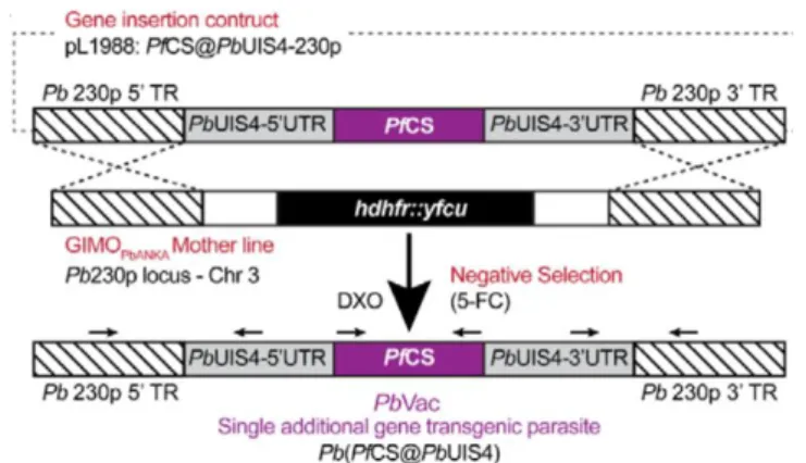
“naturally attenuated”, whole-sporozoite (Wsp) vaccine. PfCSP insertion into Pb is expected to generate humoral immunity against Pf, as well as cross species cellular immune responses, which may protect against a subsequent Pf infection. The candidate antigen was transfected to its delivery platform using ‘gene insertion/marker out’ (GIMO) methods of transfection.The gene encoding PfCS was inserted into the neutral 230p locus of the Pb genome under the control of the 5′-and 3′ regulatory sequences of Pb’s uis4
20
gene (Fig. 2), which is expressed exclusively in sporozoites and developing liver stages. The GIMO transfection method employed ensures the stable insertion of the gene encoding the heterologous PfCS and flanking regions in the Pb genome, resulting in a drug-selectable marker-free transgenic parasite. Genotyping of PbVac showed correct integration of the PfCS expression”100.
Figure 2- Generation of PbVac. Schematic representation of the transgenic line PbVac line (Pb(PfCS@UIS4) where the GIMO 199 insertion-construct (pL1988) replaces the selectable marker (SM; hdhfr::yfcu) in the GIMO 200 PbANKA mother line with the PfCS coding sequence (CDS) after negative selection using 5- 201 fluorocytosine (5-FC). Construct pL1988 integrates by double cross-over homologous 202 recombination (DXO) using 5’ and 3’ targeting sequences (TR) for the neutral 230p locus, resulting 203 in the introduction of the PfCS CDS under the control of the PbUIS4 gene promoter (5’-UTR) and 204 Pbuis4 transcriptional terminator sequence (3’- UTR) and removal of the SM. Black arrows: 205 location of primers used for diagnostic PCR; 2018, Mendes M. A., A Plasmodium berghei Sporozoite-Based Vaccination Platform Against Human Malaria
This genetically modified WSp vaccine can infect human hepatocytes but cannot continue to a blood stage infection, degenerating into unviable cryptic forms, which means that it may be non-pathogenic to humans. This evidence was further supported by studies in human hepatocytes cell lines, in liver and blood humanized mice and in New Zealand White (NZW) rabbits100. The PbVac parasite has been shown to display similar fitness to the wild type Pb parasite in terms of sporogonic development; formation of oocysts in the mosquito mid gut and of sporozoites in salivary glands; yield of exo-erythrocytic forms (EEFs); and hepatic load in infected mice and a higher human hepatocyte infectivity than Pf sporozoites in vivo100.
Studies in NZW rabbits have shown that PbVac is not able to develop a blood stage infection, and that 10 days after administration the parasite has been eliminated from the liver and all the organs101. Notwithstanding this, as a safety measure, the capacity of Malarone® to eliminate hepatic PbVac parasites, and Malarone® and Coartem® to eliminate PbVac blood stage parasitemia from infected mice has been confirmed.In
21
addition, toxicological and humoral responses have been assessed in rabbits immunized 5 times via bite of 75 PbVac-infected Anopheles stephensi mosquitoes per administration. This study has shown that PbVac administration can trigger increased IgG titers
anti-PfCS and anti-PbCS101.
All these studies demonstrated the safety and immunogenicity of PbVac vaccine candidate, prompting its evaluation in non-human primates and subsequently in a Phase I/IIa clinical trial101.
1.3.4. Immune response against vaccines
The complexity of the Plasmodium life cycle allows the conception of malaria vaccines that target different stages, and which may promote immunity by recognizing the parasite as a whole or of specific proteins. For blood-stage and transmission-blocking vaccine candidates immunity is mostly mediated by antibodies, whereas for pre-erythrocytic vaccines the type of immunity generated can vary, and involve both antibodies and CD4 T cells, such as in RTSs 102, as well as other T cells (CD8+, CD4+ and γδ T cells) such as in PfSPZ102.
Liver-stage vaccine candidates aim to prevent the progression of the parasite’s life cycle to the blood stage through induction of potent liver-stage immune responses. This protective immunity is largely mediated by CD8+ T cells due to their robust parasite-induced responses and their capacity to eliminate the parasite. In rodents, CD8+ T cell responses were shown to be an efficient anti-parasite mechanism that eliminates malaria liver stages103 and the depletion of IFN-γ or CD8+ T cells blocked RAS-mediated sterile
immunity104.
Protection mechanisms behind RAS protection starts with antibodies that aim to inhibit sporozoites from reaching the liver and infecting hepatocytes, and the efficacy of this humoral response depends on the anti-sporozoite antibody titers105. Injected sporozoites when entering in the lymph nodes are presented by DC to prime specific CD8+ T cells that migrate to the liver and aim to eliminate the parasites and generate immunity105. CD4+ T cells can contribute positively to the proliferation and survival of CD8+ T cells, or with IFN-γ production, aiming to increase this response and its
22
durability105. Some of the circulating CD8+ memory T cells migrate to the liver, where they become tissue resident memory T cells106. CD8+ T cells surround these hepatocytes in order to eliminate them by IFN- production or through direct contact by cytolytic perforin and granzyme release106. Moreover, CD8+ T cells can induce RAS-protective immunity through the role of interleukins, nitric oxide (NO), and NK cells106. IvPfSPZ
vaccination was shown to increase the levels of CD8+ T cells that produced effector cytokine molecules, such as IFN-γ, IL-2 and TNF-α, compared to pre-vaccination, as well as anti-CSP and anti-PfSPZ antibody levels92.
In GAS vaccine studies in mice, the protection conferred appears to depend on the time point at which the parasites arrest during liver-stage development107. Protective immunity was reported to be IFNγ-secreting CD8+ T cell-mediated, and long term protection was associated with CD8+ effector memory T cells. Protection studies in mice show that the immune response does not appear to be dependent on antibodies, and CD4+ T cell help is not essential to mount CD8+ T cell immune responses107. This immunization trigger antibody and B cell responses, but when B cells are depleted the mice were completely protected against a sporozoite challenge108. This study show that the immune response and protection are mainly mediated by CD8+ cells108.
CPS immunization response is primarily mediated by parasite-specific antibodies that can interfere with sporozoite motility, hepatocyte invasion and development and by long-lasting-memory B cells. Besides that, cellular immune responses represent key effector mechanisms leading to parasite elimination109. Once again, IFN-γ CD8+ T cells elicit a crucial immune response through the increase in granzyme B, but Th1 are also important for this response as shown by the increase in transcription factors after CPS110. Partially and fully protective CPS volunteers show alterations in cytokine-producing γδ T cells by CD4+ and CD8+ effector memory T cells111.
The immunity triggered by RTS,S is carried out by CSP-specific cell-mediated immunity complemented with CSP-specific antibody-mediated immunity. RTS,S cellular immune responses are essentially dependent on CD4+ T cells and IL-2 production, although other cytokines, including IFN- and TNF- have also been reported to be involved112.
23
1.3.5. Rhesus monkeys as malaria vaccine trial models
The main animal model used in biomedical research in the world is the rodent model. However, although it has many advantages, it is not ideal for accurately mimicking the cellular and molecular mechanisms of the immune response and the response to infection in humans. Nonhuman primates (NHP), are able to bridge many shortcomings of other animal models, due to their significant genetic, physiological and behavioural homology, and are thus critical to understand pathogenesis, immunity and vaccine development in humans. However, some differences exist regarding markers of immune populations, namely NK, NKT cells and memory T cells113.
Several studies evaluating malaria vaccines have been performed in rhesus monkey, such as studies on RTS,S vaccine adjuvants114, IFNγ responsesand antibody titers of RTS,S of adenovirus serotype-35 CSP prime and RTS,S boost115. CD8+ T cell responses were correlated with protection against a challenge of P. knowlesi on rhesus inoculated with irradiated P. knowlesi sporozoites, which has been replicated in CHMI model with irradiated Plasmodium falciparum sporozoite vaccine116. NHP studies using PfSPZ have shown that iv immunization triggers higher cellular immune responses than sc, especially with regard to PfSPZ-specific IFN- TNF- and IL-2-producing CD8+ T cells. Analysis of the liver cells’ immune responses 3 to 4 months after the last immunization showed that, while iv-immunized animals revealed a prevalence in magnitude of PfSPZ-specific T cell responses, sc immunization resulted in low to undetectable T cell responses 117.
24
2. Aims
The asymptomatic and highly immunogenic nature of the Plasmodium liver stage makes it an ideal target for malaria vaccination development. IMM’s MPrudêncio lab has developed PbVac, a new malaria vaccine candidate, in which the rodent parasite P.
berghei expresses the human P. falciparum antigen PfCSP in order to promote
PfCS-specific and cross-species immune responses that may protect against a subsequent P.
falciparum infection. The pre-clinical phase of the vaccine candidate included
experiments in mice, NZW rabbits and rhesus monkeys. Here we report the pre-clinical testing in rhesus monkeys of P. berghei WT and genetically modified sporozoites, as a strategy to induce immunity to P. falciparum.
The objectives established for this thesis are as follows:
- To confirm that the P. berghei (Pb) parasite infects rhesus hepatocytes in vivo.
- To analyse the humoral responses generated by specific immunization with PbWT and
PbVAC sporozoites.
- To characterize the immune population dynamics in the liver, spleen and peripheral blood cells in response to immunization.
- To study the specific immune responses of PBMC and liver cells to PbWT, PbVac and