BJRS
RADIATION SCIENCES
05-03-A (2017) 01-09Development of a methodology for the analysis of
radionu-clide impurities in radiopharmaceuticals by gamma-ray
spectrometry
E. B. de Paula
a; M. T. F. de Araújo
a,b; J. U. Delgado
a; R. Poledna
a; R. Lins
a; A. Leiras
a;
C. J. da Silva
a; A. E. Oliveira
aaInstituto de Radioproteção e Dosimetria, Av. Salvador Allende, s/n; Bairro: Recreio dos Bandeirantes, CEP:
22780-160 – Rio de Janeiro, RJ.
b Laboratório de Instrumentação Nuclear (LIN/PEN/COPPE/UFRJ), Caixa Postal 68509, CEP 21945-970 RJ, Brazil.
[email protected] / [email protected]
ABSTRACT
LNMRI has sought to develop a methodology for the identification and accurate detection of impurities emitting gamma radiation at the metrological level, aiming to meet the recommendations not only of the international pharmacopoeia, but also of CNEN and ANVISA regarding quality control that can ensure patients That the doses received by the practices are as low as practicable. As an initial goal, it was possible to obtain an efficiency curve with uncertainty around 1% necessary to start future measurements of interest applied to nuclear medicine and to begin the development of the impurities analysis technique.
1. INTRODUCTION
Radiopharmaceuticals are preparations consisting of one or more radionuclides and a pharmacologi-cal moiety. The use of these radiopharmaceutipharmacologi-cals in nuclear medicine facilitates the diagnosis and therapy of patients [1]. In the country, the largest quantity of radiopharmaceuticals is due to 99mTc,
which presents several useful properties to the patient as a gamma-ray tracer radionuclide [2]. There are thirty-one (31) fully listed radiopharmaceuticals for imaging studies of important organs such as the brain, myocardium, thyroid, lung, liver, bladder, kidneys, skeletal, blood and follow-up as well as tumor treatment. Of the 31 existing ones the main ones used in Brazil are: 67Ga, 123I, 131I, 210Tl, 18F
and 223Ra, where all of them can present impurities are emitting gamma radiation (generally
accord-ing to the production process).
It is important to note that, according to the pharmacopoeia; these impurities must not only be iden-tified but also quaniden-tified with a certain level of precision. A standardized radioactive source must al-ways be accompanied by a statement of the presence of all possible and measurable radioactive im-purities. It is also important that purity checks be continued over time for the entire working life of a reference source or even a radiopharmaceutical administered to the patient, since the impurities of long half-lives have a longer life and their Percentage of the radionuclides with short half-lives, as is the case with radiopharmaceuticals. Furthermore, it should not be forgotten that the sample ra-dioactive purities deteriorate continuously because of the rara-dioactive decay itself and due to radia-tion damage around the active material, especially labeled compounds. This problem becomes more pronounced when it comes to the case of radiopharmaceutical applications in patients [1].
The radiopharmaceutical is characterized by a short half-life, but in this mode of production the presence of radionuclide impurities, usually of larger half-lives in relation to the main one, is com-mon. For most purposes of the user of radioactive materials it is necessary to know their specific ac-tivities or concentrations of activity and purity, their decay constants and the nature of their radia-tions, including the energy of each.
Rarely, however, would it be a secondary standardization laboratory that would be assigned to mea-sure or verify more than activity and impurity, and would normally be meamea-sured with calibrated equipment using activity standards provided by national laboratories via gamma spectrometry
meth-ods; Purity (chemical and radionuclide) should be the responsibility of the producing center and nu-clear data are generally produced by specialized national metrology laboratories with their decay parameters related to radionuclides published in updated tables of nuclear data. Thinking in this light, the LNMRI has sought to develop a methodology for analyzing these impurities in order to have as an end product an internal document where concepts for approaching the subject are well delineated.
Some recent work brings us analyzes related to the control of impurities that can be seen in the work of Ferreira et. Al. [2]. Where, for example, 99Mo appears as an impurity of the 99mTc, with a
half-life of 6 h while the impurity has a half-life of 66 h, both emitting gamma radiation. The US pharmacopoeia stipulates an impurity limit with respect to the major radionuclide of 0.15 mCi / mCi (15%) of 99Mo / 99mTc at the reference date.
On the other hand, the gamma spectrometry technique not only identifies how it quantifies radionu-clides emitting gamma radiation since it allows distinguishing peaks of very close energies due to their high resolution power. Therefore, it is necessary to develop a procedure using gamma spec-trometry techniques to ensure that these limits set by the pharmacopoeia are not exceeded [4..10]
2. OBJECTIVES
The initial objective of this work was to obtain a good efficiency curve with low associated uncer-tainties (around 1%) as a starting point for the establishment of a methodology for the analysis of radionuclide impurities. With this, it is expected to be able to make feasible the determination of the activity quantity, aiming at the quantification, qualification and determination of nuclear parameters using techniques of gamma spectrometry to ensure that the limits established by the pharmacopoeia are not exceeded.
3. THEORY
In Nuclear Medicine, ionizing radiations are emitted from within the patients from incorporated ra-dionuclides and detected externally for imaging. Radiopharmaceuticals are used in well established procedures, based on their selective incorporation by the organs within the human body [3]. In order to succeed in the exam it is necessary that the value of the applied activity be well determined. A lower value may compromise the diagnosis and, therefore, the determination of the activity of the radiopharmaceuticals administered to the patients should have good accuracy. To measure it, the Nuclear Medicine Services use activity meters (activimeters). Basically, this equipment does not distinguish the energies and consists of a well-type detector and a system of processing the signals or the current generated. In the reentrant part of the meter the radiopharmaceutical is placed and may be packed in ampoules, vials or syringes [2]. To detect the activity from the radionuclide of interest it is necessary to make use of another suitable detector with resolution so that it can be identi -fied by the energy; therefore, the germanium detector (HPGe) was used here.
3.1. Radiopharmaceuticals
These are preparations containing one or more radionuclides which, in addition to meeting pharma-copeia specifications, have their production, supply, and storage use and disposal regulated by rele-vant governmental regulations. Radionuclide emissions of choice should be easily identified and perceived by nuclear detectors so that the dose of radiation to the patient is required for performing the process. About 95% of radiopharmaceuticals are used for diagnosis, while the remaining 5% are used for treatments [2].
4. MATERIALS AND METHODS
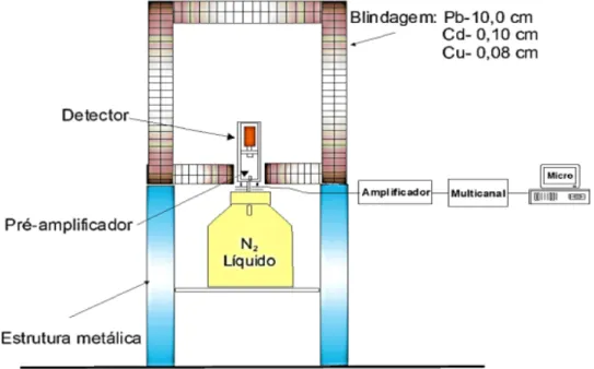
The experimental data were obtained through measurements performed at the National Laboratory of Metrology of Ionizing Radiation - LNMRI at the Institute of Radioprotection and Dosimetry. It is the first laboratory stage for the development of the work: the preparation of the measurement system through its calibration. A gamma spectrometry system was used, consisting of a hyperpure
germanium detector (GMX) that allows the identification of radiation with energy of 10 keV to 10 MeV.
Figure 1: Spectrometry system composed by AKIRA detector + Dewar + shielding +
asso-ciated electronics.
The elaboration of an efficiency curve will be necessary in order to guarantee the linearity of the detection system that will be used in the analysis of the impurities.
4.1 The Efficiency Curve Method
The Efficiency Curve method consists of a methodology to obtain the photoperiod efficiency of a gamma or X spectrometry system. The efficiency of photopeak is defined as the ratio between the rate of radiation detected by the system and the rate emitted by the source [6]. With the use of calibrated standard sources of known energies and intensities, using the same detection system and the same counting geometry, an energy efficiency curve is obtained through the relation:
Where S is energy photopeak area of a standard source at (s-1), T is the count time (s), A is activity of
standard source at (s-1), K is decay factor of the radionuclide standard source, Pγ is a probability of emission
of the gamma radiation of photopeak in (s-1) [10].
With the above data we obtain the efficiency curve that will be given by means of an adjustment of experimental points by the LABFIT® that will provide a mathematical adjustment for each type of curve. The
format of the mathematical adjustment of the efficiency curve used is given arbitrarily by those who use the program. Some criteria of choice are taken into account such as: physical behavior of the quantities or parameters involved and the mathematical expression in which the value of the correlation factor (r²) of the adjustment should be as close as possible to 1.
5. EXPERIMENTAL PROCEDURES
In order to make the efficiency curve, the AKIRA detector of HPGe model Gem-45P4 (detector with 40% relative efficiency for 1.33MeV of 60Co), series: 47-JP12399A, positive operating voltage
was used. Radionuclide patterns in the puntual geometry of: 166mHo, 152Eu, 133Ba, 137Cs, 134Cs, 241Am, 54Mn and 22Na were also used to determine the Energy versus Efficiency points.
Table 1. Some energies of radionuclidic standadrds
Radionuclides Principal energies
166mHo 184,4 keV 711,7 keV 152Eu 121.78 keV 244.69 keV 344.28 keV 133Ba 81,00 keV 356,01 keV 137Cs 661.66 keV 134Cs 604,70 keV 795,85 keV 241Am 59.54 keV 54Mn 1022,21 keV 1115.87 keV 22Na 511 keV 1274 keV
The respective standards were selected in the LNMRI, each one in the point sources geometry. The sources were positioned at a distance d = 20 cm from the top of the detector and a geometry fixture
was used to ensure reproducibility of the measurements. The data acquisition was performed considering MAESTRO® software associated to a multichannel of 16000 channels. Counts were obtained by considering a 24 hours measurement time interval for each standard. The spectrum obtained gives the counts for each peak of interest with good accuracy in addition to providing data such as live time and real-time counting and dead time.
6. RESULTS AND DISCUSSION
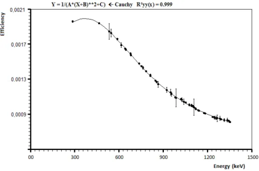
In this first preliminary step, the patterns in the point geometry were measured during a 1-day aver-age time interval at a fixed source-detector distance of 20 cm. It is important to emphasize that when performing long measurements, we are improving the counting rate and decreasing some of the associated uncertainties since the dead time is very low (less number of pulse stacks) [1].
With the measured energy it was possible to construct an efficiency curve that presented uncertain-ties of the order of 1% for the calculated efficiencies. Figure 2 represents the efficiency curve for the AKIRA detector that will be used in the next.
Figure 2: Efficiency curve obtained for the spectrometry system using patterns in
7. CONCLUSIONS
In this first step we were able to first establish an overview of the bibliographies that carried out studies for the quantification and / or analysis of radionuclide impurities each with a particular crite-rion. Here, it was possible to establish an efficiency curve that will be needed to start future mea-surements of radiopharmaceuticals of interest in nuclear medicine. In initial criterion, this stage was important because it will act as mediator of the next steps of the development of said methodology of analysis of radionuclide impurities.
REFERENCES
1. Journal
[1] Araújo, M.T.F., Poledna, R., Delgado, J.U., Silva, R.L., Iwahara, A., Silva, C.J., Tauhata, L., Oliveira, A.E., Almeida, M.C.A., Lopes, R.T., 2016. Absolute Standardization of the impurity 121Te associated to the
production of the radiopharmaceutical 123I. Applied Radiation and Isotopes 109, 389-392.
[2] Ferreira, F.C.L., Cardoso, L.X., Costa, M.J.C, Cunha, C.J., Souza, D.N., 2008. Avaliação de 99Mo em
amostra de 99mTc em clínica de medicina nuclear de Sergipe. Scientia Plena 4, 11482.
[3] L. Carballo Silva, F. Campos Añón, M.A. Roque Alegre, C. Santos Montero, M. Martínez Seguer, M.N. Campos Llamazares, E. Marcos Segura, F. Pons Pons, 2014. Determinación de
impurezas Radionucléidicas de Európio em 153Sm. Revista Española de Medicina Nuclear e Imagen
Molecular 33 (Supl 1), 249.
[4] M.F Catanoso, J.A. Osso Jr., 2009. Evaluation of the radionuclidic purity of 123I and 131I samples. Iba
Radiopharmasolutions, publications.
[5] Bernardes, E.M.O., Delgado, J.U., Tauhata, L., da Silva, C.J., Iwahara, A., Poledna, R., Paschoa, A.S., 2002. 166Ho: a multi-γ standard for the calibration of the Ge spectrometers. Appl. Radiat. Isot. 56,157–161.
[6] De Almeida, M.C.M., Iwahara, A., Poledna, R., da Silva, C.J., Delgado, J.U., 2007. Absolute
disintegration rate and 320keV γ-ray emission probability of 51Cr. Nucl. Instrum. Method Phys. Res.
A580,165–168.
[7] LNHB/CEA, Laboratoire National Henri Becquerel, 2004.Table of Radionuclides. 〈www.nucleide.org/DDEP_WG/123_tables〉.
[8] Lépy, M.C., Pearce, A., Sima, O., 2015. Uncertainties in gamma-ray spectrometry. Metrologia 52, S123– S145.
[9] Schoetzig, U., Schrader, H., 1989. Halbwertszeitenund Photon-Emissions wahrsheinlichkeiten von haeufigverwendeten radionukliden.PTB-Ra-16/3.
[10] Silva, M.A.L., Poledna, R., Iwahara, A., Silva, C.J., Delgado, J.U., Lopes, R.T., 2006. Standardization