Universidade de Lisboa
Faculdade de Farmácia
Antineoplastic Therapy
Multidrug Resistance
Luís Paulo Antunes da Silva
Mestrado Integrado em Ciências Farmacêuticas
Universidade de Lisboa
Faculdade de Farmácia
Antineoplastic Therapy
Multidrug Resistance
Luís Paulo Antunes da Silva
Monografia de Mestrado Integrado em Ciências Farmacêuticas apresentada à Universidade de Lisboa através da Faculdade de Farmácia
Orientador: Doutora Noélia Maria da Silva Dias Duarte, Professora
Auxiliar
5
Resumo
A multirresistência à terapêutica antineoplásica (MDR) constitui atualmente, um dos principais desafios no âmbito clínico. A MDR é o resultado de vários mecanismos de ação concomitantes, derivados da adaptação das células estaminais cancerígenas (CSCs) do tumor, promovida pela exposição a stress celular. Prognósticos da doença face à resistência à terapia antineoplásica agravam drasticamente aquando do desenvolvimento de um fenótipo resistente pelo tumor, para os quais os regimes terapêuticos atuais não mostram qualquer eficácia. Os mecanismos responsáveis pela multirresistência são vários e incluem: transportadores ABC (ATP binding cassette), modulação pelo microambiente tumoral, vias de sinalização Hippo, epigenética, exossomas, reparação de danos na cadeia de DNA, resposta a proteínas unfolded (UPR), stress no retículo endoplasmático (ERS), mito e autofagia, e metabolismo celular tumoral. Nesta monografia, procura-se abordar estes mecanismos de resistência, conferindo uma perspetiva atual e transversal sobre o impacto do cancro na sociedade e em específico para o doente oncológico, numa visão de como a terapia convencional e novas terapias promissoras em estudo possam ter a capacidade de superar esta problemática.
O cancro, considerado como uma das duas grandes doenças do século XXI, é a segunda causa de morte a nível mundial e totaliza aproximadamente 100 milhões de casos que, em algum momento da sua vida, desenvolveram a doença. Uma em cada seis mortes tem como causa o cancro, e a grande etapa a atingir é um diagnóstico precoce, que potencia a probabilidade do doente obter uma remissão do tumor.
A estratégia terapêutica a adoptar deve ser cuidadosamente avaliada consoante o perfil do doente, e cada vez mais se procura abordar um esquema terapêutico polivalente, ao incorporar associações de fármacos, a fim de melhorar outcomes e evitar resistências. Para além das convencionais cirurgia, quimioterapia e radioterapia, a necessidade de individualizar a terapêutica para cada doente tem promovido o despertar dos fármacos biológicos que fundamentam a imunofarmacologia.
Dos mecanismos mencionados que promovem a multirresistência à terapêutica antineoplásica, os transportadores ABC são os que há mais tempo têm sido alvos de inúmeros estudos. A sua capacidade em reduzir as concentrações dos fármacos citotóxicos
6
do meio intracelular através do efluxo dos mesmos são a base desta resistência pelo tumor, permitindo à célula tumoral adaptar-se a uma concentração sub-terapêutica destes fármacos, a qual não reproduz o efeito de stress celular que repercuta em morte celular. Já o microambiente tumoral destaca-se por vários fatores: um estado de hipoxia conjugado com inúmeras moléculas de índole neoplásica, como as pró-angiogénicas, promove o crescimento e metastização tumoral, enquanto impossibilitam que os fármacos penetrem as camadas mais internas do núcleo tumoral, tornando-os ineficazes.
As células estaminais tumorais têm ainda a capacidade de, quando expostas a situações de stress celular, desenvolver mutações reversíveis nas proteínas membranares – dito epigenoma – que impedem a ligação de determinados fármacos citotóxicos às células-alvo. Também os exossomas têm um papel fulcral no desenvolvimento tumoral, na medida em que promovem a comunicação entre células tumorais sem sofrer degradação, possibilitando a transferência de material genético premetastático e metastático para células fora do local inicial do tumor, enquanto permitem também alguma plasticidade celular na medida em que modulam a interconversão entre células estaminais tumorais e células tumorais diferenciadas, para além de promoverem angiogénese.
Por outro lado, as vias de sinalização têm também sido alvo de estudo, destacando-se a via de Hippo. Esta é responsável por regular diversos processos celulares, dos quais se incluem a proliferação celular, apoptose e respostas celulares a stress, três grandes bases da modulação tumoral e processos intrínsecos aos mecanismos de resistência.
As células tumorais são também passíveis de se adaptar e se tornar resistentes à radioterapia, mediante uma via celular de reparação aos danos provocados pela terapêutica à cadeia de DNA. As células estaminais cancerígenas são extremamente competentes no que toca a preservar o DNA, através da modulação de enzimas responsáveis pela reparação de material genético, impedindo a desejada morte celular das mesmas.
Outro método celular que promove resistência à terapêutica é a via de resposta a proteínas
unfolded. Esta via é despoletada por stress no retículo endoplasmático, que por sua vez é
provocado por um incorreto folding proteico, e permite às células tumorais contrariar este stress através de um número de vias celulares.
7
Temos ainda o processo de autofagia, que permite às células estaminais cancerígenas a reutilização de organitos celulares e consequentemente, regular a homeostasia intracelular, mesmo quando exposto a fármacos citotóxicos, e preservar a sua pluripotência.
Ao evitar o processo de apoptose, o tumor consegue, através de inúmeros processos mais detalhados nesta monografia, estabelecer-se sem ser eliminado no doente, e ainda evitar o processo de anoikis, que possibilitam o desenrolar metastático.
Por fim, as vias metabólicas do tumor diferenciam-se das células saudáveis, o que lhes permite uma enorme capacidade de adaptação e sobrevivência, bem como um rápido crescimento e proliferação celular.
A busca incessante de novas estratégias que possam colmatar a persistência tumoral face às terapêuticas atuais tem mostrado resultados promissores. Curiosamente, alguns fármacos já aprovados para determinadas patologias, provaram eficácia ao estimularem sensibilização das células alvo à quimio, imuno e radioterapia, e reverterem a resistência desenvolvida pelo tumor, como é o exemplo das estatinas, a metformina, a melatonina, e compostos de origem natural, como os flavonoides e isoflavonoides.
Ainda assim, o desenvolvimento de novos fármacos mais específicos, com menor toxicidade e menos efeitos adversos, tem sido uma prioridade neste ramo. Uma terapêutica cada vez mais direcionada, de índole biológica, é atualmente a aposta com melhores
outcomes, sejam elas anticorpos monoclonais, células CAR-T, inibidores enzimáticos,
como a tirosina cinase, e ainda vacinas.
Palavras-chave: cancro; multirresistência; terapêutica antineoplásica; células cancerígenas
8
Abstract
Multidrug resistance (MDR) in oncology stand as one of the main challenges in clinical treatment, as a result of several concomitant yet different mechanisms developed by carcinoma stem cells (CSCs). Outcomes in antineoplastic therapy drastically worsen when the tumour displays a resistant phenotype and therapy regimens lack efficacy. Several mechanisms have been discovered that contribute to this multiple resistance: ATP binding cassette (ABC) transporters, microenvironment modulation, the Hippo signalling pathway, epigenome, exosomes, unfolded protein response (UPR), endoplasmic reticulum stress (ERS), autophagy and mitophagy, and metabolism.
Of the mentioned mechanisms above that promote multidrug resistance to antineoplastic therapy, ABC transporters have been the target of numerous studies for the longest time. Their ability to reduce intracellular cytotoxic drug concentrations through efflux is the basis of this resistance by the tumor, allowing the tumor cell to adapt to a sub-therapeutic concentration of antineoplastic drugs.
The tumour microenvironment, by promoting a state of hypoxia coupled with numerous neoplastic molecules, such as pro-angiogenic, promotes tumour growth and metastasis, while preventing drugs from penetrating the innermost layers of the tumour core.
On the other hand, tumour stem cells also have the ability, when exposed to cellular stress, to develop reversible mutations in membrane proteins – the epigenome - that prevent the binding of certain cytotoxic drugs to target cells.
Here, we further detail these resistance mechanisms, while providing a perspective towards how cancer is perceived nowadays, as well as conventional therapy, and future potential therapies in study that may have the ability to overcome this issue.
9
Agradecimentos
Começar por expressar a minha gratidão revelou ser talvez a tarefa mais árdua.
A sensação agridoce do fechar de um capítulo e o desfolhar de um novo que se avizinha a largos passos remete-me para um dissabor nostálgico de um passado não tão distante, do qual não poderia encerrar sem umas breves palavras.
À professora doutora Noélia Duarte, agradeço-lhe por ser a bússola deste projeto, pela orientação, disponibilidade, o saber que me soube transmitir, opiniões e críticas, e um total apoio.
À família, por ser o meu mais forte pilar de sustento. Aos meus pais, por serem modelos de perseverança, pelo apoio megalómano, a paciência e a força que me deram para desbravar estes cinco anos de MICF. Espero poder retribuir sempre todo o carinho e generosidade. A eles dedico esta monografia.
Aos amigos, o meu outro porto de abrigo. Obrigado pelo companheirismo, a eterna alegria, mas também na melancolia, foram a família longe da família.
Por fim, à alma mater, por ser a base da profissão. Por me acolher, edificar e ver-me crescer no universo da saúde.
10
Abreviaturas
ABC – ATP binding cassette
EMT – epithelial-mesenchymal transition CSCs – carcinoma stem cells
DDR – DNA damage response UPR – unfolded protein response ERS – endoplasmic reticulum stress
c-FLIP - cellular FLICE (FADD-like IL-1β-converting enzyme)-inhibitory protein OXPHOS – oxidative phosphorylation
CML – chronic myelogenous leukaemia CAF – cancer associated fibroblasts
MIF – macrophage migration inhibitory factor MDSCs – myeloid-derived suppressor cells lncRNA – long non-coding RNA
miRNA – microRNA
ATM – Ataxia-Telangiesctasia mutated protein
ATR – Ataxia-Telangiectasia and Rad3-related protein ESA - epithelial-specific antigen
ALDH1 - aldehyde dehydrogenase1 TK - tyrosine kinase
TME - tumor microenvironment
MHC – major histocompability complex HLA – human leukocyte antigens
NSCLC – non small cell lung cancer SCLC – small cell lung cancer
11
RT – radiotherapy CT – chemotherapy PK – pharmacokinetics
EGFR - epidermal growth factor receptor FGFR - fibroblast growth factor receptor EMT - epithelial–mesenchymal transition HMG-CoA - β-Hydroxy β-methylglutaryl-CoA MMPs - matrix metalloproteinases
TNBC – triple negative breast cancer 5-FU - fluorouracil
LGL – large granular lymphocyte CLL - chronic lymphocytic leukaemia
12 Índice: 1 Introduction ... 14 1.1 EPIDEMIOLOGY ... 15 1.1.1 Worldwide paradigm ... 15 1.1.2 European paradigm ... 17 1.1.3 Portugal paradigm ... 18 1.2 DIAGNOSIS ... 18 1.2.1 TNM classification ... 19
1.2.2 Histology and differentiation level ... 19
1.2.3 Cellular receptors ... 21 1.2.4 Genetics ... 21 1.3 CONVENTIONAL THERAPY ... 22 1.3.1 Surgery ... 22 1.3.2 Radiotherapy ... 23 1.3.2.1 Radiobiology ... 24 1.3.3 Chemotherapy ... 25 1.3.3.1 Alkylating agents ... 26 1.3.3.2 Platin-derived ... 27 1.3.3.3 Antimetabolites ... 27
1.3.3.4 Topoisomerase I and II inhibitors ... 28
2 MULTIDRUG RESISTANCE ... 32
2.1 Resistance mechanisms ... 33
2.1.1 ATP-binding cassette transporters ... 33
2.2.2 Microenvironment modulation ... 34
2.1.3 Hippo pathway ... 34
2.1.4 Epigenome ... 34
2.1.5 Exosomes ... 36
2.1.6 DNA damage response ... 36
2.1.7 Unfolded protein response/Endoplasmic reticulum stress ... 37
2.1.8 Autophagy ... 37 2.1.9 Metabolism ... 38 2.1.10 Apoptosis evasion ... 38 3 THERAPEUTIC APPROACHES ... 40 3.1 ABC transporters ... 40 3.2 Microenvironment ... 41 3.3 Hippo pathway ... 42 3.4 Epigenome ... 42 3.5 Exosomes ... 43 3.6 DDR ... 43 3.7 ERS/UPR ... 43 3.8 Autophagy/mitophagy ... 43 3.9 Metabolism ... 43 4 Conclusions ... 45 5 Referências Bibliográficas ... 47
14
1 Introduction
Cancer is described as a clinical condition in which cells with abnormal behaviour divide with no regulation and may develop the ability to invade nearby tissues. Cancer cells may as well spread to distant parts of the body via bloodstream and lymphatic system. Also called malignancy, cancer is classified according to six major types: carcinoma, for tumours that begin in the skin or tissues that outline or cover internal organs; sarcoma, for tumours that begin in connective and/or supportive tissues, such as bone, cartilage, fat, muscle and blood vessels; leukaemia is a type of cancer which begins in blood-forming tissue, namely the bone marrow, and is responsible for a great amount of abnormal blood cells to be produced and spread throughout the blood; lymphoma and multiple myeloma consist of cancers whose origin is in the immune system cells; and the central nervous system type of cancer, originated in the brain and spinal cord tissues. (1)
During the process of carcinogenesis, a multi-stage mechanism where a healthy cell evolves into a tumour cell, throughout a series of multiple epigenetic and genetic events that are staged as the following: initiation - results from an irreversible modification in the genome due to a strand break or adduct promoted by the carcinogen with the DNA chain, which under specific circumstances the altered cell may show a selective growth behaviour; promotion - a reversible stage where promoters, a distinct class of incomplete carcinogens, play a role in increasing growth of initiated cells, which may then proliferate and result in a benign tumour; and progression - where additional exposure to the carcinogen, or resulting from spontaneous mutation, or even thanks to instabilities caused in the initiating process, may lead to a malignant conversion. (2)
15
1.1 EPIDEMIOLOGY
The oncological disease sits at the top, in both Portugal and the whole occidental world, as the second leading cause of death for both men and women, placing only behind cardiovascular diseases. Statistics from the World Health Organization (WHO) state that in 2018, 9.6 million people died of cancer, where numbers rise in countries with low- to middle-economies. By 2040, it is predicted a 64% rise in new cancer cases worldwide. Despite the massive efforts in research and development of novel therapies, cancer is still a major threat to health. As a result, urges the need to continuously develop new therapeutic strategies to ameliorate patients’ outcomes who suffer from aggressive or treatment-resistant carcinomas (3)(4).
1.1.1 Worldwide paradigm
From a global perspective, cancer is estimated to have risen up to 18.1 million new cases. One out of 5 men and one out of 6 women worldwide will develop the disease throughout their lifespan, and one out of 8 men and one out of 11 women will die from cancer. Worldwide, an estimated number of 43.8 million people are alive within 5 years of a cancer diagnosis. (5)
Asia represents 48.1% of new cases of cancer worldwide (in part because Asia represents around 60% of the global population), followed by Europe, with 23.5% estimated new cases, North America (13.2%), Latin America (7.9%), Africa (5.9%) and lastly, Oceania (1.4%). Asia also dominates the incidence statistics when it comes to each specific type of cancer, where Europe shows higher percentages only in melanoma (53.3%), bladder (34.8%), vulvar (37.1%), and kidney (34.9%) cancers; Non-melanoma skin cancer is highest in North America, accounting for 44.3% of worldwide’s incidence, and Kaposi sarcoma (77.8%) in Africa. Breast and lung cancers share the topmost incident cancers with 11.6% for both, followed by colorectum (10.2%), prostate (7.1%) and stomach (5.7%). (3,5)
16
Figura 1.1 Estimated number of new cases of cancer in 2018 for both sexes and all ages – Data Source: GLOBOCAN 2018; Graph production: Global Cancer Observatory (International Agency for Research on Cancer 2019).
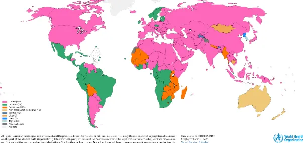
Figura 2 Top cancer per country, estimated age-standardized incidence rates (World) in 2018 for both sexes and all ages – Data Source: GLOBOCAN 2018; Graph production: Global Cancer Observatory (International Agency for Research on Cancer 2019).
As for mortality, lung cancer was, for 2018, the deadliest type of cancer with 18.4% estimated percentage of deaths, next to colorectum (9.2%), stomach (8.2%), liver (8.2%) and breast (6.6%). (5)
17
Lung, prostate, colorectal, stomach and liver cancer are the most common types of cancer in men, while breast, colorectal, lung, cervix uteri and thyroid cancer are the most common among women. This noted gender disparity is often reported due to lifestyle differences, mainly to exposure to risk factors such as alcohol and tobacco consumption. WHO describes however a “worrying” shift in the rise of lung cancer in women, mostly due to changes in social behaviour.
1.1.2 European paradigm
It has been estimated that in Europe, in 2018, there were 3.9 million new diagnosed cases of cancer and a total of 1.9 million deaths.(6) The five most incident types of cancer in Europe are breast (522513 reported new cases in 2018), followed by colorectum (499667), lung (470039), prostate (449761), and bladder (197105) cancers, which accounted for 50.6% of total new cancer cases in 2018, and 48.4% of cancer-associated deaths in 2018. (5)(7)
Figura 3 Estimated number of new cases in 2018 in Europe for all cancer, both sexes and all ages – Data Source: GLOBOCAN 2018; Graph production: Global Cancer Observatory (International Agency for Research on Cancer 2019).
Lung has the highest rate of mortality, with estimated 387913 deaths as of 2018. Colorectal cancer comes next, with 242483 estimated deaths, followed by breast (137707), pancreas (128045) and prostate (107315).(3,5)
18
1.1.3 Portugal paradigm
As for Portugal, colorectum cancer is, for both genders and all ages, the most incident type of cancer, placing fourth in European statistics, and the second deadliest cancer, placing only behind lung cancer. Then follows breast cancer, the most incident type of cancer in women, and prostate cancer, the most incident in men (Figure 1.1). (5,8)
Figura 4 Estimated number of incident cases and deaths in Portugal, both sexes and all ages – Data Source: GLOBOCAN 2018; Graph production: Global Cancer Observatory (International Agency for Research on Cancer 2019).
In terms of prevalence, it is estimated that in 2018, 28130 people were ever diagnosed and/or live with colorectal cancer in a 5-year perspective. Breast cancer follows next, with 26329 cases, followed by prostate (22111), bladder (7144), and thyroid cancers (6504), and non-Hodgkin lymphoma (5774). Although lung cancer is one of the most incident types of tumour in Portugal, its overall 5-year survival is extremely low.(5,9) This reverts into a 5,5% of total health expenses, accounting as 84 euros per capita, in a total of 867 million euros, where antineoplastic drugs stand as 31,5% of total expenditure.(10)
1.2 DIAGNOSIS
An early cancer identification can effectively decrease the risk of mortality associated with the disease. Late-stage diagnosis often occurs in resource-poor settings and is associated with a potentially higher morbidity and increased costs of treatment. Cancer outcomes can be improved with public health strategies aiming to early diagnosis, which imply handling patients by healthcare professionals at the earliest stage of the disease, reverting into a more anticipated, appropriate approach in terms of treatment.(11)(12)
19
1.2.1 TNM classification
The American Joint Committee on Cancer and the Union Internationale Contre le Cancer established a classification based on the three major stages in the tumour’s evolution: local growth of the tumour - T; lymphatic nodes relation to the tumour in the draining area – N – and lastly, the presence of metastasis - M. For each specific organ, there are further specifications for T or N classifications, whereas for M, it is based solemnly on the presence or absence of metastasis (M1 or M0, respectively). For each different possible TNM combination, there is a respective staging, that is also specific for each organ.(13) This classification allows for patients to be classified in a way that allows the healthcare professional in charge to evaluate and compare the progression of the disease, and particularly, the impact of each chosen therapeutic. (14)
1.2.2 Histology and differentiation level
Cancers can be classified in two ways: by primary site – meaning where the tumour was first originated, through the TNM classification – and by type of tissue in which the tumour originates – its histological classification. The conventional international standardized histology classification and nomenclature is the International Classification of Diseases for Oncology, Third Edition (ICD-O-3). (15)
From a histological point of view, there is a great amount of possibilities when it comes to cancer variability, which are grouped into six major categories:
Carcinoma is the most common type of cancer accounting for 80 to 90 percent of total
cancer cases. Originated in the epithelial tissue, carcinoma refers to a malignant neoplasm of the internal or external lining of the bodyand are divided into two major subtypes: adenocarcinoma, referring to a tumour in an organ or gland, and squamous cell carcinoma, which origin is in the squamous epithelium.
Adenocarcinomas are mostly associated with mucus membranes and are first observed as a thickened plaque-like white mucosa. They are often easily spread through the soft tissue, whereas squamous cell carcinomas may occur in several areas of the body.
Most carcinomas inflict secreting organs or glands, including the breasts, the lungs, colon, prostate or bladder.
20
Sarcoma is the term used to describe cancer whose origin is in supportive and connective
tissues - bones, tendons, cartilage, muscle, and fat. Mostly incident in young adults, the most common sarcoma consists of a painful mass on the bone. Sarcoma tumours usually resemble the tissue in which they grow: Osteosarcoma or osteogenic sarcoma (bone); chondrosarcoma (cartilage); leiomyosarcoma (smooth muscle); rhabdomyosarcoma (skeletal muscle); mesothelial sarcoma or mesothelioma (membranous lining of body cavities); fibrosarcoma (fibrous tissue), angiosarcoma or haemangioendothelioma (blood vessels), liposarcoma (adipose tissue), glioma or astrocytoma (neurogenic connective tissue), myxosarcoma (primitive embryonic connective tissue), mesenchymous or mixed mesodermal tumour (mixed connective tissue types)
Myeloma refers to cancer originated in the plasma cells of bone marrow. The plasma cells
are responsible for producing some of the proteins found in blood, namely antibodies, which are found faulty in the bloodstream, and may accumulate in the bone marrow, damaging the organ
Leukemias described as “liquid cancers" or "blood cancers", is the term used to refer to
cancers also in the bone marrow.This clinical condition leads to the excessive production of immature white blood cells, leaving the patientsusceptible to infection. Leukaemia also inflicts red blood cells damage and may cause low blood clotting and fatigue due to anaemia. Examples of this type of cancer include: Myelogenous or granulocytic leukaemia (malignancy of the myeloid and granulocytic white blood cell series), lymphatic, lymphocytic, or lymphoblastic leukaemia (malignancy of the lymphoid and lymphocytic blood cell series), polycythaemia vera or erythraemia (malignancy of various blood cell products, but with red cells predominating)
Lymphomas refer to cancer in the glands or nodes of the lymphatic system, a network of
vessels, nodes, and organs - the spleen, tonsils, and thymus, responsible for filtering bodily fluids and producing infection-fighting lymphocytes. Unlike the leukaemia lymphomas are "solid cancers." Lymphomas may also occur in specific organs such as the stomach, breast or brain and are defined as extranodal lymphomas. There are two subclassifications of a lymphoma: Hodgkin lymphoma and Non-Hodgkin lymphoma, which is characterized by
21
the presence of Reed-Sternberg cells in Hodgkin lymphoma, or absence in the non-Hodgkin lymphoma.
Mixed Types
The cancer may, however, exhibit components that may be characteristic to one category, or from different categories – such as the adenosquamous carcinoma, mixed mesodermal tumour, carcinosarcoma and teratocarcinoma.
Histological classification can even undergo into further specifications according to the primary type of the tumour. For instance, in prostate cancer, healthcare professionals developed an even more specific score to aid in choosing the best therapy – the Gleason score.(14)(16)(17)
1.2.3 Cellular receptors
Every tumour cell is equipped with an array of biologically active surface molecules, and several of these function as receptors for various ligands. They include the major histocompatibility complex (MHC), or in the case of humans, human leukocyte antigens (HLA), cytokine receptors, cell-adhesion molecules, growth factor receptors, and Fas/Fas-ligand molecules, among others. Their expressions are a subject to alterations, usually to the advantage of tumour growth and spread. Some appear on tumour cells de novo, having no counterparts on the respective normal cells. Detailed knowledge about the expression of tumour-cell receptors and their genotypes, in particular of cancerous ones, may provide information essential for the creation of tools for specific tumour immunotherapy.(18) (19)
1.2.4 Genetics
Cancer is itself a genetic condition - that is, the cause of cancer is due to certain alterations in genes that maintain and control the normal function of cells, namely their cell cycle. Certain gene changes can lead to cells evasion in normal growth controls and become malignant. For instance, some cancer-causing genes – oncogenes - increase production of a specific protein that promotes cell growth, while others lead to the production of a misshapen, non-functional form of a protein, in normal conditions, would repair cellular damage.
Cancer-inducing genetic mutations can also be acquired during the patient’s lifetime, as a result of errors that occur in cells division or from exposure to carcinogenic substances with
22
DNA-damage activity, such as chemicals in tobacco smoke, or ionizing radiation – e.g. ultraviolet rays. Genetic changes that occur post-conception are called somatic (or acquired) changes.
There are several types of DNA changes. Some changes affect just one unit of DNA, called a nucleotide. One nucleotide may be replaced by another, or it may be missing entirely. Other changes involve larger stretches of DNA and may include rearrangements, deletions, or duplications of long stretches of DNA.
Sometimes the changes are not in the actual sequence of DNA. For example, the addition or removal of chemical marks, called epigenetic modifications, on DNA can influence whether the gene is “expressed”—that is, whether and how much messenger RNA is produced. (Messenger RNA in turn is translated to produce the proteins encoded by the DNA.)
In general, cancer cells have more genetic changes than normal cells. But each person’s cancer has a unique combination of genetic alterations. Some of these changes may be the result of cancer, rather than the cause. As the cancer continues to grow, additional changes will occur. Even within the same tumour, cancer cells may have different genetic changes. Presence of oncogenes, tumour suppressing genes, apoptosis regulator genes, proteases or angiogenic factors. (20)(21)(14,22,23)
1.3 CONVENTIONAL THERAPY
There are several types of cancer treatment. The types of treatment that each patient will receive will depend on the type of cancer and how advanced it is. Today, we can talk about surgery, radiotherapy, chemotherapy, immunotherapy, targeted therapy, hormone therapy and stem cell transplants processes that can be used to treat cancer. (14)
1.3.1 Surgery
Surgery is considered the most efficient yet ancient procedure to treat non-haematological cancers () with curative intent. Surgery stands as not only a treatment procedure option, but also as complementary diagnosis, support and/or reconstruction methods. It is indicated, in most cases when the tumour is limited (with no evidence of metastasis). The chances of
23
achieving a complete resection of the tumour without compromising other organs; the tumour’s nature (e.g.: SCLC); and the morphological and/or functional mutilation caused by the procedure should all be taken in account when evaluating the patient’s condition and admission for surgery.(15,24)
The oncological surgery’s success rate is determined by the surgeon’s experience and how the approach is made: the safety borders play a crucial role in the success of the procedure, whereas it may be required an extemporaneous exam in order to guarantee that this peripheric region is tumour-free. It is also a common procedure to perform a regional lymphadenectomy (e.g.: armpit for breast cancer), as a way of establishing prognostic factors, as well as a complementary treatment for the primary tumour. In some cases, the extent of the lymphadenectomy is itself a prognostic factor (e.g.: colorectal carcinoma). (25)
For certain types of cancer (specifically breast and skin tumours), there has been introduced the Sentinel lymph node search technique. The sentinel node is the first lymph node that is directly affected when cancer spreads from the primary tumour site to the lymphatic system. In breast cancer, the sentinel lymph nodes (SLN) would generally be the lymph node that receives direct drainage from the breast. Once this node has been identified, it is biopsied. If this node is free from cancer cells, it is unlikely that the cancer has spread. If, however, the SLN contains cancer cells, then an axillary lymph node dissection (ALND), dissection of many nodes throughout the axilla) or complete lymph node dissection (CLND) may be indicated. (26)
There are a few exceptions, where surgery has been indicated in metastatic tumours - these indications depend on the type of tumour, the metastasis extent and the patient’s condition.(24) Metastatic breast surgery, for example, has shown improved women’s survival, although it does not stand as standard treatment. Surgery can, however, display a contradictory behaviour by potentializing tumour cells shedding into circulation, reducing anti-tumour immunity and promote tumour tissues changes that can induce migration and invasion towards the target site.(27)
1.3.2 Radiotherapy
Radiotherapy (RT) plays a relevant role in oncology therapy. It is defined as the controlled use of ionizing radiations (X-ray produced by linear accelerators) for therapeutic purposes, which main usage is its appliance for tumours. (28)
24
The most common administration is cutaneous (external RT), followed by intracavitary and endoluminal RT, interstitial, and lastly, plesiotherapy.
Radiotherapy can be prescribed as monotherapy in many circumstances, when it is considered the best therapeutic option or as valid alternative for surgery and/or chemotherapy. However, studies show that patients more frequently show a better prognostic when in combined therapy.
Still, tumours inflicting the oral cavity, skin or lips in early stages, have similar responses to RT and surgery, with the advantage that RT is non-mutilatory and therefore, does not cause any significative local deformity.
Combining RT with surgery may occur in two ways: pre-op, in order to enable a then-impossible surgery, reducing the risk of contamination on the operating room, or allowing a less mutilating procedure; post-op, in case of incomplete resection, or when the risk of recurrence locally is high; intra-op (more commonly in abdominal tumour), where transcutaneous administration of efficient doses on the patient is nearly impossible.
Combining RT with chemotherapy has demonstrated great results, by combining the systemic effect of chemo with the local efficacy of RT (e.g. digestive or respiratory ducts). (14)(29)
1.3.2.1 Radiobiology
The relative radiosensitivity of each tumour is related with the specific characteristics of its cells, which determine their capacity to repair genome damage induced by radiation. There are, however, external factors that can be modulated in order to achieve higher therapeutic results. Intervention in the cell cycle and the employment of drugs that are able to alter the quantity of free radicals formed are possible strategies.
It is known for a fact that oxygenation is key, since local hypoxia is one of the main factors for tumour resistance. A tissue in hypoxia conditions may show a resistance to radiation 3 times higher. Different types of radiation, that are able to produce a higher number of ionizations through their track (higher linear energy transfer) are, therefore, more efficient in reaching and attacking neoplastic cells (neutrons, pi mesons, alpha particles). These kinds of particles, for their technical difficulty in production, are however not used in routine/more common schemes of treatment.(14,30)
25
A tumour’s radiocurability - possibility of controlling a tumour in real life cases - depends on many factors that go from the tumour’s intrinsic sensibility and its volume, to the health state of the patient, that are directly related to the ability of recovery of the healthy tissues surrounding neoplastic cells. (31)
The extent of the tumour throughout tissues, such as bone or cartilage for instance, determine perfusion variabilities, that result in the so-called relative hypoxia, a resistance’s factor. The tumour’s location near vital structures with low tolerance to radiation also prevents the correct administration and delivery of efficient drug therapeutic doses.
In practice, it is possible to establish a scale in terms of radiosensitivity and radiocurability for the most frequent types of tumours: hemolymphatic tumours, leukaemia and lymphomas are typically the most sensitive, responding to doses of 40 Gy in 4 weeks; on the other hand, melanomas show exceptional resistance to conventional radiotherapy; For solid tumours, these show different sensitivities among themselves, thanks to the factors mentioned above. (14,32)
1.3.3 Chemotherapy
The different types of chemotherapy are divided according to timing and in which context they are used in:
● Induction therapy: the initial chemotherapy procedure, used to obtain the maximum cytoreduction (debulking) possible. It is usually reserved for acute leukaemia. ● Consolidation/intensification therapy: also referred to leukaemia only, it is the
therapy used to optimize and maintain the therapeutic response.
● Adjuvant therapy: used after surgery and/or RT with curative intent, in order to reduce the risk of relapse and with the preventive purpose for micrometastasis. Most common in breast, colon, ovarian and lung cancer.
● Neoadjuvant (primary) therapy: often associated with RT, it is referred as the one used before a local definitive intervention (usually surgery). Its indication aims to boost the chances of achieving a more efficient resection of the tumour, with fewer chances of relapse (e.g. rectal tumour) or organ preservation (e.g. larynx tumour) or better cosmetic results (e.g. breast cancer).
● Maintenance therapy: associated with leukaemia, it refers to long periods of treatment (months or even years) with usually lower levels of cytostatic drugs.
26
● Palliative therapy: used as treatment for metastasized disease, whose purpose is to increase the average survival time of the patient and reduce symptoms, improving the patient’s overall quality of life. (33,34)
The two main obstacles for chemotherapy are achieving a specific cytotoxicity, selective for neoplastic cells, and the arising of drug resistances.
It is important to bear in mind that different drugs show different actions towards the tumour. While some display a cytostatic effect, where the drug stops the cellular division, and does not induce apoptosis (which results in a higher risk of developing resistances), others show a cytotoxic effect, where besides stopping cellular division, it does induce cell death.(33)
Table 1.1 – Most common chemotherapy drugs in clinical usage according to drug classification
1.3.3.1 Alkylating agents
This class of drugs is characterized by establishing covalent bonds with DNA chain (alkylation process), which prevent the chain from duplicating, therefore interfering with the mitotic activity, growth and cellular differentiation - cytostatic activity. (35)
Cyclophosphamide consists in a prodrug activated in the liver by cytochrome p450, whose cytotoxic metabolite is Phosphoramide mustard. It alkylates the nucleophylic groups of DNA bases (N-7 guanine position), inducing cross-link and/or DNA helix fragmentation. In terms of pharmacokinetics, it shows a good absorption rate via per os, with a half-life of 1.3 to 16h (patient variability). This drug’s excretion is mainly urinary, with less than 10%
27
excreted in its active form, and 90% being metabolites. Cyclophosphamide active form can pass through the blood-brain barrier and the placenta, and is excreted through breast milk. Adverse effects associated with this drug include nausea and vomit, loss of appetite, alopecia, metallic taste during the injection, bone marrow depression, and haemorrhagic cystitis, due to acrolein - a metabolite. It is advised to administer concomitantly Mesna ( 2-mercaptoethane sulfonate Na), that reacts specifically to acrolein, reducing the risk of developing this kind of drug-associated effect.
It stands as part of first line therapeutic scheme in large granular lymphocyte leukaemia (LGLL), chronic lymphocytic leukaemia (CLL) and advanced breast cancer. (36–38) Other drugs with alkylation features include Chlorambucil, Melphalan and Busulfan.(39)
1.3.3.2 Platin-derived
The most common therapeutic regimens used currently.(40) This class of drugs establish adducts in DNA through cross-linking process intra and interstrand DNA chains, preventing the process of DNA chain separation. Adverse effects include nephro and ototoxicity, peripheral neuropathy, nausea and vomit, anaemia and nail modifications. First line therapeutic in association with other drugs for most tumours. Cisplatin, Carboplatin, Oxaliplatin and Nedaplatin all belong to this class of drugs. (41)(42)
1.3.3.3 Antimetabolites
These antineoplastic agents are natural metabolites analogues (purines, pyrimidines and folic acid), which are required for DNA replication. They can be incorporated in the DNA, or bond irreversibly to enzymes who play a crucial role in cell division. Therefore, they are able to inhibit the synthesis of DNA and RNA essential compounds, allowing this type of drug to inhibit DNA synthesis, specifically in the S phase of the cell cycle. This class of drugs shows cytotoxic properties, depending on the dosage and time of exposure.(43) Its selectivity is owed to the fact that tumour cells divide more quickly than normal cells, with the exception of bone marrow cells, hair follicles and some GI cells.
● Methotrexate is a structural analogue of the folic acid, constituted by a pteridine ring, a p-aminobenzoic acid (PABA) and glutamyl residues, differing in a NH3 group located in the pteridine ring (OH for folic acid), that allows a much stronger bond (10^4x stronger) to DHFR (dihydrofolate reductase), preventing folic acid from producing tetrahydrofolic acid (essential to thymine synthesis). Used in several types of cancer –
28
breast, leukaemia, lung and lymphoma, it showed exceptional potential in autoimmune disorders.
As for its pharmacokinetics, it showed high oral absorption (F=90%), with a half life of 8 to 15 hours for higher doses, and 3 to 10 hours for smaller doses. Less than 10% of the drug is metabolized, and its main excretion is through renal (80-90%) and bilis (10%).
Adverse effects include nausea, vomit, diarrhea, mucositis (ulcer, more common and earlier signs of toxicity), bone marrow suppression, anaemia and folic acid deficit.(43,44)
● Fluorouracil (5-FU) inhibits DNA synthesis via inhibition of pyrimidines synthesis, by blocking thymidylate synthetase. It also prevents protein synthesis.
Its main clearance is via urine and lungs. Side effects go from alopecia, to myelosuppression, mucositis, nausea and vomit, diarrhea, and hand, foot and mouth disease (HFMD). (45–47)
● Capecitabine is a prodrug that converts into 5-FU via thymidine phosphorylases. It has the advantage of having minimal systemic effects, since it is non toxic until it is converted to 5-FU. Also, this drug has a high specificity to tumour cells, due to having a selective activation to the tumour (higher concentrations of the enzyme). First line treatment in metastatic colorectal cancer, as CapeOx (combined scheme with oxaliplatin). (48,49)
Other drugs with this mechanism of action include cytarabine; gemcitabine - first line in squamous cells lung cancer, in combination with cisplatin and necitumumab, and in recurrent ovarian cancer; and mercaptopurine, which is extremely toxic to patients. (43)
1.3.3.4 Topoisomerase I and II inhibitors
These enzymes cut the DNA chain, preventing it from forming supercoiled DNA during replication and transcription. (50)
Irinotecan is a topoisomerase I inhibitor specific for the S phase. Its adverse effects include severe diarrhea (leading to dehydration), leukopenia, fever and rise of serum transaminases, alkaline phosphatase and bilirubin levels. First line drug when in combination with 5-FU and leucovorin (FOLFIRI) and bevacizumab for metastatic colorectal cancer. Topotecan stands as a topoisomerase I inhibitor alternative.(50,51)
29
As for etoposide, it is a topoisomerase II inhibitor, active during S phase and G2 of the cell cycle. Side effects include alopecia, cutaneous rash, nausea, vomit and myelosuppression. Indicated as first line treatment in small cells lung cancer, in combination with cisplatin. Teniposide is its alternative. (50,51)
1.3.3.5 Tubulin inhibitors
Vinca alkaloids prevent the formation of tubulin dimers, and their incorporation into the mitotic spindle (spindle apparatus). Side effects include neurotoxicity, with manifestation such as ataxia, paresthesia and loss of reflexes, and bone marrow suppression. Vinblastine is a first line therapy option in invasive bladder cancer, in association with cisplatin and methotrexate. Vincristine and bindesine also belong to this antineoplastic subfamily. (52) The most common antineoplastic drug from this class in terms of clinical usage, and that has shown best outcomes is paclitaxel, which mechanism of action works as the following: they block the mitotic spindle by inhibiting tubulin depolymerization, stacking unusual stable molecules that freeze the cell during metaphase. Neutropenia, thrombocytopenia and peripheral neuropathies are often associated with this type of therapeutic. Proven to be the most efficient as first line treatment in ovarian cancer, when in combination with a platin-derived agent. Docetaxel is also indicated as a first line option for ovarian cancer, with carbo or cisplatin. (52,53)
1.3.3.6 DNA damaging agents
Doxorubicin is an antibiotic with cytotoxic properties: it intercalates itself in the DNA’s double helix, blocking synthesis of DNA and RNA and decreasing topoisomerase II activity; and establishing bonds with phospholipids from the cellular membrane (charged negatively), it is able to alter the membrane’s fluidity and ion transport. Side effects include nausea, vomit, mucositis, bone marrow suppression, alopecia and cardiomyopathy. First line therapy for metastatic breast cancer, in combination with docetaxel or cyclophosphamide. Other antineoplastic drugs from this category include daunorubicin, epirubicin, idarubicin, dactinomycin, mitoxantrone, bleomycin and mitomycin. (54,55)
1.3.3.7 Hormonal and anti-hormonal drugs
Mostly indicated in hormonal-sensitive types of tumour, including breast, endometrial and prostate cancers. Therapeutic options go from corticosteroids, with lymphocytic effect - capacity of suppressing mitosis in lymphocytes; anti-estrogens, as tamoxifen stands as first
30
line in hormone positive breast cancer; progestagenes, such as medroxyprogesterone and megestrol; and lastly, androgen suppression, which include LHRH agonists or antagonists such as goserelin and leuprolide, or anti-androgens like bicalutamide and flutamide, and is often associated with prostatic cancer and orchiectomy. (56,57)
1.3.3.8 Chemotherapy toxicity
Most common acute toxicity associated with CT includes myelosuppression, alopecia, mucositis, allergic and cutaneous reactions, ulceration (due to IV leakage of the drug, causing local necrosis), nausea and vomit (proactive, acute and retarded) - use of antiemetic drugs: antagonists of the serotonin receptors (hidrodolasetron, granisetron, ondansetron, palonosetron), corticosteroids (dexamethasone, metilprednisolone, prednisolone), antagonists of the neurokinin receptors (aprepitant) and benzodiazepines (for proactive emesis).
As for chronic toxicity, these late effects can go from changes in growth and development in children and teenagers, to changes in fertility, possible irreversible cardiac, renal and or lung alterations and teratogenic effects. (58,59)
1.3.3.9 Conventional drug limitations
Chemotherapy has, however, many limitations when it comes to its usage. Short scope of safety, the fact that it acts only on dividing cells and on rapid proliferation healthy tissues, that eventually lead to adverse effects and the development of resistances to this therapy are the major limitations to this therapeutic approach.
In order to decrease the risk of developing resistances, it is often used drug associations with different mechanisms of action, and that act on different phases of the cell cycle. Such associations are, for example: ABVD, PVB, CMF. These associations should take into account that each drug must be active against that type of neoplasia when used individually; and the drugs in question must have different mechanisms of action. Crossed resistance among drugs should be minimal and these drugs must not overlap adverse effects. (60,61)
1.3.4 Immunotherapy
Immunotherapy main focus is to support the immune system in order to act directly against the cancer, and includes checkpoint inhibitors, adoptive cell transfer, monoclonal antibodies, treatment vaccines, and cytokines. Even though it has shown great advantages
31
when compared to conventional therapy methods, immunotherapy is not, at the moment, as widely accepted and recurred to as surgery, chemotherapy, and radiation therapy. Many ongoing clinical trials for novel immunotherapies are currently being studied. (62)
Precision medicine owes its basis to targeted therapy. In general, targeted therapies are either small-molecule drugs or monoclonal antibodies, whose main focus is to help the immune system to eradicate cancer cells, inhibit their growth, stop signalling pathways that promote angiogenesis, deliver cytotoxic substances to target cells, lead to apoptosis, and inhibit hormones that promote tumour growth. The main downside of targeted therapy is tumour resistance to the therapy and trouble in the development of drugs to certain specific targets.
In addition, genetic understanding of the patient and its disease will aid doctors in choosing the most adequate and specific treatment that will undoubtedly improve therapeutic incomes. (63,64)
The most used monoclonal antibodies for the time being are trastuzumab (anti HER-2), which is first line for HER-2 positive breast cancer; bevacizumab (anti-VEGF), first line in non-small lung cancer with EGFR mutation (NSLCEGFRm) and metastatic colorectal tumours; and rituximab (anti-CD20), first line in several stages of non-Hodgkin’s lymphoma. (30)
As for tyrosine kinase inhibitors, we have imatinib, a drug that is first line approved for chronic myeloid leukaemia; gefitinib; erlotinib; sorafenib and sunitinib. (30)
32
2 MULTIDRUG RESISTANCE
The major obstacle to the therapeutic arsenal against cancer consists on the resistance of cancer cells to the therapeutic range of anticancer drugs used clinically. Defined as several and simultaneous ineffectiveness of multiple anticancer drugs with different mechanisms of action due to resistance of target cells, multidrug resistance can either be classified as intrinsic - namely referred to failure in response to initial chemotherapy; or acquired - when tumour cells develop a MDR phenotype.(65) (66)
Tumour cells show multidrug resistance through classical/typical mode, where alterations in intracellular drug distribution by inadequate expression of specific transport proteins that lead to changes in the process of accumulation for the anticancer drug inside the target cell. (66,67)
Other mechanisms of MDR may include drug targets’ modifications, alterations in the pharmacokinetics (such as increased elimination of the drug through increased levels of glutathione and excretion enzymes), alterations in DNA repair and failure of apoptosis initiation. These mechanisms may happen simultaneously, preventing single-target antitumour drugs to be effective, and therefore, therapeutic schemes combining different drugs with different mechanisms of actions are used in order to prevent these resistances. (67)
Antitumour drugs are mostly administered via IV (except for methotrexate and some biologics, in which administration is subcutaneous), whose systemic effect imply variations in the pharmacokinetics, and therefore individual for each patient and its comorbidities. (68)
Tumours display a biological feature that, much like healthy tissues, allows them to differentiate and adapt through cancer stem cells. This allows the tumour to show heterogeneity in terms of genetic information in cellular subpopulations, conferring the
33
tumour both functional and phenotypic distinctions among it. This potentiates its self-renewal, increased tumorigenic and intrinsic resistance properties. Cancer cells and their succession line present, however, distinct epigenetic profiles, meaning numerous slight changes in signalling pathways, which consequently have the ability to adapt according to microenvironment stress, caused by antineoplastic therapy. (66,68)
Cell surface and functional markers are hard to trace in carcinoma stem cells (CSCs), as there is a major variability among tumours and within the same tumour. CDD133, CD44, CD24, CD29, CD90, ESA and ALDH1, also found in normal stem cells, have been adapted to identify CSCs, standing however as a not so reliable source, considering they are tissue type-specific and further tumour subtype-specific, and susceptible to downregulation. (67) The best studied and most compromised mechanism of MDR is the active efflux, which involve ATP-binding cassette (ABC).
2.1 Resistance mechanisms
2.1.1 ATP-binding cassette transporters
The major factor contributing to chemotherapy failure is the overexpression of ATP-binding cassette (ABC) transporters in CSCs. ABC transporters export a broad range of toxic drugs from the intracellular environment and has been proven that these transporters are upregulated in number in carcinoma stem cells. They can be classified as proteins with oncogenic features, since overall, most of these proteins lead directly to antineoplastic drugs resistance, and their inhibition tends to reverse this described resistance. The ABC cassette is a complex formed by two ATP-binding domains - nucleotide binding folds (NBF), and two sets of transmembrane (TM) domains, usually bearing six membrane-spanning α-helices.. Its family include 3 main subtypes: PGP, P-glycoprotein; MRP1 - multidrug resistance-associated protein 1; and BCRP/ABCG2 - breast cancer resistance protein. Antineoplastic drugs such as doxorubicin, taxol and vinblastine have all shown diminished intracellular concentrations due to ABC inducted efflux. For instance, breast cancer has shown an ABCG2 overexpression in breast CSCs, whereas ovarian CSCs upregulated ABCB1, and increased levels of ABCB5 were found in malignant melanoma initiating cells (MMIC). It is also known that certain oncogenic proteins (e.g. E6 and E7 from HPV) enhance ABC transporters expression in oropharyngeal cancer. (67–69) (70) (71,72) (73)(74)
34
2.2.2 Microenvironment modulation
An individual CSC is not able to survive by itself but requires others in order to be preserved due to microenvironmental properties. Changes in the microenvironment, due to conversion of epithelial cells into mesenchymal-like cells, that promote neovasculature genesis and tumour growth, by releasing TGF-beta1 and periostin (tumour outgrowing promoting factors), promote metastasis and therapy resistance.(68,75)
Cells that go through this EMT gain migration and invasion abilities. One important factor that is key is hypoxia. The lack of oxygen inside this microenvironment activates several signalling pathways through HIF1alpha and HIF2alpha activation (hypoxia-inducible factors) or PI3K/AKT pathway, that cascade down to release of interleukins and modulate proliferation. (68,76,77)
2.1.3 Hippo pathway
The Hippo pathway consists of a core kinase cascade in which Hpo phosphorylates the protein kinase Warts (WTS). Responsible for regulating several cellular processes, including cell proliferation, apoptosis, and various stress responses, this biological pathway has two specific proteins known to facilitate the activation of Wts: Salvador (Sav) and Mob as tumour suppressor (Mats). (78)
Expression of several genes that promote organ growth, such as cyclin E, that promotes cell cycle progression, and diap1 (Drosophila inhibitor of apoptosis protein-1), which, as its name suggests, prevents apoptosis. (79)
It is a tumour suppressor pathway, regulated mostly by phosphorylation-dependent protein kinase cascade signalling, by extracellular ligands (TK receptors), cell-to-cell junctions (cadherin), cell-to-matrix contact (integrins or CD44), or cell polarity. (68,80)
2.1.4 Epigenome
Epigenetic regulation ranges from DNA methylation, histone modifications and nucleosome relocation, and unlike genetic alterations, epigenetic are reversible, allowing tumour cells to be susceptible to alternative therapies. (68)
The phenotypic diversity present in a tumour allows it to survive based on a small non susceptible/drug-resistant cancer cell population that persist and later multiply and repopulate de tumoral mass. However, there is another mechanism that subordinates cancer
35
cells to undergo through a reversible resistance after first contact with antineoplastic drugs, resulting in a resistance drug-induced that protects the tumour core from eradication via acquiring undifferentiated, pluripotent and drug-resistant phenotypes. This modulation of the epigenome is most likely to occur when CSCs face environmental stress - where chemotherapy is included - and is directly linked to unregulated methylation. Hypermethylation has been described in initial phases of the tumour as a response to downregulate promoters responsible for tumour suppressor genes. (81) However, hypomethylation prevails in CSC and stands as a common feature correlated to tumour’s time of prevalence. PI3K-AKT signalling is associated to these conditions and stands as one of the differentially deregulated pathways that play a key role in reducing apoptosis and allowing cancer cells proliferation. It has been shown that an increase in transcriptional variability favours the acquisitions of resistance, powered via these same signalling pathways, such as the Hedgehog pathway. (81,82)
Strategies to counterattack epigenetic resistance have been developed, especially for aggressive variants such as pancreatic, ovarian, multiple myeloma and breast, and include:
▪ inhibition of KDM1A, a a lysine-specific histone demethylase, which decreases Wnt/beta-catenin pathway activity and therefore eliminating hepatocarcinoma resistant.
▪ DNMT inhibitor 5-aza-2’-deoxycytidine concomitantly with radiotherapy are able to diminish CSC properties, such as sphere-forming. (83)
Another common evidence in different cancer cell population is an altered state of chromatin, which may be a first acute response for survival of cancer cells as a temporary resistance. Therefore, chromatin-modifying agents can be a smart strategy to overcome this transient resistant state by sensitizing these cells, making them more vulnerable to drugs. (68,84)
Bear in mind that miRNAs and overall small RNA fragments, or long noncoding RNAs also play a role in CSC resistance, by modulating mRNA translation and therefore, stemness differentiation. Examples of this behaviour are hypoxia induction via increased levels of miR-495 that induce a decrease in E-cadherin and REDD1; cisplatin resistance provoked by Wnt/beta-catenin and ABCG2 signalling pathways due to influence of miR-199a in colorectal carcinoma. Even so, microRNAs with tumour suppression activity with
36
inhibiting and chemosensibilizating properties, such as miR-205 or miR-99a, may be the solution for pancreatic or lung cancers. (85) (86,87)
2.1.5 Exosomes
Short and long-range communications between cells are mediated by exosomes. They may contain DNA, miRNA, ncRNA, lipids and proteins, and their ability to transport these molecules to different parts of the body while under protection of degradation in the bloodstream works as a pathway to cancer metastasis and therapy resistance. (88)
Exosomes have a crucial role in cell plasticity, allowing preservation and favouring interconversion among CSCs and tumour cells, standing as a potential desired target for novel therapies against cancer. These vehicles promote angiogenesis via release of chemo and cytokines, as well as factors that act in cancer-associated fibroblasts and endothelial cells, including TGF-beta, TNFalpha, COX2, LOX and MIF, through paracrine and juxtacrine signalling. (68,89)
Metastatic establishment is facilitated by cancer stem cells, which secrete exosomes that may contain: microRNAs, that are able to confer drug resistance to the tumour (e.g. miR2222-3p for gemcitabine or miR-155 for taxol drugs); oncogenic mRNAs with pro-tumour survival characteristics (e.g. telomerase reverse transcriptase) ; or long non-coding RNA (e.g. MALAT1, HOTAIR, H19, GAS5). This exosomal content stand as an intermediate between premetastatic and metastatic phases.(85,90,91)
Microenvironment can also be modulated through exosomes by induction of apoptosis in cytotoxic T-cells and activation of suppressor T-cells. (68)
2.1.6 DNA damage response
Radiotherapy is based on the principle that tumour cells have their DNA repairment ability diminished, and where radiation causes a build-up of mutations and consequently genomic instability that leads to cell death that, with a deficient repair system - the DDR pathway (DNA-damage response), sparing surrounding healthy cells with competent DNA repair processes. (92,93)
However, unlike differentiated tumour cells, CSCs display a complex DDR system that, much like healthy stem cells, is extremely competent in preserving DNA. Cells have two essential checkpoint activators in cell cycle: ATM and ATR related protein kinases, where ATM is associated with DNA double-strand breakage and chromatin structure disruption,
37
whereas ATR is associated with stalled replication forks. These kinases eventually downstream targets through a cascade that ends in cell cycle arrest, DNA repair or apoptosis. Studies have shown that CSCs show a fundamental activation of ATM and CHK1(plays a key role in the signalling cascade and stands as a predictive marker of radiotherapy resistance when phosphorylated) kinases, besides other repair mechanisms: NER, MMR; BER; DSB (HR and NHE). (94,95)
Notably, in comparison with non-CSCs, CSCs strong DDR grants them survival over endogenous and exogenous toxins who target DNA. BMI1, one of the transcription factors involved in both radio and chemoresistance, also plays a part in CSCs DDRs active proteins in CSC that are directly related to DNA repair, and who are suggestive of an efficient DDR in these cells. Among these proteins are CHK1 and 2, ATR, MSI1, RAD50 and 51. (68,94)
2.1.7 Unfolded protein response/Endoplasmic reticulum stress
Studies have shown that solid tumours show an overexpression of the unfolded protein response (UPR) pathway and its components. This pathway is triggered by stress of the endoplasmic reticulum provoked by incorrect protein folding, which leads to a series of processes that culminate in the decrease of protein load in the endoplasmic reticulum (ER) via regulation of mRNA translation; increase of protein folding capacity; and increase of misfolded proteins degradation. Cancer cells require this continuous induction in order to survive. (96,97)
More especifically, in melanoma, recent studies have shown that resistance to inflammation-mediated immunotherapy triggers a stress response which further intensifies tumour invasiveness, and also promotes a proinflammatory phenotype in preneoplastic cells, implying that this UPR switch is decisive when it comes to cancer induction. (98,99) Further investigation has found that the UPR regulator ATF6 is continuously activated via mutant TP53 (most prevalent mutated gene in cancer), resulting in augmented cancer cells resistance. Components intrinsic to this protein processing in the ER have been found to be deregulated, specifically IRE1 and PERK (both ERS response pathways). (68,99,100)
2.1.8 Autophagy
Autophagy plays a role in regulating intracellular homeostasis, and tumours take advantage of this biological process to reuse cellular organelles in order to preserve their integrity and
38
survival - such as the mitochondria, as to maintain an energy source. Therefore, autophagy stands as a mechanism of resistance of cancer stem cells that, for example, promotes ROS removal in metastatic prostate cancer and consequently, prevents tumour cells from apoptosis.
However, CSCs require, in order to maintain stem properties and survival, basal levels of this process and when inhibited, studies show that CSC stemness properties decrease, leading to a decline in CSC population in the tumour. (100–103)
2.1.9 Metabolism
Cancer cells differ from normal cells by using a different metabolic pathway – described as the Warburg effect, these cells showed high intracellular glycolytic metabolism (even in no stress conditions). This upregulation in this metabolic process shows promoted glucose uptake and incremented fermentation (transforming glucose into lactate), which contributes to rapid cellular growth and proliferation, and often followed by diminished ATP production by the mitochondria. Therefore, tumour cells are able to adapt to sudden changes in energy demand, benefiting overall survival – the so called plasticity – allowing these cells to modulate their own metabolism without loss of their properties. (75,87,104) Classified as either quiescent – lowest metabolism activity state – or proliferative, CSCs may in fact coexist in both these phenotypes in the same tumour type, depending on the absence or presence of OXPHOS, respectively. Quiescent CSCs have their acetyl Co-A production inhibited by HIF-1alpha due to PDK2 and PDK4 activation, increasing however glycolytic enzymes, plus CAIX and GLUT1 (mediators in glycolysis). (68,105,106) (https://www.ncbi.nlm.nih.gov/pubmed/24146383)
2.1.10 Apoptosis evasion
Through different mechanisms, cancer stem cells can surpass death signalling: • modified regulation of the cell cycle;
• unbalanced programmed cell death proteins
• upregulation and overexpression of apoptosis inhibitors • decreased mitochondria-mediated apoptosis.
39
• upregulation of anti-apoptotic regulator c-FLIT, which consequently suppresses the activation of caspases, and therefore inhibiting the death receptor-initiated pathway, preventing apoptosis.
• downregulation of death receptors such as CD95, TRAIL receptors, and TNF receptor 1 (TNFRI).
• extended G2/M phase, which promotes DNA damage repairment by checkpoint proteins CHK1 and CHK2 instead of apoptosis. (107–109)
BAD, the apoptotic protein, can be inhibited if cell membrane receptors in CSCs are found upregulated (such as. EGFR, FGFR and HER2R). (110)
Another way of apoptosis evasion, namely as a method of survival for tumour cells in circulation, consists in avoiding anoikis. Anoikis occurs in anchorage-dependent cells and is a type of programmed cell death that happens when these cells detach from the surrounding extracellular matrix (ECM). This anchoring process allows the organism to prevent cells that are detached from the tissue where they initially grow to migrate and colonize other organs, and therefore, preventing metastization of the tumour. Anoikis is promoted by pro-survival factors, hypoxia or adverse microenvironmental conditions that boost the EMT process upon contact. However, cancer stem cells may develop an anoikis resistant phenotype, displaying clear resistance to cell death and lastly, favouring metastasis. CSCs also have the ability to shield cancer cells from anoikis, through affecting gap junctions and Erk activation. (68,107–109) (111)