UNIVERSIDADE TÉCNICA DE LISBOA
Faculdade de Medicina Veterinária
BOVINE MASTITIS DUE TO COAGULASE-NEGATIVE STAPHYLOCOCCI AND THE ROLE OF MINOR PATHOGENS ON MASTITIS
JOSÉ RICARDO DIAS BEXIGA
CONSTITUIÇÃO DO JÚRI PRESIDENTE:
Reitor da Universidade Técnica de Lisboa VOGAIS:
Doutora Kathryn Amanda Ellis
Doutor Francisco Xavier Miranda de Avillez Doutor José Augusto Gamito Melo Cristino Doutor António Luís Mittermayer Madureira Rodrigues Rocha
Doutor Luis Manuel Morgado Tavares Doutora Ana Cristina Gaspar Nunes Lobo Vilela
ORIENTADOR
Doutora Ana Cristina Gaspar Nunes Lobo Vilela
CO-ORIENTADOR Doutora Kathryn Amanda Ellis
2011
UNIVERSIDADE TÉCNICA DE LISBOA
Faculdade de Medicina Veterinária
BOVINE MASTITIS DUE TO COAGULASE-NEGATIVE STAPHYLOCOCCI AND THE ROLE OF MINOR PATHOGENS ON MASTITIS
JOSÉ RICARDO DIAS BEXIGA
TESE DE DOUTORAMENTO EM CIÊNCIAS VETERINÁRIAS ESPECIALIDADE DE SANIDADE ANIMAL
CONSTITUIÇÃO DO JÚRI PRESIDENTE:
Reitor da Universidade Técnica de Lisboa VOGAIS:
Doutora Kathryn Amanda Ellis
Doutor Francisco Xavier Miranda de Avillez Doutor José Augusto Gamito Melo Cristino Doutor António Luís Mittermayer Madureira Rodrigues Rocha
Doutor Luis Manuel Morgado Tavares Doutora Ana Cristina Gaspar Nunes Lobo Vilela
ORIENTADOR
Doutora Ana Cristina Gaspar Nunes Lobo Vilela
CO-ORIENTADOR Doutora Kathryn Amanda Ellis
2011
DECLARAÇÃO
Nome _____________________________________________________________ Endereço electrónico ______________________Telefone __________/__________ Número do Bilhete de Identidade__________________________
Título: Dissertação Tese
___________________________________________________________________ ___________________________________________________________________ ___________________________________________________________________ Orientador(es) ___________________________________________________________________ _________________________________________Ano de conclusão____________ Designação do Mestrado ou do ramo de conhecimento do Doutoramento
___________________________________________________________________
Nos exemplares das teses de doutoramento ou dissertações de mestrado entregues para a prestação de provas na Universidade e dos quais é obrigatoriamente enviado um exemplar para depósito legal na Biblioteca Nacional e pelo menos outro para a Biblioteca da FMV/UTL deve constar uma das seguintes declarações:
1. É AUTORIZADA A REPRODUÇÃO INTEGRAL DESTA TESE/TRABALHO APENAS PARA EFEITOS DE INVESTIGAÇÃO, MEDIANTE DECLARAÇÃO ESCRITA DO INTERESSADO, QUE A TAL SE COMPROMETE.
2. É AUTORIZADA A REPRODUÇÃO PARCIAL DESTA TESE/TRABALHO (indicar, caso tal seja necessário, nº máximo de páginas, ilustrações, gráficos, etc.) APENAS PARA EFEITOS DE INVESTIGAÇÃO, MEDIANTE DECLARAÇÃO ESCRITA DO INTERESSADO, QUE A TAL SE COMPROMETE.
3. DE ACORDO COM A LEGISLAÇÃO EM VIGOR, (indicar, caso tal seja necessário, nº máximo de páginas, ilustrações, gráficos, etc.) NÃO É PERMITIDA A REPRODUÇÃO DE QUALQUER PARTE DESTA TESE/TRABALHO.
Faculdade de Medicina Veterinária da UTL, ___/___/_____
II
Acknowledgements
To the Foundation for Science and Technology, to the Interdisciplinary Centre of Research in Animal Health (CIISA) and to the Calouste Gulbenkian Foundation for financial support. To Prof. Cristina Vilela for her supervision and for giving me the right amount of support and independence to let me grow as a researcher.
To Kathryn Ellis for her supervision, friendship, sense of humour and, of course, for her bike! To the farmers and practitioners that allowed access to the farms where the sampling was performed. Among these, a special thanks to João Cannas da Silva for all the input he had and still has in my carrer as a veterinarian and for his friendship.
To the milkers that had to put up with my sampling for 48 weeks – hope I didn’t get in the way too often…
To David Taylor, David Logue and David Barrett for the initial discussions on this subject that finally led to this project being developed. To David Logue also for being a true inspiration in so many ways.
To Dominic Mellor for his supervision, for his expert opinions on statistics and economics and for his hospitality.
To Mikko Koskinen and to Jani Holopainen for their great job at Finnzymes and for contributing with a great innovation in mastitis diagnosis.
To Dr Suvi Taponen for kindly providing the staphylococcal reference strains and for interesting discussions on this subject.
To the staff at the Scottish MRSA reference laboratory, in particular to Dr. Giles Edwards, Kirsty Girvan, Derek Brown and Andrew Robb, for their expert views and technical support in interpreting PFGE profiles.
To Natacha Couto and to Prof. Constança Pomba, her supervisor, for kindly providing the Staphylococcus pseudintermedius reference strain.
To Octávio Pereira and André Leitão for their collaboration in study III.
To Márcia Rato for a good partnership and for teaching me all I know about PFGE, as well as to Prof. Ilda Santos-Sanches, her supervisor.
To the Lemsaddek couple, Abdou and Teresa, for their support and for teaching me all about PCR, gene sequencing, Bionumerics and molecular biology in general.
To Luisa, Sofia, António and Hugo for their discussions on several technical subjects and for their support.
To Carla Carneiro and Elisabete Silva, colleagues and good friends, for all their help and contagious good mood.
To Helena Pereira for all her statistical work, for the bike rides and cakes, and for being such a good friend.
III
BOVINE MASTITIS DUE TO COAGULASE-NEGATIVE
STAPHYLOCOCCI AND THE ROLE OF MINOR PATHOGENS ON
MASTITIS
Abstract
Bovine mastitis is the most common disease of dairy cows. To implement efficient control measures, it is generally necessary to diagnose the relevant aetiologic agents. Coagulase-negative staphylococci (CNS) and Corynebacterium spp. are considered minor mastitis pathogens because of the mild impact they have on udder health. Despite that, they are the most common agents isolated from milk samples in several large scale surveys worldwide. The objectives of this study were to evaluate if there were differences in pathogenicity between individual CNS species, to evaluate if alternative sampling or diagnostic techniques could more accurately determine in which cases the aforementioned minor pathogens were responsible for mastitis, and to determine if treatment of CNS infected quarters was a cost-effective control measure.
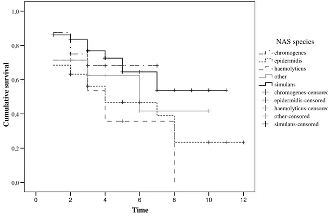
We followed 111 intramammary infections due to CNS for up to 48 weeks in four commercial dairy farms. Duration of infection had a mean of 188 days and was not significantly different between CNS species; geometric mean quarter somatic cell count (SCC) overall was 132,000 cells/ml and was also not significantly different between CNS species. There were differences in diversity between CNS species, with Staphylococcus epidermidis and Staphylococcus simulans showing less diversity than Staphylococcus haemolyticus, the epidemiological significance of which is debated.
Freezing milk samples overnight at -20ºC did not increase detection of intramammary bacteria in milk samples. Use of a real-time PCR-based test allowed for detection of udder pathogens beyond the ones identified using conventional bacteriology in milk samples with a high SCC that were culture negative or that yielded CNS or Corynebacterium bovis shown by conventional bacteriology.
Single quarter milk samples were collected in duplicate from 132 dairy cows in a commercial dairy farm, with the standard technique and by use of a cannula surpassing the teat canal. There was a significant difference between the two sampling techniques for recovery of Corynebacterium spp. and for culture-negative samples. The observed difference could not be attributed to a particular sampling order and no significant change was observed in quarter SCC between the sampling day and seven days later, indicating iatrogenic IMI following use of the alternative technique was not an issue.
Use of a deterministic economic model allowed to determine that in most situations lactational treatment of subclinical mastitis due to CNS would result in a net financial loss, on average of €38.74 per treated quarter.
Taken together, these results indicate that CNS have a low impact in terms of udder health, with little differences between individual species. Treating CNS subclinical mastitis during the lactation is not cost-efficient and should therefore not be advised. Using a RT-PCR and sampling with a teat cannula might improve diagnosis of mastitis etiology.
Keywords: minor mastitis pathogens, diagnosis, economics, coagulase-negative staphylococci, Corynebacterium spp., diversity, PFGE, impact, udder health, bovine
IV
MASTITE BOVINA POR STAPHYLOCOCCI COAGULASE-NEGATIVOS
E O PAPEL DE AGENTES PATOGÉNICOS MENORES NA MASTITE
Resumo
A mastite bovina é a doença mais comum das vacas leiteiras. Para implementar medidas de controlo eficientes, é geralmente necessário identificar os agentes etiológicos relevantes. Os staphylococci coagulase-negativos (SCN) e Corynebacterium spp. são considerados agentes patogénicos menores porque têm um impacto ligeiro na saúde do úbere. Apesar disso, são os agentes mais frequentemente isolados a partir de amostras de leite em vários estudos de larga escala, em diversos pontos do mundo. Os objectivos deste estudo foram avaliar se havia diferenças em termos de patogenicidade entre espécies individuais de SCN, avaliar se um método de colheita e de diagnóstico alternativos podiam determinar mais precisamente em que casos os agentes mencionados acima eram responsáveis por mastite, e determinar se o tratamento antimicrobiano durante a lactação de quartos infectados com SCN era uma medida de controlo eficiente em termos de custos.
Acompanhámos 111 infecções intramamárias por SCN durante até 48 semanas em quatro explorações leiteiras comerciais. A duração média de infecção foi de 188 dias e não foi significativamente diferente entre espécies de SCN; a média geométrica de contagens de células somáticas (CCS) global foi de 132.000 células/ml de leite e também não foi significativamente diferente entre espécies de SCN. Houve diferenças na diversidade entre espécies de SCN, tendo Staphylococcus epidermidis e Staphylococcus simulans mostrando menor diversidade do que Staphylococcus haemolyticus, sendo discutida o possível significado epidemiológico destas observações.
A congelação de amostras de leite durante a noite a -20ºC não permitiu o aumento da detecção de bactérias em amostras de infecções intramamárias. A utilização de um PCR em tempo real permitiu a detecção de agentes patogénicos mamários para além dos identificados após utilização de técnicas bacteriológicas convencionais em amostras de leite com CCS elevadas que se mostraram negativas após cultura ou que levaram ao isolamento de SCN ou Corynebacterium bovis.
Foram colhidas amostras de leite de quartos individuais em duplicado de 132 vacas leiteiras numa vacaria comercial, com a técnica convencional e com a utilização de uma cânula permitindo ultrapassar o canal do teto. Observou-se uma diferença significativa entre as duas técnicas de colheita para a recolha de Corynebacterium spp. e para amostras sem crescimento após cultura. A diferença observada não era atribuível a uma ordem de colheita particular e não foram observadas alterações significativas na CCS dos quartos entre o dia de colheita e sete dias mais tarde, indicando que a infecção intramamária iatrogénica após a utilização da técnica alternativa não constituiu um problema.
A utilização de um modelo económico determinístico permitiu determinar que na maioria das situações o tratamento durante a lactação de mastites subclínicas por SCN resultaria numa perda finaceira líquida, em média de €38.74 por quarto tratado.
Considerados em conjunto, estes resultados indicam que os SCN têm um impacto baixo em termos de saúde do úbere, com poucas diferenças entre espécies individuais. Tratar mastites subclínicas por SCN durante a lactação não é eficiente em termos de custos e não deve portanto ser aconselhado. A utilização de um RT-PCR e a colheita de amostras com uma cânula de tetos pode melhorar o diagnóstico das causas de mastite.
Palavras-chave: agentes patogénicos menores de mastites, diagnóstico, estudo económico, staphylococci coagulase-negativos, Corynebacterium spp., diversidade,
V Contents Acknowledgments……….……….II Abstract………..III Resumo……….……….IV Contents……….………....V List of figures……….………VI List of tables……….………VII List of abbreviations……..……….VIII Introduction……….1 Literature review……….3 Coagulase-negative staphylococci………3 Identification………3
Effects on udder health………....5
Epidemiology……….7
Antimicrobial treatment……….10
Control……….11
Virulence factors………12
Corynebacterium spp………..17
Effects on udder health……….17
Epidemiology and control……….19
Main objectives of the study…….……….21
Studies……….……….22
I – Bovine intramamary infection due to coagulase-negative staphylococci in four farms: impact of individual species………25
II – Diagnosis of intramammary infection in samples yielding negative results or minor pathogens in conventional bacterial culturing…...……….53
III – Observed reduction in recovery of Corynebacterium spp. from bovine milk samples by use of a teat cannula………..…63
IV – Deterministic model to evaluate the impact of lactational treatment of subclinical mastitis due to coagulase-negative staphylococci……...71
Discussion……….………...97
Identification of non-aureus staphylococci………..97
Impact of different non-aureus staphylococci species on udder health...100
Economic impact of lactational therapy for non-aureus staphylococci intramammary infection………105
Are we dealing with true udder pathogens?...106
Sampling to reduce contamination form teat canal flora……….109
Improving detection of true pathogens………...110
Improving mastitis diagnosis………113
Conclusions.………116
Further studies………117
VI List of figures
Figure 1. Quarter SCC for NAS infected quarters in the current
study, and mean SCC for a meta-analysis………...108 Figure 2. Quarter SCC for Corynebacterium bovis infected quarters in the
VII List of tables
Table 1. CNS characteristics as a group or for individual species
and respective supportive references………..15 Table 2. Bacteriological cure rates for clinical and subclinical CNS
mastitis observed after antimicrobial treatment, and spontaneous
VIII
List of abbreviations
AFLP……….. amplified fragment length polymorphism CCS…………...contagens de células somáticas CFU...colony forming units CNS………....coagulase-negative staphylococci CT...cycle threshold IMI……….intramammary infection ITS-PCR……….internal transcribed spacer region polymerase chain reaction MLST………..multilocus sequence typing NAS………...non-aureus staphylococci NMC………National Mastitis Council PCR……….polymerase chain reaction PFGE………...pulsed-field gel electrophoresis RT-PCR………..real time polymerase chain reaction SCC………somatic cell count SCN………..staphylococci coagulase-negativos SD………..standard deviation SDI………Simpson diversity index SNP………single nucleotide polymorphism TSST-1…. ……….toxic shock syndrome toxin-1 UPGMA…. ………...unweighted pair group method using arithmetic averages
1
Introduction
Mastitis is an inflammation of the mammary gland, most often of infectious origin. It is considered the most frequent disease in dairy farms and one of the main reasons for culling dairy cows (Gröhn, Eicker, Ducrocq & Hertl, 1998; Hortet & Seegers, 1998). It may be clinical if it can be detected on examination by the animal handler, or subclinical if an ancillary diagnostic test is needed for its diagnosis. Mastitis has a great economic impact for farmers: the average cost of a clinical mastitis episode has been estimated to be around 210€ and the average cost of a subclinical mastitis, for a farm with a bulk tank somatic cell count (SCC) between 250 and 400,000 cells/ml, to be around 94€ (Huijps, Lam & Hogeveen, 2008). Intrammammary infections (IMI) are infections of the mammary gland, detected more often through culture of milk samples, but not necessarily associated with inflammation.
Traditionally, microorganisms causing mastitis were divided into major and minor mastitis pathogens. Major mastitis pathogens included microorganisms that frequently led to clinical mastitis episodes, had low cure rates or were easily transmitted from cow to cow, hence having a high impact on udder health. These comprised Staphylococcus aureus, Streptococcus agalactiae, Streptococcus uberis, Streptococcus dysgalactiae, Escherichia coli and Mycoplasma bovis (Hassan, Samarasinghe & Lopez-Benavides, 2009). Minor mastitis pathogens had none of the aforementioned attributes and included Corynebacterium bovis as well as other species of the genus Corynebacterium and a group collectively termed as coagulase-negative staphylococci (CNS).
The latter group has progressively been acquiring more attention from researchers and milk quality advisors, with seminars being dedicated exclusively to it (Seminar on coagulase-negative staphylococci, Ghent, Belgium, 15-16 September 2010) as well as whole issues of scientific journals (Veterinary Microbiology 2009, volume 134, issues 1-2, dedicated to heifer and CNS mastitis). In the late 1960’s the five-point mastitis control plan started to be applied (Neave, Dodd & Kingwill, 1966), including postmilking teat disinfection, antimicrobial treatment of clinical mastitis, dry cow therapy, culling chronically infected cows and correctly maintaining the milking machine. With its effective implementation in most farms, prevalence of many of the major pathogens has been greatly reduced and continues to be so (Makovec & Ruegg, 2003; Pitkälä, Haveri, Pyörälä, Myllys & Honkanen-Buzalski, 2004). This led to more emphasis being put on the role of minor mastitis pathogens. Over the last decade or so, several large scale studies around the world have identified CNS as the most frequently isolated microorganisms from milk samples submitted either from clinical or subclinical mastitis cases or for mastitis surveillance programmes (Wilson, Gonzalez & Das, 1997; Makovec & Ruegg, 2003; Pitkälä et al., 2004; Bexiga, Cavaco & Vilela, 2005; Tenhagen, Koster, Wallmann & Heuwieser, 2006; Bradley, Leach, Breen, Green & Green, 2007). As for C. bovis and other species from the same genus, not as much attention has been devoted to them, despite their frequent isolation in the same studies.
2
Despite CNS being frequently isolated from milk samples, their role as mastitis pathogens is not clearly defined, with some authors giving them little importance (Schukken et al., 2009) whereas others question if their role in terms of udder health is significantly different from that of major pathogens (Taponen & Pyörälä, 2009).
Common to both CNS and C. bovis is the fact that they seldom lead to clinical episodes of mastitis (Bradley et al., 2007) and that they lead to fairly low SCC in quarters where they are causing intramammary infections (Djabri, Bareille, Beaudeau & Seegers, 2002).
The current thesis aims to better characterise the impact of these minor pathogens on udder health and mastitis diagnosis. Aspects related to CNS and to C. bovis are under different headings for convenience.
3
Literature review
Coagulase-negative staphylococci
IdentificationHistorically, CNS were not always identified to species level because that distinction was not easily or reliably performed, and because different species within this group were viewed as having very similar characteristics and clinical relevance.
There are several ways of identifying CNS to species level. The method commonly used to discriminate between this group of microorganisms and S. aureus is the coagulase test. This test detects the presence of coagulase, an enzyme commonly expressed by S. aureus, S. intermedius, S. pseudintermedius, S. delphini, S. schleiferi subsp. coagulans and by some strains of S. hyicus (Blaiotta, Fusco, Ercolini, Pepe & Coppola, 2010). Coagulase forms an active molecular complex with prothrombin, which converts fibrinogen to a fibrin clot. For detection of coagulase, rabbit plasma is generally inoculated with isolated colonies and tested for gel or clot formation (Roberson, Fox, Hancock, Gay & Besser, 1996).
In terms of mastitis herd investigations it is often viewed as sufficient and more economical to know what is the incidence of S. aureus and of CNS as a group, because clinical relevance, expected cure rates and control measures differ between the two groups (Taponen & Pyörälä, 2009). The following review focuses on the identification of CNS, not on S. aureus.
Use of biochemical reactions to identify species of CNS is the basis of the method described by Kloos and Schleifer (1975) cited as the phenotypic reference method (Watts & Washburn, 1991; De Paulis, Predari, Chazarreta & Santoianni, 2003). This method includes tests for coagulase activity, hemolysis, lysostaphin and novobiocin susceptibility, phosphatase activity, nitrate reduction and aerobic acid production from fructose, xylose, arabinose, ribose, maltose, lactose, sucrose, trehalose, mannitol, and xylitol. This method could only distinguish nine different species of CNS, far from the more than 40 species currently described (NCBI Taxonomy, 2010). Biochemical reactions are the basis of most commercial tests for the species level identification of CNS which include the API Staph, API Staph ID 32 and Vitek (all bioMérieux), the BBL Crystal Gram-positive (Becton & Dickinson) and the Staph-Zym (Rosco).
More recently, genotypic methods have become widespread to either detect the presence of CNS in milk samples (Koskinen et al., 2009) or to identify CNS to species level after isolation in culture (Poyart, Quesne, Boumala & Trie-Cuot, 2001; Drancourt & Raoult, 2002). Some of the latter methods are based on the recognition of species-specific sequences including sequencing of housekeeping genes (Poyart et al., 2001; Drancourt & Raoult, 2002) or amplified fragment length polymorphism (AFLP, Piessens et al., 2010a), while others measure the variation in, or close to, genomic sequences present in all species,
4
which include internal transcribed spacer-PCR (Couto, Pereira, Miragaia, Santos-Sanches & Lencastre, 2001), tRNA intergenic length polymorphism analysis (Maes et al., 1997), transfer RNA-intergenic spacer PCR (Supré et al., 2009), (GTG)5-PCR (Braem et al, 2010) or
ribotyping (Carretto et al., 2005).
Commercial biochemical identification systems often provide unreliable results for CNS species, as a result of the variability of metabolic profiles within species and the subjective nature of their interpretation (Carretto et al., 2005). The fact that commercially available biochemical methods of identification are not generally conceived to identify veterinary pathogens probably contributes to this problem.
Genotypic methods seem to be generally more discriminatory than commercially available biochemical methods in differentiating between CNS species (Zadoks & Watts, 2009). Sampimon et al. (2009), comparing the performance of two biochemical identification systems with that of sequencing a housekeeping gene, found that for Staphylococcus chromogenes, the species with the highest number of representatives in their study, 0 and 36.5% of the isolates were correctly identified by the two commercial systems, respectively. Taponen, Simojoki, Haveri, Larsen and Pyörälä (2006) and Taponen, Koort, Björkroth, Saloniemi and Pyörälä (2007) mention an agreement between API Staph ID 32 and AFLP CNS identifications of 66 and 71.9%, respectively. Carretto et al. (2005) were able to identify correctly 93.8% of CNS isolates (n=177) through ribotyping, whereas only 36.7% were identified correctly with an API 20 Staph system. A faster diagnosis has also been claimed for some of the genotypic methods of diagnosis, as well as the identification of genes responsible for resistance to antimicrobials (Koskinen et al., 2009). Economical considerations, the technical possibilities of staff and equipment and the final objective of the diagnostic should also be taken into account in the choice of the diagnostic test. If diagnosing only to CNS group level remains the most useful information, with no need for identification to species level, then phenotypic methods currently in use may suffice (Zadoks & Watts, 2009).
Differentiation between bacterial strains within a certain species is essential for epidemiological surveillance and to identify possible cases of bacterial cross-transmission (van Belkum et al., 2007). Several methods have been proposed to distinguish between different strains of CNS species. These include use of antibiograms, pulsed-field gel electrophoresis (PFGE) or phage typing (Kloos & Bannerman, 1994; Bjorland et al., 2005; Taponen, Björkroth & Pyörälä, 2008).
5
Effects on udder health
Coagulase-negative staphylococci have been isolated from milk samples from clinical and subclinical mastitis cases. Isolation of CNS in cases of clinical mastitis, defined as mastitis apparent on examination by the animal handler or veterinarian (IDF, 1999), has been reported for CNS as a group (Smith et al., 1985; McDougall, 1998) or for individual species (Jarp, 1991; Birgersson, Jonsson & Holmberg, 1992; Todhunter, Cantwell, Smith, Hoblet & Hogan, 1993; Waage et al., 1999; Taponen et al., 2006). Simojoki et al. (2009) performed an experimental infection with S. chromogenes in six primiparous cows and all developed mild signs of clinical mastitis. All but one of the animals cleared the infection spontaneously within a week of being infected and there were no significant differences in average daily milk yields pre and post challenge, despite a 16.3% reduction in production for the challenged quarters during the seven-day observational period.
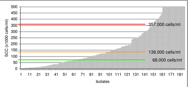
Likewise, isolation of CNS in cases of subclinical mastitis, defined as inflammation of the mammary gland that is not visible and requires a diagnostic test for detection (IDF, 1999), has also been reported for CNS as a group (Timms & Schultz, 1987; Rainard, Ducelliez & Poutrel, 1990; Chafer et al., 1999; Borm et al., 2006) and for individual species (Jarp, 1991; Birgersson et al., 1992; Taponen et al., 2006). Several criteria are used to define subclinical mastitis including increased SCC (Djabri, Bareille, Beaudeau & Seegers, 2002), increased N-acetyl-β-D-glucosaminidase (NAGase) activity (Myllys, 1995) or increased conductivity (McDougall, 1998). Generally papers seem to indicate a small increase in quarter SCC for infected quarters, when compared to uninfected quarters. Djabri et al. (2002), in their meta-analysis reporting on the effects of several intramammary pathogens on SCC, mention a mean quarter value of 138,000 cells/ml for staphylococci other than S. aureus, with 68,000 cells/ml being reported for culture-negative samples.
Besides being isolated from clinical and subclinical mastitis cases, several CNS species have also been found in samples from animals with no clinical signs and a low cell count. Birgersson et al. (1992) observed no significant differences in frequency of isolation for most CNS species between the three categories they cultured: clinical mastitis, subclinical mastitis and non-mastitic quarters, even though their case definition for non-mastitic was no clinical signs of inflammation and a SCC<300,000 cells/ml, a somewhat high threshold to define presence of inflammation.
It is well established that increases in SCC beyond a certain level lead to decreased milk production (Green, Schukken & Green, 2006), so milk production losses would be expected for CNS infected quarters. Timms and Schultz (1987) report a mean loss of 821 kg for 305-day milk production when comparing CNS infected animals with controls, whereas Wilson et al. (1997) and Schukken et al. (2009) actually report higher milk production for animals with IMI due to CNS, which seems somewhat surprising. This could be linked with confounding factors not yet identified: Wilson et al. (1997) suggest that higher producing cows may be
6
more likely to contract CNS mastitis and avoid major losses in milk production, whereas Schukken et al. (2009) suggest that this effect might be mediated through a protective effect of CNS against clinical mastitis by major pathogens. In fact, Piepers, Opsomer, Barkema, de Kruif and De Vliegher (2010) observed that a high proportion of CNS IMI in their study was short-lasting and that the affected quarters had a lower incidence of clinical mastitis and a higher milk production.
Several pathogenic changes have been reported in mammary quarters infected with CNS including leukocyte infiltration and increased connective tissue stroma. One study on nuliparous heifers with naturally acquired infection with CNS concluded that there were significant differences in stroma percentage and leukocyte infiltration, but not in epithelium or lumen percentages (Trinidad, Nickerson & Alley, 1990). Another study analysed microbiological results and histopathological changes of udders collected at abattoirs (Benites, Guerra, Melville & Costa, 2002), which are of debatable significance considering that some of the histological changes might have been produced by other pathogens that had previously infected the mammary gland. The authors concluded that no differences were observed between the main histopathological results associated with CNS and coagulase-positive staphylococci.
Persistence of intramammary infection has been studied for CNS, with reported values of 46% (Taponen et al., 2007), 66% (Seymour, Jones & McGilliard, 1989), 75.6% (Rainard et al., 1990), 84.5% (Chaffer et al., 1999) and 85% (Timms & Schultz, 1987) of infections persisting until dry-off or until the cows left the herd. Todhunter et al. (1993) mention an average length of IMI with CNS of 222 days.
Several studies addressed differences in pathogenicity between CNS species. Myllys (1995) found Staphylococcus simulans and S. hyicus more frequently associated with clinical infections and with increased inflammatory response than other CNS species. Myllys, Honkanen-Buzalski, Virtanen, Pyörälä and Müller (1994) also mention that S. hyicus was more effective in causing teat canal infections than Staphylococcus epidermidis. Jarp (1991) found that Staphylococcus haemolyticus was the CNS species more often associated with clinical mastitis, even though no significant difference was observed in clinical severity between different species. A Danish study (Aarestrup & Jensen, 1997) indicated that S. simulans produced more persistent and stable infections than the other CNS species, whereas a Finnish study (Taponen et al., 2007) found no significant differences between clinical characteristics and persistence of infection between the two most frequently isolated CNS species causing bovine mastitis. Hogan, White and Pankey (1987) observed no significant differences among SCC for quarters infected with different CNS species, indicating that the same type of inflammatory response is elicited by the several species that constitute the group. Chaffer et al. (1999) similarly found no differences in SCC or differential cell counts between CNS species. To the author’s knowledge there have been no published
7
studies on differences in impact on udder health between strains of individual CNS species. Immunity to CNS at the level of the mammary gland is also not sought on the aforementioned studies focusing on differences in impact between CNS species. Both these factors could influence results of these studies and have not been investigated.
The overall impact of CNS on bulk tank SCC has also been addressed. Rainard et al. (1990) found that quarters infected by CNS contributed to 18.1% of bulk tank SCC, whereas Schukken et al. (2009) found that CNS IMI contributed 12% to the bulk tank SCC in herds in which this was between 200 and 400.000 cells/ml and 8% in herds with a bulk tank SCC above 400,000 cells/ml. Of the 4,200 full herd samplings analysed in this study, less than 2% of the herds would implicate CNS IMI as an important contribution to a bulk tank SCC higher than 400,000 cells/ml. The financial impact of mastitis caused by CNS has been estimated to be lower than mastitis caused by other pathogens (Ruegg & Dohoo, 1997).
Epidemiology
Prevalence of CNS is generally highest at calving for all parities (Mathews, Harmon & Langlois, 1992; Todhunter et al., 1993; Honkanen-Buzalski, Myllys & Pyörälä, 1994) and is higher in primiparous than in multiparous cows (Mathews et al., 1992; Rajala-Schultz, Smith, Hogan & Love, 2004). Their role as aetiological agents of mastitis in heifers has been highlighted, especially before parturition and in the peripartum period (Trinidad, Nickerson & Alley, 1990; Pankey, Drechsler & Wildman, 1991; Myllys, 1995; Nickerson, Owens & Boddie, 1995; Fox et al., 1995; Aarestrup & Jensen., 1997).
Due to the fact that many such studies address the isolation of CNS prior to calving, mostly they either report results on bacteriology of gland secretions (Trinidad et al., 1990a; Birgersson, et al., 1992; Aarestrup, Larsen & Jensen, 1999) or from skin and teat canal (Woodward, Ward, Fow & Corbeil, 1988; White, Harmon, Matos & Langlois, 1989; Matthews et al., 1992), often making no correlation between isolation of the organisms and pathologic effects. It is debatable if the terms mastitis or intramammary infection should be used in these circumstances. However, Trinidad et al. (1990a) found that the species more frequently involved in clinical mastitis were also the most frequently isolated from teat canal keratin samples: S. chromogenes and S. hyicus besides S. aureus.
Coagulase-negative staphylococci have been found on the skin and other body sites of cattle (White et al., 1989) and on the skin and mucous membranes of humans (Kloos & Bannerman, 1994), with no deleterious effects in these circumstances. They have also been isolated from several environmental sources (Matos, White, Harmon & Langlois, 1991) including several feeds and bedding material. There is evidence that the most frequently isolated CNS species from extramammary sites are not the same as the most frequently isolated from milk samples. Taponen et al. (2008) found that Staphylococcus equorum, Staphylococcus sciuri, Staphylococcus saprophyticus, and Staphylococcus xylosus
8
predominated in extramammary samples but were not isolated from milk samples. The same authors also found that for S. chromogenes, common PFGE pulsotypes were observed for environmental and intramammary isolates and that S. simulans was more often found in milk samples than isolated from environmental samples. This could be an indication that S. chromogenes IMI is acquired more often from environmental sources and that S. simulans is more often transmitted from cow to cow. Thorberg et al. (2006) observed that the most common S. epidermidis PFGE pulsotype isolated from milk samples for each of the two farms in their study, was also found on the milkers’ hands, suggesting that the S. epidermidis IMI observed originated from humans. Aarestrup et al. (1999) found that in seven out of nine herds from which multiple S. simulans were isolated from milk samples, more than one ribotype was present, which can be seen as an indication that several sources of infection are present.
Despite the aforementioned evidence that at least some CNS IMI may originate from environmental sources, the most frequent form of infection acquisition is still not known. The classical differentiation between environmental and contagious mastitis pathogens no longer makes sense for several microorganisms (Bradley & Green, 2001; Zadoks et al., 2003; Rato et al., 2008), but it is still important to understand what are the main sources of infection in a particular farm when performing milk quality advisory work, so that control measures can be applied to reduce new infections. To our knowledge there is no evidence in the literature of what the most frequent source of infection for CNS as a group is, or if there are differences between CNS species.
There is some evidence of a low (Østerås, Sølverød & Reksen, 2006) or moderate (Barkema et al., 1997) herd-level clustering effect for CNS. Scientific evidence of a herd-level clustering effect for individual CNS species is still missing and the identification of herd and cow-level risk factors for IMI with CNS has not been addressed by many. Sampimon et al. (2009) found that an increased prevalence of CNS IMI was associated with the source of drinking water not being tap water, housing of dry cows in one group instead of multiple groups, monthly milk recording, udder health monitoring by the veterinarian, pasturing during outdoor season, percentage of stalls contaminated with milk and bulk tank SCC>250,000 cells/ml. It needs to be reminded that this was an epidemiological study and therefore no causality can be established from the observed associations.
Another subject that may contribute to our understanding of the epidemiology of CNS mastitis is the occurrence of seasonal variation of CNS IMI, which has been addressed by two publications, both originating in Norway. Waage et al. (1999) found a significantly lower percentage of cases from September to December, which agrees with Østerås et al. (2006) who found significantly higher prevalences of CNS IMI in April through to July. Seasonal variation might be linked with a number of factors (Østerås et al., 2006): feed quality when grazing (vitamin E intake), climatic and hygienic conditions.
9
Most CNS IMI would appear to be self-limiting. Aarestrup and Jensen (1997) found that S. chromogenes was isolated from 15% of heifers before parturition, but from only 1% four weeks after parturition. Different CNS species displayed different patterns of infection: S. epidermidis was seldom isolated from the same quarter for two consecutive weeks, whereas S. simulans persisted for several weeks. In this study no molecular fingerprinting method was used to try to understand if the presence of the same bacterial species corresponded to the same infection or to a newly acquired infection with the same species. The same authors (Aarestrup et al., 1999) used ribotyping for that purpose and were able to demonstrate that S. simulans can cause persistent infections. Similarly, Thorberg, Danielsson-Tham, Emanuelson and Persson Waller (2009) observed that S. epidermidis, S. chromogenes and S. simulans were the species more frequently isolated from the same quarters in two months in succession. Piepers et al. (2010) found that nearly half of CNS IMI detected at 1 to 4 days post-partum, could not be detected 5 to 8 days after calving. Several other authors have reported a decrease in infection levels after parturition (Hogan, Pankey & Smith, 1987b; Mathews et al., 1992; Myllys, 1995). It would appear therefore that different species have different spontaneous cure rates, but that this is probably influenced by lactational stage.
There is still some debate as to whether CNS might have a protective role against IMI caused by major pathogens, with some authors reporting evidence of such an effect (Schukken, Van de Geer, Grommers, Smit & Brand, 1989; Matthews, Harmon & Smith, 1990; Green et al., 2005), and some reporting evidence of the opposite effect (Hogan, Smith, Todhunter & Schoenberger, 1988; Davidson, Dohoo, Donald, Hariharan & Collins, 1992; Myllys, 1995; Aarestrup & Jensen, 1997). Matthews et al. (1990) performed a trial in which 35 quarters were challenged by infusion of S. aureus into the teat sinus, 18 that were culture negative and 17 that were infected with S. chromogenes. The outcome was that 100% of the previously non-infected quarters got infected, whereas 47% of the quarters harboring S. chromogenes became infected. Several theories have been proposed to justify this potential protective effect. One is the production of inhibitory substances with antagonistic effect for S. aureus or other major pathogens. A study by De Vliegher et al. (2004) seems to support this theory. They assessed the in vitro growth of mastitis pathogens in the presence of ten strains of S. chromogenes. Two of the S. chromogenes strains showed inhibitory activity against all S. aureus, S. dysgalactiae and S. uberis isolates, but not against E. coli. Another study (Nascimento, Fagundes, Paiva Brito, Santos & Freire Bastos, 2005) demonstrated that 6.4% of 188 CNS strains isolated from bovine mastitis cases exhibited antimicrobial substance production against an indicator strain of Corynebacterium fimi. The same study showed that up to 78% of S. agalactiae indicator strains were inhibited by one strain of CNS. Another possible explanation would be the protective effect of an increased SCC against infection by major pathogens. This is not a established theory, largely based on the again not well-established theory that the incidence of clinical mastitis in low SCC herds is higher in cows
10
with a lower SCC (Peeler, Green, Fitzpatrick & Green, 2002). A study that tried to explore this hypothesis introduced inert devices into glands leading to increases in SCC; however this showed no protective effect against experimental infection with S. uberis (Nickerson, Boddie, Owens & Watts, 1990). The significance of these findings should be questioned because even if a protective role is proven, the deleterious effects would have to be measured and a cost-to-benefit analysis performed. Maintaining or inducing a protective teat apex flora should be preferred over causing IMI as an IMI with the protective pathogen would result in an increased SCC and potential production losses (De Vliegher et al., 2004). In this sense, Woodward et al. (1988) tested the persistence of teat apex colonisation by several bacteria that had been previously proven to inhibit growth of mastitis pathogens in vitro. A strain of S. hominis that inhibited the growth of several Gram-positive bacteria was recovered from the same animals for up to 28 days. Conversely, if the risk of new infections is greater for CNS infected quarters, this could be due to their alteration of normal protective effects against other pathogens (Hogan et al., 1988), although these effects are not well understood. Overall, the current literature seems far from clear as to whether CNS infection is, or is not, protective against major pathogen IMI in the cow in field conditions.
Antimicrobial treatment
Irrespective of the discussion on their impact in terms of udder health, the fact is that both clinical and subclinical CNS IMI are treated with antimicrobials.
The bacteriologic cure rates reported for clinical mastitis therapy (McDougall, 1998; Waage et al., 2000; McDougall, 2003; McDougall, Arthur, Bryan, Vermunt & Weir, 2007a; McDougall, Agnew, Cursons, Hou & Compton, 2007b) vary between 53.3% and 100% depending on the study and on the antimicrobial used. Reported bacteriologic cure rates (McDougall, 1998; Wilson, Gonzalez, Case, Garrison & Gröhn, 1999; Taponen et al., 2006) for subclinical mastitis therapy vary between 81% and 88.9%. Cure rates for CNS infected quarters with dry cow therapy are not frequently reported in the literature. Rajala-Schultz, Torres, DeGraves, Gebreyes and Patchanee (2009) report a cure rate of 82% for quarters infected with CNS after dry cow therapy.
Several papers report results for antimicrobial treatments performed on pre-calving heifers (Oliver, Lewis, Gillespie & Dowlen, 1992; Oliver et al., 2003; Oliver et al., 2004; Borm et al., 2006) with cure rates for CNS infected quarters varying from 73 to 100% for intrammamary treatment. Despite the fairly high cure rates obtained, there is no agreement between these authors as to whether such a type of treatment should actually be performed. For Oliver et al. (2003) a net revenue of $200.64 per treated heifer was obtained when treating heifers pre-calving with intramammary cloxacillin or cephapirin, whereas for Borm et al. (2006) pre-calving treatment with intramammary cephapirin led to no significant improvements in milk production or linear somatic cell count. It should be mentioned that
11
these studies report results mainly on CNS infected quarters but also on IMI due to other pathogens. Parker, Compton, Anniss, Heuer and McDougall (2008) reporting on pre-calving treatment of heifers with parenteral tylosin found no difference between treated and untreated quarters with regards the proportion of quarters that underwent bacteriological cure.
Results of parenteral treatment of CNS IMI are also available in the literature. McDougall et al (2007b) reporting on cure rates for clinical mastitis mention a 75.9% cure rate for penethamate hydriodide and 90.5% for tylosin and Pyörälä and Pyörälä (1998) report cure rates of 78.8 and 58.3% for parenteral treatment with penicillin G and spiramycin respectively.
Cure rates for antimicrobial treatment seem therefore to be fairly high. This is partly explained by a low resistance to antimicrobials. Several papers address antimicrobial susceptibility testing either through the Kirby-Bauer method (Costa, Benites, Guerra & Melville, 2000; Cattell, Dinsmore, Belshner, Carmen & Goodell, 2001; Makovec & Ruegg, 2003) or by determination of minimum inhibitory concentrations (Rajala-Schultz et al., 2004; Lüthje & Schwarz, 2006; Pol & Ruegg, 2007). Antimicrobial resistance seems mostly to be low but some exceptions have been reported including one study that reports 90.9% of penicillin resistant strains (Costa et al., 2000). Other potentially important issues are the observation of changes in resistance patterns through time (Makovec & Ruegg, 2003), differences in resistance levels seen with different production systems (Pol & Ruegg, 2007) or the existence of methicillin-resistant strains of CNS (Sawant, Gillespie & Oliver, 2009; Fessler, Billerbeck, Kadlec & Schwarz, 2010). Besides resistance to antimicrobials, there is also the possibility of resistance to quaternary ammonium compounds among CNS (Bjorland et al., 2005), which could have an impact on udder health as several pre and post milking teat sanitizers are based on these compounds.
Control
Despite the literature on risk factors for CNS IMI being scarce, there are other options besides antimicrobial treatment for mastitis control. Initially mastitis control programmes targeted contagious mastitis pathogens. The increasing frequency of isolation of CNS led to the discussion about the causes of such a change in relative frequency of isolation (Zecconi, Piccinini & Fox, 2004). After an effective control of contagious pathogens, did environmental pathogens simply became relatively more prevalent, or did control measures aimed at contagious pathogens favour the environmental pathogens, causing an actual increase in their infection level? Zecconi et al. (2004) tested the hypothesis that strict adoption of contagious mastitis control practices by nine commercial dairy herds would result in an increase in the prevalence of non-contagious mastitis pathogens. There was no significant increase in the prevalence of coliforms, environmental streptococci or CNS at the end of the
12
trial period, even though the risk for CNS mastitis was significantly lower at the beginning of the adoption of the contagious pathogens control programme.
Included in most control programmes to prevent infection with udder pathogens is the use of post-milking teat disinfection. Different germicide solutions seem to have different effects on the prevalence of CNS IMI: both Goldberg et al. (1994) and Foret, Corbellini, Young and Janowicz (2005) found significant differences in the reduction of new IMI between two iodine-based formulations. The relative distribution of different CNS species can also be influenced by the type of sanitizer used (Hogan et al., 1987a). The effect of pre-milking teat sanitization has also been addressed. Ruegg and Dohoo (1997) observed no difference in the isolation of CNS between the group of animals submitted to pre-dipping and the group where this was not performed and likewise, Oliver et al. (1993) found no difference in new CNS IMI between performing pre and post-milking teat disinfection and post-milking disinfection alone.
Vaccination has also been addressed as a possible tool in the control of CNS IMI. Development of vaccines against mastitis problems is a considerable technical challenge because of the idiosyncratic immunological nature of the mammary gland and the multitude of microorganisms causing the disease (Denis, Wedlock, Lacy-Hulbert, Hillerton & Buddle, 2009). In 1994, Amorena, Baselga and Albizu (1994) reported on results of an experimental challenge following immunization against S. aureus and CNS in ewes. Despite showing some promising results for certain CNS species, this study was not followed up by a field trial. More recently, Middleton, Luby and Adams (2009) reported on results of a commercial vaccine against S. aureus mastitis (Lysigin®, Boehringer Ingelheim Vetmedica, Inc.) in field conditions. No significant differences were observed between vaccinated cows and unvaccinated controls with regards to number of quarters that developed new CNS IMI, time to CNS IMI or milk SCC. To the author’s knowledge, currently there is only one vaccine licensed in Europe for the prevention of CNS mastitis (Startvac®, Laboratorios Hipra, S. A.), which is also indicated for the prevention of coliform and S. aureus mastitis. The manufacturer reports a significant difference in the number of CNS IMI between the placebo and vaccinated groups both for primiparous and multiparous cows. A benefit-to-cost analysis of the use of this vaccine is not yet available.
Virulence factors
Evidence of the production of a number of different virulence factors, or of the possibility to express them, has been evaluated for several CNS species. These factors could contribute to their infective or damaging potential to the mammary gland, to their capability of escaping the host’s natural defences or the effects of antimicrobial therapy, or to negative consequences in terms of Public Health.
Being frequently found on the skin, the ability to establish themselves in the mammary gland is what confers CNS their infectious potential. This ability depends on the capacity to
13
bind tissue matrix and plasma proteins, which may be exposed in the traumatized or toxin-damaged mammary epithelium (Mamo, Froman & Wadstrom, 1988), but probably also on the capacity to avoid host defence mechanisms by way of biofilm production (Oliveira et al., 2006). Watts, Naidu and Wadstrom (1990) studied the abilities of several CNS species of bovine origin to bind collagen, degrade elastin (elastase production) and produce biofilm. Comparison between animals infected by different strains showed no significant difference in SCC between animals affected by strains producing biofilm, elastase or binding collagen and the remaining strains. Jarp (1991) suggested that haemolysin could be another important virulence factor owing to the fact that S. haemolyticus was more often associated with clinical mastitis in his study. Watts and Owens (1987) reported production of a delta-like toxin (cytolysin) by several species of CNS. Production of this toxin was associated with a higher increase in SCC for S. epidermidis, S. hominis and S. warneri but not for S. hyicus or S. chromogenes. Other potentially virulent toxins and enzymes produced by CNS strains include leucocidin, lipase, proteases and DNAse (Zhang & Maddox, 2000). Surface characteristics such as hydrophobicity and capsule formation have also been described as potential virulence factors (Matthews, Oliver and Guidry, 1991; Birgersson et al., 1992). Adhesion to and internalisation into bovine mammary epithelial cells could also contribute to the CNS infective potential. Different species display different levels of adhesion and internalization, with S. xylosus showing the highest values for both. S. epidermidis was the least efficient. Distinct pathways for internalization may be involved for different CNS species (Almeida & Oliver, 2001).
Coagulase-negative staphylococci have also been found to produce toxins that pose a potential risk for Public Health. Staphylococcal enterotoxins and toxic shock syndrome toxin-1 (TSST-toxin-1) are toxins produced by staphylococcal species responsible for food poisonings. Their production by 40 mastitis isolates was investigated by Orden et al. (1992). Enterotoxins were only produced by two strains of S. xylosus and TSST-1 was only produced by five strains of the same species, one strain of S. sciuri and two strains of S. epidermidis. Another aspect contributing to CNS virulence and potentially to Public Health concerns is transfer of antimicrobial resistance among mastitis isolates. Muhammad, Hoblet, Jackwood, Bech-Nielsen and Smith (1993) mention that of nine strains representing three species of CNS, two could transfer streptomycin resistance. A S. hominis strain transferred streptomycin resistance to a S. chromogenes lacking resistance, and to a S. aureus strain carrying penicillin and tetracycline resistance.
There is a potential for differences in expression of virulence factors according to in vitro or in vivo conditions experienced. Therefore, more recently a number of genotypic studies have addressed several potential virulence factors: presence of the bap (biofilm-associated protein) gene and the capability of its horizontal transfer (Tormo, Knecht, Götz, Lasa & Penadés, 2005), presence of staphylococcal superantigen genes (Nemati et al., 2008; Park
14
et al., 2010), and presence of genes responsible for antimicrobial resistance (Lüthje & Schwarz, 2006), which may all contribute to competitive advantages for the strains expressing these genes.
Despite the multitude of studies on virulence factors, there are very few studies on the correlation of these with clinical characteristics in vivo. Haveri, Taponen, Vuopio-Varkila, Salmenlinna and Pyörälä (2005), Fournier et al. (2008) and Graber et al. (2009) found that certain S. aureus genotypes were more often associated with certain clinical characteristics such as persistence or contagiousness. However, Haveri, Roslöf, Rantala and Pyörälä (2007) in a follow up study, failed to correlate persistence or other clinical signs with presence of a particular gene or group of virulence genes. Specifically for CNS, Simojoki, Hyvönen, Taponen and Pyörälä (2010) observed no correlation between biofilm production and persistence of CNS IMI.
Table 1 displays a summary of CNS characteristics and relevant references that support those characteristics.
15
Table 1. CNS characteristics as a group or for individual species and respective supportive references.
Characteristic CNS/individual species Reference Isolated from cases of clinical
mastitis CNS Smith, Todhunter & Schenberger, 1985 McDougall, 1998 Individual species Jarp, 1991
Birgersson et al., 1992 Todhunter et al., 1993 Waage et al., 1999 Taponen et al., 2006 Simojoki et al., 2009 Isolated from cases of
subclinical mastitis CNS Timms and Schultz, 1987 Rainard et al, 1990 Chafer et al., 1999
Borm et al., 2006 Individual species Jarp, 1991
Birgersson et al., 1992 Taponen et al., 2006 Isolated from clinically normal
samples with low cell count Individual species Birgersson et al., 1992 Isolated from extramammary
sites
Individual species White et al., 1989 Matos et al., 1991 Thorberg et al., 2006 Taponen et al., 2008 Infected cows had lower milk
yield CNS Timms and Schultz, 1987
Infected cows had higher milk
yield CNS Wilson et al., 1997 Schukken et al., 2009 Piepers et al., 2010 Leads to histopathological
changes CNS Trinidad, Nickerson & Adkinson, 1990 Benites et al., 2002 Observed differences in
pathogenicity between species
S. haemolyticus more often associated with clinical mastitis
Jarp, 1991 S. hyicus more effective
in causing teat canal infections than S. epidermidis
Myllys et al., 1994
S. simulans and S. hyicus more often associated with clinical infections and increased inflammatory response
Myllys, 1995
S. simulans produced more persistent infections
Aarestrup & Jensen, 1997
No difference among SCC for quarters infected with different species
Hogan et al., 1987a Chaffer et al., 1999 No difference in clinical
characteristics and persistence between two most frequent species
16 Table 1 (continued).
Characteristic CNS/individual species Reference Protective effect against
mastitis by major pathogens CNS Schukken et al., 1989 Green et al., 2005 Individual species Matthews et al., 1990
De Vliegher et al., 2004 No protective effect against
mastitis by major pathogens CNS Hogan et al., 1988 Davidson et al., 1992 Myllys, 1995
Aarestrup & Jensen, 1997
Table 2. Bacteriological cure rates for clinical and subclinical CNS mastitis observed after antimicrobial treatment and spontaneous cure rates.
Characteristic Observed cure rates Reference Response to antimicrobial
treatment - clinical 53.3 and 91.7%* 89.6% McDougall, 1998 Waage et al., 2000 80 and 100%* McDougall, 2003
84.3% Taponen et al., 2006
76, 92.9 and 93.8%* McDougall, 2007a 75.9 and 90.5%* McDougall, 2007b Response to antimicrobial
treatment - subclinical 81.8% 68, 75, 76, 87 and 89%* McDougall, 1998 Wilson et al., 1999
88.9% Taponen et al., 2006
Spontaneous cure for
subclinical mastitis 64.5% 72% McDougall, 1998 Wilson et al., 1999
39.5% Taponen et al., 2006
Response to antimicrobial
treatment – dry period 82% Rajala-Schultz et al., 2009 * Depending on which antimicrobial was used.
17
Corynebacterium
spp.
Effects on udder health
Like CNS, Corynebacterium spp. have been isolated from cases of both clinical and subclinical mastitis. Isolation from clinical mastitis cases is not very frequent, with authors reporting percentages of 2.5% (Botrel et al., 2010) or 3.5% (Bradley et al., 2007) of the total number of clinical mastitis samples. Typically, Corynebacterium spp. are isolated from cases of clinical mastitis that are less severe than cases from which other pathogens are isolated, but that are slightly more severe than mastitis cases that are culture-negative (Morin & Constable, 1998).
Isolation from subclinical mastitis cases is not very frequently reported in the literature. The author found Corynebacterium spp. in 12.4% of samples from sublinical mastitis cases (Bexiga et al., 2005). Most large scale studies refer to bacteriology results of clinical and subclinical mastitis samples without differentiating between them; sometimes including samples which are not necessarily mastitic, but that were submitted to laboratories as part of mastitis control programmes. This type of study reports isolation frequencies of 2.7% (Makovec & Ruegg, 2003), 7.2% (Wilson et al., 1997), 7.3% (Tenhagen et al., 2006) and 11.5% (Pitkälä et al., 2004) for C. bovis as a percentage of all samples (including culture-negative samples).
The impact of C. bovis IMI in terms of SCC is low. Djabri et al. (2002) on a meta-analysis of individual quarter SCC resulting from IMI with different pathogens, report a value of 105,000 cells/ml for C. bovis infected quarters. This was the lowest value for the pathogens studied and not so distant from the geometric mean obtained for culture-negative samples which was 68,000 cells/ml.
This may be because not all C. bovis isolated from milk samples will actually be causing mastitis (Honkanen-Buzalski and Bramley, 1984). The term intramammary infection is sometimes used interchangeably with subclinical mastitis and there is great variability between authors in defining criteria for these (Andersen et al., 2010). Reports on the isolation rates of C. bovis from milk samples are therefore not always linked to a measure of inflammation of the mammary gland (Wilson et al., 1997; Tenhagen et al., 2006). Teat canal colonisation with C. bovis would lead to contamination of milk samples submitted for mastitis diagnosis. There is evidence of this in the scientific literature for a long time. Black, Marshal and Bourland in 1972, observed that when sampling milk directly from the teat cistern by use of a transdermic needle, large differences were observed in the recovery of C. bovis: only 5 out of 20 samples previously positive with the conventional sampling technique were positive for C. bovis when sampling directly from the teat cistern and the average number of colony forming units (cfu) per ml in those 5 samples was 66, whereas it was 1900 for the 20 samples collected with the standard technique. Honkanen-Buzalski and Bramley in 1984
18
report on the results of an experimental infection with C. bovis on 72 quarters. No clinical mastitis was recorded, even though the microorganism was being excreted three days after the initial challenge in 73.6% of quarters. Again using a transdermic needle to collect milk samples from quarters that were excreting C. bovis, 44.4% of the samples were negative, indicating that colonisation did not go further than the teat canal.
Evidence about duration of infection is scarce. Sordillo, Oliver, Doane, Shull and Maki (1989c) performed experimental infections with C. bovis. In one of the trials, infection during the lactation persisted for 93 days, the duration of the study, and in the other study, infection just before drying off led to infections that persisted through the whole dry period and into early lactation. Rainard and Poutrel (1982) performed three-weekly milk sampling for one year on three herds and found that of 161 IMI with C. bovis, only 27 were eliminated. Approximately one third (34.8%) of C. bovis IMI were detected at calving, another third (33.5%) in the first three months post-calving and another third (31.7%) from three months onwards.
The impact of C. bovis infection on milk yield has been addressed by LeVan, Eberhart and Kesler (1985). These authors compared the milk yield of 53 quarters experimentally infected with C. bovis with their contralateral quarters and observed a reduction in yield of 0.18kg/day, a non-significant difference.
Corynebacterium bovis contribution to the bulk tank SCC is also low, with estimated values varying between 7.1 and 8.6% of the total SCC, depending on the overall level of bulk tank SCC (Schukken et al., 2009).
There are at least two descriptions of the pathological changes associated with IMI due to C. bovis. Sordillo, Doymaz, Oliver and Dermody (1989a) observed that despite C. bovis colonised glands showing higher numbers of most inflammatory cells, the differences observed in the proportions of epithelium, lumen and stroma were not significantly different from control uninfected glands. Ngatia, Jensen and Berg (1991) studied mammary glands naturally infected with C. bovis alone and found inflammatory changes affecting teat cisterns, Furstenberg’s rosettes and for some, but not all the glands, changes in the mammary parenchyma.
Other species of the Corynebacterium genus have been implicated in bovine mastitis including C. amycolatum, C. ulcerans, C. pseudotuberculosis, C. minutissimum and C. xerosis (Hommez et al., 1999; Watts, Lowery, Teel & Rossbach, 2000). These identifications were confirmed through genotypic methods, since the performance of phenotypic methods seems to be poor for corynebacteria (Watts et al., 2000). No differences in pathogenicity of corynebacteria have been reported regarding udder health.
Corynebacteria causing intramammary infection have been reported to present a low resistance to antimicrobials, with Watts & Rossbach (2000) observing a good in vitro activity
19
for the 15 antimicrobials tested and a much lower resistance than the correspondent human isolates.
There has also been some discussion about the potential protective effects of colonisation with C. bovis. Some authors have found evidence of a protective effect (Schukken et al., 1989; Lam et al., 1997; Green et al., 2005), whereas others found no evidence of such an effect or in fact found an opposite effect (Pankey, Nickerson, Boddie & Hogan, 1985; Hogan et al., 1988; Berry & Hillerton, 2002). There is also some in vitro evidence to support a possible protective effect of some Corynebacterium spp. (Woodward, Besser, Ward & Corbeil, 1987). The stage of the lactation may influence the protective role of
Corynebacterium spp. as Green, Green, Medley, Schukken and Bradley (2002) observed
that its presence at drying off was associated with an increased risk of new clinical mastitis, whereas its presence in early lactation was associated with a reduced risk.
Epidemiology and control
There is not much literature available on the epidemiology of C. bovis as an intramammary pathogen. It has been considered a contagious mastitis pathogen (Fox & Gay, 1993) but the scientific evidence to back that statement is scarce and more recent studies, resorting to molecular biology techniques, are not straightforward. García-Crespo, Navas, Medley and Juste (2005) used PFGE to strain type 162 C. bovis isolates from 57 dairy herds and observed only seven pulsotypes, which might suggest that the criteria used to discriminate between different strains of C. bovis might need to be modified, to be sufficiently discriminatory and a useful tool to study its molecular epidemiology. Pulsed-field gel electrophoresis is generally a useful tool for epidemiological analysis, but interpretation criteria need to be adapted for bacterial species that typically are highly conserved, such as E. coli O157, or that have high genetic macrodiversity, such as Helicobacter pylori (Goering, 2010). Busato, Trachsel, Schällibaum and Blum (2000) observed that the quarter prevalence of C. bovis increased nearly 20% between samples collected in the period 7-100 days in milk and 100-305 days in milk. An increase in the level of infection as the lactation progresses could be seen as more characteristic of a contagious type of infection – the longer the animals are exposed to the milking risk factor, the more they will get infected – but this is just a supposition.
Corynebacterium bovis has also been viewed as occurring mainly in farms where post-milking teat disinfection is sub-optimal (Bramley, Kingwell, Griffin & Simkin, 1976; Hillerton, Staker & Shearn, 1995) or not performed (Brooks, Barnum & Meek, 1983). Certainly there is evidence that use of post-milking teat disinfection leads to a lower number of IMI due to C. bovis being acquired in naturally challenged quarters (Oliver et al. 1989; Erskine, Sears, Bartlett & Gage, 1998). Likewise, use of a backflush system at milking with an iodine solution significantly reduced the number of new IMI due to C. bovis (Smith et al., 1985a).