Full paper published online: May 31, 2008 ISSN 1678-9199.
ANTI-COAGULASE-NEGATIVE Staphylococcus ACTIVITY OF ETHANOLIC
EXTRACTS OF PROPOLIS FROM TWO BRAZILIAN REGIONS AND SYNERGISM
WITH ANTIMICROBIAL DRUGS BY THE E-TEST METHOD
MANTOVANI R. P. (1), RALL V. L. M. (1), BATALHA J. E. N. (1), FERNANDES A.
A. H. (2), FERNANDES JUNIOR A. (1)
(1) Department of Microbiology and Immunology, Botucatu Institute of Biosciences,
IBB, São Paulo State University-UNESP, Botucatu, São Paulo State, Brazil; (2)
Department of Chemistry and Biochemistry, IBB, UNESP, Botucatu, São Paulo State,
Brazil.
ABSTRACT: Propolis is a natural resinous substance collected by bees from vegetal
sources and its therapeutic properties have been investigated. In this work, we evaluated the inhibitory activity of ethanolic extracts of propolis (EEP) from the Southeast and South of Brazil on coagulase-negative Staphylococcus (CNS) growth as well as the EEP in vitro synergism with antimicrobial drugs by using the diffusion method (E-test). The EEP chemical characteristics (dry weight, pH, flavonoid and phenolic compounds) were determined. Seven drugs were tested, and synergism was observed between three drugs and Southeast EEP, six drugs and South EEP, and one drug and ethanol control. Ethanolic extracts of propolis from the South of Brazil presented the greatest flavonoid content and synergism rate, while EEP from the Southeast presented the greatest anti-CNS activity and phenolic compound content. Results showed the correlation among anti-CNS activity, synergism rate and chemical characteristics of propolis.
KEY WORDS: Brazilian propolis, coagulase-negative Staphylococcus, E-test,
antimicrobial drugs, synergism.
CONFLICTS OF INTEREST: There is no conflict.
CORRESPONDENCE TO:
ARY FERNANDES JUNIOR, Departamento de Microbiologia e Imunologia, Instituto
de Biociências, UNESP, Campus de Botucatu, SP, Brasil, Phone: +55 14 3811
INTRODUCTION
Propolis is a complex resinous material produced by honeybees from plant exudates,
beeswax, and bee secretions (17) and has protective function on honeycombs,
especially against microrganisms (3). The chemical composition of Apis mellifera
propolis and its wide spectrum of biological activities (hepatoprotective, antitumour,
antioxidative, antimicrobial, and anti-inflammatory properties) have attracted the
attention of researchers (2). It is composed of 50% resin and vegetable balsam, 30%
wax, 10% essential and aromatic oils, 5% pollen, and 5% several substances,
including organic debris; but this composition varies according to the vegetal source
(4). In the last few years propolis from tropical regions, especially from Brazil, has
become a subject of increasing interest, and investigations revealed that flavonoids
are important components of these samples (1).
The antimicrobial mechanism of propolis is complex and could be attributed to the
synergistic activity between phenolic and other compounds (16), mainly to the
flavonoids pinocembrin, galangin, and pinobanksin (5). Also, several mechanisms of
the activity of propolis on bacterial growth have been reported: (1) inhibition of cell
division; (2) bacterial cytoplasm, cell membranes, and cell walls collapse; (3)
bacteriolysis; and (4) protein synthesis inhibition (27). Galagin and caffeic acid from
EEP are enzyme inhibitors of the bacterial metabolism (11, 12, 15).
A stronger activity on Gram-positive bacteria growth has been reported (4) and a
correlation between this activity and flavonoids content were related (10).
Antimicrobial activity was observed against Staphylococcus aureus (8, 16, 25),
Streptococcus pyogenes and S. mutans (3, 15), Gram-positive and Gram-negative
bacteria species and Candida (7, 25, 26), anaerobic bacteria of human oral cavity
(23), Salmonella (21,22) and on miscellaneous microorganisms including
Mycobacterium (2).
In vitro synergism between propolis and antimicrobial drugs has been investigated (6,
8, 13, 16, 21, 24, 26) and the protein synthesis inhibitors in bacterial metabolism
showed the greatest synergism rate with ethanolic extract of propolis (8).
Antimicrobial susceptibility methods in clinical laboratories utilize the principle of
diffusion, known as the Kirby and Bauer (disk diffusion) and E- test (strip diffusion)
methods. The E-test method uses the combination of both principles – dilution and
established in an agar plate, and the minimal inhibitory concentration (MIC), in
mg/ml, is evaluated (18).
Staphylococci species are initially differentiated by the coagulase test. The following
coagulase-producing Staphylococcus are S. aureus, S. intermedius, S. delphini, and
some strains of S. hyicus and S. schleiferi. Staphylococci that do not produce
coagulase are referred to as coagulase-negative Staphylococcus (CNS). The most
clinically significant species in this group are S. epidermidis and S. saprophyticus
which have been known to be responsible for a variety of hospital-acquired infections
and to be associated with urinary tract infections (18).
The aim of the present work was to investigate in vitro synergism between two
ethanolic extracts of Brazilian propolis (EEP) and seven drugs employed against
CNS strains using the susceptibility test denominated E-test method.
MATERIALS AND METHODS
Ethanolic Extract of Propolis (EEP)
A crude sample of Apis mellifera propolis was collected from Botucatu, São Paulo
State, Southeast of Brazil (latitude 22o53’09” south, longitude 48o26’42” west) and
Uribici, Santa Catarina State, South of Brazil (latitude 28o00’48” south, longitude
49o35’22” west); EEP were obtained treating 25g crude propolis in 100ml of 70%
ethanol, and extracted at room temperature. After three days, the extract was filtered
(Whatman paper) and stored at 5°C. Some physicochemical characteristics of EEP
(dry weight, pH, phenolic and flavonoid content) were measured according to the
methodology of Gonsales et al. (10).
Coagulase-Negative Staphylococcus (CNS) Strains
Thirty-two CNS strains were isolated from clinical specimens of newborns admitted to
the Neonatal Unit of the University Teaching Hospital, Botucatu, São Paulo State,
Brazil. Strains were isolated in Sheep’s Blood Agar and after biochemical
identification (14) they were stored in brain heart infusion (BHI) plus agar. The strains
tested were S. haemolyticus (n=6), S. warneri (n=4), S. epidermidis (n=13), S.
Minimal Inhibitory Concentration (MIC) of EEP
MIC of EEPs were carried out for 32 CNS strains by diluting the extracts in
Mueller-Hinton Agar (MHA) media (%v/v), as recommended by the CLSI/NCCLS (19,20).
Petri plates containing EEP concentrations varying from 0.1 to 5.0%v/v and control
plates with MHA and MHA plus 70% ethanol were inoculated with CNS strains (105
CFU) using a Steers replicator and were incubated at 37oC/24h. The MIC was
determined as the lowest concentration of EEP that inhibited the growth of CNS
strains, and the MIC 90% was calculated for two EEP and ethanol 70% control.
Synergism Assays between EEP and Antimicrobial Drugs
In vitro synergism was performed by E-test diffusion method on Mueller Hinton Agar
(MHA) (20) on ten CNS strains - S. epidermidis (5) and S. saprophyticus (5).
One-fourth of MIC 90% to CNS strains was considered as sub-inhibitory concentration of
EEP and ethanol 70% in the synergism assays (18). Seven drugs were evaluated:
oxacillin (OXA), vancomycin (VAN), cephalothin (CFL), chloramphenicol (CLO),
netilmicin (NET), tetracycline (TET), and clindamycin (CLI). Three susceptibility tests,
in duplicate, were performed for each CNS strain in control plates with plain MHA
and in plates containing MHA plus one-fourth of MIC 90% of EEP and ethanol 70%. The MIC values in the E-test method (μg/ml) were recorded by observing the
elliptical inhibitory areas for each strip after incubation at 37oC/18h.
Statistical Analysis
Results from synergism assays were subjected to the Mann-Whitney non-parametric test in order to compare the values (μg/ml) of the inhibitory zone in the strip diffusion
method (Minitab Statistical Software version 13.32). Results were considered
significant when p<0.05.
RESULTS AND DISCUSSION
The results presented in Table 1 indicate that EEP from the South of Brazil (Urubici,
Santa Catarina State) showed the greatest amount of flavonoid content, and EEP
from the Southeast of Brazil (Botucatu, São Paulo State) showed the greatest
phenolic compounds content. These results also show that EEP from the Southeast
although presenting the lowest flavonoid content. The antimicrobial mechanism of
propolis seems to be complex. It may vary according to the composition (2, 9) and
can be explained by the synergism between the active substances found in propolis.
In vitro synergism between antimicrobial drugs and propolis, using E-test, has been
reported (6, 8, 13, 16, 24, 26), however, no reports of the EEP chemical characteristics
and synergism profile was related. Results from in vitro synergism assays between the
two EEP and the seven antimicrobial drugs performed by the E-test method are
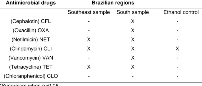
presented in Table 3. The propolis samples showed the potential to enhance the
antimicrobial drugs efficacy, but the greatest synergism profile was observed in the
propolis from the South of Brazil. This propolis sample also showed the greatest
flavonoid content although it was less active against CNS strains than Southeast EEP.
The sub-inhibitory MICs determined in the synergism assays were 0.21%v/v for
Southeast and 0.41%v/v for South EEP samples, and this protocol resulted in greater
flavonoid content in Petri plates prepared with South EEP than in those with Southeast
EEP. The results support the conclusion that EEP chemical characteristics, especially
the flavonoid content, can make clear the synergism between EEP and antimicrobial
drugs, but the establishment of the molecular basis of the synergistic effect between
EEP and antimicrobial drugs along with further microbiological, pharmacological and
clinical trials is needed.
Thus, the results were encouraging and confirmed the strong anti-CNS activity of
propolis; determination of the chemical characteristics of EEP showed that the phenolic
compounds were responsible for the greatest anti-CNS activity of Southeast EEP, and
the flavonoid content was responsible for the greatest synergism rate of South EEP.
Since bacteria may be resistant to several antimicrobial drugs, the synergism reported
here is relevant, and propolis may constitute an alternative for treating these
Table 1. Physicochemical characteristics of ethanolic extracts of propolis from two
regions of Brazil.
Propolis samples Dry weight
(mg/ml)
pH Flavonoids
%
Phenolic compounds
(%)
Botucatu
(São Paulo State-Southeast) 80.3 5 0.597 5.9
Urubicí
(Santa Catarina State-South) 87.7 4 0.871 4.7
Table 2. MIC 90% (*) from ethanolic extracts of propolis (EEP) and ethanol (%v/v)
control against 32 coagulase-negative Staphylococcus strains and one-fourth of MIC
90% values used in synergism assays.
Brazilian Regions
Southeast sample
(%v/v)
South sample (%v/v)
Ethanol Control (%v/v)
MIC 90 % (1/4 of MIC 90%) 0.83 (0.21) 1.63 (0.41) 12.72 (3.18)
Range 0.35–1.0 0.4–5.0 5.0–13.0
(*) Antimicrobial activity of Southeast EEP > South EEP > Ethanol control: p<0.05
Table 3. Synergism (X)* between ethanolic extracts of propolis (South and Southwest
samples); ethanol control and antimicrobial drugs by the E-test method against 10
coagulase-negative Staphylococcus strains: S. epidermidis (5) and S. saprophyticus
(5).
Antimicrobial drugs Brazilian regions
Southeast sample South sample Ethanol control
(Cephalotin) CFL - X -
(Oxacillin) OXA - X -
(Netilmicin) NET X X -
(Clindamycin) CLI X X X
(Vancomycin) VAN - X -
(Tetracycline) TET X X -
(Chloranphenicol) CLO - - -
ACKNOWLEDGMENTS
The authors are grateful to Dra. Lidia Raquel de Carvalho (Department of
Biostatistics /IBB/UNESP/Botucatu) for statistical analysis, and to FAPESP (The São
Paulo State Research Foundation).
REFERENCES
1 BANKOVA VS., CASTRO SL., MARCUCCI MC. Propolis: recent advances in
chemistry and plant origin. Apidologie., 2000, 31, 3-15.
2 BANSKOTA AH., TEZUKA Y., KADOTA S. Recent progress in pharmacological
research of propolis. Phytother. Res., 2001, 15, 561-71.
3 BOSIO K., AVANZINI C., D’AVOLIO A., OZIMO O., SAVOIA D. In vitro activity of
propolis against Streptococcus pyogenes. Lett. Appl. Microbiol., 31, 2000, 174-7.
4 BURDOCK GA. Review of the biological properties and toxicity of bee propolis
(Propolis). Food Chem. Toxicol., 1998, 36, 347-63.
5 CASTALDO S., CAPASSO F. Propolis, an old remedy used in modern medicine.
Fitoterapia, 2001, 73, S1-S6.
6 DETOMA P., OZINO OL. Azione della propoli su microrganismi dell’ambiente
ospedalino. Ann. Microbiol. Enzimol., 1991, 41, 231-6.
7 DRAGO L., MOMBELLI B., DE VECCHI E., FASSINA MC., TOCALLI L.,
GISMONDO MR. In vitro antimicrobial activity of propolis dry extract. J. Chemother.,
2002, 12, 390-5.
8 FERNANDES JUNIOR A., BALESTRIN EC., BETONI JEC., ORSI RO., CUNHA
MLRS., MONTELLI AC. Propolis: anti-Staphylococcus aureus activity and synergism
with antimicrobial drugs. Mem. Inst. Oswaldo Cruz, 2005, 100, 563-6.
9 GEBARA ECE., LIMA LA., MAYER MPA. Propolis antimicrobial activity against
periodontopathic bacteria. Braz. J. Microbiol., 2002, 33, 365-9.
10 GONSALES GZ., ORSI RO., FERNANDES JUNIOR A., RODRIGUES P.,
FUNARI SRC. Antibacterial activity of propolis collected in different regions of Brazil.
J. Venom. Anim. Toxins incl. Trop. Dis., 2006, 12, 276-84.
11 HAVSTEEM B. Flavonoids, a class of natural products of high pharmacology
potency. Biochem. Pharmacol., 1983, 32, 1141-8.
12 IKENO K., IKENO T., MIYAZAWA C. Effects of propolis on dental caries in rats.
13 KEDZIA B., HOLDERNA E. Investigations upon the combined action of antibiotics
and propolis on Staphylococcus aureus. Herba Polonica, 1986, 32, 187-95.
14 KONEMAN EW., ALLEN SD., JANDA NM., SCHRECKEMBERGER PC., WINN
JR WC. Diagnóstico Microbiológico: Texto e Atlas Colorido. 5.ed. Rio de Janeiro:
Medís, 2005.
15 KOO H., ROSALEN PL., CURY JA., PARK YK., BOWEN WH. Effects of
compounds found in propolis on Streptococcus mutans growth and on
glucosiltransferase activity. Antimicrob. Agents Chemother., 2002, 46, 1302-9.
16 KROL W., SCHELLER S., SHANI J., PIETSZ G., CZUBA Z. Synergistic effect of
ethanolic extract of propolis and antibiotics on the growth of Staphylococcus aureus.
Arzneimittel-Forsch., 1993, 43, 607-9.
17 KUSUMOTO T., MIYAMOTO RH., DOI S., HIROYUKI S., YAMADA H. Isolation
and structures of two new compounds from the essential oil of Brazilian propolis.
Chem. Pharm. Bull., 2001, 49, 1207-9.
18 MAHON CR., MANUSELIS JR G. Textbook of Diagnostic Microbiology.
Philadelphia: WB Saunders, 1995.
19 NATIONAL COMMITTEE FOR CLINICAL LABORATORY STANDARDS.
Methods for dilution antimicrobial susceptibility tests for bacteria that grow
aerobically. 6.ed. Wayne: NCCLS, 2003. [Approved Standard M7.A6]
20 NATIONAL COMMITTEE FOR CLINICAL LABORATORY STANDARDS. Clinical
and Laboratory Standards Institute. Performance standards for antimicrobial
susceptibility testing: fifteenth informational supplement. Wayne: CLSI, 2005.
M100-S15.
21 ORSI RO., SFORCIN JM., FUNARI SRC., FERNANDES JUNIOR A., BANKOVA
V. Synergistic effect of propolis and antibiotics on the Salmonella typhi. Braz. J.
Microbiol., 2006, 37, 108-12.
22 ORSI RO., SFORCIN JM., RALL VLM., FUNARI SRC., BARBOSA L.,
FERNANDES JUNIOR A. Susceptibility profile of Salmonella against the antibacterial
activity of propolis produced in two regions of Brazil. J. Venom. Anim. Toxins incl.
Trop. Dis., 2005, 11, 109-16.
23 SANTOS FA., BASTOS EMA., UZEDA B., CARVALHO MAR., FARIAS ESA.,
BRAGA FC. Antibacterial activity of Brazilian propolis and fractions against oral
24 SCHELLER S., DWORNICZAK S., WALDEMAR KK., RAJCA M., TOMCZIK A.,
SHANI J. Synergism between ethanolic extract of propolis (EEP) and
anti-tuberculosis drugs on growth of mycobacteria. Z. Naturforsch., 1999, 54, 549-53.
25 SFORCIN JM., FERNANDES JUNIOR A., LOPES CAM., BANKOVA V., FUNARI
SRC. Seasonal effect on Brazilian propolis antibacterial activity. J. Ethnopharmacol.,
2000, 73, 243-9.
26 STEPANOVIC S., ANTIC N., DAKIC I., SVABIC-VLAHOVIC M. In vitro
antimicrobial activity of propolis and synergism between propolis and antimicrobial
drugs. Microbiol. Res., 2003, 158, 353-7.
27 TAKAISI-KIKUNI NB., SCHILCHER H. Electron microscopy and microcalorimetric
investigations of the possible mechanism of the antibacterial action of a defined