periderm invasion contributes to
epithelial formation in the teleost
pharynx
Joana teixeira Rosa
1,2, Veronika oralová
1,3, Daria Larionova
1, G. T. Eisenhoffer
4,
P. Eckhard Witten
1& Ann Huysseune
1The gnathostome pharyngeal cavity functions in food transport and respiration. In amniotes the mouth and nares are the only channels allowing direct contact between internal and external epithelia. In teleost fish, gill slits arise through opening of endodermal pouches and connect the pharynx to the exterior. Using transgenic zebrafish lines, cell tracing, live imaging and different markers, we investigated if pharyngeal openings enable epithelial invasion and how this modifies the pharyngeal epithelium. We conclude that in zebrafish the pharyngeal endoderm becomes overlain by cells with a peridermal phenotype. In a wave starting from pouch 2, peridermal cells from the outer skin layer invade the successive pouches until halfway their depth. Here the peridermal cells connect to a population of cells inside the pharyngeal cavity that express periderm markers, yet do not invade from outside. The latter population expands along the midline from anterior to posterior until the esophagus-gut boundary. Together, our results show a novel role for the periderm as an internal epithelium becomes adapted to function as an external surface.
Anatomically, the vertebrate pharynx corresponds to the part of the digestive tube that is surrounded by the post-mandibular visceral arches1. In teleosts, the most speciose group of vertebrates, the pharynx functions in food transport and manipulation. For respiration, water taken up via the mouth passes over the internal gills and is expelled via the gill slits. Thus, different from amniotes, the pharynx has channels allowing direct communi-cation between internal and external environment. Yet, as in amniotes, the pharynx is lined by endoderm, an epithelium typically paving the (internal) alimentary tube2. Whether the teleost pharyngeal epithelium is mod-ified during early development to prepare for direct interaction with the external environment is not known. In particular, if opening of the endodermal pouches (prospective gill slits) affects the composition of the pharynx lining has not been properly investigated.
Apart from its function in feeding and as a respiratory channel, the pharynx lining forms the anlagen of widely divergent organs such as lungs, gills, or teeth, and an array of endocrine organs, such as thyroid glands, ultimobranchial bodies, and the thymus (reviewed in3,4). Moreover, signaling from the pharyngeal epithelium is required for proper development of the visceral skeleton (e.g.5,6; reviewed in7,8). With respect to teleosts and other primary aquatic gnathostomes, the question raises if the original endoderm fulfills these functions. Possibly, because of the early contact with the external environment the epithelium must first undergo secondary modifi-cations, such as stratification. The origin of the cell layers that contributes to the pharyngeal epithelium has cap-tured the interest of generations of researchers. Discussions focused on how far ectoderm invades the oral cavity via the mouth in different vertebrate lineages (e.g.9,10) and how much it contributes to organs derived from the oropharyngeal lining, such as gills11–13 or taste buds14. Little attention has been paid to the pouches as potential passageways for ectoderm to invade the pharynx and to potentially contribute to pharyngeal derivatives.
1Research Group Evolutionary Developmental Biology, Biology Department, Ghent University, K.L. Ledeganckstraat
35, B-9000, Gent, Belgium. 2Present address: Comparative, adaptive and functional skeletal biology (BIOSKEL),
Centre of Marine Sciences (CCMAR), Building 7, University of Algarve, Campus Gambelas, 8005-139, Faro, Portugal.
3Present address: Institute of Animal Physiology and Genetics, v.v.i., Czech Academy of Sciences, Veveri 97, 602
00, Brno, Czech Republic. 4Department of Genetics, The University of Texas MD Anderson Cancer Center, 1515
Holcombe Blvd, Unit 1010, Houston, Texas, 77030, USA. Joana Teixeira Rosa and Veronika Oralová contributed equally. Correspondence and requests for materials should be addressed to A.H. (email: Ann.Huysseune@UGent.be) Received: 20 August 2018
Accepted: 17 June 2019 Published: xx xx xxxx
www.nature.com/scientificreports
www.nature.com/scientificreports/
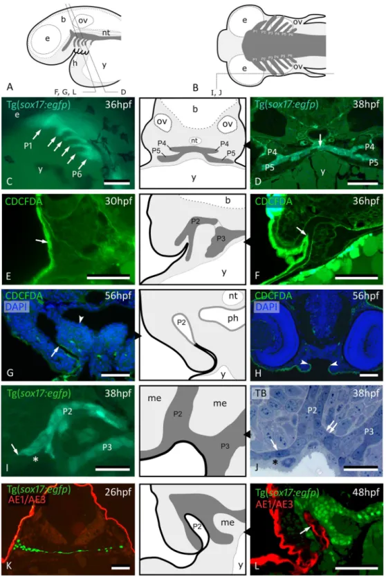
Figure 1. Peridermal cells invade pouch 2. (A,B) Generalized scheme of zebrafish pouches (dark grey) in a lateral (A) and horizontal (B) view, with sections D-L as indicated. (C) Lateral view of Tg(sox17:egfp) embryo showing pouches 1 to 6 (compare to (A)). (D) GFP labeled endodermal cells along the midline (white arrow) extend into pouches 4 and 5 (P4 and P5, resp.); corresponding schematic attached. (E) CDCFDA labeling at 26 hpf, sacrifice at 30 hpf; labeled cells are exclusively located in the superficial skin layer or periderm (arrow, level of pouch 2). (F) CDCFDA labeling at 26 hpf, sacrifice at 36 hpf; peridermal cells have partially invaded pouch 2 (arrow); corresponding schematic attached. (G) CDCFDA labeling at 26 hpf, sacrifice at 56 hpf; peridermal cells (arrow) line the now open gill slit; elsewhere the pharyngeal epithelium remains free of peridermal cells (arrowhead); corresponding schematic attached. (H) CDCFDA labeled cells only cover part of the epithelial lining of the mouth. Their boundary is indicated by arrowheads and coincides with the reduction of two to one epithelial cell layer. (I,J) Contact area of pouches 2 and 3 with skin; note that sox17+ cells spread out (arrows) and appear to displace the basal cell layer. In this way, the distal parts of the pouches even become connected
Here we examine how the zebrafish pharyngeal epithelium becomes modified in early development. We test if, and how far, cells of the outer body covering (ectoderm and/or periderm) enter the pharynx through the pharyngeal pouches and gill slits. We also examine if and how cells in the pharynx midline change during early development.
Results
Peridermal cells enter pouches 2 to 6.
The pharyngeal endoderm becomes distinct from the mesoderm at about 101/4 hours post fertilization (hpf)15. Using a transgenic line (Tg(sox17:egfp)) that marks the endoder-mal cells, pharyngeal pouches can be observed to develop sequentially from anterior (pouch 1, P1) to posterior (pouch 6, P6) between 18 hpf and 38hpf (Fig. 1A–C), confirming earlier reports16–18. Initially, the endoderm con-stitutes a single layer of flattened cells along the midline of the body, overlying the yolk. Soon after, this monolayer expands into two apposed layers of sox17+ cells (Fig. 1D). Each pouch is constituted of two simple closely apposedsox17+ epithelia, extending pairwise on each side of the midline endoderm and reaching up to the body surface
(Fig. 1B,D). Because of the intimate contact between pouch endoderm and body surface, presaging the opening of the gill slit, we investigated if, when and how cells from the embryonic skin (either the superficial, periderm layer or the basal, ectodermal layer) colonize the pouches and the midline endoderm. To do so, we monitored cell dynamics using [5-(and-6)-carboxy-2’,7’-dichlorofluorescein diacetate, succinimidyl ester, mixed isomers] (CDCFDA) as a cell tracer and investigated the distribution of ectoderm/and or periderm using different markers.
Labeling of wildtype (AB line) embryos using CDCFDA for 4 hrs, starting at 26 hpf, exclusively marked the outer, i.e., peridermal, skin layer (Fig. 1E). Labeled peridermal cells start to appear in pouch 2 (P2) at around 32 hpf and extend inwards until about halfway the pouch (Fig. 1F), but never reach the midline endoderm, not even as late as at 56 hpf (Fig. 1G). Peridermal cells also expand into the stomodeum. Similar to the invasion of P2 (and other pharyngeal pouches, see below), migration of CDCFDA-labeled cells in the forming mouth is arrested. Labeled cells do not cover the roof of the future mouth (Fig. 1H). The limit of CDCFDA labeled cells coincides with a reduction from two to one epithelial cell layers resting on the basal lamina. The basal ectodermal cells are not observed at any time within P2. Rather, they appear to be pushed away from the prospective gill slits by
sox17+ cells from the expanding distal tip of the pouch (Fig. 1I,J).
Immunolabeling with the pan-cytokeratin AE1/AE3 antibody at 26 hpf marked periderm only (Fig. 1K). In contrast, immunostaining at 48 hpf showed labeled cells extending halfway P2 (Fig. 1L, compare to 1G), confirm-ing the results obtained by cell tracconfirm-ing.
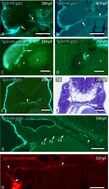
The fate of the cells labeled by CDCFDA was further confirmed using the Tg(krt4:gfp) line19. Keratin 4-positive (krt4+) peridermal cells start to appear in the distal part of pouch 2 (P2) at around 32 hpf (Fig. 2A), extending into the pouch for about halfway of its length (Fig. 2B). Cells squeeze between the two apposed layers of endoderm, thus separating them and paving each single layer of endoderm with a layer of krt4+ cells. Lumen for-mation in P2 starts at 36 hpf, and typically occurs where peridermal cells have covered the endoderm (Fig. 2C–F). By 60 hpf, the pouch has completely opened, creating the first open gill slit (Suppl. Fig. S1). The opercular flap is now a distinct structure, its surface completely covered with labeled cells. Following the invasion of P2, but considerably later, krt4+ peridermal cells sequentially enter P3-P6 from the outside (Fig. 2G,H). Invasion occurs at 56 hpf for P3 and P4 and at 72 hpf for P5 and P6, respectively (Suppl. Fig. S5). As in P2, invasion is arrested about halfway in each pouch (Fig. 2H). Double transgenic embryos revealing both endoderm and periderm (Tg(sox17:egfp; krt4:tomatoCAAX)) confirmed that, by 55 hpf, periderm has partially invaded P2 and covers
sox17-positive endodermal cells (Fig. 2I,J).
krt4
+ cells expand through the midline posteriorly to cover the entire endodermal lining.
Surprisingly, cells expressing the peridermal marker (krt4+) were also observed along the midline, trapped between the two endodermal layers (Fig. 3A,B). Because cell lineage tracing had clearly shown that peridermal cells from the skin do not reach the midline, these krt4+ cells (further called midline krt4+ cells) must have another origin.
First, we confirmed the independent origin of these midline krt4+ cells from the periderm by removing the periderm with EDTA from Tg(krt4:gfp) embryos aged 26 hpf, i.e. well before peridermal cells start to invade P2 (Fig. 3C). This experiment revealed an internal cluster of cells showing marked krt4 expression present at this time point (Fig. 3D). To investigate if the cluster of midline krt4+ cells derives from periderm at an earlier stage of development, we labeled embryos with CDCFDA for 4 hrs starting at 6 hpf, every two hours until 22 hpf. In none of these specimens did we ever observe periderm entering that could explain the cluster of krt4+ cells observed along the midline at the level of P1-P2 (data not shown).
At 26 hpf, the midline krt4+ cells are first located anteriorly, in a region potentially coinciding with endoderm (Fig. 3E,F). The midline krt4+ cells are initially separated from the peridermal cells invading P2 but contact them later (Fig. 3A,B, Supplementary Fig. S1). The domain of midline krt4-expressing cells is then seen to expand from the region of P2 posteriorly along the midline between the two layers of endodermal cells, reaching pouch 3 (P3), P4, P5 and P6 at 38 hpf, 42 hpf, 46 hpf and 48 hpf, respectively (Supplementary Fig. S2). At 54 hpf, the entire (double arrow). Only periderm (asterisks) covers the expansion of sox17+ endodermal cells. Corresponding schematic attached. (K,L) Immunolabeling using the pan-cytokeratin marker AE1/AE3 on Tg(sox17:egfp) embryos labels periderm only at 26 hpf (K), but also marks the lining of P2 at 48 hpf (arrow) (L); corresponding schematic attached. In all schemes, endoderm is dark gey, periderm is a black line. (C) Whole mount embryo; (D–H,K,L) cross sections; (I,J) sagittal sections. b: brain; e: eye; h: heart; me: mesenchyme; nt: notochord; ov: otic vesicle; P2 – P6: pouches 2 to 6; ph: pharyngeal lumen; TB: toluidine blue staining; y: yolk. Scale bar (C) = 500 μm, (D–H,K,L) = 50 μm, (I,J) = 20 µm.
www.nature.com/scientificreports
www.nature.com/scientificreports/
pharyngeal lumen, as well as P2, is covered by krt4 expressing cells (Fig. 3G). This was confirmed by Tg(sox17:egfp;
krt4:tomatoCAAX) embryos (Fig. 3H). The appearance of krt4+ cells in the pharynx midline anteriorly, and their expansion into the posterior pharynx, was also confirmed through time lapse imaging of Tg(krt4:gfp) embryos using dual photon laser scanning microscopy (Supplementary Fig. S3 and Supplementary Movie).
The krt4+ and thus periderm-like phenotype of the midline cells was further corroborated by using embryos of the Et(Gal4-VP16)zc1044A;Tg(UAS-E1b:nsfB-mCherry)c264 line, validated as having expression in the
peri-derm20, crossed to the Tg(krt4:gfp) line. In embryos sacrificed at 56 hpf, both the periderm and the midline
krt4+ cells strongly express mCherry in patterns overlapping with the krt4:GFP + cells, indicating that the same
enhancer is active in both cell types, peridermal cells and midline cells (Fig. 4A–C).
On the other hand, staining of Tg(sox17:egfp) embryos with the pan-cytokeratin antibody AE1/AE3 yielded a strong signal in the periderm but did not label midline krt4+ cells (Fig. 1K,L). This indicates that not all keratins Figure 2. Peridermal cells sequentially invade pouches 2 to 6 but do not reach the midline endoderm. (A–C)
krt4+ cells are present in progressively deeper parts of pouch 2 (P2) (arrows). (D) Peridermal cells are arrested
approximately halfway the pouch (arrow). (E,F) At 44 hpf P2 is open to the exterior. Peridermal cells extend inside the pouch (black stars); a lumen forms only where these cells cover the endodermal layer. In (F), outer dotted lines mark contour of the pouch; inner dotted line marks contour of peridermal cells. (G) While peridermal cells have entered through P2 (arrow), krt4+ cells remain excluded from P3 (arrowhead) and more posterior pouches. (H) At 56 hpf, peridermal cells have started to enter P3 (arrow), and progressively extend inwards. (I,J) Double transgenics Tg(sox17:egfp;krt4:tomato) show opening of P2 with periderm entering and covering sox17+ endodermal cells (I, green channel only; J, overlay). P2–P6: pouches 2 to 6. (A–H) cross sections, (I,J) sagittal sections. TB: toluidine blue staining. Scale bars (A–E) and (G–J) = 50 μm, (F) = 5 μm.
are shared by both cell populations. Zn-8, which detects Alcama protein and is an established endodermal pouch marker, labels pouches but not the midline endoderm. Hence, it could not be used to perform a reciprocal colo-calization experiment (staining Tg(krt4:gfp) embryos with an endoderm marker).
To examine whether mitoses contribute to stratification of the pharynx along the midline, we assessed prolif-eration on semithin sections and by 5-bromo-2′-deoxyuridine (BrdU) labeling (Fig. 4D–F). Using a short pulse at 36 hpf, proliferating cells were observed along the midline, albeit more frequent where the endoderm had not yet stratified into two opposing monolayers (compare Fig. 4D,E). This suggests that mitoses contribute to estab-lish the endoderm bilayer, but not (or little) to the krt4+ cells that cover them. Proliferation was abundant in the pouches proper (Fig. 4F).
Figure 3. krt4+ cells expand along the midline and connect to invaded peridermal cells. (A) At 38 hpf, krt4+ cells are visible along the midline (arrowhead), separated from invading peridermal cells (arrow). (B) Shortly after, the midline krt4+ cells (arrowhead) make contact with peridermal cells that have entered through P2 (arrow). (C) Lateral view on Tg(krt4:gfp) embryo in the early phase of periderm removal with EDTA. Part of the periderm is still attached (arrow). (D) At an advanced stage of EDTA treatment, the periderm is completely removed, and an internal krt4+ structure has become visible (arrowhead). (E,F) This internal krt4+ structure (arrowhead on E) coincides with the midline endoderm at the level of pouch 1–2 (arrowhead on F). (G) By 54 hpf, midline krt4+ cells cover the entire pharynx lining (boundary indicated by arrowhead). P2 is wide open (at another level of sectioning) but other pouches (P3–P6) are still free of krt4+ cells. (H) Double transgenic embryo Tg(sox17:egfp;krt4:tomato) of 55 hpf shows lining of pharyngeal epithelium with krt4+ cells (arrowheads) (red channel only). Note continuity, albeit with sharp boundary (arrow), with peridermal cells entering via P2. (A,B,E,F) Cross sections; (C,D) whole mount embryos; (G,H) sagittal section. TB: toluidine blue staining. e: eye; y: yolk; P2–P6: pouches 2 to 6. Scale bars (A,B,E–G) = 50 μm; (C,D) = 100 μm; (H) = 20 µm.
www.nature.com/scientificreports
www.nature.com/scientificreports/
The midline cells maintain krt4 expression for an extended period (Fig. 4G), and even at 5 dpf, the signal is still very strong. Remarkably, while the basal endodermal cells turn off sox17 expression at around 72 hpf, the midline
krt4+ cells that cover them start expressing sox17 strongly at around 56 hpf (Fig. 4H,I). The ultrastructure of the two cell types also differs (Fig. 4J). Superficial cells (corresponding to the krt4+ cells) develop microridges, are more squamous, with a flattened nucleus, and possess a more electron-dense cytoplasm than the underlying endodermal cells. Double transgenic embryos (Tg(sox17:egfp; krt4:tomatoCAAX) confirmed overlapping krt4 and sox17 expression in the superficial layer, as opposed to exclusive, yet fading, sox17 expression in the basal endodermal layer (Fig. 4K). Again, as with P2, a lumen forms only when and where krt4+ cells have covered the endoderm (Fig. 4K–M).
Midline krt4+ cells expand up to the gut entrance and join peridermal cells invaded through
pouches 3–6.
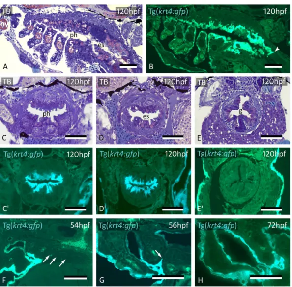
The domain of krt4+ cells has a sharp posterior limit at the end of the esophagus (Fig. 5A,B). The boundary coincides with the transition from a bilayered to a single-layered epithelium that lines the gut Figure 4. Midline krt4+ cells are distinct from peridermal and endodermal cells. (A–C) In embryos of GET-periderm x Tg(krt4:gfp) line, at 56 hpf, both the GET-periderm and the midline krt4+ cells (arrowhead) strongly express mCherry in patterns overlapping with the krt4:GFP + cells. (D,E) Mitoses (arrowheads) revealed by BrdU pulse labeling at 36 hpf in the midline endoderm at more anterior (D) and posterior (E) cross sectional level. (F) Abundant mitoses in the distal part of the pouch (arrows) but not in the midline endoderm (thick arrow). (G) Midline cells (star) maintain a strong krt4+ expression at 60 hpf. (H,I) Downregulation of sox17+ expression in the endodermal layer (delimited by the outer dotted line in H, and by arrowheads in I) but upregulation in the superficial layer surrounding the pharyngeal lumen (delimited by inner two dotted lines). (J) Cells of the superficial layer are flattened and more electron-dense in TEM than the basal endodermal layer, composed of cuboidal cells, and they develop microridges (arrows). Epidermis to the left; midline to the right. (K–M) Double transgenic embryo Tg(sox17:egfp;krt4:tomato) of 55 hpf; krt4+ cells are positive forsox17; conversely, only some cells of the basal endodermal layer have retained a sox17 signal (arrowheads).
(A–M) cross sections. en: endodermal layer; ph: pharyngeal lumen; TB: toluidine blue staining. Scale bars (A–F) = 50 μm, (G–I) = 25 µm, (J–M) = 10 µm.
(Fig. 5C–E’). The single-layered gut epithelium is krt4-negative (Fig. 5E’). Where the epithelial lining is bilayered, the superficial krt4+ cells display long cell extensions that reach down to the basal lamina in-between the endo-dermal cells (Fig. 5C’,D’).
During invasion of P2 by peridermal krt4+ cells, and during expansion of midline krt4+ cells towards the posterior pharynx, the other pouches persist as two apposed simple endodermal epithelia, free of krt4+ cells (Fig. 5F, Supplementary Fig. S2, S4). Midline krt4+ cells, present before peridermal cells enter P3 to P6, extend outwards through pouches 3–6. Eventually, they join the krt4+ cells that enter these pouches from outside, between 56 and 72 hpf (Fig. 5G,H), in a way similar as observed for P2 (compare with Fig. 3B; Supplementary Fig. S5). Thus, the simple endodermal epithelium resting on the basal lamina in the midline, as well as in each pouch, becomes completely overlain by an uninterrupted layer of krt4+ cells. This double lining is completed at 72 hpf for P3 and P4 and at 84 hpf for P5 and P6 (Supplementary Fig. S5, S6). Again, a lumen appears in the pouch only after it is invested with krt4+ cells. After the midline krt4+ cells that extend outwards in each pouch have joined with the peridermal krt4+ cells extending inwards, a boundary cannot be observed anymore between both gfp + cell populations, although it remains visible between krt4:tomatoCAAX + periderm and midline cells in double transgenic (Tg(sox17:egfp; krt4:tomatoCAAX) embryos (Fig. 3H). Thus, the entire pharyngeal cavity as well as the opened gill slits at 56 hpf and beyond are lined by a continuous krt4+ layer, covering the original endodermal layer.
Figure 5. Midline krt4+ cells cover pharynx and esophagus, and meet peridermal cells invading through P3-P6. (A,B) krt4+ cells cover hyoid arch (hy) and branchial arches 1 to 5 (numbered 1–5), as well as roof and floor of the pharynx and esophagus. Note sharp boundary (arrowhead) between krt4+ esophagus and krt4- lining of the gut. (C,C’,D,D’) krt4+ cells cover the lining of the pharynx (C,C’) and the esophagus (D,D’), resp. Note long cell extensions issuing from the krt4+ cells (E,E’) Absence of krt4+ cells in the gut coincides with single-layered epithelium. (F) At 54 hpf, pouches 3–6 are still free of krt4+ cells. (G,H) Progressive invasion of P3 (arrow) by peridermal cells that meet midline krt4+ cells. (A,B,F) sagittal sections, (C–E’,G,H) cross sections. es: esophagus; g: gut; ph: pharyngeal lumen; TB: toluidine blue staining. Scale bars (A–H) = 50 μm.
www.nature.com/scientificreports
www.nature.com/scientificreports/
Cells resembling periderm cells line the pharynx in other teleost species.
To investigate if a layer of flattened cells with periderm-like phenotype covers the pharyngeal endoderm in other teleosts, we did a pre-liminary survey of three widely divergent teleost species: European eel (Anguilla anguilla, Anguilliformes, a basal teleost species), Pacific salmon (Oncorhynchus tshawytscha, Salmoniformes), and the Jewel cichlid (Hemichromisbimaculatus, Perciformes, a highly derived teleost species). In the absence of markers or transgenic lines for these
species, we compared the histology of the skin with the region of pouches 2–3 in early developmental stages (Fig. 6). In all three species, pouch 2, and the opened portion of pouch 3, appeared to be lined by flattened cells, resembling peridermal cells in the superficial skin cover, and overlying a layer of cuboidal cells, inferred to be the endoderm. Where pouch 3 was not open yet, only a double layer of cuboidal endodermal cells was observed.
Discussion
We show here that the pharynx and the gill slits in early post-embryonic zebrafish are lined by a double-layered epithelium: a basal layer resting on the basal lamina, consisting of sox17+ endoderm, and a superficial krt4+ layer, derived partly from the skin periderm (in the pouches), and partly from internal anteriorly located cells that start to express krt4. Lineage tracing experiments along with observations on transgenic embryos clearly reveal Figure 6. Cells resembling periderm cells in the pharynx of other teleost species. Cross sections through the region of pouch 2, pouch 3 and comparison to the skin in three teleost species: Anguilla anguilla (total length, TL, 6.0 mm), Oncorhynchus tshawytscha (unhatched embryo, 15 dpf), and Hemichromis bimaculatus (TL 4.0 mm, 1 day post-hatching, dph). Pouch 2 (A,D,G) is lined by flattened cells (black arrows) overlying a layer of cuboidal cells. Where pouch 3 is not open yet (B,E), only a double layer of cuboidal endodermal cells is present (between white arrowheads). Where pouch 3 starts to open (H), similar cells can be seen to cover the pouch endoderm (black arrows). Note resemblance, for each of the three species, to peridermal cells in the superficial skin cover (white arrows, C,F,I). In (A,B,D,E,G,H), the epidermis is to the left and the pharyngeal lumen to the right; in (C,F,I) the external surface is to the right. (A–I) cross sections. ph: pharyngeal lumen. Scale bars (A–I) = 10 μm.
that peridermal cells enter all pouches, in succession from anterior (P2) to posterior (P6), albeit not up to the midline. Instead, the cells are met by krt4+ cells, whose domain expands along the midline, likewise from ante-rior to posteante-rior (Fig. 7). The midline cells turn on krt4 expression before the start of invasion of krt4+ perider-mal cells through pouch 2. Our observations confirm the dynamic pattern of krt4 expression observed by in situ hybridization: expression in P2 and midline first, followed by expression in the other pouches (fig. 11 in21). The midline krt4+ cells bear similarities to periderm: (1) they express some of the markers (krt4, zc1044) expressed by true peridermal cells; (2) they develop microridges similar to those in the periderm; (3) the krt4+ cell layer Figure 7. Interpretative scheme, showing development of the pharyngeal lining. (A) Before 30 hpf, peridermal (krt4+) cells are only seen covering the embryo; internal krt4+ cells are only present in the midline at the level of P1-P2. (B) Around 32 hpf peridermal cells start to invade P2. The domain of midline krt4+ cells starts to spread posteriorly and laterally towards P2. (C) Peridermal cells invade P2 until halfway and midline krt4+ cells extend outwards reaching peridermal krt4+ cells. Also midline krt4+ cells continue to extend through the pharynx midline until the level of P3. (D) The invasion and expansion of both peridermal and midline krt4+ cells continue, following the same pattern seen for P2. (E) Finally, the entire pharynx midline is invested by
krt4+ cells and all the pouches, until halfway, are covered in peridermal cells. The domain of midline krt4+ cells
also expands anteriorly until the mouth, and posteriorly until the end of the esophagus. P2: pouch 2; P3: pouch 3; P4: pouch 4; P5: pouch 5; P6: pouch 6.
www.nature.com/scientificreports
www.nature.com/scientificreports/
merges imperceptibly with peridermal cells invading from outside, suggesting compatible adhesion. They furthermore display ultrastructural features distinctive from the endodermal cells on which they rest. Expression of krt4 (and its disputed homologues krt8 and zf-K8) has been reported before in the pharynx of zebrafish19,22,23. Yet, none of the previous studies addresses the origin of these cells, nor how their domain expands, nor makes any suggestion regarding their periderm-like phenotype or association with the endoderm-derived epithelium. Our observations that the endoderm is covered by peridermal cells (in the pouches) and cells with a periderm-like phenotype (along the midline), generating two apposed, double-layered, epithelia in which then a lumen appears, fit with observations of Waterman & Kao24. These authors described the endoderm of the zebrafish pharynx as two-layered, becoming four-layered (a bilayer on each side of the lumen) in more advanced developmental stages. They also reported that superficial cells surrounding the lumen stain more intensely than the basal cells (visible on their Fig. 9). The superficial dark staining layer corresponds to the krt4+ and electron-dense superficial layer described here. Edwards25 already reported the invasion of the pharynx by ectodermal cells in the carp, a close relative of the zebrafish, and suggested it to be a feature common to all pouches. We agree with Edwards’ obser-vations, but we differ from Edwards’ interpretation in that we distinguish periderm from ectoderm cells, and that invading cells do not reach the midline, but are joined instead by midline krt4+ cells. The situation in carp and zebrafish may be common for teleosts, as Miyake et al.26 describe an ectoderm-derived inner lining of the opercu-lar cavity in cod (Gadus morhua) and Yamamoto et al.27 report the presence of flattened epithelial cells covering the hatching gland cells in the medaka pharynx (and see also Fig. 6). In contrast, Shone & Graham28 report only endoderm in the pouches of zebrafish, reaching out to the exterior, covering the inside of the opercular flap at 72 hpf. However, they used the endodermal marker sox 17 only and did not investigate the number of cell layers. Our observation that the pouches are invaded by peridermal cells that subsequently constitute part of the per-manent lining of the pharyngeal arches provides further evidence that the zebrafish periderm is more than just a transient embryonic feature29. It confirms that cells of the extra-embryonic enveloping layer (EVL, the source of the periderm) survive well into later embryonic stages or even juvenile life, as demonstrated previously for the epidermis30,31. While the periderm in zebrafish, by origin, is an extra-embryonic layer, periderm in mammals derives from ectoderm. Yet expression of some orthologous keratin genes, such as K8, appears to be shared31,32. That the midline krt4+ cells have an origin distinct from the skin periderm appears to be well established: (1) the cells are present prior to any periderm migrating into the pouches (as inferred from the cell tracing experiments); (2) while forming a continuous sheet, there is a sharp boundary between periderm cells and midline krt4+ cells, as revealed by double transgenic embryos; this lack of gradient strongly suggests a different origin; (3) while sharing several markers, they also differ in immunoreactivity when stained with the AE1/AE3 keratin antibody. The source of the midline krt4+ cells is intriguing. Initially, the cells form a cluster located in the midline region at the level of P1-P2, a region typically harboring sox17+ cells. The weak expression of krt4 in the midline cells of early double transgenic (endoderm/periderm) embryos however precluded colocalization of sox17+ endodermal cells and midline krt4+ cells. If the midline krt4+ cells derive from the same source as the pharyngeal endoderm proper and adopt a periderm-like phenotype this could represent a case of co-option33. It could also explain why the cells turn on sox17 expression at a later stage of embryonic development. From the area of origin, krt4+ cells could spread into the pharynx by migrating out of this region. Alternatively, krt4 expressing cells could arise locally. Due to the deep internal position of the cells, dual photon microscopy could not rule out any of the two alternatives. Most evidence speaks against a local origin of the krt4+ midline cells. First, it is unlikely that the two apposed monolayers of endodermal cells itself upregulate krt4 simply because krt4+ cells are present only where more than two cell layers are found along the midline, with krt4+ cells trapped between sox17 expressing cells. If derived locally, krt4+ cells should issue from the division of endodermal cells perpendicular to their apicoba-sal axis, generating a baapicoba-sal cuboidal endodermal cell and an apically positioned flattened cell, instantaneously expressing krt4. On semithin sections, metaphases indicating such polarized division within the endodermal cells were not detected. Mitoses were rare along the midline once the two opposing endodermal monolayers were established, but more frequent in the distal parts of the endodermal pouches, where cell division likely con-tributes to lateral pouch outgrowth. Thus, the more likely explanation is that cells migrate from a source located at the level of P1-P2. That the midline cells are rich in Krt4 raises the question whether this keratin could play a role in such a migration. Intermediate filaments, and in particular keratins, are increasingly considered to play a key role in epithelial cell migration34. For example, appropriate recruitment of keratin intermediate filaments is required for the directed protrusive behavior of individual cells within the collectively migrating mesendoderm in Xenopus35.
A likely function of the midline krt4+ cells and the peridermal cells in the pouches is in lumen formation. Lumina in the pharynx and pouches arise in a non-confluent way but appear only where krt4-expressing cells are present. These cells may thus function in an analogous way as the mammalian periderm. In mammals, the periderm prevents premature adhesion between closely apposed epithelia and needs to be removed in order for epithelia to fuse, as in palatogenesis; reviewed in36. In the zebrafish pharynx and in the pouches, the presence of cells with a peridermal phenotype appears equally to be required to move apposed endodermal layers apart and to allow lumen formation. If, and to what extent, the midline krt4+ cells participate in the formation of pharyngeal derivatives is not known at present but is worthwhile investigating, considering the timing of their appearance. For example, the first thyroid follicle develops from the ventral midline epithelium of the pharynx at around 60 hpf37 and the thymus appears at 54 hpf38, i.e. right after the midline krt4+ cells have covered the endoderm. Likewise, whether the midline krt4+ cells exchange signals with the endoderm, and/or underlying mesenchyme and play a role in the development of ectomesenchymal derivatives, is currently unknown. The presence of long cell extensions reaching down to the basal lamina between endodermal cells is intriguing in this respect. While interactions between periderm and endoderm have not been reported before, signaling between ectoderm and endoderm is crucial for several developmental processes in zebrafish, such as for the development of gill fila-ment buds39,40, or for epibranchial neurogenesis41,42. Ectodermally expressed Wnt4a signals to endodermal pouch
epithelium and acts together with EphrinB signaling to stabilize mature bilayers in the pouch endoderm43,44. In amphibians, ectodermal taste buds are induced by endoderm45. In mice endoderm-ectoderm signaling has been reported during early jaw morphogenesis46. The thymus defect in the nude mouse has been attributed to an arrest of development of ectoderm in the third cleft, depriving the endoderm from “its normal inducing agent”47. Clearly, the potential role of the periderm and of the midline krt4+ cells in the development of pharyngeal deriv-atives needs to be investigated. Advantage can be taken from the powerful tools that have been recently developed to study ectoderm and periderm fate20 and that have also been used here.
The boundary between germ layers and the delimitation of endoderm has received most attention in relation to mouth formation and mouth opening. Using lineage tracing, we observed that peridermal, krt4 expressing cells also enter the stomodeum, but only for a limited distance and after krt4+ cells have appeared along the midline at the level of P1-P2. Subsequently, the two populations meet and merge into an unin-terrupted layer, indicating striking similarities in the events of mouth and pouch formation. These obser-vations may further fuel debates on the homology of chordate mouth and gill slits, as e.g., recently revived for Amphioxus48,49. Once the mouth opens and the pouches open into gill slits, the oropharyngeal cavity is communicating with, and forms an extension of, the external environment. From a functional perspective, it is beneficial to have a pharynx that is lined by tissue adapted to interact with the aqueous environment, similar to what periderm in the skin does. From an evolutionary perspective, one would expect such a role to be fulfilled by invading ectoderm. For example, the capacity of tooth formation in the oropharynx has been proposed to be linked to an invasion of ectoderm50. It is thus somehow surprising that in zebrafish expansion of periderm into the oropharynx is arrested in the distal parts of the mouth and pouches, and that elsewhere endoderm has possibly been co-opted into a peridermal fate. That other (both basal and derived) teleost species display a two-layered pharyngeal epithelium early in development, with superficial flattened cells different from basal cuboidal cells, warrants investigations into the commonality of invasion and co-option in the development of the pharynx lining.
Materials and Methods
Transgenic zebrafish lines.
Tg(sox17:egfp)51 and wildtype (AB line) zebrafish were obtained from the lab-oratory of R. Opitz (VUB, Brussels, Belgium). Tg(krt4:gfp) fish19 and Tg(krt4:tomatoCAAX) were a gift from M. Hammerschmidt52 (University of Köln, Germany). The Tg(sox17:egfp) was crossed to the Tg(krt4:tomatoCAAX) line. Et(Gal4-VP16)zc1044A;Tg(UAS-E1b:nsfB-mCherry)c264, abbreviated as GET-periderm line (GET, Gal4enhancer trap)20 was crossed to the Tg(krt4:gfp) line. Adult fish were maintained and spawned according to53. Embryos were raised in egg water at 28.5 °C and staged according to15.
CDCFDA labeling.
For cell lineage tracing, WT embryos were soaked in CDCFDA [5-(and-6)-carboxy-2′,7′-dichlorofluorescein diacetate, succinimidyl ester, mixed isomers] (cat. No.: 22026, AAT Bioquest, Inc.). This mol-ecule, an isomer of the molecule used by Shone & Graham28 is taken up by any cell that is in direct contact with the agent and does not diffuse further. It is thus limited to cells exposed to the aqueous environment. CDCFDA was dissolved to 50 mM in anhydrous DMSO (dimethylsulfoxide) and stored at −20 °C. A working concentration of 250 µM CDCFDA was dissolved in egg water and embryos were soaked for 4 hours starting at 6 hpf, and then every two hours until 18 hpf, as well as at 26 hpf. After 4 hours in CDCFDA staining, the embryos were rinsed in fresh egg water and kept in petri dishes with fresh egg water until sacrifice. Embryos were fixed in 4% PFA and processed for glycol methacrylate (GMA) embedding.Periderm removal.
Periderm was removed from Tg(krt4:gfp) fish by soaking Tg(krt4:gfp) embryos at 26 hpf for 15 minutes in 12.5 mM EDTA dissolved in Ca2+ and Mg+ free Tyrode solution. Afterwards, they werewashed several times and maintained in regular Tyrode solution. Embryos were sacrificed at various intervals and processed for GMA embedding.
BrdU incorporation.
Proliferation at the level of the pharynx was examined through BrdU (5-bromo-2′-deoxyuridine) administration followed by immunostaining on whole-mount embryos. Embryos were treated with BrdU at 36 hpf according to the protocol described in54, sacrificed immediately after BrdU administration and fixed in 4% PFA for GMA embedding.Immunohistochemistry.
Immunohistochemistry on whole-mount embryos was performed as described in54 using as primary antibodies an anti-BrdU (mouse, BD Biosciences, 1:100), a Zn-8 (mouse, ZIRC, 1:100) or a pan-cytokeratin (AE1/AE3) (mouse, Santa Cruz, 1:200) and as secondary antibody an Alexa Fluor®
594 (goat anti-mouse IgG, Abcam, 1:200).Histology & TEM.
All animals were sacrificed by an overdose of 1% ethyl 3-aminobenzoate methansul-fonate (MS-222) (E10521-10G, Sigma Aldrich) and fixed overnight in 4% paraformaldehyde (PFA in 1xPBS, pH 7.2) at 4 °C. The embryos were processed for GMA embedding after immunohistochemistry, vital staining or without pretreatment (transgenic zebrafish), and serially sectioned at 3 µm. After proper drying of the sec-tions, some sections were counterstained with DAPI for nuclear staining. Fluorescent signals (GFP, DyLight, or CDCFDA) were visualized by placing a drop of 1xPBS per slide, and coverslipping. After analysis, sec-tions were stained with toluidine blue. For high resolution histology and transmission electron microscopy (TEM), embryos were fixed in a mixture of paraformaldehyde and glutaraldehyde, embedded in epon, serially sectioned at 1 µm55, and stained with toluidine blue. Ultrathin sections were prepared on an ultratome, con-trasted with uranyl acetate and lead citrate, and observed under a Jeol JEM 1010 transmission electron micro-scope (Jeol Ltd., Tokyo, Japan) operating at 60 kV. Microphotographs were taken with a Veleta camera (Emsis, Muenster, Germany).www.nature.com/scientificreports
www.nature.com/scientificreports/
Toluidine blue stained sections from GMA or epon embedded material of Anguilla anguilla, Oncorhynchus
tshawytscha, and Hemichromis bimaculatus, are part of the collections maintained in the Research Group
Evolutionary Developmental Biology (Ghent University).
Dual photon microscopy.
The Tg(krt4:gfp) fish were dechorionated and were immobilized at 30 hpf in agar diluted in egg water to a concentration of 0.1%. The embryo was positioned to obtain a dorso-lateral view and a sequence was recorded on a Zeiss LSM 780 confocal microscope of 32 Z-stacks every 20 minutes for a total of 18 hours. The pictures and video were extracted from records using Zeiss Axio Imager Z1 (www.zeiss.com) and Image J (version 2.0.0-rc-29, plus Bio-Formats 5.1.0 in Fiji, general public license).Observations and microphotography.
Observations of epon or GMA sections were done on a Zeiss Axio Imager Z1 (www.zeiss.com). Photomicrographs were taken with an MRC camera and processed using ZEN software (Zeiss, www.zeiss.com). Computer-generated images were processed for color balance, contrast and brightness only, and applied to all parts of the figures equally.Ethical statement.
Animal care, experimentation and sacrifice complied with European Directive 2010/63/ EU of 22 September, 2010. The experimental protocol and all animal procedures used in this study were approved by Ghent University (laboratory permit number LA1400452).Data Availability
All sections used for this study are kept in the slide collection of the Research Group ‘Evolutionary Developmen-tal Biology’ at the Biology Department of Ghent University, and are available for inspection upon request.
References
1. Hildebrand, M. Analysis of vertebrate structure. 4th Ed. 657 pp, John Wiley & Sons, New York (1995).
2. Gilbert, S. F. & Barresi, M. J. F. Developmental Biology, 11th Ed. 810 pp. Sinauer Associates (2016).
3. Grevellec, A. & Tucker, A. S. The pharyngeal pouches and clefts: development, evolution, structure and derivatives. Semin. Cell Dev.
Biol. 21, 325–332 (2010).
4. Adams, A., Mankad, K., Offiah, C. & Childs, L. Branchial cleft anomalies: a pictorial review of embryological development and spectrum of imaging findings. Insights Imaging 7, 69–76 (2016).
5. David, N. B., Saint-Etienne, L., Tsang, M., Schilling, T. F. & Rosa, F. M. Requirement for endoderm and FGF3 in ventral head skeleton formation. Development 129, 4457–4468 (2002).
6. Crump, J. G., Maves, L., Lawson, N. D., Weinstein, B. M. & Kimmel, C. B. An essential role for Fgfs in endodermal pouch formation influences later craniofacial skeletal patterning. Development 131, 5703–5716 (2004).
7. Graham, A. & Richardson, J. Developmental and evolutionary origins of the pharyngeal apparatus. EvoDevo 3, 24 (2012). 8. Frisdal, A. & Trainor, P. A. Development and evolution of the pharyngeal apparatus. Wiley Interdiscip. Rev. Dev. Biol. 3, 403–418
(2014).
9. Rothova, M., Thompson, H., Lickert, H. & Tucker, A. S. Lineage tracing of the endoderm during oral development. Dev. Dyn. 241, 1183–1191 (2012).
10. Soukup, V., Horacek, I. & Cerny, R. Development and evolution of the vertebrate primary mouth. J. Anat. 222, 79–99 (2013). 11. Goette, A. Über die Kiemen der Fische. Z. Wissenschaftl. Zool. 69, 533–577 (1901).
12. Janvier, P. Homologies and evolutionary transitions in early vertebrate history. In Major Transitions in Vertebrate Evolution (Anderson, J. S. & Sues, H.-D. eds) pp. 57–121, Indiana University Press, Bloomington (2007).
13. Gillis, J. A. & Tidswell, O. R. A. The origin of vertebrate gills. Curr. Biol. 27, 729–732 (2017).
14. Barlow, L. A. Progress and renewal in gustation: new insights into taste bud development. Development 142, 3620–3629 (2015). 15. Kimmel, C. B., Ballard, W. W., Kimmel, S. R., Ullmann, B. & Schilling, T. F. Stages of embryonic development of the zebrafish. Dev.
Dyn. 203, 253–310 (1995).
16. Crump, J. G., Swartz, M. E. & Kimmel, C. B. An integrin-dependent role of pouch endoderm in hyoid cartilage development. PLoS
Biol 2(9), e244 (2004).
17. Kopinke, D., Sasine, J., Swift, J., Stephens, W. Z. & Piotrowski, T. Retinoic acid is required for endodermal pouch morphogenesis and not for pharyngeal endoderm specification. Dev. Dyn. 235, 2695–2709 (2006).
18. Choe, C. P. et al. Wnt-dependent epithelial transitions drive pharyngeal pouch formation. Dev. Cell 24, 296–309 (2013).
19. Gong, Z. et al. Green fluorescent protein expression in germ-line transmitted transgenic zebrafish under a stratified epithelial promoter from Keratin8. Dev. Dyn. 223, 204–215 (2002).
20. Eisenhoffer, G. T. et al. A toolbox to study epidermal cell types in zebrafish. J. Cell Sci. 130, 269–277 (2017).
21. Rauch, G. J. et al. Submission and curation of gene expression data. ZFIN Direct Data Submission. (http://zfin.org) (2003). 22. Imboden, M., Goblet, C., Korn, H. & Vriz, S. Cytokeratin 8 is a suitable epidermal marker during zebrafish development. C.R. Acad.
Sci. Paris, Sciences de la vie/Life Sciences 320, 689–700 (1997).
23. Chen, C.-F. et al. Establishment of a transgenic zebrafish line for superficial skin ablation and functional validation of apoptosis modulators in vivo. PLoS One 6(5), e20654, https://doi.org/10.1371/journal.pone.0020654 (2011).
24. Waterman, R. E. & Kao, R. Formation of the mouth opening in the zebrafish embryo. Scanning Electron Microscopy III, 1249–1257 (1982).
25. Edwards, L. F. The origin of the pharyngeal teeth of the carp (Cyprinus carpio Linnaeus). Ohio J. Sci. 29, 93–130 (1929).
26. Miyake, T., Von Herbing, I. H. & Hall, B. K. Neural ectoderm, neural crest, and placodes: Contribution of the otic placode to the ectodermal lining of the embryonic opercular cavity in Atlantic Cod (Teleostei). J. Morphol. 231, 231–251 (1997).
27. Yamamoto, M., Iuchi, I. & Yamagami, K. Ultrastructural changes of the teleostean hatching gland cell during natural and electrically induced precocious secretion. Dev. Biol. 68, 162–174 (1979).
28. Shone, V. & Graham, A. Endodermal/ectodermal interfaces during pharyngeal segmentation in vertebrates. J. Anat. 225, 479–491 (2014).
29. Lee, R. T. H., Asharani, P. V. & Carney, T. J. Basal keratinocytes contribute to all strata of the adult zebrafish epidermis. PLoS One 9(1), e84858, https://doi.org/10.1371/journal.pone.0084858 (2014).
30. Fukazawa, C. et al. Poky/chuk/ikk1 is required for differentiation of the zebrafish embryonic epidermis. Dev. Biol. 346, 272–283 (2010).
31. Fischer, B. et al. p53 and TAp63 promote keratinocyte proliferation and differentiation in breeding tubercles of the zebrafish. PLoS
Genet. 10(1), e1004048, https://doi.org/10.1371/journal.pgen.1004048 (2014).
32. Bragulla, H. H. & Homberger, D. G. Structure and functions of keratin proteins in simple, stratified, keratinized and cornified epithelia. J. Anat. 214, 516–559 (2009).
33. True, R. J. & Carroll, S. B. Gene co-option in physiological and morphological evolution. Annu. Rev. Cell. Dev. Biol. 18, 53–80 (2002). 34. Leduc, C. & Etienne-Manneville, S. Intermediate filaments in cell migration and invasion: the unusual suspects. Curr. Opin. Cell Biol.
32, 102–112 (2015).
35. Weber, G. F., Bjerke, M. A. & DeSimone, D. W. A mechanoresponsive cadherin-keratin complex directs polarized protrusive behavior and collective cell migration. Dev. Cell 22, 104–115 (2012).
36. Lan, Y., Xu, J. & Jiang, R. Cellular and molecular mechanisms of palatogenesis. Curr. Top. Dev. Biol. 115, 59–84 (2015). 37. Alt, B. et al. Analysis of origin and growth of the thyroid gland in zebrafish. Dev. Dyn. 235, 1872–1883 (2006).
38. Willett, C. E., Cherry, J. J. & Steiner, L. A. Expression of zebrafish rag genes during early development identifies the thymus. Dev. Biol. 182, 331–341 (1997).
39. Hogan, B. M. et al. Zebrafish gcm2 is required for gill filament budding from pharyngeal ectoderm. Dev. Biol. 276, 508–522 (2004). 40. Hall, C., Flores, M. V., Murison, G., Crosier, K. & Crosier, P. An essential role for zebrafish Fgfrl1 during gill cartilage development.
Mech. Dev. 123, 925–940 (2006).
41. Holzschuh, J. et al. Requirements for endoderm and BMP signaling in sensory neurogenesis in zebrafish. Development 132, 3731–3742 (2005).
42. McCarroll, M. N. & Nechiporuk, A. V. Fgf3 and Fgf10a work in concert to promote maturation of the epibranchial placodes in zebrafish. PLoS One 8(12), e85087, https://doi.org/10.1371/journal.pone.0085087 (2013).
43. Choe, C. P. & Crump, J. G. Eph-Pak2a signaling regulates branching of the pharyngeal endoderm by inhibiting late-stage epithelial dynamics. Development 142, 1089–1094 (2015a).
44. Choe, C. P. & Crump, J. G. Dynamic epithelia of the developing vertebrate face. Curr. Opin. Genet. Devel. 32, 66–72 (2015b). 45. Barlow, L. A. Taste buds in ectoderm are induced by endoderm: implications for mechanisms governing taste bud development. In
Regulatory processes in Development: The legacy of Sven Hörstadius. Proceedings of the Wenner-Gren International Symposium (Olsson, L., and Jacobson, C.-O., eds) pp. 185–190, Portland Press (2000).
46. Haworth, K. E., Healy, C., Morgan, P. & Sharpe, P. T. Regionalisation of early head ectoderm is regulated by endoderm and prepatterns the orofacial epithelium. Development 131, 4797–4806 (2004).
47. Cordier, A. C. & Haumont, S. M. Development of thymus, parathyroids, and ultimo-branchial bodies in NMRI and Nude mice. Am.
J. Anat. 157, 227–263 (1980).
48. Lacalli, T. Interpreting amphioxus, and thoughts on ancestral chordate mouths and brains. Int. J. Dev. Biol. 61, 649–654 (2017). 49. Nielsen, C. Evolution of deuterostomy – and origin of the chordates. Biol. Rev. 92, 316–325 (2017).
50. Huysseune, A., Sire, J.-Y. & Witten, P. E. Evolutionary and developmental origins of the vertebrate dentition. J. Anat. 214, 465–476 (2009).
51. Mizoguchi, T., Verkade, H., Heath, J. K., Kuroiwa, A. & Kikuchi, Y. Sdf1/Cxcr4 signaling controls the dorsal migration of endodermal cells during zebrafish gastrulation. Development 135, 2521–2529 (2008).
52. Richardson, R. et al. Re-epithelialization of cutaneous wounds in adult zebrafish combines mechanisms of wound closure in embryonic and adult mammals. Development 143, 2077–2088 (2016).
53. Westerfield, M. The Zebrafish Book: A Guide for the Laboratory Use of Zebrafish Danio (Brachydanio rerio). University of Oregon Press, Oregon (2000)
54. Verduzco, D. & Amatruda, J. F. Analysis of Cell Proliferation, Senescence and Cell Death in Zebrafish Embryos. Methods Cell Biol. 101, https://doi.org/10.1016/B978-0-12-387036-0.00002-5 (2013).
55. Huysseune, A. & Sire, J.-Y. Development of cartilage and bone tissues of the anterior part of the mandible in cichlid fish: a light and TEM study. Anat. Rec. 233, 357–375 (1992).
Acknowledgements
The authors gratefully acknowledge Dr. R. Opitz (VUB, Brussels, Belgium) for the gift of Tg(sox17:egfp) zebrafish, Dr. M. Hammerschmidt (University Köln, Germany) for the gift of Tg(krt4:gfp) and Tg(krt4:tomatoCAAX), allele fr32Tg zebrafish, and Huan Liu, Greg Bonde and Robert Cornell (University of Iowa) for the gift of Tg(sox17:egfp;krt4:mCherry) embryos, respectively. TEM was carried out at the Ghent University TEM-Expertise center (Life Sciences), facility Nematology Research Unit. Dr. C. Guérin (VIB, Bioimaging Core Facility, VIB-UGent Center for Inflammation Research, Technologiepark 927, Gent, Belgium) is gratefully acknowledged for expert technical assistance with dual photon recording and image processing. M. Soenens provided valuable technical help. J.T.R., O.V. and A.H. acknowledge a grant of the Ghent University Research Fund (n° BOF24J2015001401). G.T.E. was supported by the Cancer Prevention Research Institute of Texas (RR140077).
Author Contributions
J.T. Rosa, V. Oralová, P.E. Witten and A. Huysseune designed research; J.T. Rosa, V. Oralová, and D. Larionova performed research; J.T. Rosa, V. Oralová, D. Larionova, and A. Huysseune analyzed data; J.T. Rosa, V. Oralová, P.E. Witten and A. Huysseune wrote the paper. G.T. Eisenhoffer contributed analytical tools.
Additional Information
Supplementary information accompanies this paper at https://doi.org/10.1038/s41598-019-46040-y. Competing Interests: The authors declare no competing interests.
Publisher’s note: Springer Nature remains neutral with regard to jurisdictional claims in published maps and institutional affiliations.
Open Access This article is licensed under a Creative Commons Attribution 4.0 International License, which permits use, sharing, adaptation, distribution and reproduction in any medium or format, as long as you give appropriate credit to the original author(s) and the source, provide a link to the Cre-ative Commons license, and indicate if changes were made. The images or other third party material in this article are included in the article’s Creative Commons license, unless indicated otherwise in a credit line to the material. If material is not included in the article’s Creative Commons license and your intended use is not per-mitted by statutory regulation or exceeds the perper-mitted use, you will need to obtain permission directly from the copyright holder. To view a copy of this license, visit http://creativecommons.org/licenses/by/4.0/.


