THE VENOM GLAND OF THE SCORPION SPECIES Euscorpius mingrelicus
(SCORPIONES: EUSCORPIIDAE): MORPHOLOGICAL AND
ULTRASTRUCTURAL CHARACTERIZATION
YIGIT N (1), BENLI M (2)
(1) Department of Biology, Faculty of Science and Arts, Kirikkale University, Kirikkale,
Turkey; (2) Department of Biology, Faculty of Science, Ankara University, Ankara,
Turkey.
ABSTRACT: The histology and ultrastructure of venom glands in the scorpion
Euscorpius mingrelicus (Kessler, 1874) are described and illustrated in the current
study for the first time by employing light microscopy and transmission electron
microscopy (TEM). The venom apparatus is composed of a pair of venom glands and
a stinger, both situated in the last segment of the metasoma. The venom glands are completely separate but similar. The two glands are segregated within the telson by
striated muscle bundles, and their outer surfaces are surrounded by a cuticle. An
internal layer constitutes the secretory epithelium. This epithelium is made up of
simple columnar cells. The nucleus and organelles involved in cellular synthetic
activity are situated basally. In the apical portion, near the gland lumen, there are
many secretory granules of different sizes, shapes and electron densities.
KEY WORDS: Euscorpius mingrelicus, scorpion, telson, venom gland, histology,
ultrastructure.
CONFLICTS OF INTEREST: There is no conflict.
CORRESPONDENCE TO:
MEHLIKA BENLI, Department of Biology, Faculty of Science, Ankara University,
INTRODUCTION
Scorpions have long been of interest to humans, primarily because of their ability to
give painful and sometimes life-threatening stings. These animals are also an
important and beneficial component of many ecosystems and they are one of the
oldest known terrestrial arthropods. Scorpions present an elongated body. The
abdomen consists of 12 distinct segments and the last five segments of abdomen
form the metasoma that is commonly called as a tail. The telson, situated at the end
of the metasoma, is a bulb-shaped structure that contains the venom glands and a
sharp, curved stinger to deliver venom. The scorpion venom is used for both prey
capture and defense. It is a complex mixture of neurotoxins and other substances;
each species has a unique composition. Despite their intimidating reputation, about
30 species worldwide have venom sufficiently potent to be considered dangerous to
humans (1).
Scorpion venoms are rich sources of bioactive peptides but they contain only some
biogenic amines (21). Most scorpion toxins, which are ligand peptides, are described
in some literatures. They can be recognized and can interact specifically with ion
channels (Na+, K+, Cl– and Ca2+) of excitable or non-excitable cells (1, 6, 16, 20). In scorpion venom, there are also many toxins that present antimicrobial activity against
both gram-negative and gram-positive bacteria (3, 13, 19).
Venoms or whole tails of scorpions have been used as a traditional Chinese
medicine to treat some neurological diseases such as stroke, rheumatism and
tetanus for more than 2,000 years. Therefore, it is very interesting to identify the
pharmacologically effective components, and gain further insight into their functions
and action mechanisms. Up to now, numerous disulfide-bridged toxins have been
identified and characterized from several scorpion species. However, there are many
compounds yet to be discovered in venom (22).
For the aforementioned reasons, there are many studies on scorpion venoms of
various species. Little is known about the histology and the ultrastructure of venom
glands, although the histology has been investigated by several researchers (7, 9-11,
17). Nevertheless, these studies were limited to a few scorpion species whose stings
While the venom is not sufficiently potent to harm humans, it presents a mixture that
can affect prey.
E. mingrelicus belongs to the Euscorpiidae family which is widespread in central and
southern Europe, and also found in Africa (Mediterranean coast), North America
(Mexico), Central America (Guatemala), South America (Brazil, Peru, and
Venezuela), and Asia (west, central, south and southeast). One species has become established in some parts of southern England. The countries where E. mingrelicus
can be found are: Georgia, Syria, Turkey, Bosnia and Herzegovina, Bulgaria,
Croatia, Greece, Italy, Romania, Russia, Slovenia and Yugoslavia (4). In Turkey, the
scorpion is widely distributed throughout the following regions: Marmara,
Mediterranean, Aegean and Black Sea (5).
MATERIALS AND METHODS
Scorpions
In the present study, 12 adult E. mingrelicus scorpions were used. They were
collected from the Çamlıdere – Çamkoru Forest (33°E, 40°N, Ankara, Turkey),
having been found under stones in September, 2005. The animals were identified,
reared in special cages and fed insects and grasshoppers at the Biology Department
of Kirikkale University. Their telsons were removed, and the venom glands were
employed as microscopic specimens under a stereo microscope (SMZ800®, Nikon
Instech Co. Ltd., Japan).
Transmission Electron Microscopy (TEM)
The telson was fixed in 3% glutaraldehyde buffered with 0.1 M sodium phosphate
(pH 7.2) for 2 hours at 4°C and then washed four times with the same buffer; after
that, it was postfixed in 1% osmium tetroxide for an hour at room temperature. To
remove osmium tetroxide, the samples were washed four times in sodium phosphate
buffer. They were then dehydrated in a graded ethanol series. After dehydration, the
telson was embedded in Araldite® CY 212 (Agar Scientific Ltd., UK). From the blocks
thereby prepared, thin sections (60 to 70-nm-thick) were cut with glass knifes on
RMC MT-X® ultramicrotome (Boeckeler Instruments, USA) and mounted on copper
grids (8). Thin sections were stained with uranylacetate, followed by lead citrate and
Light Microscopy
The araldite blocks prepared for TEM were also used to obtain semithin sections for
light microscopic examination that were mounted on slides. Then they were stained
with 1% toluidine blue and examined under a Leica-DM LS2® light microscope (Leica
Microsystems, Germany).
RESULTS
The venom apparatus of E. mingrelicus is composed of a pair of venom glands and a
stinger. The venom glands that produce venom fill the telson and are a pair, with
equal size and shape (Figure 1A); they are completely separate but similar to each other. The two glands were segregated within the telson by striated muscle bundles;
their outer surfaces are surrounded by a cuticle (Figure 1B). In the cross sections,
the telson is covered by a cuticle as well as all the body. The telson cuticle is
composed of two layers: the exocuticle and the endocuticle. Telson endocuticle
consists of lamellar layers of chitin; while the exocuticle is a homogeneous layer.
There are some hemolymph vessels and cuticle canals in the endocuticle (Figure
1C).
The venom glands are covered by the innermost muscle layer and by cuticle outside.
The muscle layer terminates at the cuticle, where the glandular epithelium is attached
by two layers of cuboidal cells. In other words, a muscle layer is not present on the
side of the glandular epithelium facing the cuticle. And there are two layers of
cuboidal cells between cuticle and glandular epithelium. These cells form a
continuous layer under the telson cuticle and are attached to the basal lamina of
secretory epithelium. Their nuclei are round and centrally located (Figure 1C). In the
thin sections, two layers of the cuboidal cells can be seen easily by TEM (Figure 1D).
Muscle bundles surrounding the secretory epithelium adhere tightly to the telson
cuticle (Figure 2). In this region of connection, myosins that are found in muscle
bundles terminate with thick tendons. The attachment of muscle bundles to the telson
cuticle is mediated by dense intercalated tendons that firmly attach the muscle
bundles to the cuticle (Figures 2A, 2B and 2C).
The venom quickly goes through ducts by the contraction of the muscle bundles that
surround secretory glands. Ultrastructural examination of muscle tissue reveals that
and H bands are indistinguishable (Figure 3). The basal lamina separates muscle
layer from glandular epithelium (Figure 4).
An internal layer constitutes secretory epithelium that is made up of simple columnar
cells. Along with a lack of epithelial folding, there is a narrow lumen in the center of
the venom gland. The secretory cells present a nucleus placed close to their basal
portion. There are many secretory granules of different sizes, shapes and electron
densities in the apical portion of the cells near the gland lumen (Figure 5A).
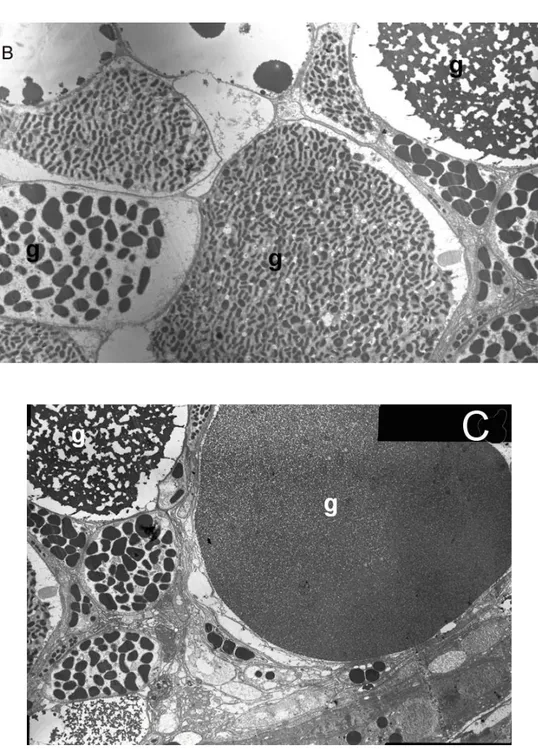
Venomous secretory materials produced within the venom gland were subdivided
into several types according to their locations and structures. These granules vary in
their reactions to the same stain. One type is coarse-grained electron-dense
granules. Another type of granule has a spongy structure and large electron-dense
granules in secretory cells. Still another one is electron lucent with transparent
granules (Figures 5B and 5C). That the secretory granules demonstrate such variety
Figure 1. (A) General view of transverse section of E. mingrelicus’s telson consisting
of a pair of venom glands. The two glands were segregated within the telson by
gross striated muscle bundles; the outer surfaces of venom glands are covered by
cuticle. (B) One of the venom glands. Muscle bundles converge to regions where the
venom glands contact telson cuticle. Cuticle (c), muscle tissue (m), secretory
epithelium (se) and secretion granules are visible (167x). (C) The telson cuticle
consists of exocuticle that is a homogeneous layer and endocuticle composed of
lamellar layers. There are some hemolymph vessels and cuticle canals in the
endocuticle. The cytoplasm of glandular epithelium is filled with many secretory
granules of different sizes, shapes and densities. (D) In the region where cuticle and
gland merge, there are two layers of cuboidal cells (cc) between cuticle (c) and
Figure 2. Attachment region of muscle bundles and telson cuticle. (A) Muscle
bundles joined to telson cuticle (c). (B) The attachment is mediated by intercalated
tendons (t) (2,652x). (C) Intercalated tendons, muscle myofibrils (mf) and cuticle at
higher magnification (7,200x).
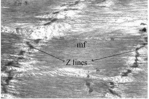
Figure 3. Two Z lines and myofibrils (mf) are shown in muscle layer in a longitudinal
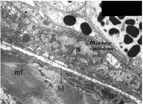
Figure 4. The muscle layer and glandular epithelium are separated by basal lamina
(bl). Secretory cells present a nucleus (n) placed close to their basal portion, while
Figure 5. (A) Thick muscle layer, glandular epithelium, beginning of lumen, glandular
epithelium made up of simple columnar secretory cells and their cytoplasm filled with
many secretory granules of different sizes, shapes and densities under light
microscope (397x). (B, C) The different granules (g) in secretory cells under TEM (B:
DISCUSSION
The venom apparatus of E. mingrelicus is composed of a pair of venom glands
producing the venom that is, in turn, injected by stingers. The venom apparatus of Leiurus quinquestriatus (Hemprich et Ehrenberg, 1827) is composed of two
completely separate but similar glands, each with its own canal (18). The differentiation of the venom glands into lobes was observed by Kanwar et al. (10) in Buthus tamulus gland where the glands were divided longitudinally into parts by a septum. Quiroga et al. (17) studied the venom gland histology in Tityus caripitensis (Quiroga, 1988) adult females. The venom gland of T. caripitensis consists of two
ovoid lobes that fill the vesicle except for a small cavity where the venom
accumulates; the cavity continues distally into an excretory duct. Similarly, our histological results showed that E. mingrelicus presents two distinct venom glands
that were completely separate but similar to each other, and each venom gland
opened separately into its own venom duct.
The scorpion needs to quickly eject the poison produced in its venom glands or inject
it into its victim. For this, each venom gland is surrounded by a striated muscle layer.
Striated muscle fibers vary in their biochemistry, ultrastructure and manner of
contraction. Some undergo a slow, continuous contraction (type I) and are
fatigue-resistant, while others undergo a fast contraction (type II) for a short period of time
and fatigue quickly. According to this classification, type I is the red muscle tissue
and type II is the white muscle tissue. Type I has ultrastructurally regular Z lines and
abundant mitochondria. Type II has irregular Z lines, M lines and H bands that cannot
be distinguished and has few mitochondria (2). In our study, as clearly visible under
light and electron microscopy, the striated muscles surrounding the venom gland of E. mingrelicus have irregular Z lines and M lines, whereas H bands cannot be
distinguished. Besides, this muscle tissue is white. When these characteristics are
taken into consideration, the muscle tissue of the venom gland is included in type II.
Given their short and rapid contraction characteristics, type II muscles offer an
advantage to scorpions because they can inject their venom into their victims as
soon as they catch them.
The secretory epithelium of E. mingrelicus is a simple glandular epithelial type (type I
narrow lumen in the center of the venom gland. Pawlowsky (14, 15) reported on six
of seven known families and found that the morphology followed a generalized
scheme, with the major differences consisting of the presence versus the absence of
folds in the secretory epithelium. Two main types were listed: type I (primitive gland)
possesses a smooth and indented epithelium and type II (complex gland) presents
true folds.
Quiroga et al. (17) determined that T. caripitensis’s venom glands are made of a
simple, pseudostratified epithelium. The epithelium contains secretory cells that have
either coarse-grained or thin granules, basal cells or nonsecretory cells. Taib and Jarrar (18) reported that the secretory epithelium of L. quinquestriatus venom glands
is made up of three cell types: venom-producing cells, mucous cells and supporting cells. The venom apparatus of Centruroides sculpturatus (Ewing) was studied under
light and electron microscopy by Mazurkiewicz and Bertke (12). Each of the paired
glands is lined by secretory epithelium made up of a single layer of columnar cells.
The secretory products were observed in single epithelium cells and in the gland lumen. Similarly, we found that E. mingrelicus venom gland is composed of different
cell types. One type is the venom-producing cells that have several granules of
different sizes, shapes and electron densities. Another type, the supporting cells, is
found between glandular epithelium and cuticle or between glandular epithelium and
muscle bundles. Still another type is goblet cells, found among secretory epithelium
cells, whose function is to secrete mucus. To summarize, it is possible to mention
three types of cells in E. mingrelicus venom glands.
Taib and Jarrar (18) studied L. quinquestriatus venom glands using histochemical
methods and at least five types of granular secretory products were determined
inside the cells. As it can clearly be seen by light and electron microscopy, there are
granules with different morphologies and different locations in the secretory cells.
This is a plain proof that the venom secretion which has a complex structure is the
combination of many secretion products.
In the present study, E. mingrelicus venom glands were studied in terms of their
histology and ultrastructure. The venom gland structural aspects registered by light
and electron transmission microscopy will constitute a basis for future research on
REFERENCES
1 BROWNELL P., POLIS G. Introduction. In: BROWNELL PH., POLIS GA. Eds. Scorpion biology and research. New York: Oxford University Press, 2001. p.3-12. 2 CEBESOY S., AYVALI C. Myotis myotis 'in ( Borkhausen) esas uçma kaslarında
morfolojik ve histokimyasal arastırmalar. Gazi Üniv. Fen Bil. Enst. Derg., 2003,16,
245-52.
3 CORZO G., ESCOUBAS P., VILLEGAS E., BARNHAM KJ., HE W., NORTON RS.,
NAKAJIMA T. Characterization of unique amphipathic antimicrobial peptides from venom of the scorpion Pandinus imperator. Biochem. J., 2001, 359, 35-45.
4 FET V., BRAUNWALDER ME. The scorpions (Arachnida: Scorpiones) of the Aegean area: current problems in taxonomy and biogeography. Belg. J. Zool., 2000,
130, 17-22.
5 FET V., KARATAS AY., FET EV., KARATAS A. First data on the molecular
phylogeny of Euscorpius (Scorpiones: Euscorpiidae) from Turkey. Zool. ZH., 2003,
82, 1518-21.
6 FROY O., SAGIV T., POREH M., URBACH D., ZILBERBERG N., GUREVITZ M.
Dynamic diversification from a putative common ancestor of scorpion toxins affecting sodium, potassium, and chloride channels. J. Mol. Evol., 1999, 48, 187-96.
7 HALSE SA., PRIDEAUX PL., COCKSON A., ZWICKY KT. Observations on the morphology and histochemistry of the venom glands of a scorpion, Urodacus novaehollandiae Peters (Scorpionidae). Aust. J. Zool., 1980, 28, 185-94.
8 HAYAT MA. Principles and techniques of electron microscopy. London: Edward
Arlond, 1981.
9 KANWAR U., NAGPAL N. Studies on the secretory cells in the glands of the scorpions, Heterometrus scaber and Buthus hendersoni. Toxicon, 1983, 3, 207-10.
10 KANWAR U., SHARMA A., NAGPAL N. Morphological and cytochemical studies
on the venom secreting cells of the scorpion Buthus tamulus. J. Anim. Morphol.
Physiol., 1981, 28, 206-9.
11 KEEGAN LH., LOCKWOOD WR. Secretory epithelium in venom glands of two species of scorpion of the genus Centruroides Marx. Am. J. Trop. Med. Hyg., 1971,
12 MAZURKIEWICZ JE., BERTKE EM. Ultrastructure of the venom gland of the scorpion, Centruroides sculpturatus (Ewing). J. Morphol., 1972, 137, 352-83.
13 MOERMAN L., BOSTEELS S., NOPPE W., WILLEMS J., CLYNEN E., SCHOOFS
L., THERVISSEN K., TYGAT J., VAN ELDERE J., VAN DER WALT J., VERDENCK
F. Antibacterial and antifungal properties of alpha-helical, cationic peptides in the venom of scorpions from southern Africa. Eur. J. Biochem., 2002, 268, 4799-810.
14 PAWLOWSKY EN. Skorpiontomische Mitteilungen. I. Ein Beitrag zur Morphologie der Giftdrüsen der Skorpione. Z. Wiss. Zool., 1913, 105, 157-77.
15 PAWLOWSKY EN. Studies on the organization and development of scorpions. Quart. J. Micr. Sci., 1924, 68, 615-40.
16 POSSANI LD., BECERIL B., DELEPIERRE M., TYTGAT J. Scorpion toxins
specific for Na+-channels. Eur. J. Biochem., 1999, 264, 287-300.
17 QUIROGA M., MARVAL MJ., PARRILLA-ALVAREZ P., SOUSA L. Tityus caripitensis n. sp. Scorpion venom gland histology. Toxicon, 1998, 36, p.1269.
18 TAIB NT., JARRAR BM. Histological and histochemical characterization of the venom apparatus of Palestine yellow scorpion, Leiurus quinquestriatus Hemprich & Ehrenberg 1828. Trop. Zool., 1993, 6, 143-52.
19 TORRES-LARIOS A., GURROLA GB., ZAMUDIO FZ., POSSANI LD. Hadrurin, a
new antimicrobial peptide from the venom of the scorpion Hadrus aztecus. Eur. J.
Biochem., 2000, 267, 5023-31.
20 ZAMUDIO FZ., GURROLA GB., AREVALO C., SRREEKUMAR R., WALKER JW.,
VALDIVIA HH., POSSANI LD. Primary structure and synthesis of imperatoxin A
(IpTxa) a peptide activator of Ca2+ release channels/ryanodine receptors. FEBS Lett.,
1997, 405, 385-9.
21 ZENG XC., PENG F., LUO F., ZHU SY., LIU H., LI WX. Molecular cloning and
characterization of four scorpions K+ channel toxins: a new subfamily of venom
peptides (alpha-KTx-14) and genomic analysis of a member. Biochimie, 2001, 83,
883-9.
22 ZENG XC., WANG SX., ZHU Y., ZHU SY., LI WX. Identification and functional