UNIVERSIDADE TÉCNICA DE LISBOA
FACULDADE DE MOTRICIDADE HUMANA
Indicadores Neuromusculares de Recuperação da Funcionalidade do Joelho Após Cirurgia do Ligamento Cruzado Anterior
Dissertação elaborada com vista à obtenção do Grau de Doutor no ramo de Motricidade Humana na especialidade de Fisioterapia
Orientador: Professor Doutor Pedro Luís Camecelha de Pezarat Correia Co-orientador: Professor Doutor Jan Maria Hendrick Cabri
Júri Presidente
Reitor da Universidade Técnica de Lisboa
Vogais
Doutor João Paulo Vilas-Boas Soares de Campos, Professor Catedrático da Faculdade de Desporto da Universidade do Porto;
Doutor Pedro Luís Camecelha de Pezarat Correia, Professor Associado com Agregação da Faculdade de Motricidade Humana da Universidade Técnica de Lisboa;
Doutor Jan Maria Hendrick Cabri, Professor Associado com Agregação da Norwegian School of Sports Sciences, Department of Physical Performance, Oslo, Noruega;
Doutora Maria Margarida Marques Rebelo Espanha, Professora Associada da Faculdade de Motricidade Humana da Universidade Técnica de Lisboa;
Doutor Raúl Alexandre Nunes da Silva Oliveira, Professor Auxiliar da Faculdade de Motricidade Humana da Universidade Técnica de Lisboa;
Doutor José Carlos Pereira Pinto Noronha, Médico Ortopedista do Hospital Ordem da Trindade do Porto.
Nuno do Carmo Antunes Cordeiro
UNIVERSIDADE TÉCNICA DE LISBOA
FACULDADE DE MOTRICIDADE HUMANA
Indicadores Neuromusculares de Recuperação da Funcionalidade do Joelho Após Cirurgia do Ligamento Cruzado Anterior
Dissertação elaborada com vista à obtenção do Grau de Doutor no ramo de Motricidade Humana na especialidade de Fisioterapia
Orientador: Professor Doutor Pedro Luís Camecelha de Pezarat Correia
Co-orientador: Professor Doutor Jan Maria Hendrick Cabri
Júri Presidente
Reitor da Universidade Técnica de Lisboa
Vogais
Doutor João Paulo Vilas-Boas Soares de Campos, Professor Catedrático da Faculdade de Desporto da Universidade do Porto;
Doutor Pedro Luís Camecelha de Pezarat Correia, Professor Associado com Agregação da Faculdade de Motricidade Humana da Universidade Técnica de Lisboa;
Doutor Jan Maria Hendrick Cabri, Professor Associado com Agregação da Norwegian School of Sports Sciences, Department of Physical Performance, Oslo, Noruega;
Doutora Maria Margarida Marques Rebelo Espanha, Professora Associada da Faculdade de Motricidade Humana da Universidade Técnica de Lisboa;
Doutor Raúl Alexandre Nunes da Silva Oliveira, Professor Auxiliar da Faculdade de Motricidade Humana da Universidade Técnica de Lisboa;
Doutor José Carlos Pereira Pinto Noronha, Médico Ortopedista do Hospital Ordem da Trindade do Porto.
Agradecimentos
Para a concretização desta Tese foram co-responsáveis várias pessoas, relativamente às quais estou muito grato:
Ao Professor Doutor Pedro Pezarat Correia, é um enorme privilégio ser orientado por tão Distinto Pedagogo que está sempre presente em todos os passos do trabalho. Pela forma competente com que sempre me orientou, por todos os ensinamentos que me transmitiu (que desde o primeiro momento foram preciosos) e pela forma que os mesmos me influenciam quer pessoal quer profissionalmente. A ele o meu mais profundo agradecimento.
Ao Professor Doutor Jan Cabri, por ter acreditado no projecto e pela preciosa ajuda que deu, especialmente por ter partilhado connosco a sua enorme experiência na recolha de dados. A ele muito obrigado e votos de rápidas melhoras.
Aos Professores Doutores Orlando Jesus Fernandes e Nelson Cortes, o seu contributo na realização dos trabalhos empíricos foi imprescindível, a sua actuação, sem dúvida, permitiu a realização dessa parte do trabalho. Obrigado amigos, um abraço.
Ao Professor Doutor João António Neves Gil, também o seu contributo na realização deste trabalho foi determinante. Infelizmente o seu final de vida precoce não permitiu que
chegasse a ver o trabalho final, que decerto (desculpem a ousadia) iria apreciar… Até sempre
Terapeuta João Gil.
Aos agentes médicos e desportivos que tornaram possíveis as amostras em estudo e aos elementos que compõem as amostras, sem eles este trabalho não existia mesmo. A todos eles, desculpem e obrigado.
A toda a minha família, que sempre me apoiou. E em especial à Susel, por todo o conforto e suporte que me deu, o que também tornou possível este trabalho. Obrigado a todos.
Lista de abreviaturas
º/s – Graus por Segundo;
ACL – Anterior Cruciate Ligament;
ACLR – Anterior Cruciate Ligament reconstruction; Ang. – Ângulo de obtenção do peak torque;
BF – Bicipete Femoral ou Bíceps Femoralis; CCI – Coeficiente de Correlação Intraclasse; CG – Control Group;
CLBP - Chronic Low Back Pain; CVM – Contração Voluntária Máxima; EVA – Escala Visual Analógica; EMG – Eletromiografia;
ICC – Intraclass Correlation Coefficient; IT – Ísquio-tibiais
kg/cm2 – Quilograma por Centímetro Quadrado; KOOS – Knee injury and Osteoarthritis Outcome Score; LBP – Low Back Pain;
LCA – Ligamento Cruzado Anterior do Joelho; LCP – Ligamento Cruzado Posterior do Joelho; ms – Milissegundos;
PT – Peak torque;
Rad.s-1 – Radianos por segundo; RF – Recto femoral ou Rectus Femoris; RMS - Root Mean Square;
ROM – Rang of Movement; SNC – Sistema Nervoso Central;
ST – Semitendinoso ou Semitendinosos;
TSK-13 – Tampa Scale for Kinesiophia – 13 Items;
TSK-PT – Escala Tampa de cinesiofobia – versão para língua e cultura portuguesa – 13 items; TSKJ-13 e TSK-J – Escala Tampa de cinesiofobia – 13 items – versão específica de joelho; VAS – Visual Analog Scale;
VL – Vasto Lateral ou Vastus Lateralis;
Resumo Geral
Esta tese centrou-se no estudo de respostas neuromotoras associadas a movimentos rápidos do joelho em cadeia cinética aberta, efetuados por jogadores profissionais de futebol, seis meses após ligamentoplastia ao ligamento cruzado anterior do joelho (LCA).
Foram realizados quatro estudos, dois relacionados com o interesse de medir a cinesiofobia, donde resultou a validação da versão portuguesa da Tampa Scale for Kinesiophobia 13 itens (TSK-13 ou TSK–PT) e outro para a avaliação psicométrica duma versão específica do mesmo instrumento para disfunções do joelho (TSKJ-13). Os restantes dois estudos foram laboratoriais e observaram o padrão de movimento da extensão balística do joelho dum remate de futebol e o perfil isocinético dos movimentos de extensão/flexão a 60º, 180º e 300º/segundo, seis meses após a cirurgia.
A versão portuguesa da TSK-13 apresentou propriedades psicométricas adequadas à medição do grau de cinesiofobia, inclusivamente em condições do joelho quando comparada com a TSKJ-13. Apesar da maior cinesiofobia, a realização dum remate de futebol seis meses após ligamentoplastia apresentou na fase de extensão, um padrão de movimento semelhante ao movimento efetuado por atletas sem história anterior de lesão no joelho. Também a análise do pico de força no perfil isocinético mostrou ser inferior em todas as velocidades e com o ângulo em que acontece o pico de força dos extensores maior para a velocidade mais rápida. Apesar do maior grau de cinesiofobia, menor funcionalidade e maior grau de hipotrofia do quadricipite em especial nos movimentos rápidos, os resultados obtidos apontam para um padrão neuromotor recuperado nestes movimentos, nos atletas seis meses após a sua cirurgia ao LCA.
Abstract
This thesis focused on the neuromuscular pattern obtained in open kinetic chain fast movements, performed for professional soccer players of the Portuguese major soccer league, six months after surgery for Anterior Cruciate Ligament reconstruction (ACLR).
Four studies were performed, two for the intercultural validation of the Tampa Scale for kinesiophobia (TSK-13 or TSK-PT) and psychometric exploration of the same instrument adapted to knee dysfunctions (TSKJ-13). The other studies focused on the instep soccer kick to the ballistic knee extension movement pattern obtained and on the isokinetic profile to the strength parameters on the 60º, 180º and 300º/second knee flexion/extension, six months after ACLR.
The TSK-PT presented adequate psychometrics properties for measure kinesiophobia, even better on the knee dysfunction than TSKJ-13. With higher kinesiophobia, the ballistic extension on the instep soccer kick six months after ACLR presented similar movement pattern to the movement performed for players without prior knee injury. On the isokinetic profile, the obtained peak torque (PT) values were lower for the ACLR players in all velocities in both movements and the angle of PT obtainment was larger on the faster movement velocity. But even with higher kinesophobia level, lower functionality level and quadriceps hypotrophy, the results are in agreement with the movement pattern recuperation on the fast knee movements six months after ACLR.
Índice
Agradecimentos ... v
Lista de abreviaturas ... vii
Resumo Geral ... ix
Abstract ... xi
Capitulo 1 – Introdução ... 15
Capítulo 2 – Portuguese Language Version of the Tampa Scale for Kinesiophobia [13 Items] ... 31
Capítulo 3 – Utilização da Tampa Scale for Kinesiophobia 13 Itens Após Ligamentoplastia do Cruzado Anterior do Joelho: Versão Genérica Versus Versão de Condição Especifica ... 39
Capítulo 4 – Dynamic Knee Stability and Ballistic Knee Movement after ACL Reconstruction: An Application on Instep Soccer Kick ... 49
Capítulo 5 – Specific Isokinetic Angle Peak Torque of Fast Angular Knee Velocities on Soccer Players after Anterior Cruciate Ligament Reconstruction ... 63
Capítulo 6 – Discussão Geral ... 75
Capítulo 7 – Conclusões ... 85
ENQUADRAMENTO DA TESE
A articulação do joelho é uma das mais complexas estruturas anatómicas do corpo humano, sendo sujeita a uma frequente e intensa solicitação, permite uma grande amplitude articular e é sujeita a grandes cargas mecânicas.
No joelho, distinguem-se duas subarticulações: a fémuro-patelar e a fémuro-tibial. A articulação fémuro-tibial, que articula os côndilos do fémur com as cavidades glenoideias da tíbia, é uma articulação do tipo bicondilomeniscartrose, enquanto a articulação fémuro-patelar define-se como uma trocleartrose.
A articulação do joelho está essencialmente preparada para funcionar no plano sagital, em sequências de movimentos de extensão e de flexão. O músculo extensor do joelho é o quadricipite, formado por quatro segmentos musculares: o crural, o vasto externo e o vasto interno (mono-articulares) e o bi-articular recto femoral. Os flexores do joelho são os músculos isquiotibiais: bicípite crural, gracilis, semitendinoso, semimembranoso e o costureiro. Também participam na flexão, o poplíteo e os gémeos.
Como meio passivo de suporte na articulação do joelho temos a cápsula articular, que é um delgado e potente invólucro fibroso, que envolve a extremidade inferior do fémur e a extremidade superior da tíbia, mantendo os dois segmentos em contacto um com o outro. Na face posterior do joelho, esta cápsula é membranosa, tem as suas inserções no contorno da superfície articular dos côndilos fémurais e linha intercondiliana. Lateralmente ao joelho, fixa-se ao longo das linhas obliquas, estendendo-se até à tuberosidade anterior da tíbia. Uma grande parte da sua área anterior é formada por: tendão quadricipital, patela e tendão patelar. A parede capsular é dupla e profunda devido à membrana sinovial que a reveste internamente [1].
O joelho é muito rico em ligamentos, sendo este facto devido à necessidade de existirem fortes estruturas passivas que dêem direcção ao movimento [2]. Como ligamentos extra-capsulares temos os ligamentos laterais. Estes reforçam a cápsula articular dos lados interno e externo da articulação, assegurando assim a estabilidade lateral quando joelho está em extensão. O ligamento lateral interno estende-se do côndilo interno à face superior da tíbia, a sua camada mais profunda liga-se à cápsula articular e ao menisco interno. O ligamento lateral externo estende-se do côndilo externo à cabeça da peróneo [1]. Os ligamentos laterais aumentam de tensão durante a extensão e diminuem-na durante a flexão [2].
estiramento em todos os movimentos, no entanto, obtêm estiramento máximo com o joelho em extensão. O LCP insere-se atrás da espinha da tíbia, dirigindo-se anteriormente para a face interna do côndilo interno do fémur, impedindo assim, o deslocamento posterior da tíbia sobre o fémur. O LCA, insere-se na superfície pré-espinhal da tíbia, dirige-se posteriormente para a face externa do côndilo externo do fémur e tem uma acção inversa ao LCP, ou seja, impede o deslocamento anterior da tíbia sobre o fémur [1, 2]. Apesar de intra-articular e extra-sinovial, de entre as estruturas ligamentares, o LCA é a única sem qualquer inserção capsular. Na tíbia, insere-se numa fosseta localizada anteriormente e externamente à espinha tibial anterior, por trás do corno anterior do menisco externo, ao qual adere por uma pequena expansão (nalguns casos, feixes da parte posterior da inserção tibial do LCA aderem ao corno posterior do menisco externo). A sua área de inserção tibial é oval, e tem cerca de 3 cm de diâmetro antero-posterior e no fémur insere-se na fase média do côndilo fémural externo (numa localização muito posterior), com uma orientação quase vertical, curvilínea, convexa posteriormente e paralela ao rebordo articular posterior do côndilo fémural externo. Esta área de inserção tem cerca de 2 cm2 [1].
O número de feixes que constituem o LCA é um ponto controverso. Há autores que defendem a existência de um único feixe constituído por fascículos. Outros autores defendem a existência de dois feixes. No entanto, estudos efectuados no cadáver por dissecação do LCA, confirmam a existência de dois feixes sem distinção entre si [3].
A orientação deste ligamento desde a inserção fémural à tibial é antero-interna e distal. Devido à orientação das inserções (sagital no fémur e transversal na tíbia) há uma torção externa dos fascículos iniciada cerca dos 5mm da inserção fémural, condicionando assim desde a flexão à extensão do joelho uma certa tensão permanente do LCA [4]. Procurando a unanimidade, definimos que o LCA é assim formado por um conjunto de fascículos reunidos em dois feixes, conjectura mais frequentemente vista na bibliografia. Um feixe antero-interno (A.I) com inserção fémural (proximal) e inserção distal antero-interna na tíbia e um segundo feixe, postero-externo (P.E), com origem na parte mais posterior e distal da inserção fémural que termina na zona postero-externa da inserção tibial. Quando o joelho está em extensão ambos os feixes estão tensos. Quando o joelho está em flexão a inserção fémural do LCA torna-se mais horizontal, o que mantêm o feixe A.I em tensão e o feixe P.E em relaxamento [3]. Em resumo, na extensão do joelho ambos os feixes do LCA estão em tensão, a qual reduz com a flexão mais acentuadamente no feixe P.E..
por fibrocartilagem e funciona como estabilizador em todos os movimentos do joelho (nas três rotações e nas três translações) [1]. Tem a seu cargo 90% do controlo da translação anterior da tíbia sobre o fémur, quando se realiza flexão entre os 30 e os 90° [2]. Permite ainda suportar algum peso antes da rutura, tem uma resistência mecânica de 50kg/cm2 [3] e a sua tolerância à rutura está intimamente ligada à sua capacidade visco-elástica. Apesar de todo o comportamento de proteção dado pela arquitetura anatomo-fisiológica e pela componente reflexa, a rutura do LCA é uma lesão muito comum, em especial no futebol [5]. Em situações de procedimento cirúrgico, é necessário substituir eficazmente o LCA com uma plastia sólida. Poderão ser consideradas diversas estruturas como fonte de autoenxerto (plastia), sendo o uso do tendão rótuliano com osso nas extremidades uma das mais utilizadas, sendo essa cirurgia conhecida por osso-tendão-osso [6]. A experiência permite a realização desta cirurgia em menos de uma hora, estando atualmente de tal forma otimizada que alguns autores defendem inclusivamente que o sucesso da recuperação completa dependerá mais da fisioterapia pós operatória do que propriamente da cirurgia [3, 7, 8].
Uma recuperação bem orientada, fundamentada em bases biomecânicas e respeitando o normal processo de ligamentação, permite a obtenção de um neo-ligamento saudável, com características mecânicas e histológicas compatíveis com função quase normal, até mesmo na atividade desportiva [6].
A retoma anatómica da arquitetura do neo-ligamento não é suficiente, tendo a fisioterapia papel preponderante na reeducação propriocetiva, imprescindível ao bom funcionamento da função neuromuscular e base fundamental para a normal função articular após a cirurgia [6]. Durante todo processo de recuperação, qualquer falha nesta função poderá comprometer o resultado final.
No âmbito dos processos coordenativos envolvidos é particularmente importante a coordenação entre músculos agonistas e antagonistas. Existem dois tipos de coordenação da musculatura agonista/antagonista: uma presente em movimentos mais lentos e em situações em que o sistema de controlo procura privilegiar a estabilidade articular, que utiliza a contração simultânea dos dois grupos antagonistas, designada por cocontração; a outra, característica de movimentos mais rápidos, caracteriza-se por um padrão fásico constituído por ativações curtas e bem definidos dos músculos agonistas e antagonistas, organizado através de um processo de inervação recíproca, com o início da contração do músculo antagonista a ocorrer muito chegado ao final da contração do agonista, de tal forma que o período de contração simultânea dos dois grupos antagonistas é muito reduzido [11]. Apesar de o padrão fásico ter sido no século passado amplamente estudado em movimentos do membro superior, ele manifesta-se também em movimentos do membro inferior, nomeadamente no joelho [12, 13].
Num regime de cocontração, a ação frenadora que a musculatura antagonista desencadeia no início do movimento não permite movimentos rápidos servindo essencialmente para proteger a articulação [14, 15]. No caso de tarefas não muito treinadas,
logo pouco “aprendidas”, mesmo que a intenção para a realização da tarefa seja um
movimento rápido, o sistema nervoso central usa a cocontração como medida de segurança e estabilidade para a articulação, obviamente com prejuízo da velocidade de execução. À medida que determinada tarefa começa a ser conhecida, este regime de segurança dá lugar a um tipo de coordenação intermuscular mais característico de inervação recíproca, compatível com a grande velocidade a que o movimento é realizado [12, 13].
Os clássicos protocolos de recuperação da ligamentoplastia do LCA apresentam em comum o entendimento de que a reabilitação propriocetiva é fundamental no sucesso de um programa como medida de treino de coordenação intermuscular [20]. Assim, o trabalho propriocetivo está presente em todos os programas, existindo variações de programa para programa que estão normalmente associadas a diferenças na fase em que este trabalho específico é implementado. Para a fisioterapia, as vinte e quatro semanas após a cirurgia constituem um tempo comummente aceite para o início da retoma de todas as atividades funcionais [6] incluindo as desportivas e de elevada carga cinética [20]. Subsistem no entanto alguma incerteza relativa à total reorganização, nesta fase, do sistema de controlo motor de modo a garantir o movimento normal e, por outro lado a proteção efetiva do neo-ligamento.
Torna-se portanto necessário sistematizar indicadores neuromusculares que permitam a perceção duma correta função neuromuscular, condição essencial para a obtenção de um normal grau de estabilidade no período pós cirúrgico. É conhecida a capacidade de reinervação do enxerto patelar colocado cirurgicamente [21] e, por isso, consideramos existir um perfil de função neuromuscular próprio para o período pós reconstrução, que passe a considerar também informações vindas do novo ligamento. Vários autores têm estudado diferentes características desta função em período pré reconstrução do LCA [22, 23], assistindo-se ultimamente a uma maior frequência ao seu estudo em período pós cirúrgico [24] utilizando indicadores de tónus muscular, atividade muscular, força do quadricipite e coordenação intermuscular agonista-antagonista entre o quadricipite crural e os músculos isquiotibiais.
Uma tarefa comparável a um remate de futebol exige a atividade dos músculos com influência no joelho. Esta tarefa permite observar a atividade fásica do grupo muscular extensor e a forma como este se coordena com o grupo antagonista flexor. Pensamos que o padrão de coordenação observado esteja relacionado com a velocidade angular imposta à articulação do joelho, na sequência da tarefa a realizar.
intensa quanto maior for a ação a desacelerar, sendo que, nos indivíduos não treinados não exige atividade acima dos 60%, mas, em sujeitos treinados a mesma pode ir até aos 80% da CVM [25].
Num período pós-cirúrgico em que a aferência neuromuscular está altamente afetada [29], o silêncio neuromuscular proveniente do novo ligamento é percebido pelo sistema de controlo motor, obrigando a uma resposta dos níveis superiores mais cuidada, no que respeita aos graus de liberdade neuromuscular concedidos à articulação. Assim, qualquer tarefa funcional mais exigente é caracterizada como tarefa desconhecida, logo é mais controlada e apresenta-se com menor grau de liberdade [12]. Sendo o remate de futebol um movimento rápido da articulação do joelho, o sistema de controlo motor deve dar primazia a um padrão motor do tipo de inervação recíproca mesmo que o controlo neuromuscular possa ser diferente do mecanismo existente antes da lesão, ele deverá reestruturar-se de forma a permitir um padrão de movimento desse tipo e torná-lo possível no final do período de recuperação e, tal como anteriormente referido, integrando as informações vindas do novo ligamento na reestruturada rede reflexa de controlo de movimento.
Também não podemos esquecer o impacto psicológico que a lesão e a cirurgia provocam e que pode adicionalmente condicionar a forma de funcionamento da articulação intervencionada e contribuir para que o nível funcional pré-lesão não seja recuperado [30]. O medo de executar movimento deve ser encarado como uma complicação deste tipo intervenção, devendo ser considerado esse fator pelos profissionais que lidam com esta tipologia, particularmente pelo fisioterapeuta, por ser o responsável pelo restabelecimento do movimento normal. Este medo poderá traduzir uma alteração do padrão de coordenação agonista/antagonista presente nos movimentos balísticos, dando lugar a uma maior cocontração muscular que, como vimos, é estratégia normalmente utilizada pelo SNC para produzir aumento de estabilidade articular em situações de maior insegurança nas tarefas [31].
A Tampa Scale for Kinesiophobia [32] permite a graduação do nível do medo e do conforto para desempenhar movimento, possibilitando a ponderação deste fator em cada individuo e, consequentemente, acautelar a sua influencia para o movimento [30].
do mesmo, quer para inferir a magnitude de fraqueza [34, 35], quer para descortinar quais os mecanismos que conduzem a essa fraqueza [36-40].
OBJETIVOS
Apesar de muito ter sido já debatido sobre o estado funcional de indivíduos após substituição cirúrgica do LCA, não existe conhecimento em como tarefas comuns e importantes na prática do futebol estão a ser realizadas pelos profissionais que o praticam, aquando da sua reintegração na atividade desportiva ao nível pré-lesional, principalmente nas tarefas efetuadas a grande velocidade angular e em cadeia cinética aberta. O padrão de movimento obtido na extensão balística do joelho no remate do futebol é disso um exemplo, bem como a caracterização de indicadores de confiança para a execução de movimentos com o joelho e diferentes abordagens na determinação da força da musculatura com influência na articulação em função da modalidade desportiva praticada. O conhecimento do comportamento destes indicadores no período pós-cirúrgico, torna a decisão para a completa retoma das atividades do sujeito a um nível pré-lesional mais esclarecida, já que habitualmente esta decisão resulta em função do tempo decorrido desde a cirurgia, permitindo depois a adaptação e controlo dos programas de fisioterapia a aplicar nestas condições.
Assim, o objetivo geral desta tese centrou-se na caracterização de indicadores neuromusculares em atletas profissionais de futebol, seis meses após a sua ligamentoplastia ao LCA. A verificar-se que estes indicadores se encontram alterados no período pós-cirúrgico, essa alteração é passível de causar alterações nos padrões de movimento, adiando a decisão para a completa retoma das atividades do sujeito a um nível pré-lesional.
Do que precede, delineámos como objetivos específicos para a presente tese:
- A tradução semântica e cultural da Tampa Scale for Kinesiophobia (TSK-PT) para a língua e cultura portuguesa e avaliação das capacidades psicométricas da versão traduzida numa amostra de indivíduos com dor lombar crónica. Essa tradução é necessária para que a TSK-PT possa ser um instrumento de avaliação do grau de cinesiofobia válido, fiável e disponível para a população portuguesa, para utilização posterior em diferentes condições crónicas, nomeadamente nas disfunções do joelho;
- Após a validação da versão genérica da TSK-PT e relacionado com a avaliação do grau de cinesiofobia em disfunções do joelho, pretende-se fazer a comparação das capacidades psicométricas da TSK-PT aplicada a uma amostra de indivíduos em processo de fisioterapia para recuperação de ligamentoplastia ao LCA, com as obtidas da aplicação na mesma amostra, duma versão por nós adaptada especificamente para condições do joelho (TSK-J);
padrão de movimento obtido na extensão balística do joelho integrada num remate de futebol a partir da posição de parado, entre atletas da liga portuguesa de futebol profissional, seis meses após a sua ligamentoplastia do LCA na perna dominante e, atletas do mesmo nível competitivo sem quaisquer problemas anteriores no joelho;
- Por ultimo, após o conhecimento do tipo de padrão de movimento obtido na extensão balística do joelho e dirigindo o interesse no estudo do perfil de força dos músculos com influência no joelho em função da modalidade desportiva praticada e nos gestos desportivos nela executados, é nossa intenção comparar o perfil de força, com relevo do máximo momento de força e ângulo articular em que o mesmo acontece, nos movimentos extensão e flexão isocinética do joelho a 60º, 180º e 300º/segundo, entre atletas da liga portuguesa de futebol profissional seis meses após a sua ligamentoplastia do LCA na perna dominante e, atletas de igual nível competitivo mas sem anteriores registos de problemas no joelho.
ESTRUTURA DA TESE
O capítulo 1 inclui o enquadramento do tema e os objetivos da tese.
Os capítulos 2 e 3, apresentam respetivamente os processos de validação da TSK-PT e a comparação da exploração psicométrica da TSK-PT e da TSKJ-13 quando aplicadas em sujeitos em período pós-ligamentoplastia do LCA.
O capítulo 4 versa sobre a comparação do padrão de movimento obtido na extensão balística do joelho que serve de base a um remate de futebol, efetuado por jogadores da liga portuguesa de futebol profissional, seis meses após ligamentoplastia do LCA do seu membro dominante e outros atletas do mesmo nível competitivo sem história anterior de lesão do joelho.
O capítulo 5 compara os perfis de força obtidos nos extensores e flexores do joelho em condições isocinéticas de movimento a 60º, 180º e 300º/s, considerando o momento máximo de força e o ângulo articular em que o mesmo acontece, produzidos por jogadores da liga portuguesa de futebol profissional, seis meses após ligamentoplastia do LCA e indivíduos da mesma competição sem história anterior de lesão do joelho.
O capítulo 6 apresenta a discussão geral dos resultados e o Capítulo 7 as conclusões da tese.
os capítulos 2, 3, 4 e 5, apresentam formatações do texto de acordo com as revistas a que foram submetidos para publicação.
Quadro sinóptico
Indicadores de funcionalidade do joelho após ligamentoplastia do LCA
Capitulo 2 Capitulo 3 Capitulo 4 Capitulo 5
Validação semântica e cultural do instrumento
de medição de cinesiofobia
TSK-PT
Exploração psicocométrica de instrumento de medição
de cinesiofobia especifico do joelho
TSKJ-13
Coordenação intermuscular agonista/antagonista à extensão do joelho num remate de futebol a partir
da posição de parado
Força e estrutura muscular com influência no joelho
Sujeitos com dor lombar crónica (n=166,
teste- reteste n=41)
Sujeitos em recuperação de ligamentoplastia
(n=53)
Futebolistas profissionais: A) – 6 Meses após ligamentoplastia (n=8); B) – Sem qualquer lesão anterior (n=9)
Futebolistas profissionais: A) – 6 Meses após ligamentoplastia (n=15 incluindo os 8 sujeitos do capitulo 4); B) – Sem qualquer lesão anterior (n=69) Variáveis independentes
Nível de dor;
Nível de confiança para o movimento.
Variáveis independentes Nível de confiança para o movimento;
Grau de cinesiofobia dado pela TSK-PT.
Variáveis independentes Situação do sujeito: A - Situação de seis meses
pós-ligamentoplastia ao LCA;
B – Ausência de lesão anterior.
Variáveis independentes Situação do sujeito: A - Situação de seis meses
pós-ligamentoplastia ao LCA;
B – Ausência de lesão anterior.
Variáveis dependentes Grau de cinesiofobia dado pela TSK-PT.
Variáveis dependentes Grau de cinesiofobia dado pela TSKJ-13.
Variáveis dependentes Cinemáticas:
Duração do movimento; Instante do contacto com a bola;
Instante e valor da velocidade angular máxima;
Amplitude de
movimento, de flexão e extensão máxima; Ângulo articular do contato e do pico de velocidade;
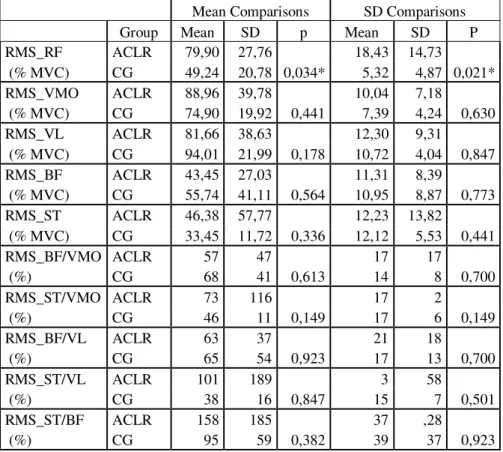
Duração da fase de aceleração/desaceleração; Tempo decorrido desde o pico de velocidade ao contato com a bola. Electromiográficas: RMS – RF, VI e VE; RMS – BF e ST; RMS – BF/VMO; RMS – ST/VMO; RMS – BF/VL; RMS – ST/VL; RMS – ST/BF.
Variáveis dependentes PT Qua 60º/s;
REFERÊNCIAS
1. Testut, L., Compendio de anatomía descriptiva. 1977.
2. Kapandji, A.I., Physiology of the Joints: Volume 2 Lower Limb, 6e. 2010.
3. Noyes, F.R., et al., Intra-articular cruciate reconstruction. I: Perspectives on graft strength, vascularization, and immediate motion after replacement. Clin Orthop Relat Res, 1983(172): p. 71-7.
4. Shelbourne, K.D., D.V. Patel, and D.J. Martini, Classification and management of arthrofibrosis of the knee after anterior cruciate ligament reconstruction. Am J Sports Med, 1996. 24(6): p. 857-62.
5. Roos, H., et al., Soccer after anterior cruciate ligament injury--an incompatible combination? A national survey of incidence and risk factors and a 7-year follow-up of 310 players. Acta Orthop Scand, 1995. 66(2): p. 107-12.
6. Shelbourne, K.D. and D.V. Patel, Rehabilitation after autogenous bone-patellar tendon-bone ACL reconstruction. Instr Course Lect, 1996. 45: p. 263-73.
7. Beynnon, B.D. and R.J. Johnson, Anterior cruciate ligament injury rehabilitation in athletes. Biomechanical considerations. Sports Med, 1996. 22(1): p. 54-64.
8. Brandsson, S., et al., A prospective four- to seven-year follow-up after arthroscopic anterior cruciate ligament reconstruction. Scand J Med Sci Sports, 2001. 11(1): p. 23-7.
9. Sernert, N., et al., Comparison of functional outcome after anterior cruciate ligament reconstruction resulting in low, normal and increased laxity. Scand J Med Sci Sports, 2002. 12(1): p. 47-53.
10. Lund-Hanssen, H., et al., Isokinetic muscle performance in healthy female handball players and players with a unilateral anterior cruciate ligament reconstruction. Scand J Med Sci Sports, 1996. 6(3): p. 172-5.
11. Wadman, W., Denier van der Gon, J., Geuze, R., & Mol, C. , Control of fast goal-directed arm movements. Journal of Human Movements Studies, 1979. 5: p. 3-17. 12. Brown, J.M. and W. Gilleard, Transition from slow to ballistic movement:
development of triphasic electromyogram patterns. Eur J Appl Physiol Occup Physiol, 1991. 63(5): p. 381-6.
13. Cordeiro, N., Pezarat-Correia,P., Fernandes, O., Cabri, J., Agonist/antagonist Pattern and Movement Characteristics During Ballistic Knee Extension 12 Weeks After ACL Reconstruction. Medicine and Science in Sports and Exercise, 2007(No. 5 Supplement): p. pp S264.
14. Markolf, K.L., A. Graff-Radford, and H.C. Amstutz, In vivo knee stability. A quantitative assessment using an instrumented clinical testing apparatus. J Bone Joint Surg Am, 1978. 60(5): p. 664-74.
15. Aagaard, P., et al., Antagonist muscle coactivation during isokinetic knee extension. Scand J Med Sci Sports, 2000. 10(2): p. 58-67.
16. Roos, E.M., Outcome after anterior cruciate ligament reconstruction--a comparison of patients' and surgeons' assessments. Scand J Med Sci Sports, 2001. 11(5): p. 287-91.
17. Zatterstrom, R., et al., Rehabilitation following acute anterior cruciate ligament injuries--a 12-month follow-up of a randomized clinical trial. Scand J Med Sci Sports, 2000. 10(3): p. 156-63.
19. Kvist, J. and J. Gillquist, Sagittal plane knee translation and electromyographic activity during closed and open kinetic chain exercises in anterior cruciate ligament-deficient patients and control subjects. Am J Sports Med, 2001. 29(1): p. 72-82. 20. Shaw, T., Accelerated Reahabilitation Follwing Anterior Cruciate Ligament
Reconstruction. Physical Therapy in Sports, 2002. 3(1): p. 19-26.
21. Barrack, R.L., H.B. Skinner, and S.L. Buckley, Proprioception in the anterior cruciate deficient knee. Am J Sports Med, 1989. 17(1): p. 1-6.
22. Steele, J.R., G.J. Roger, and P.D. Milburn, Reproducibility of knee laxity assessment results using the dynamic cruciate tester. J Sci Med Sport, 1998. 1(4): p. 245-59. 23. Boerboom, A.L., et al., Atypical hamstrings electromyographic activity as a
compensatory mechanism in anterior cruciate ligament deficiency. Knee Surg Sports Traumatol Arthrosc, 2001. 9(4): p. 211-6.
24. Bryant, A.L., J. Kelly, and E. Hohmann, Neuromuscular adaptations and correlates of knee functionality following ACL reconstruction. J Orthop Res, 2008. 26(1): p. 126-35.
25. De Proft, E., Clarys, J., Bollens, E., Cabri, J. and Dufour, W. , Muscle activity in the soccer kick. .Science and Football, 1988: p. 434-440.
26. Kellis, E., Biomechanical characteristics and determinants of instep soccer kick. Journal of Sports Sciences and Medicine, 2007(6): p. 154-165.
27. Lees, A. and L. Nolan, The biomechanics of soccer: a review. J Sports Sci, 1998. 16(3): p. 211-34.
28. Dorge, H.C., et al., EMG activity of the iliopsoas muscle and leg kinetics during the soccer place kick. Scand J Med Sci Sports, 1999. 9(4): p. 195-200.
29. Grigg, P., Peripheral neural mechanisms in proprioception. Journal of Sport Rehabilitation, 1994. 3: p. 2-17.
30. Kvist, J., et al., Fear of re-injury: a hindrance for returning to sports after anterior cruciate ligament reconstruction. Knee Surg Sports Traumatol Arthrosc, 2005. 13(5): p. 393-7.
31. Enoka, R.M., Neural adaptations with chronic physical activity. J Biomech, 1997. 30(5): p. 447-55.
32. Kori SH, M.R., Todd DD, Kinesiophobia: a new view of chronic pain behavior. Pain Manag., 1990. 3: p. 35-43.
33. Brasileiro, J.S., et al., Functional and morphological changes in the quadriceps muscle induced by eccentric training after ACL reconstruction. Rev Bras Fisioter, 2011. 15(4): p. 284-90.
34. Krishnan, C. and G.N. Williams, Factors explaining chronic knee extensor strength deficits after ACL reconstruction. J Orthop Res, 2011. 29(5): p. 633-40.
35. Ingersoll, C.D., et al., Neuromuscular consequences of anterior cruciate ligament injury. Clin Sports Med, 2008. 27(3): p. 383-404, vii.
36. Pua, Y.H., et al., Isokinetic dynamometry in anterior cruciate ligament injury and reconstruction. Ann Acad Med Singapore, 2008. 37(4): p. 330-40.
37. Renstrom, P., et al., Non-contact ACL injuries in female athletes: an International Olympic Committee current concepts statement. Br J Sports Med, 2008. 42(6): p. 394-412.
38. Eitzen, P., ACL deficient potential copers and non-copers reveal different isokinetic quadriceps strength profiles in the early stage after injury. American Journal Sports of Medicine, 2010. 38(17): p. 586
40. Ageberg, E., et al., Knee extension and flexion muscle power after anterior cruciate ligament reconstruction with patellar tendon graft or hamstring tendons graft: a cross-sectional comparison 3 years post surgery. Knee Surg Sports Traumatol Arthrosc, 2009. 17(2): p. 162-9.
41. Guilhem, G., C. Cornu, and A. Guevel, A methodologic approach for normalizing angular work and velocity during isotonic and isokinetic eccentric training. J Athl Train, 2012. 47(2): p. 125-9.
42. Ebert, J.R., et al., Isokinetic knee extensor strength deficit following matrix-induced autologous chondrocyte implantation. Clin Biomech (Bristol, Avon), 2012. 27(6): p. 588-94.
43. Silder, A., D.G. Thelen, and B.C. Heiderscheit, Effects of prior hamstring strain injury on strength, flexibility, and running mechanics. Clin Biomech (Bristol, Avon), 2010. 25(7): p. 681-6.
Capítulo 2 – Portuguese Language Version of the Tampa Scale for
Kinesiophobia [13 Items]
ARTICLE
Portuguese Language Version of the Tampa Scale for
Kinesiophobia [13 Items]
Nuno Cordeiro1, Pedro Pezarat-Correia1, João Gil2 and Jan Cabri3
1
Faculty of Human Kinetics, Center for Research in Physiotherapy, Technical University of Lisbon, Lisbon, Portugal,2Faculty of Economics, Centre for Health Studies and Research, University of Coimbra, Coimbra, Portugal,3Department Physical
Performance, The Norwegian School of Sport Sciences, Oslo, Norway
A
Objectives:The aim of this study was to translate and adapt the Tampa Scale for Kinesiophobia to the Portuguese language and culture [TSK-PT], and validate its use in Portuguese patients with chronic low back pain [CLBP].
Methods: The procedures were performed following published international guidelines. Patients with CLBP were recruited to complete the TSK-PT and visual analog scales [VAS] for pain and for confidence in low back movement. A portion of the same patients also completed a second TSK-PT questionnaire after a seven-day interval to establish test-retest reliability. After three months, all of the original patients completed the questionnaire again to assess its respon-siveness using standardized effect size and standardized response mean.
Results:At baseline, 166 patients with chronic LBP completed the series of questionnaire instruments. After an interval of seven days, forty one patients again completed the series of instruments. Three months after baseline, all 166 patients again completed the questionnaire series. The Cronbach alpha of 0.82 indicated good internal consistency of the TSK-PT total score, and the one-week intraclass correlation coefficient of 0.99 indicated exceptional test-retest reliability. A three-factor solution could be found on factorial analysis. There were significant correlations between the TSK-PT total score and both VAS Pain and VAS Confidence scores. The standardized effect size of the TSK-PT was 0.50 and the standardized response mean was 0.67.
Conclusions:The TSK-PT has proven to be a valid and reliable tool in the assessment of kinesiophobia in patients with CLBP.
K: Kinesiophobia, pain, fear of movement
INTRODUCTION
Self-reporting measures are very common instru-ments for exploring different impacts of impairinstru-ments in bodily functions. Kinesiophobia is defined as an excessive and debilitating fear of physical movement and activity, resulting from a feeling of vulnerability to painful injury or reinjury (1). It is a type of impair-ment that simultaneously affects neurobiological and psychological pain factors, as well as all components of movement confidence (2). The fear of movement is an important factor in increasing painful symptoms associated with chronic low back pain [CLBP]. Avoid-ing movement maintains incorrect posture, exagger-ates tension, and extends the impairment (3). The
Tampa Scale for Kinesiophobia [TSK] (4) was specifi-cally developed for the assessment of kinesiophobia (1) and collects information on the degree of comfort, security, and preparation for movement.
The original English version of the TSK is a set of 17 questions, using a four-point Likert scale, assessing the subjective perception of the subjects related to their security and confidence to perform movements. The sum of the scores obtained on each issue will typically range between 17 and 68, where higher scores represent greater levels of perceived fear. Short versions of the TSK, such as the TSK-13 or TSK-11 items, are also used (5). It has been postulated that the use of the TSK without the four inverse items
Journal of Musculoskeletal Pain, Vol. 21(1), 2013 © 2013 Informa Healthcare USA, Inc. ISSN: 1058-2452 print / 1540-7012 online DOI: 10.3109/10582452.2012.762966
J Muscoskeletal Pain Downloaded from informahealthcare.com by 85.138.99.67 on 04/29/13
[items 4, 8, 12, and 16] gives better psychometric measures and has the advantage of being a shorter version (6). These factors were significant in the decision to opt for the shorter 13-item TSK for Por-tuguese validation. The TSK has been validated for different cultures, including Norwegian (7), Dutch (2), Swedish (8), French (9), and Brazilian (10). However, no adaptation and validation of the TSK for the Portuguese culture could be found in the lit-erature and there is no other self-report questionnaire validated for the Portuguese language and culture for measuring kinesiophobia.
The aim of this study was to cross-culturally adapt the TSK-13 to the Portuguese-speaking culture, using a sample of patients with CLBP, in order to explore its reliability and validity, including its factor structure and responsiveness.
MATERIALS AND METHODS
Ethical Oversight
The study protocol was approved by the ethics com-mittee of the Scientific Board of the Faculty of Human Kinetics– Technical University of Lisbon.
Cross-Cultural Adaptation
The cross-cultural process was carried out following established guidelines (11). Prior to the validation process, the original English TSK-13 items were translated to a Portuguese version [TSK-PT] by two native Portuguese-speaking independent professional translators [T1 TSK-PT and T2 TSK-PT] at the Language Centre of the Polytechnic Institute of Castelo Branco. After combining these into a single Portuguese version [T12 TSK-PT], two other pro-fessional translators, without previous contact with the original version of TSK, performed the T12 TSK-PT back-translation to English. A review group consisting of two physiotherapists [from the research group, one of them a methodologist], two of the pro-fessional translators, and one language expert then agreed on a pre-final version. In order to ensure that the pre-final TSK-PT version was comprehensi-ble, it was also reviewed and tested on a panel of 10 patients with CLBP. In their opinion, the TSK-PT was a questionnaire that was short, quick and easy to answer, understandable, useful, and suitable for the target population. They unanimously found that the language was simple, clear, and colloquial. The questionnaire took between four and six minutes to
relation to the content of the translated items. The TSK-PT is provided in Appendix 1.
Patients with nonspecific CLBP were recruited from 14 Portuguese physiotherapy clinics [nine in the center, four in the south, and one in the north of the country]. Subjects were selected after obtaining informed consent and checking inclusion and exclu-sion criteria. To be included in the study, patients were required to undergo physical therapy treatments [related to CLBP] for at least four weeks, and to have experienced low back pain for at least 12 weeks. Sub-jects were excluded if they had neurological diseases or any other disabling condition, or if they were unable to read or write Portuguese fluently.
The 13-item TSK-PT was administered as part of a package that also included a socio-demographic ques-tionnaire [age, gender, level of education, work status], a medical questionnaire [physical therapy treatment duration and/or prior back surgery], and a patient-reported outcome instrument visual analog scale [VAS] to measure low back pain [0–100 mm VAS Pain] and low back movements confidence [0–100 mm VAS Confidence]. The VAS Pain form states
“On the line, mark the pain that you have at this moment – consider the left side to be complete absence of pain and the right side the worst imaginable pain.”The VAS Confidence form states,“On the line, mark your level of confidence to perform movements, based on your perceived security of movement –
consider the left side to be no confidence and the right side to be total confidence.”
The Statistical Package for Social Sciences version 16 was used for the data analysis. Principal com-ponent analysis was used to evaluate the factor struc-ture of the TSK-PT. Factors with eigenvalues >1 were extracted, and the factor structure was explored by Varimax orthogonal rotation. Factors loading ≥0.30 were included in the representation.
Construct validity was tested by relating the TSK-PT total score with variables that could be expected to have an association with them [VAS Pain, VAS Confidence, and duration of pain episode]. Construct validity was tested using Spearman’s correlation. Spearman’s corre-lation coefficients were interpreted as an excellent relationship when r≥0.91; good, 0.90–0.71; fair, 0.70–
0.51; weak, 0.50–0.31, and little, r≤0.30. A P-value of 0.05 was considered the level of significance (12).
In order to calculate test–retest reliability, a sub-group of patients were asked to fill in the TSK-PT one week after their first response. The reproducibility of the test was evaluated using an intraclass corre-lation coefficient [ICC], and a reliability coefficient
Portuguese Version of TSK-13
J Muscoskeletal Pain Downloaded from informahealthcare.com by 85.138.99.67 on 04/29/13
Internal consistency was calculated using Cron-bach’s alpha and corrected item-total scale corre-lations averaged for all items of the TSK-PT. An alpha value between 0.70 and 0.95 was considered to represent acceptable reliability (13).
Responsiveness three months after the first measure-ment was calculated using the standardized effect size and standardized mean response. A value of 0.80 or higher was considered high responsiveness (13).
RESULTS
A total of 166 patients were recruited and participated in the baseline and three-month assessments. Forty-one of the original patients also completed the series of instruments one week post-baseline to estab-lish test–retest reliability. The characteristics of the patients are presented in Table 1, as well as the characteristics of the test–retest group. A three-factor solution was found in the three-factorial analysis of TSK-PT. Eigenvalues greater than 1 were 5.4, 1.8, and 1.3, respectively, and the factors explained 65 percent of the variance. Principal component analysis suggested that items 2, 3, and 11 loaded on two factors were included on the component where they had the highest load [Table 2]. There were significant correlations between the total score of the TSK-PT and both the VAS Pain and VAS Confidence scores [Table 3]. The correlation between the TSK-PT total score and patients’ pain was positive, but negative for movement confidence.
The TSK-PT Cronbach’s alpha coefficient was 0.82, and the item total scale correlations were between 0.331 and 0.749, except for items 1, 10, and 13. The last two items showed values lower than 0.30 [0.016 and 0.133, respectively], and item 10 showed a value of 0.300. The one-week ICC was 0.99 for the TSK-PT total score, and ranged from 0.94 to 0.98 for the 13
group [n = 41] was 33.51 ± 7.08, but there was no stat-istical significance between these two scores.
The standardized effect size of the TSK-PT was 0.50 and the standardized response mean was 0.67.
DISCUSSION
Portuguese is the official language of Portugal and Brazil, but the two cultures are different. As an example, the expression throughout the questionnaire
“might the injury”was equivalent to semantic
Portu-T1. Characteristics of the Subjects
Total study [n = 166] Test–retest subgroup [n = 41]
Characteristics N % Mean SD N % Mean SD
Age [year] 50.55 10.80 47.46 12.32
Females 105 63.3 24 58.5
Married 157 94.6 38 92.7
Working 144 86.8 36 87.8
VAS pain score T0 62.6 19.4 61.9 20.5
VAS confidence score T0 34.2 18.8 34.3 19.8
TSK-PT total score T0 33.11 7.26 33.51 7.08
SD, standard deviation, VAS, visual analog scale, T0, baseline, TSK-PT, Portuguese version of the Tampa Scale for Kinesiophobia
T2. Main Component Analysis of the TSK-PT [N= 166] Item Activity avoidance Somatic factors Pain factor
1 0.719
2 0.595 0.598
3 0.775 0.312
4 0.790 5 0.814 6 0.636 7 0.765 8 0.823 9 0.736 10 0.694
11 0.711 0.495
12 0.611
13 0.840
T3. Relationship between the TSK Total Score and the Patient’s Pain and Movement Confidence [n = 166]
VAS pain VAS confidence TSK-PT total score VAS pain r 1.000 −0.565 0.691
P 0.000 0.000
VAS confidence
r −0.565 1.000 −0.772
P 0.000 . 0.000
VAS, visual analog scale.
Cordeiro et al.
J Muscoskeletal Pain Downloaded from informahealthcare.com by 85.138.99.67 on 04/29/13
figuratively relates to“me magoar”, so the option in Portuguese of Portugal could not be“me machucar.”
Considerations like this have motivated our choice for this validation study.
The cross-cultural adaptation process resulted in a TSK-PT version that was easily understood by the patients, and no problems were encountered during its application. We used the 13-item TSK short version according to the recommendations for its use for research and clinical purposes, due to its superior reliability level (7,10,14).
The factor structure analysis of TSK-PT showed three factors explaining 65.12 percent of the total var-iance in CLBP patients. The Norwegian version of the TSK-13 also showed a three-factor solution, which explained only 49.4 percent of the variance (7). In our case, the main component analysis of the TSK-PT showed an association model, with the first com-ponent concerning questions related to “activity avoidance,” a second component in relation to
“somatic factors,” and a third component related to
“pain factors.” In general, the TSK bibliography at-tempts two-factor solutions in different versions of the scale. These two factors, that is, somatic factors and activity avoidance, are related independently of patient condition (5,15). The results of our study are in accordance with the existing literature. However, our study also showed a third component with only two questions linked with“pain”perception. The het-erogeneity of the pain concept, much valorized by CLBP patients, could explain the discrepancy between the answers and link the two questions for one isolated component.
The TSK-PT showed good reliability and internal consistency with a Cronbach’s alpha value of 0.82. Similar values have been obtained in validations of the TSK in other languages. In a psychometric exploration of the original English version in patients with chronic back and/or neck pain, alpha values of 0.84 were obtained (16). In a Dutch version of the TSK in patients with low back pain, the authors obtained alpha values of 0.76 (14), 0.77 (2), and 0.80 (15). In a Norwegian version in patients with sciatica related to disc herniation, the authors obtained an alpha of 0.81 (7). In a Brazilian version of TSK in patients with low back pain, alpha values of 0.95 were obtained, based on interview method-ology (10). With the exception of the Norwegian (7) and Brazilian versions (10), all other authors used the 17-item TSK scale, which resulted in lower alpha values. Swinkels-Meewisse et al. (14) verified that if the TSK-13 item questionnaire was used,
The results of item-total scale correlations in TSK-PT were between 0.331 and 0.749. With the exception of three items [1, 10, and 13], there were no problems with the other items in the psychometric explorations of TSK scales in other languages. In the original English version, all the items showed good values on item-total scale correlations (16).
Our results indicate that items 1, 10, and 13 in the TSK-PT may generate confusion in the subjects regard-ing the interpretation of the question. If these items were deleted from the scale, the total alpha value would change from 0.82 to 0.86. In our opinion, this small change in the total alpha does not justify the withdrawal of these items because an alpha of 0.82 is a sufficiently high value to justify retention of all items, and it allows the scale to keep its original appearance.
High ICC for the total score and for the 13 items of the questionnaire indicated an acceptable stability of the TSK-PT over time. Also, the mean total TSK-PT score after a one-week interval was similar and without significant statistical difference between the two times. Analogous results were obtained in other studies which have observed a good stability in TSK results over similar time periods (7,10,15).
The hypothesis for construct validity was confirmed: the proposal that the TSK-PT was associated with the concept of fear and movement confidence was correct. The construct validity of the questionnaire had already been measured in other psychometric studies using patients with low back pain, through significant corre-lations with other self-reported measures, body func-tions, and behavioral performance/function tests (7,14). In the original English version of the TSK (16), even using the 17-item scale, the scores were signifi-cantly correlated with the physical activity and work sub-scales of the Fear Avoidance Beliefs Questionnaire [FABQ] [r = 0.53–0.35] and some pain measures, for example, the Pain Catastrophizing Scale [r = 0.51] and VAS Pain [r = 0.23]. Using the 17-item TSK Dutch version, Crombez (15) observed similar correlation coefficients: FABQ work [r = 0.53], FABQ physical activity [r = 0.76], and VAS Pain [r = 0.27]. In the Nor-wegian version using the 13-item TSK (7), the same ten-dency was observed: FABQ work [r = 0.38], FABQ physical activity [r = 0.51], and VAS Pain [r = 0.18]. In the case of the TSK-PT, the relationship with the VAS Pain score showed a fair correlation [r =0.69, P = 0.00], the best possible association compared with other cited validations. In our opinion, that was possible based on the characteristics of the population of our study and its sensibility to chronic pain, namely the influence of the present episode of high pain intensity,
Portuguese Version of TSK-13
J Muscoskeletal Pain Downloaded from informahealthcare.com by 85.138.99.67 on 04/29/13
The good level of inverse association between VAS Movement and VAS Confidence and the TSK-PT total score [r =−0.77, P = 0.00] suggests that the TSK-PT offers a good capacity to evaluate the confi-dence that low back pain patients feel in their ability to perform movements.
Our study showed low to moderate levels of respon-siveness on assessment of the TSK-PT three months after the first application. Other authors have found similar vulnerability in this scale in other cultures (7,10). This means that the TSK-PT has a limited capacity for identification of small changes across time in patients with nonspecific CLBP.
In our study, there were some limitations, includ-ing the lack of use of another instrument to measure fear or avoidance to assess the construct val-idity due to the fact there are no alternative measures adapted for Portuguese language and culture. In addition, this validation process only assessed a sample with CLBP, and other studies are necessary with other samples and other patients for psycho-metric exploration of the TSK-PT 13-item scale.
In conclusion, we found that the translation and cul-tural adaptation of the Portuguese version of TSK, using patients with unspecific CLBP, showed good construct validity, high internal consistency, good test–retest reliability, and low to moderate responsiveness.
Declaration of Interest: The authors did not receive any form of benefit or subsidy and have no other relationships that might lead to a conflict of interest.
ACKNOWLEDGMENT
The authors gratefully thank the Language Centre of the Castelo Branco Polytechnic Institute for their support in the translation process.
AUTHOR NOTES
Nuno do Carmo Antunes Cordeiro, MS Pt, Assist-ant Professor of physiotherapy, physiotherapy on sports.
Pedro Luis Camecelha de Pezarat Correia, PhD, Graduate Professor of human kinetics.
João António Neves Gil, PhD, Graduate Professor of physiotherapy.
Jan Maria Hendrick Cabri, PhD, Graduate Pro-fessor of physiotherapy.
REFERENCES
1. Kori SH, Miller RP, Todd DD: Kinesiophobia: a new view of chronic pain behavior. Pain Manag. 3:35–43, 1990.
2. Vlaeyen JW, Kole-Sniders AM, Boeren RG, Van Eek H: Fear of movement/ (re)injury in chronic low back pain and its relation to behavioral performance. Pain 62(3):363–372, 1995. 3. Barke A, Baudewig J, Schmidt-Samoa C, Dechent P,
Kroner-Herwig B: Neural correlates of fear of movement in high and low fear-avoidant chronic low back pain patients: an event-related fMRI study. Pain 153(3):540–552, 2012.
4. Miller RP, Kori SH, Todd DD: The tampa scale for kinisopho-bia. Unpublished Report, Tampa, 1991.
5. Woby SR, Roach NK, Urmston M, Watson PJ: Psychometric properties of the TSK-11: a shortened version of the Tampa Scale for Kinesiophobia. Pain 117(1–2): 137–144, 2005. 6. Goubert L, Crombez G, Van Damme S, Vlaeyen JW, Bijttebier
P, Roelofs J: Confirmatory factor analysis of the Tampa Scale for Kinesiophobia: invariant two-factor model across low back pain patients and fibromyalgia patients. Clin J Pain 20 (2):103–110, 2004.
7. Haugen AJ, Grovle L, Keller A, Grotle M: Cross-cultural adaptation and validation of the Norwegian version of the Tampa scale for kinesiophobia. Spine (Phila Pa 1976)33(17): 595–601, 2008. 8. Lundberg MKE, Styf J, Carlsson SG: A psychometric evaluation
of the Tampa Scale for Kinesiophobia- from a physiotherapeutic perspective. Physiother Theor Pract 20: 121–133, 2004. 9. French DJ, Roach PJ, Mayes S: Peur du mouvement chez des
accidentés du travail: L’Échelle de Kinésiophobie de Tampa (EKT). Can J Behav Sci 34(1): 28–33, 2002.
10. Souza FS, Marinho CS, Siqueira FB, Maher CG, Costa LO: Psychometric testing confirms that the Brazilian-Portuguese adaptations, the original versions of the Fear-Avoidance Beliefs Questionnaire, and the Tampa Scale of Kinesiophobia have similar measurement properties. Spine (Phila Pa 1976) 33(9): 1028–1033, 2008.
11. Beaton DE, Bombardier C, Guillemin F, Ferraz MB: Guidelines for the process of cross-cultural adaptation of self-report measures. Spine (Phila Pa 1976) 25(24):3186–3191, 2000. 12. Fermanian J: (Measuring agreement between 2 observers: a
quantitative case). Rev Epidemiol Sante Publique 32(6): 408–413, 1984.
13. Husted JA, Cook RJ, Farewell VT, Gladman DD: Methods for assessing responsiveness: a critical review and recommen-dations. J Clin Epidemiol 53(5): 459–468, 2000.
14. Swinkels-Meewisse EJ, Swinkels RA, Verbeek AL, Vlaeyen JW, Oostendorp RA: Psychometric properties of the Tampa Scale for kinesiophobia and the fear-avoidance beliefs questionnaire in acute low back pain. Man Ther 8(1): 29–36, 2003.
15. Crombez G, Vlaeyen JW, Heuts PH, Lysens R: Pain-related fear is more disabling than pain itself: evidence on the role of pain-related fear in chronic back pain disability. Pain 80(1–2): 329–339, 1999.
16. French DJ, France CR, Vigneau F, French JA, Evans RT: ear of movement/(re)injury in chronic pain: a psychometric assessment of the original English version of the Tampa scale for kinesiophobia (TSK). Pain 127(1–2):42–51, 2007.
Cordeiro et al.
J Muscoskeletal Pain Downloaded from informahealthcare.com by 85.138.99.67 on 04/29/13
APPENDIX
P
ortuguese
V
ersion
of
TSK-13
Capítulo 3 – Utilização da Tampa Scale for Kinesiophobia 13 Itens
Após Ligamentoplastia do Cruzado Anterior do Joelho: Versão
Genérica Versus Versão de Condição Especifica
> ARTIGO ORIGINAL
Utilização da Tampa Scale of Kinesiophobia 13 Itens Após
Ligamentoplastia do Cruzado Anterior do Joelho: Versão
Genérica Versus Versão de Condição Especifica
Nuno Cordeiro 1, Pedro Pezarat-Correia 1, João Gil 2 , Jan Cabri 3
Revista Portuguesa de Fisioterapia no Desporto | Volume 5 Número 2
Nuno Cordeiro Faculdade de Motricidade Humana, Centro de Estudos em Fisioterapia, Universidade técnica de Lisboa, Lisboa, Portugal 1
Pedro Pezarat-Correia Faculdade de Motricidade Humana, Centro de Estudos em Fisioterapia, Universidade técnica de Lisboa, Lisboa, Portugal 1
João Gil
Universidade de Coimbra, Centro de Estudos de Investigação em Saúde; Instituto Politécnico de Coimbra, Escola Superior de Tecnologia da Saúde de Coimbra, Portugal 2
Jan Cabri Department Physical Performance, The Norwegian School of Sport Sciences, Oslo, Norway 3
>RESUMO
Introdução: A ligamentoplastia do ligamento cruzado anterior do joelho é frequente e a cinesiofobia é um factor que o fisioterapeuta deverá conhecer na recuperação. A Tampa Scale of Kinesiophobia 13 items (TSK-13) é um instrumento capaz de monitorizar o grau de cinesiofobia. Objectivo: Comparação dos valores da consistência interna e validade de construção da TSK-13 com os obtidos na versão para disfunções do joelho (TSKJ-13) e analisar a sua reprodutibilidade. Relevância: A TSKJ-13 por ser específica para a função articular do joelho poderá ter melhor comportamento psicométrico na amostra, permitindo melhor acesso ao grau de cinesiofobia induzido numa condição desta articulação. Metodologia: Cinquenta e três indivíduos num período médio de 7,62 semanas (±1,40) após
cirurgia preencheram os questionários TSK-13, TSKJ-13 e uma Escala Visual Analógica de confiança para o movimento. Sete dias depois preencheram novamente a TSKJ-13 para análise da sua reprodutibilidade. Resultados: Os valores de alfa de Cronbach da TSK-13 e TSKJ-13 foram de 0,66 e de 0,68 respectivamente. Na TSKJ-13 os valores do coeficiente de correlação intraclasse obtidos foram de 0,933. Os valores das correlações entre as pontuações das TSK-13 e TSKJ-13 foram directos (r= 0,892) e inversos com a EVA-confiança (r= 0,472 e r=0,510 respectivamente). Discussão: Ambos instrumentos revelaram estar no limiar da aceitabilidade de coerência interna devido aos valores de Cronbach e num nível aceitável de validade de construção. Foram obtidos bons valores de reprodutibilidade. Conclusão: A aplicação da TSK-13 não produz resultados muito distintos da aplicação da TSKJ-13 pelo que se aconselha a opção pela primeira.
Palavras-Chave: Tampa Scale of Kinesiophobia; Joelho; Ligamento Cruzado Anterior; Cinesiofobia.
>ABSTRACT
Introduction: Anterior cruciate ligament reconstruction is common and the physiotherapist must control the kinesiophobia factor along the recovery time. The Tampa scale of Kinesiophobia (TSK-13) assesses the patient’s Kinsiophobia level. Objective: Compare levels of internal consistency and construct validity of the TSK-13 with those obtained in the version for dysfunctions of the knee (TSKJ-13) and analysis of its reproducibility. Relevance: The TSKJ-13 is more specific to the knee function and may have better psychometric behaviour in the sample that the TSK-13. Methodology: Fifty-three patients on average of 7,62 weeks (± 1,40) after surgery completed the two questionnaires and a Visual Analogical Scale for movement confidence (VAS-confidence). After seven days all patients performed again TSKJ-13 for her reproducibility analysis. Results: The Cronbach alpha values of TSK-13 and TSKJ-13 were 0,66 and 0,68 respectively. The Intraclass Correlation Coefficient between both TSKJ-13 applications was 0,933. Correlation values were direct between TSK-13 and TSKJ-13 (r= 0,892) and indirect for VAS-confidence (r= 0,472 for TSK-13 and r=0,510 for TSKJ-13). Discussion: Both instruments present acceptability related to his internal consistency. Both demonstrate an acceptable level on the construct validity. TSKJ-13 presents a good reproducibility level. Conclusion: The application of TSK-13 does not produce very different results from those obtained by applying the TSKJ-13 and the first seems to be a better option.