08 artigo 415
ORIGINAL ARTICLE
1 – Resident Physician at Passo Fundo Orthopedic Hospital, Passo Fundo, RS, Brazil.
2 – Physician and Preceptor of Medical Residence and the Knee Group, Passo Fundo Orthopedic Hospital, Passo Fundo, RS, Brazil. Work performed at Passo Fundo Orthopedic Hospital, Passo Fundo, RS, Brazil.
Correspondence: Av. 7 de Setembro 817, Centro, 99010-121 Passo Fundo, RS, Brazil. E-mail: [email protected] Work received for publication: September 28, 2010; accepted for publication: June 16, 2011.
ANATOMICAL RECONSTRUCTION OF ANTERIOR CRUCIATE
LIGAMENT OF THE KNEE: DOUBLE BAND
OR SINGLE BAND?
Luiz Antonio Zanotelli Zanella¹, Adair Bervig Junior¹, Augusto Alves Badotti¹, Alexandre Fróes Michelin², Rodrigo Ilha Algarve², Cesar Antonio de Quadros Martins²
AbSTRACT
Objective: To evaluate the double-band and single-band techniques for anatomical reconstruction of the anterior cruciate ligament of the knee and demonstrate that the double-band technique not only provides greater anterior stability but also causes less pain and a better subjective patient response. Methods: We selected 42 patients who underwent anterior cruciate ligament reconstruction, by means of either the single-band anatomical reconstruction technique, using flexor tendon grafts with two tunnels, or the double-band anatomical reconstruction technique, using four tunnels and grafts from the semitendinosus and gracilis tendons. All fixations were performed using interference screws. There was no variation in the sample. Before the operation, the objective
and subjective IKDC scores, Lysholm score and length of time with the injury were evaluated. All these variables were reassessed six months later, and the KT-1000 correlation with the contralateral knee was also evaluated. Results: There was no significant difference between the two groups in subjective evaluations, but the single-band group showed better results in relation to range of motion and objective evaluations including KT-1000 (with statistical significance). Conclusion: Our study demonstrated that there was no difference between the two groups in subjective evaluations, but better results were found using the single-band anatomical technique, in relation to objective evaluations.
Keywords – Anterior Cruciate Ligament; Arthroscopy; Sport Medicine
INTRODUCTION
The anterior cruciate ligament (ACL) is formed by two bands: the anteromedial (AM) and the posterolate-ral (PL)(1-4). The distal origin of both of these is in the
anterior intercondylar area of the tibia, which is located between the medial and lateral tibial spines. The proxi-mal insertion of the ACL is the area below the lateral intercondylar crest, in the lateral femoral condyle(2).
The majority of reconstruction procedures only reestablish one band of the ACL, and thus the long--term results have demonstrated that 11-30% of the patients treated with this technique present unsatis-factory results(5-7).
In 1995, biomechanical studies demonstrated
the importance of reconstruction of the PL band for increasing the rotational stability(1,4,6,7). Mott
apud Gabriel et al(8), in 1983, described the first
technique for ACL reconstruction using a double band. This technique then went on being modified over the years, with improvement of the results. Several studies have demonstrated that the double band technique is superior(9-12), but others have not
confirmed such findings(13-15).
The doubts regarding whether the technique for anatomical reconstruction of the ACL using a double band is superior are still giving rise to much debate among knee surgeons. The main arguments used by those who still avoid this new technique relate to the
The authors declare that there was no conflict of interest in conducting this work
On the postoperative return visit (15th day), the
patient was released for full weight-bearing. No tests were performed until the 180th day after the
opera-tion, when the patients underwent functional tests using KT-1000, objective IKDC, subjective IKDC and Lysholm. At this consultation on the 180th day,
the patient was given guidance regarding physical activity and released in this respect. It is important to emphasize that none of the pre or postoperative examiners knew which group the patient belonged to.
Rehabilitation
The two groups received the same physiotherapy protocol. Passive continuous movement was started on the first day, and the patients were released for par-tial load-bearing. Full load-bearing was allowed two weeks after the operation. The patients were released for sports activities six months after the operation.
Among the 30 patients who were allocated to the group receiving the technique for anatomical reconstruction of the ACL using a single band, three were excluded from the study because of transo-perative findings that were among the exclusion criteria, such as an extensive meniscal lesion (two patients) and cartilaginous lesions (one patient); five patients did not return for a new assessment after 180 days; and one patient returned after 180 days with a rerupture of the ACL graft. However, from anamnesis conducted with this patient, it was learned that he had not complied with the medical guidance and had returned to exhaustive sports ac-tivities only 90 days after the operation. Therefore, this patient was excluded from the analysis. In this manner, 21 patients were selected.
Among the 30 patients allocated to the group with ACL reconstruction using the double-band anatomical technique, three were excluded because of transope-rative findings: two due to extensive meniscal lesions and one due to cartilaginous lesions. Also during the operation, in the cases of two patients, the graft material was not big enough to perform the technique and these patients were excluded from the study. In addition, one patient returned with a torn graft and excluded. Three patients in this group were also lost from the postopera-tive follow-up. In this manner, 21 patients were selected.
Operative technique
The incisions made were identical in the two groups. Graft material from the tendons of the difficulty in performing the technique, the greater
duration of the surgical procedure, the greater cost and the possibility that insufficient graft material will be available.
Our objective in this study was to evaluate the two techniques and prove our hypothesis that the techni-que for anatomical reconstruction of the ACL using a double band not only provides greater anterior stabi-lity, but also causes less pain and a better subjective response among patients.
MATERIALS AND METHODS
In this double-blind randomized prospective cli-nical trial, 60 patients (60 knees) underwent surgi-cal reconstruction of the ACL. The inclusion criteria were that the patients should present a unilateral torn ACL, without any meniscal lesion or with a unilateral meniscal lesion covering not more than 25% of the meniscal surface; they should be between 15 and 40 years of age, without an open growth plate; and they had to have come to the referral hospital between April 2008 and January 2009. The exclusion criteria were ligament lesions on the contralateral knee; ipsi-lateral ligament lesions in the ankle; previous surgery on either of the knees; and chondral lesions (grade 3 or higher in the Outerbridge classification)(16), and this
was verified during this operation.
The patients were divided into two groups of 30, and this division was done randomly using sealed envelopes. The draw was performed by the surgical support team immediately before the operation. The operations were performed by two knee surgeons, who both had the technical knowledge and expe-rience for carrying out these procedures, and they were together in the surgical theater during all the procedures. Neither the patients nor the postoperative assessment team knew which group the patient had been allocated to. The medication used during the immediate postoperative period was standardized, and only medications for daily use were added to this. The latter were not analgesics or non-steroidal anti--inflammatory drugs (NSAIDs).
Clinical follow-up
semitendinosus and gracilis muscles was harvested by means of an oblique anteromedial incision above the pes anserinus, and for both groups, the dissec-tion was performed using a tendon-stripper (ST8850, Conmed Linvatec). The grafts were then cleaned and measured, seeking the length necessary for recons-truction using two double tendons in a double band or quadruple flexors in a single band. These were prepared using “baseball” sutures, with Vicryl® no. 1.0 absorbable thread (Polyglactin 910, Ethicon Inc), to keep the two bundles together, in the case of using double bands, or the four bands together, in the case of using single bands.
In the case of the double-band reconstruction tech-nique, the tendon of the semitendinosus muscle was used for the anteromedial band and the tendon of the gracilis muscle for the posterolateral band.
Conventional PL and AM portals were made and, after insufflation with serum, a rigorous joint inven-tory was conducted, to look for abnormalities that might be among the exclusion criteria. When neces-sary, meniscectomy and/or debridement were per-formed before starting the ligament reconstruction. The stumps of the ACL were carefully identified and debrided in cases of each of the techniques, thereby minimizing the possibility of error in positioning the tunnels, through direct viewing of the insertion sites. In the technique of anatomical reconstruction of the ACL using a single band, a guidewire was pas-sed through the tibia using a tutor, at an approximate angle of 55° to the sagittal plane. The exit point for the wire was at the center of the site of the original insertion (the attachment point).
The femoral tunnel was constructed after placement of a guidewire at the center of the site of the previous insertion of the torn ligament, under direct viewing, taking the reference point of the bifurcated intercon-dylar crest(3). It is extremely important to emphasize
that the guidewire and drill bit were passed in through the medial portal that had previously been used for joint inspection. The femoral tunnel was then construc-ted using a cannulaconstruc-ted drill bit that was chosen to be the same diameter as the graft. A depth of 30 mm was drilled into the femur. After passing the graft material through, the femoral fixation was performed with the knee at 120° of flexion, using a non-absorbable inter-ference screw. The tibial fixation was performed with the knee in extension, again using non-absorbable
in-terference screws, and these were place distally to the graft. Range-of-motion tests were conducted while the patient was still on the surgical table (Figure 1).
In the technique for anatomical reconstruction of the ACL using a double band, the graft harvesting was done using the same technique as described above. After making the joint inventory, the tibial bed was prepared for construction of the AM and PL tunnels. The AM tibial tunnel was constructed taking the basis of the anteromedial portion of the center of attach-ment of the original ACL, at an angle of 55° in the sagittal plane and 10-15° in the coronal plane, while maintaining the line of the tibial diaphysis as a guide. The PL tibial tunnel was positioned posterolaterally in relation to the original attachment, at an angle of 45° in relation to the tibial axis in the sagittal plane and 45° in the coronal plane. The tunnels were drilled using a bit of the same diameter as the graft.
The femoral tunnels were constructed using a medial transportal technique. The guidewire was positioned under direct viewing, taking the reference point of the bifurcated intercondylar crest(3). The AM
band was positioned 4 to 5 mm posteriorly to the center of the crest, and the PL band was positioned 3 to 4 mm anteroinferiorly to the central position of the crest, with the knee positioned at 90°. At this degree of knee flexion, the PL band was located at 45° anteroinferiorly in relation to the AM band. The grafts were fixed to the femur using 7 x 25 mm non-absorbable interference screws, with the knee at 120° of flexion. After this, with the knee at 45°-60° of flexion, the graft was fixed in the AM tibial tunnel. Then, with the knee flexed at 0-10°, and with posteriorization and external rotation
of the tibial plateau, the graft of the PL band was fixed in its tibial tunnel. All the patients were evaluated regarding their range of motion and joint laxity during the immediate postoperative period (Figure 2).
Postoperative lysholm and subjective IKDC
The postoperative subjective IKDC and Lysholm scores were analyzed. These assessments were made by the same examiner, using direct questions, before performing the objective assessments, so that there would not be any assessment bias. However, no statis-tically significant differences were observed between the two techniques, with p = 0.971 for the subjective IKDC and p = 0.289 for Lysholm (Table 4). Thus, the two techniques were shown to be equivalent with regard to these tests.
Objective assessment
To evaluate ligament laxity, the KT-1000 arthro-meter was used (MEDmetric, San Diego, California, USA). This instrument quantifies the anterior and pos-terior dislocation of the tibia in relation to the femur, in the sagittal plane (Figure 3). By means of applying a tension system (67 N, 89 N and 134 N), the anterior translation of the tibia in relation to the femur was quantified. This was shown to be an excellent means of assessing passive laxity(17,18). The measurements
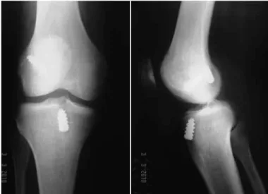
registered (in mm) were seen through a viewer. The number corresponding to the difference between the Figure 2 – Radiograph on the right knee in anteroposterior and
lateral views after the operation, in the double-band group.
STATISTICAL ANALYSIS
Student’s t test was applied to compare the clinical parameters of the two groups, and the 95% confidence interval was obtained (p < 0.05). Because not all of the grades of the objective IKDC contained patients, it was not possible to perform the chi-square test.
RESULTS
Analysis on the sample
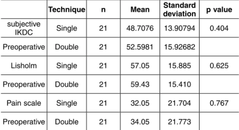
The sample consisted of 42 patients in two groups of 21 each, with mean ages of 28.71 years for the single-band group and 29.27 years for the double--band group (Table 1). There was only one female patient in the first group. Based on the data obtained, and from analysis on the preoperative assessments, the Lysholm test, subjective IKDC, length of time with the lesion and pain scale were correlated. For the single-band and double-band groups, respectively, the mean preoperative results were as follows: subjective IKDC score: 48.70 and 52.59; Lysholm score: 57.05 and 59.43; pain scale: 32.05 and 34.05; length of time from injury to surgery: 14.6 and 23.11 months (Ta-bles 2 and 3). These did not present any statistically significant difference (p > 0.05), thus showing that there was excellent homogeneity and randomization in the study groups.
Table 1 – Correlation of age with the techniques used.
Age in years
Technique n Mean Standard
deviation p value Single
band 21 28.71 6.286 0.736
Double
band 21 29.43 7.318
Table 2 – Correlation of preoperative variables in the sample.
Technique n Mean Standard
deviation p value subjective
IKDC Single 21 48.7076 13.90794 0.404 Preoperative Double 21 52.5981 15.92682
Lisholm Single 21 57.05 15.885 0.625
Preoperative Double 21 59.43 15.410
Pain scale Single 21 32.05 21.704 0.767
Preoperative Double 21 34.05 21.773
Table 3 – Correlation of length of time with the lesion between the two groups. Values in months.
Length of time with
lesion
Technique n Mean Standard
deviation p value Single 21 14.60 20.854 0.412
limbs was taken to be the amount of the knee ligament laxity, and this was deemed to be normal up to 3 mm. The test using the KT-1000 was done by the same evaluator on all the patients during their return visit after 180 days of evolution. The operated and con-tralateral limbs were compared in pairs of repeated tests, thus obtaining three values for each tension in each knee, per test. The difference in tension for each knee was obtained by subtracting the values for the operated knee from the contralateral knee. Thus, po-sitive values represented a laxer knee and negative values represented a firmer knee, in comparison with the contralateral knee (Table 5).
From careful analysis on the results, it was obser-ved that there were statistically significant differences between the techniques in relation to the forces of 67 N (0.006) and 89 N (0.001). It was also observed that all the values were lower than 3 mm (Figure 4).
Assessing the objective IKDC test
In our study, we observed before the operation that in the single-band group, there were five patients with an extension deficit of 10°. Likewise, in the
double-band group, there were four who had lost 10° and one who had lost 35° of extension. None of the patients presented flexion loss.
After the operation, in the single-band group, there was only one patient with an extension loss of 10° of range of motion. However, in the double-band group, extension loss was presented by four patients: two with losses of 5° and two with losses of 10°.
From the graph in Figure 5, the single-band group before the operation showed a greater num-ber of patients with “A” results, but not as many patients with “D” results. Thus, this represents a possible selection bias in our study.
DISCUSSION
From reviewing the literature, we found several articles on this topic. Some authors advocate using a double band, but others do not. In theory, a double band presents several advantages, such as making a greater graft-bone area available, thereby favoring greater anchorage of collagen fibers(19-21).
Construc-tion of the tunnels can be done independently, thus Table 4 – Postoperative correlation of subjective IKDC and Lysholm tests
between the groups.
Technique n Mean Standard deviation p value subjective
IKDC Single 21 78.05 11.385 0.971 Preoperative Double 21 77.89 17.717
Lisholm Single 21 87.52 11.214 0.289
Preoperative Double 21 83.00 15.707
Figure 3 – KT-1000 arthrometer.
Table 5 – Correlation of KT-1000 arthrometer test at tensions of 67 N, 89 N and 134 N (maximum) between the two groups.
Technique n Mean Standard deviation p value
67 N test Single 21 0.1024 0.75901 0.006 Double 21 0.7238 0.62782
89 N test Single 21 0.0810 0.83344 0 001 Double 21 0.8476 0.59129
134 N test Single 21 0.3452 0.94907 0.054 Double 21 0.8952 0.83933
Figure 4 – Difference in mm between KT-1000 values. Single band
ensuring that the correct angle is used for each band, as described by Yasuda et al(13). Biomechanical
stu-dies have demonstrated what individual contribution each band has towards ACL function. Sakane et al(22)
demonstrated that the AM band presented constant tension levels during knee flexion, and that the PL band presented greater variability of tension, with greatest levels between 0 and 30°, and thereafter decreasing with gradual flexion of the knee. These data were confirmed by Amis and Zavras(1), who
de-monstrated tension of 67 N (in situ) in the PL band in extension and 90 N in the AM band with the knee flexed at 60°. Another advantage was demonstrated by Gabriel et al(8), who showed that the PL band
con-tributed especially towards the rotational stability of the knee at flexion of 15° to 30°. On the other hand, we emphasize that using the technique for double--band reconstruction is a challenge for surgeons, since it requires greater expertise and experience, takes lon-ger to perform and presents patient-dependent factors regarding graft quality, such that the graft needs to be long enough and thick enough to construct the two bands, and regarding bone quality, which has to be good enough to construct the tunnels.
In 2008, in a randomized clinical study on a se-ries of 70 patients allocated into two groups, Siebold et al(11) demonstrated that the rotational and anterior
stability of knees with double-band ACL reconstruc-tion, using an anatomical technique, was greater in KT-1000 and objective IKDC tests than was the stabi-lity of ligament reconstructions using the single-band technique. However, when the data were analyzed with regard to the Cincinnati, Lysholm and subjective
IKDC scores, there was no statistically significant variation in the results between the groups.
Similar results had previously been observed by Muneta et al(12), in a series from 1992 to 1996, in
re-constructions using flexor tendons in a single or dou-ble band, in which the measurement parameters were the Lachman test and anterior stability from KT-1000. The results indicated that the results from reconstruc-tions using the double-band technique were superior.
In 2004, Yasuda et al(13) presented a technique for
double-band anatomical reconstruction in a study that compared three techniques: ACL reconstruction using a single band; ACL reconstruction using a double band and three tunnels (of which two were femoral); and lastly, ACL reconstruction using a double-band anato-mical technique with four tunnels. The results demons-trated that the anatomical technique of double-band re-construction was statistically superior, when assessed using KT-1000 and pivot-shift. The clinical results of Zhao et al(14), in 2006, further confirmed these results
with regard to anterior and rotational stability.
However, in 2004, contrary to previous studies, Adachi et al(15) did not find any differences between
the classic techniques of ACL reconstruction using a single or a double band, with tendons from the semitendinosus and gracilis muscles in both cases. Likewise, Hamada et al(16) did not find any
signifi-cant difference between the single and double band techniques, regarding anterior instability of the knees.
Also in disagreement with previous studies, in a series of 123 patients that compared the single and double band techniques and analyzed knee laxity, range of motion, extension and flexion strength (de-termined using Cybex), anterior instability (KT-1000) and Lysholm score, Asagumo et al(17) (2006) did not
find any significant differences except in the range of motion, in which the double band was inferior. Thus, these authors did not corroborate adoption of the double band technique.
In our study, we made observations in a satisfac-tory sample, albeit with evaluation bias due to the impossibility of objectively measuring the internal rotation of the tibial plateau after the reconstruction. Our results with the double band were not as good as we had expected. We did not observe any significant improvement in any subjective score, but even thou-gh we achieved knees that were less rigid, they were firm, as shown by the results from the KT-1000 and Figure 5 – Pre and postoperative values in each group correlated
with objective IKDC classes. Preoperative
objective IKDC tests. The reason for this may lie in the fixation angle of the PL band. This structure be-came fragile and may have been responsible for the loss of extension, which was initially greater in the double band group.
One explanation for these results may be that we were comparing the double band technique with an anatomical single band technique, rather than with the conventional single band technique (transtibial), thereby leading to a result that was not as good as ex-pected. Like Asagumo et al(17), we observed a greater
loss of extension in the double band group, which is making us think again about our use of this technique.
CONCLUSION
In the present study, we did not find any difference between the two groups in the subjective evaluations. However, in the objective evaluations, we observed better results from the single-band technique. Thus, we were unable to prove that anatomical reconstruc-tion using a double-band was superior. However, it should be emphasized that there is a need for studies with longer follow-up in order to make assessments regarding the development of gonarthrosis (a late complication from instability), which might perhaps be avoided with this new technique.
REFERENCES
1. Amis AA, Zavras TD. Isometricity and graft placement during anterior cruciate ligament reconstruction. Knee. 1995;2(1):5-17.
2. Harner CD, Baek GH, Vogrin TM, Carlin GJ, Kashiwaguchi S, Woo SL. Quantitative analysis of human cruciate ligament insertions. Arthroscopy. 1999;15(7):741-9.
3. Martins CAQ, Kropf EJ, Shen W, van Eck CF, Fu FH. The concept of anatomic an-terior cruciate ligament reconstruction. Oper Tech Sports Med. 2008;16:104-115.
4. Carneiro M, Navarro RD, Nakama GY, Barreto JM, Queiróz AAB, Luzo MVM. Reconstrução do ligamento cruzado anterior com duplo feixe utilizando os tendões dos músculos semitendíneo e grácil: fixação com dois parafusos de interferência. Rev Bras Ortop. 2009;44(5):441-5.
5. Bach BR Jr, Tradonsky S, Bojchuk J, Levy ME, Bush-Joseph CA, Khan NH. Arthroscopically assisted anterior cruciate ligament reconstruction using patel-lar tendon autograft. Five- to nine-year follow-up evaluation. Am J Sports Med. 1998;26(1):20-9.
6. Aglietti P, Buzzi R, D’Andria S, Zaccherotti G. Long-term study of anterior cruciate ligament reconstruction for chronic instability using the central one-third patellar tendon and a lateral extraarticular tenodesis. Am J Sports Med. 1992;20(1):38-45.
7. Lohmander LS, Ostenberg A, Englund M, Roos H. High prevalence of knee os-teoarthritis, pain, and functional limitations in female soccer players twelve years after anterior cruciate ligament injury. Arthritis Rheum. 2004;50(10):3145-52.
8. Gabriel MT, Wong EK, Woo SL, Yagi M, Debski RE. Distribution of in situ forces in the anterior cruciate ligament in response to rotatory loads. J Orthop Res. 2004;22(1):85-9.
9. Yagi M, Wong EK, Kanamori A, Debski RE, Fu FH, Woo SL. Biomechanical analysis of an anatomic anterior cruciate ligament reconstruction. Am J Sports Med. 2002;30(5):660-6.
10. Mott HW. Semitendinosus anatomic reconstruction for cruciate ligamentinsuf-ficiency. Clin Orthop Relat Res. 1983;(172):90-2.
11. Siebold R, Dehler C, Ellert T. Prospective randomized comparison of double-bundle versus single-double-bundle anterior cruciate ligament reconstruction. Arthros-copy. 2008;24(2):137-45.
12. Muneta T, Koga H, Morito T, Yagishita K, Sekiya I. A retrospective study of the midterm outcome of two-bundle anterior cruciate ligament reconstruction using
quadrupled semitendinosus tendon in comparison with one-bundle reconstruc-tion. Arthroscopy. 2006;22(3):252-8.
13. Yasuda K, Kondo E, Ichiyama H, Kitamura N, Tanabe Y, Tohyama H, et al. Anatomic reconstruction of the anteromedial and posterolateral bundles of the anterior cruciate ligament using hamstring tendon grafts. Arthroscopy. 2004;20(10):1015-25.
14. Zhao J, Peng X, He Y, Wang J. Two-bundle anterior cruciate ligament recon-struction with eight-stranded hamstring tendons: four-tunnel technique. Knee. 2006;13(1):36-41.
15. Adachi N, Ochi M, Uchio Y, Iwasa J, Kuriwaka M, Ito Y. Reconstruction of the anterior cruciate ligament. Single- versus double-bundle multistranded ham-string tendons. J Bone Joint Surg Br. 2004;86(4):515-20.
16. Hamada M, Shino K, Horibe S, Mitsuoka T, Miyama T, Shiozaki Y, et al. Single-versus bi-socket anterior cruciate ligament reconstruction using autogenous multiple-stranded hamstring tendons with endoButton femoral fixation: A pro-spective study. Arthroscopy. 2001;17(8):801-7.
17. Asagumo H, Kimura M, Kobayashi Y, Taki M, Takagishi K. Anatomic recon-struction of the anterior cruciate ligament using double-bundle hamstring ten-dons: surgical techniques, clinical outcomes, and complications. Arthroscopy. 2007;23(6):602-9.
18. Outerbridge RE. The etiology of chondromalacia patellae. J Bone Joint Surg Br. 1961;43:752-7.
19. Tomita F, Yasuda K, Mikami S, Sakai T, Yamazaki S, Tohyama H. Comparisons of intraosseous graft healing between the doubled flexor tendon graft and the bone-patellar tendon-bone graft in anterior cruciate ligament reconstruction. Arthroscopy. 2001;17(5):461-76.
20. Cho S, Muneta T, Ito S, Yagishita K, Ichinose S. Electron microscopic evalua-tion of two-bundle anatomically reconstructed anterior cruciate ligament graft. J Orthop Sci. 2004;9(3):296-301.
21. Gali JC, Mod MSB, Mimura HM, Kushiyama W. Reconstrução anatômica do ligamento cruzado anterior com dupla banda: estudo prospectivo com segui-mento de dois anos. Rev Bras Ortop 2011;46(1):31-6.