ESCOLA DE CIÊNCIAS E TECNOLOGIA
DEPARTAMENTO DE MEDICINA VETERINÁRIA
COAGULOPATHY IN SEPSIS AND THE
PROGNOSTIC VALUE OF ABNORMAL
COAGULATION TIMES
Luís Miguel Manita Rodrigues
Orientação | Professora Doutora Sandra Maria
da Silva Branco
Orientação Externa | Dra. Ângela Martins
Mestrado Integrado em Medicina Veterinária
Dissertação
II
ESCOLA DE CIÊNCIAS E TECNOLOGIA
DEPARTAMENTO DE MEDICINA VETERINÁRIA
COAGULOPATHY IN SEPSIS AND THE
PROGNOSTIC VALUE OF ABNORMAL
COAGULATION TIMES
Luís Miguel Manita Rodrigues
Orientação | Professora Doutora Sandra Maria
da Silva Branco
Orientação Externa | Dra. Ângela Martins
Mestrado Integrado em Medicina Veterinária
Dissertação
I
To my brightest stars: Schtotsu, Daini, Inu. To my brightest light, Mew.
To my brightest self; you’ve made it kiddo!
“It’s time to try defying gravity.”
II
A
CKNOWLEDGEMENTS
I would like to show my appreciation and gratitude to those who made this whole crazy journey possible:
To my family for their never-ending support, particularly my brothers Tavinho and Pedro, my Mom, and my Avó. I am also grateful to my godparents, Mika and Orlando, for being my second set of parents and for lending me their laptop when mine decided to take a nap while writing this dissertation.
To my supervisor Prof. Sandra Branco for the ongoing guidance and encouragement, for the prompt availability to help me with my doubts, and for always replying to my emails within minutes.
To my supervisor Dr. Ângela Martins for all the knowledge passed on to me throughout my internship period, for the continuous guidance, for always being a role model of hard work, and for believing in me.
To Prof. Inês Viegas for so eagerly helping me out with the statistical work of this dissertation.
To my internship colleagues and all the staff from Hospital Veterinário da Arrábida that put up with me and my uncontrollable urge to hug the patients; I swear it was all part of the job!
To my hometown friends, who have always been a source of cheerfulness and encouragement. A special thank you goes to my buddies Vasco, Rute, and Raquel, who have been by my side since we were next to toddlers, and to Pat, Inês, and Catarina, who also put up with me just as much.
To my friends who are scattered around the globe for being a source of laughter and enjoyment, even when I am feeling the lowest. A shout out to John for being a sweetheart and reading this dissertation beforehand.
III
To my friends and colleagues with whom I shared the last 6 years of this veterinary rollercoaster of a course. A special thank you goes to David, Inês, Cláudio, Ana Lurdes, and Flôr for the many moments shared.
To Mafalda and Carolina for keeping my sanity in check throughout one of the toughest times of this journey.
To Diogo for the ongoing support, comfort, and warmth throughout every obstacle. To my academic family. You lot are amazing individuals, and it is plain silly how much I cherish each and every one of you guys. A special mention goes to my academic godmother Sara and godson Luís. I will make sure to continue to be as clingy as I have always been!
IV
A
BSTRACT
Sepsis is a hard to define condition associated with the deleterious systemic inflammatory response syndrome (SIRS) which ultimately leads to the failure of multiple organs. The mediators released throughout this exaggerated inflammatory reaction activate coagulation pathways and generate a dysfunctional response that results in coagulopathy. The present dissertation includes a literature review regarding the subject of sepsis and associated coagulopathy, along with a study that primarily aims to investigate the use of abnormal coagulation times as biological markers of coagulation dysfunction and as predictors of outcome in veterinary patients who are at risk of developing sepsis. The results suggest that pairing coagulation time data with an organ failure scoring system may be advantageous in the prediction of outcome. Furthermore, critically ill patients should be given a five-day time frame following admission before euthanasia is considered, as most tend to survive their illnesses once they get past this period.
V
R
ESUMO
C
OAGULOPATIA NA SÉPSIS E O VALOR DE PROGNÓSTICO DE
TEMPOS DE COAGULAÇÃO ALTERADOS
A sépsis é uma síndrome de difícil definição e que está associada à síndrome da resposta inflamatória sistémica (SIRS) que leva à falha de múltiplos órgãos. Os mediadores libertados durante esta reação inflamatória exagerada levam à ativação disfuncional da coagulação sanguínea, o que resulta em coagulopatia.
A presente dissertação inclui uma revisão bibliográfica sobre o tema da sépsis e a coagulopatia associada, bem como um estudo cujo objetivo primário é o de investigar a utilização de tempos de coagulação alterados, tanto como marcadores biológicos de disfunção da coagulação sanguínea bem como fatores de prognóstico em pacientes veterinários em risco de sépsis.
Os resultados do estudo realizado mostram vantagem em associar a avaliação dos tempos de coagulação com sistemas de pontuação de falha orgânica para a realização do prognóstico. Estes sugerem também que os pacientes críticos que ultrapassam os primeiros cinco dias após a sua admissão hospitalar tendem a sobreviver.
VI
T
ABLE OF
C
ONTENTS
ACKNOWLEDGEMENTS . . . II ABSTRACT . . . IV RESUMO . . . V LIST OF TABLES . . . VIII LIST OF FIGURES . . . IX LIST OF SYMBOLS AND ABBREVIATIONS . . . X PREFACE . . . XIV
1. LITERATURE REVIEW . . . 1
1.1.INTRODUCTION TO THE DEFINITION OF SEPSIS . . . 1
1.2.THE SYSTEMIC INFLAMMATORY RESPONSE SYNDROME (SIRS) . . . 4
1.2.1SIGNS OF SIRS IN VETERINARY PATIENTS . . . 4
1.2.2.PATHOPHYSIOLOGY OF SIRS . . . 6
1.2.2.1.PATHOGEN AND TISSUE DAMAGE RECOGNITION . . . 8
1.2.2.2.THE HYPERINFLAMMATORY RESPONSE IN SIRS . . . 10
1.2.2.3.THE COMPENSATORY ANTI-INFLAMMATORY RESPONSE SYNDROME (CARS) . . . 11
1.2.2.4.SEPTIC SHOCK . . . 13
1.2.2.5.THE MULTIPLE ORGAN DYSFUNCTION SYNDROME (MODS). . 15
1.3.COAGULOPATHY IN SEPSIS. . . . 19
1.3.1.FROM SYSTEMIC INFLAMMATION TO THE ACTIVATION OF BLOOD COAGULATION . . . 19
1.3.2. IMPAIRMENT OF ANTICOAGULANT MECHANISMS . . . 22
1.3.3.SUPPRESSION OF FIBRINOLYSIS . . . 24
1.3.4.ADDITIONAL INTERACTIONS BETWEEN INFLAMMATION AND COAGULATION . . . 25
1.3.5.DISSEMINATED INTRAVASCULAR COAGULATION (DIC) IN SEPSIS . . . 26
1.3.6.ABNORMAL COAGULATION TIMES IN SEPSIS . . . 27
1.4.RECOMMENDED SCORING SYSTEMS FOR THE ASSESSMENT OF ORGAN DYSFUNCTION IN SEPSIS . . . 29
VII
2. STUDY – THE PROGNOSTIC VALUE OF ABNORMAL COAGULATION TIMES . . . 33
2.1.STUDY INTRODUCTION . . . 33
2.2.OBJECTIVES . . . 33
2.3.MATERIALS AND METHODS . . . 34
2.3.1.STUDY POPULATION . . . 34
2.3.2.STUDY DESIGN . . . 34
2.3.3. BLOOD SAMPLING AND COAGULATION TESTING . . . 35
2.3.4.STATISTICAL ANALYSIS . . . 36
2.4.RESULTS . . . 36
2.4.1.DIAGNOSIS . . . 36
2.4.2. BREED, AGE, AND SEX . . . 37
2.4.3. QSOFA, ACTIVATED PARTIAL THROMBOPLASTIN TIME, AND PROTHROMBIN TIME . . . 38
2.4.4. LENGTH OF TREATMENT AND OUTCOME . . . 40
2.5.DISCUSSION . . . 41 2.6.CONCLUSIONS . . . 46 LIST OF REFERENCES . . . 48 APPENDICES . . . i APPENDIX A . . . ii APPENDIX B . . . iii
VIII
L
IST OF TABLES
Table 1. Systemic inflammatory response syndrome (SIRS) criteria for dogs and cats
(data collected from references 6 and 7)...………..4
Table 2. The sequential organ failure assessment (SOFA) score criteria for veterinary
patients (adapted from references 84 and 279)……….30
Table 3. The quick sequential organ failure assessment (qSOFA) score criteria (adapted
from reference 24)………31
Table 4. The mortality rate associated with each quick sequential failure assessment
(qSOFA) score in the present study……….39
Table 5. Mean length of hospitalisation and treatment of survivors and
IX
L
IST OF FIGURES
Figure 1. The interrelationship between the systemic inflammatory response syndrome
(SIRS), infection, and sepsis (reprinted from reference 5 with permission from Elsevier)...……….….7
Figure 2. Recognition of infection or tissue injury by a macrophage (original figure)…..9 Figure 3. The pathophysiology of septic shock (original figure)...…..14 Figure 4. The current concept of coagulation in sepsis (original figure)…..………21 Figure 5. The identification process for sepsis and septic shock according to Sepsis-3
(adapted from reference 24)……….32
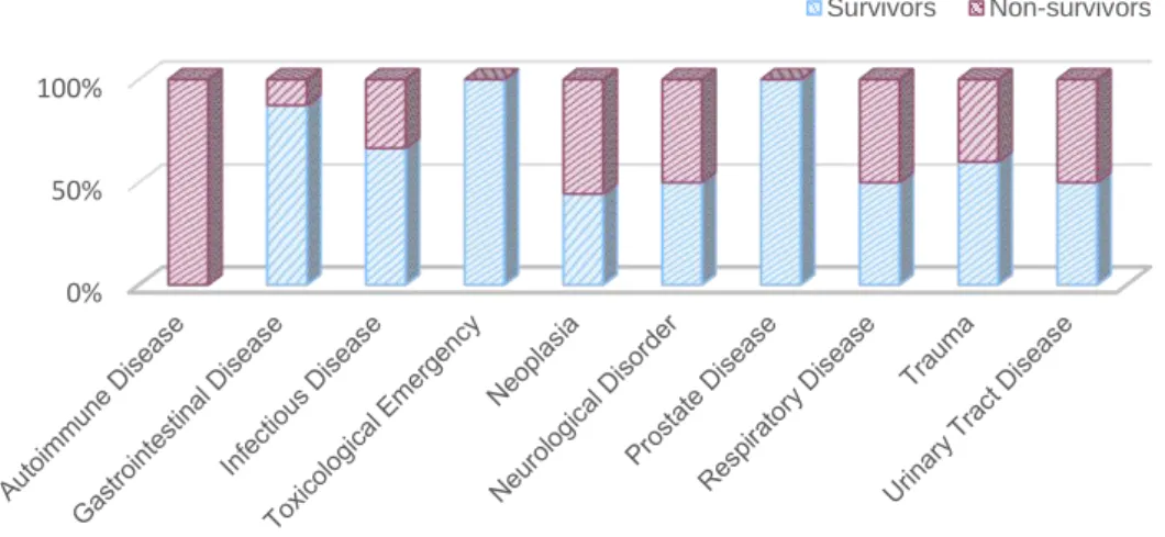
Figure 6. qLabs® Vet Coag Panel 2 device and test strips (original figure)………35 Figure 7. Bar graph representing survival and non-survival rates of each of the diagnosed
underlying causes of illness or injury………...37
Figure 8. Bar graph representing the number of survivors and non-survivors of each
sex...38
Figure 9. Graphical representation of mean activated partial thromboplastin time (aPTT)
values associated with each quick sequential organ failure assessment (qSOFA) score…...39
Figure 10. Scatter graph representing the positive correlation between activated partial
X
L
IST OF SYMBOLS AND ABBREVIATIONS
ACh – Acetylcholine AKI – Acute Kidney Injury ANOVA – Analysis of Variance ANS – Autonomic Nervous System AP-1 – Activator Protein 1
aPC – Activated Protein C
aPTT – Activated Partial Thromboplastin Time ARDS – Acute Respiratory Distress Syndrome AT – Antithrombin
ATC – Acute Traumatic Coagulopathy ATP – Adenosine Triphosphate
C1 – Complement Component 1 C3 – Complement Component 3 C5 – Complement Component 5
CARS – Compensatory Anti-inflammatory Response Syndrome CIRCI – Critical Illness-Related Corticosteroid Insufficiency CNS – Central Nervous System
d - Days
XI DIC – Disseminated Intravascular Coagulation DNA – Deoxyribonucleic Acid
EPCR – Endothelial Protein C Receptor FV – Factor V or Proaccelerin
FVa – Activated Factor V
FVII – Factor VII or Proconvertin FVIIa – Activated Factor VII
FVIII – Factor VIII or Antihaemophilic Factor A FVIIIa – Activated Factor VIII
FIX – Factor IX or Christmas Factor FIXa – Activated Factor IX
FX – Factor X or Stuart-Prower Factor FXa – Activated Factor X
FXI – Factor XI or Plasma Thromboplastin Antecedent FXII – Factor XII or Hageman Factor
FXIII – Factor XIII or Fibrin-Stabilising Factor FiO2 – Fraction of Inspired Oxygen
GIT – Gastrointestinal Tract
HPA – Hypothalamic-Pituitary-Adrenal (axis) HSD – Honest Significant Difference (Tukey’s test) ICU – Intensive Care Unit
XII IL-1 – Interleukin 1
IL-6 – Interleukin 6 IL-10 – Interleukin 10 LPS – Lipopolysaccharide MAP – Mean Arterial Pressure
MGCS – Modified Glasgow Coma Scale
MODS – Multiple Organ Dysfunction Syndrome NF-KB – Nuclear Factor Kappa B
NK – Natural Killer (cell) NO – Nitric Oxide
PAI – Plasminogen Activator Inhibitor
PAI-1 – Plasminogen Activator Inhibitor Type 1 PAMP – Pathogen-Associated Molecular Pattern PaO2 – Partial Pressure of Oxygen
PAR – Protease-Activated Receptor
PIRO – Predisposition, Infection, Response, Organ Dysfunction (sepsis staging system) PRR – Pattern Recognition Receptor
PT – Prothrombin Time
qSOFA – Quick Sequential Organ Failure Assessment (score) ROS – Reactive Oxygen Species
Sepsis-1 – 1991 Sepsis Definitions Sepsis-2 – 2001 Sepsis Definitions
XIII Sepsis-3 – 2016 Sepsis Definitions
SIRS – Systemic Inflammatory Response Syndrome SOFA – Sequential Organ Failure Assessment (score) T3 – Triiodothyronine
TAFI – Thrombin-Activatable Fibrinolysis Inhibitor TF – Tissue Factor
TFPI - Tissue Factor Pathway Inhibitor Th – T Helper (cell)
Th1 – T Helper 1 Th2 – T Helper 2 Th17 – T Helper 17 TLR – Toll-Like Receptor TNF – Tumour Necrosis Factor
TNFα - Tumour Necrosis Factor Alpha
TNM – Classification of Malignant Tumours (cancer staging system) TPA – Tissue Plasminogen Activator
UPA – Urokinase-Type Plasminogen Activator
UPAR – Urokinase-Type Plasminogen Activator Receptor vWF – von Willebrand Factor
XIV
P
REFACE
The present dissertation was written following a six-month internship, from September 2016 to March 2017, at Hospital Veterinário da Arrábida and Centro de Reabilitação Animal da Arrábida, a small animal hospital connected to a referral rehabilitation centre in the civil parish of Azeitão in Portugal.
Many areas of veterinary medicine were explored throughout this internship, including diagnostics, emergency and critical care medicine, orthopaedic and soft tissue surgery, internal medicine, and small animal rehabilitation. It was during this period that the author determined the topic of his research and began collecting the data that would be subsequently analysed, after developing a particular interest in the subject of sepsis in critically ill patients.
1
1.
L
ITERATURE REVIEW
1.1.
I
NTRODUCTION TO THE DEFINITION OF SEPSIS
Defining sepsis is not an easy task. The word “sepsis” is as old as ancient Greece when it was originally used to describe decomposition in the presence of bacteria1, long before
anything was known about this serious condition.2 Prior to 1989, sepsis was merely believed to be associated with bacteraemia.3
In 1989, Bone et al.4 defined sepsis syndrome as “the systemic manifestations of presumed sepsis”. However, this definition of a systemic response to infection was based on a set of clinical signs which could be found in the absence of infection. This fact led to the creation of the concept of a “Systemic Inflammatory Response Syndrome” (SIRS) in a consensus conference held by the American College of Chest Physicians and the Society of Critical Care Medicine in 1991.5 SIRS was created to describe the inflammatory response found in sepsis, regardless of its cause. It was established that infection, as well as trauma, pancreatitis, and other non-infectious insults, could trigger this response. It was also suggested that the term sepsis should only be used if SIRS was the result of a confirmed infectious process. SIRS was associated with variables such as altered temperature (hypothermia or hyperthermia), heart rate (bradycardia or tachycardia), respiratory rate (bradypnea or tachypnea), and white blood cell count (leukocytosis or leukopenia), and would be diagnosed if a human patient was positive for at least two of these four criteria.5 It has later been suggested that dogs should also meet two of these criteria to be diagnosed with SIRS whereas cats would need to fulfil three criteria for the same purpose.6,7 In addition to the definition of SIRS, the notions of
“severe sepsis” and “septic shock” were introduced to describe different stages of sepsis. The concept of a “Multiple Organ Dysfunction Syndrome” (MODS) was also established
2
to describe the presence of altered organ function in an acutely ill patient such that homeostasis could not be maintained without intervention.5
Despite the general acceptance of this new definition of sepsis, many clinicians did not fully agree with it.8–10 This new approach did not seem to provide a precise definition of sepsis and many considered the SIRS criteria to be too sensitive and nonspecific for its diagnosis since a large number of patients admitted to intensive care units would meet such criteria and would thus be considered septic.11–16 In 2001, an International Sepsis Definitions Conference was held in an attempt to tackle these issues by revisiting the previous definitions surrounding sepsis.9 It was recognised that, while still useful, the diagnostic criteria for SIRS were overly sensitive and nonspecific. Thus, a list of additional signs and symptoms of systemic inflammation in response to infection was presented to more accurately reflect the host’s clinical response. However, this list was arguably too long to be universally adopted, and the SIRS criteria continued to be used to diagnose sepsis.2 A conceptual staging system for sepsis called PIRO, inspired by the Classification of Malignant Tumours (TNM) system, was also proposed at this conference as a potential tool for sepsis patient stratification. In the PIRO (an acronym for Predisposition, Infection, Response and Organ dysfunction) model, P refers to all predisposing factors which may impact the outcome of sepsis, such as genetic variability, age, the presence of concomitant diseases, and nutritional status. In veterinary patients, racial predisposition would fit into this component. I refers to the description of the infection, which includes its etiologic agent, location, and extent. R concerns the host’s inflammatory response to sepsis. Finally, O corresponds to the number of failing organs and the degree of dysfunction. Although promising, PIRO was yet to be fully developed and required further investigation.9,17–19
SIRS criteria continued to be criticised for their inadequacy, and the need for a new definition of sepsis remained.20–23 In 2016, the authors of the Third International Consensus Definitions for Sepsis and Septic Shock released newly updated definitions for sepsis and septic shock. It was suggested that these would be regarded as Sepsis-3, while the 1991 and 2001 versions would be known as Sepsis-1 and Sepsis-2, respectively. Improved understanding of sepsis pathobiology led to its current definition of a “life-threatening organ dysfunction caused by a dysregulated host response to infection”.24
3
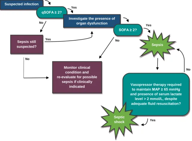
Furthermore, septic shock was considered a subset of sepsis associated with a higher mortality rate and was characterised by the need for vasopressor therapy to maintain the mean arterial pressure (MAP) of 65 mmHg or more, as well as the presence of serum lactate levels higher than 2 mmol/L despite appropriate fluid resuscitation.25
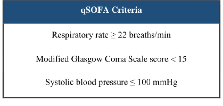
To assess organ dysfunction severity and recognise sepsis in critically ill patients with suspected infection, the authors of Sepsis-3 suggested the use of the Sequential Organ Failure Assessment (SOFA) score. A SOFA score of at least two points is indicative of organ dysfunction and is associated with a higher mortality rate when compared with lower scores. However, SOFA is rather complex and requires laboratory testing, and therefore using it to quickly identify sepsis outside of an intensive care unit (ICU) setting is not realistic. To address this issue, the authors of Sepsis-3 proposed the use of a new straightforward scoring system called “quick SOFA” (qSOFA). This simplified SOFA variant can be used to promptly identify patients with suspected infection who are likely of developing poor outcomes. To determine these patients, qSOFA analyses the existence of altered mentation, hypotension and tachypnea. Each of these clinical signs represents one point, and a score equal to or greater than two points is suggestive of organ dysfunction.24,26,27
Controversy has always surrounded the definition of sepsis, and Sepsis-3 is no exception to this as many clinicians are not in full agreement with its foundation.28–31 Many clinicians believe that the newly recommended criteria for the identification of sepsis require further testing before replacing their antecedents.32–35 Despite the most recent
approach to the definition of sepsis, the SIRS criteria are still considered to be of great utility in the identification of infected patients, as well as any other patients suffering from sterile SIRS.24,36–39 Sepsis is a very complex condition, and there is yet to exist a widely
4
1.2.
T
HE SYSTEMIC INFLAMMATORY RESPONSE
SYNDROME
(SIRS)
1.2.1
S
IGNS OFSIRS
IN VETERINARY PATIENTSSIRS is a complex and systemic response to an infectious or non-infectious insult that may occur in both human and veterinary patients.41 The concepts of sepsis and SIRS and all surrounding discussion were initially concerning the human patient. Studies have been conducted in an attempt to adapt the SIRS criteria to veterinary patients and establish limits for each criterion.6,7 Table 1 shows the suggested criteria, based on such research.
Table 1. Systemic inflammatory response syndrome (SIRS) criteria for dogs and cats
(data collected from references 6 and 7).
Clinical Parameters Dogs
(must meet two criteria)
Cats
(must meet three criteria)
Heart rate (beats/min) >120 < 140 or > 225
Respiratory rate (breaths/min) > 20 > 40
Rectal temperature (Cº) < 38.1 or >39.2 < 37.8 or > 39.7 Leukogram (white blood cells/ µL; % band
cells) <6000 or >16,000; > 3 < 5000 or > 19.500; > 5
The suggested SIRS criteria for cats are slightly different from the ones suggested for dogs. Besides tachycardia, low heart rates are also frequently found amongst critically ill feline patients and should be considered when applying the SIRS criteria. Furthermore, cats must express much higher respiratory rates to be diagnosed with tachypnea, when compared to dogs. Interestingly, it is also suggested that cats must satisfy at least three criteria for the identification of SIRS.7 Dogs, however, are only required to meet two criteria for SIRS to be identified, much like what happens with human patients.6 A study
5
performed by Okano et al.42 suggests that, in canine patients, the prognosis worsens as more SIRS criteria are met. In that same study, the results also indicated that some alterations in the parameters included in the SIRS criteria might be related to worse outcomes when compared to others. Abnormalities in body temperature and white blood cell count seemed to be linked to a poorer prognosis and were considered to be the most reliable of the four parameters to be evaluated since external stimuli can easily influence both respiratory and heart rates. Contrary to these findings, a study by Declue et al.43, performed with cats, revealed that the number of satisfied SIRS criteria was not correlated with prognosis. The dissonance in the results of reports such as these shows how dogs and cats can respond differently to inflammation and sepsis and should not be evaluated as being part of the same species.
Infection, heat stroke, pancreatitis, immune-mediated disease, neoplasia, trauma, and burns are the most common causes of SIRS in veterinary patients. Clinical signs of SIRS are usually nonspecific and can change depending on the underlying disease process. They tend to mimic the manifestations of sepsis and are generally treated similarly.44 It is
important to mention that dogs and humans tend to display clinical signs of an initial hyperdynamic phase of sepsis such as loss of appetite, depression, hyperemic mucous membranes, bounding peripheral pulses, tachycardia, tachypnea, and fever. Cats, however, rarely manifest this hyperdynamic state. Thus, the clinical signs of sepsis found in cats tend to be related to a secondary hypodynamic phase and may include lethargy, diffuse abdominal pain, pale mucous membranes, tachypnea, bradycardia, hypotension and hypothermia. Cats are also more likely to experience hypotension, hypoglycaemia and hyperbilirubinemia than dogs.7,44,45
Blood cell count alterations, such as neutrophilic leukocytosis, and toxic cytologic changes of the neutrophils are common in patients with SIRS, as well as a variety of other changes on a biochemical level. Blood glucose levels tend to fluctuate between hyperglycaemia in the early phase of inflammation when gluconeogenesis is increased, and subsequent hypoglycaemia once glucose levels drop as a result of excessive use. Albumin concentration levels are likely to drop secondarily to reduced albumin production by the liver, in favour of acute phase proteins. Changes in endothelial permeability found in SIRS also lead to plasma protein leakage and consequently loss of
6
albumin.44 The resulting hypoalbuminemia may cause the development of pulmonary and peripheral oedema, which was evident in a study performed with cats suffering from sepsis, by Brady et al.7 Liver enzymes, such as alanine aminotransferase and aspartate aminotransferase, are inclined to increase in concentration due to changes in perfusion and decreased tissue oxygenation. Serum bilirubin may also suffer alterations, usually as a result of cholestasis.44 Haemolysis may also be responsible for icterus in cats with sepsis considering how common anaemia seems to be present in these patients.7 A study by Schaefer et al.46 showed that proteinuria is also present in dogs with SIRS, as a result of altered urinary protein excretion due to glomerular and tubular malfunction.
1.2.2.
P
ATHOPHYSIOLOGY OFSIRS
In ancient Rome, Celsus was the first to introduce the four signs widely used to describe an inflammatory response: redness (rubor), swelling (tumor), heat (calor), and pain (dolor). Many centuries later, a fifth sign, loss of function (function laesa), was added to this list.47,48 These terms characterise the visual changes that occur in a localised
inflammatory response to tissue damage or infection.49 Local blood vessel dilation and increased permeability result in the passage of an additional number of erythrocytes and fluids into the damaged area resulting in redness, heat, and swelling. Cells also infiltrate into the affected area, and prolonged inflammatory responses may generate deposits of connective tissue, further increasing the swelling. Resulting oedema leads to the stretching of sensory nerves, which results in pain. Pain is also a consequence of the initial tissue damage as well as the resulting inflammatory response itself and the effects of its mediators. Loss of mobility in structures such as the joints, due to pain and oedema, and replacement of once functional cells with scar tissue are examples of circumstances that lead to loss of function.48
The local hemodynamic changes in the inflammatory response are aimed at defending the host and eliminating harmful agents and damaged cells.49 Thus, localised inflammation is a physiological protective response, controlled by inflammatory mediators. However, overactivation of this inflammatory reaction or loss of its local control may result in the exaggerated systemic response we know as SIRS.50
7
SIRS is a dysregulated inflammatory response to injury or microbial invasion. Even though this syndrome is an essential part of sepsis when triggered by infectious agents, it can also occur in the absence of infection. Regardless of the initial insult, the resulting inflammatory response is considered to be fairly similar.51 When infection is the cause of SIRS, both gram-negative and gram-positive bacteria, as well as parasitic, fungal, protozoan and viral microorganisms, can be responsible for inciting the systemic response (Figure 1).52 However, infections caused by gram-negative bacteria seem to be both the most prevalent and dangerous, in cases of sepsis.53,54 Escherichia coli is the most commonly isolated microorganism in dogs and cats with sepsis.7,55–60 Interestingly, in the particular case of sepsis associated with pyothorax, members of the genus Pasteurella appear to be more commonly isolated in cats amongst facultative bacteria, whereas
Escherichia coli continues to be more prevalent in dogs.61,62
Figure 1. The interrelationship between the systemic inflammatory response syndrome
8
In human patients, the infectious processes that represent the most common causes of sepsis are pneumonia, urinary tract infections, intra-abdominal infections, and bacteraemia.63 In dogs, sepsis has been linked with conditions such as septic peritonitis, pancreatitis, pneumonia, pyometra, prostatitis, and wound infections.6,57 In our domestic felines, sepsis has been associated with conditions including septic peritonitis, pneumonia, bacteraemia, endocarditis, pyelonephritis, hepatic abscessation, and pyothorax.7,58,59,62
1.2.2.1.
P
ATHOGEN AND TISSUE DAMAGE RECOGNITIONMammals, such as humans and their small animal companions, possess an immune system with the task of protecting them against the invasion of harmful microorganisms. This immune system includes both innate and acquired immunity. While the innate immune system represents the first line of host defence against infection, the acquired immune system is associated with later phases of pathogen elimination and with the development of immunological memory.52 The innate immune system is responsible for containing the infection and delivering antigens to local lymph nodes, which results in the activation of the acquired immune system and consequent eradication of infection.64
For an invading microorganism to be able to successfully disseminate and cause sepsis and septic shock, both innate and acquired immune defences must be breached.65,66
The innate immune system includes the activity of many different cells such as macrophages, neutrophils, natural killer cells (NK), endothelial and epithelial cells, and dendritic cells.52,67,68 These cells can detect the presence of molecular structures associated with microbial pathogens and tissue damage, as well as endogenous molecules released during cellular injury, through a group of surface proteins named pattern recognition receptors (PRRs).67–69 Many of these PRRs have been identified and extensively studied, and one of the best-understood families of PRRs is the Toll-like receptors (TLRs) family.70,71
PRRs, such as TLRs, can recognise particular components expressed by microorganisms known as pathogen-associated molecular patterns (PAMPs), as well as endogenous mediators released during tissue injury and cell death known as “alarmins” or
danger-9
associated molecular patterns (DAMPs).66,67,71 Some authors seem to consider that the term DAMPs includes both PAMPs and alarmins 69,72, but the previous distinction will be the one used in the present dissertation.
Cell wall components, such as lipopolysaccharide (LPS) expressed by gram-negative bacteria (one of the most potent PAMPs), flagellin, and bacterial deoxyribonucleic acid (DNA) are some examples of PAMPs, which tend to be closely related to the survival or pathogenicity of the invading microorganism.68 Examples of DAMPs include heat shock proteins, fibrinogen, hyaluronic acid, and components of the endothelial glycocalyx.52,73,74
The recognition of PAMPs and DAMPs by PRRs results in the activation of the cell through a downstream of signalling cascades that culminate in a transcriptional response, via the mobilisation of transcription factors such as nuclear factor-kappa B (NF-kB) and activator protein 1 (AP-1). This cell activation results in the production and secretion of inflammatory mediators like cytokines, chemokines and complement-activating products.68,71,75–80 Figure 2 exemplifies this response.
Figure 2. Recognition of infection or tissue injury by a macrophage (original figure).
DAMP, Danger-associated molecular pattern; PAMP, pathogen-associated molecular pattern; TLR, Toll-like receptor.
TLR Macrophage
PAMP
DAMP
10
1.2.2.2.
T
HE HYPERINFLAMMATORY RESPONSE INSIRS
Cytokines are small protein mediators of low molecular weight (usually less than 40 kDa) that initiate, modulate, and sustain inflammatory interactions.76,78 The main proinflammatory cytokines responsible for inducing a systemic inflammatory response are those of the tumour necrosis factor (TNF) family and some interleukins (ILs), namely tumour necrosis factor alpha (TNFα), interleukin 1 (IL-1), and interleukin 6 (IL-6).68,71,79 Once released into circulation, these cytokines will signal endothelial cells to upregulate adhesion molecules that promote the migration of leukocytes from the microcirculation into sites of tissue injury or infection, recruiting them to perform the phagocytosis of pathogens and removal of damaged and dead host cells. 81–83 This proinflammatory
environment leads to the secretion of additional cytokines as well as secondary mediators such as nitric oxide (NO), reactive oxygen species (ROS), and lipid factors.64,77,84 Under
controlled inflammatory responses, this process would ultimately result in the clearance of infection and tissue healing.67,80
During SIRS, there is an overstimulation of immune cells as a response to extremely high levels of DAMPs from injured host tissue or PAMPs from invading microorganisms.77 This leads to an uncontrolled production and secretion of proinflammatory mediators, also known as “cytokine storm”, that enter the systemic circulation and travel to organs distant to the initial site of tissue damage or infection, resulting in the global activation of the inflammatory system.80,85
The acquired immune system is also involved in the production of cytokines and development of SIRS and sepsis.85,86 Antigen-presenting cells, such as monocytes and dendritic cells, activate the acquired immune response by interacting with naïve T cells and driving them to proliferate and differentiate into T helper (Th) cells. T helper 1 (Th1) and T helper 17 (Th17) cells are responsible for producing additional proinflammatory cytokines whereas T helper 2 (Th2) cells produce anti-inflammatory cytokines. Shifts in the balance between Th1/Th17 and Th2 cells dictate the nature of the immune response.85–
87 Early stages of SIRS have been associated with increased proinflammatory cytokine
production while anti-inflammatory activity and immune suppression are more characteristic of later phases of the syndrome.86
11
The autonomic nervous system (ANS) takes part in the inflammatory response as well. Immune cells are capable of producing and secreting neurotransmitters, as well as expressing receptors for such mediators, allowing the nervous and immune systems to communicate during inflammation.77 Released cytokines also provide the central nervous system (CNS) with updated information regarding the ongoing inflammatory response.88 Vagus nerve stimulation triggered by inflammatory stimuli has been shown to suppress inflammation.89,90 Efferent activity in the vagus nerve results in acetylcholine (ACh) secretion in organs of the reticuloendothelial system such as the liver, heart, spleen, and gastrointestinal tract. Exposure of tissue macrophages to ACh inhibits the release of proinflammatory cytokines. This anti-inflammatory mechanism is called the “cholinergic anti-inflammatory pathway” and is an important part of the “inflammatory reflex” carried out by the nervous system to control acute inflammation.91,92 Failure of mechanisms such as these due to CNS dysfunction in SIRS may contribute to the exacerbation of the inflammatory response.88 Furthermore, some authors have suggested that the release of
catecholamines by phagocytes and cells of the sympathetic branch of the ANS, in early phases of the syndrome, may amplify the proinflammatory responses of macrophages, neutrophils and dendritic cells. However, this subject appears to be controversial and not yet fully understood.77,93–95
The hyperinflammatory response developed in SIRS is further aggravated by the systemic activation of the complement system, which results in the generation of large amounts of proinflammatory peptides that act as leukocyte chemoattractants, enhance adhesion molecule expression, increase vascular permeability, and stimulate cytokine production.77,84,96,97 Excessive complement activation has also been previously linked to neutrophil dysfunction and increased mortality in cases of severe trauma.98,99
1.2.2.3.
T
HE COMPENSATORY ANTI-
INFLAMMATORYRESPONSE SYNDROME
(CARS)
Following the recognition of PAMPs and DAMPS, proinflammatory cytokines are not the only ones to be released. In fact, anti-inflammatory cytokines, such as interleukin 10
12
(IL-10), and proinflammatory cytokine receptor antagonists are also secreted by immune cells in an attempt to control the resulting inflammatory response and prevent it from becoming excessive and causing damage.100,101 In SIRS, however, this regulatory mechanism is overwhelmed, and the development of the exaggerated proinflammatory response takes place.49,102 Following the systemic inflammation generated in SIRS, an opposing exaggerated anti-inflammatory response may also develop, leading the organism to a state of “immune paralysis” and to what is known as the compensatory anti-inflammatory response syndrome (CARS).102–106 Many patients that survive the initial hyperinflammatory phase of SIRS may later succumb to the effects of this status of immunological depression.107,108
There is a large number of phenomena that contribute to the development of CARS, but like many other topics surrounding SIRS and sepsis, a great deal of them are still under research.104–106,109 One of the hallmarks of CARS is the depletion of many types of immune cells via dysregulated apoptosis induced by mediators such as TNFα, 1, IL-6, NO and ROS.66,110–114 An adjusted version of this interaction would represent a regulatory mechanism to mediate inflammatory responses. Following SIRS, however, it ends up resulting in the death of a lot of immune cells, rendering the organism unprotected against secondary infections.64,111,115 Many other types of cells such as neurons, epithelial and endothelial cells, thymocytes, and cardiac myocytes also display accelerated apoptosis during systemic inflammation.111,116 Additionally, this increased level of
apoptosis stimulates some of the remaining immune cells to secrete anti-inflammatory cytokines such as IL-10.113
An overall increased production of IL-10 is characteristic of CARS.100,105,106 High levels of this cytokine are responsible for decreasing proinflammatory cytokine synthesis by Th1 cells, monocytes, neutrophils, and dendritic cells, as well as inhibiting monocytes of their ability to present antigens and activate cells of the acquired immune system.113,117–
122 Following systemic inflammation, there is also an increase in the number and
suppression ability of regulatory T cells. These cells are a subpopulation of T cells that contribute to the development of CARS by reducing Th1 proliferation and inducing further apoptosis of monocytes and neutrophils.68,101,113,123 Interactions such as these encourage the shift towards a Th2 predominant response which results in the release of
13
additional IL-10 and other anti-inflammatory cytokines, further boosting immunosuppression.101,113 The CNS may also contribute to the development of CARS by inhibiting the release of proinflammatory cytokines by macrophages through the previously mentioned cholinergic anti-inflammatory pathway.91,92,101,109 Catecholamines and cortisol released as a result of the activation of the hypothalamic-pituitary-adrenal (HPA) axis, triggered by SIRS, also contribute to the shift towards Th2 predominance by inhibiting Th1 cytokine synthesis and upregulating Th2 cytokine production.88,106,113,124–
126
Throughout the years, many theories have been made regarding the interactions between the hyperinflammatory and hypoinflammatory states observed in SIRS.102,103,127 Current models of SIRS suggest the occurrence of a cycle between each state with both contributing to patient morbidity and mortality.107,108,128 The development of secondary infections may be responsible for the generation of new proinflammatory responses and thus, the longer SIRS goes on, the more likely a patient is to experience profound immunosuppression.107 Regardless of which state is predominant, it appears that both
proinflammatory and anti-inflammatory responses are concurrently active during the syndrome.107,108,127,128
1.2.2.4.
S
EPTICS
HOCKSeptic shock is the most severe form of sepsis.129,130 According to its most recent definition, septic shock is considered “a subset of sepsis in which underlying circulatory, cellular, and metabolic abnormalities are associated with a greater risk of mortality than sepsis alone”.25 As mentioned earlier, septic shock involves persistent hypotension and is
characterised by the need for vasopressor therapy to maintain the minimum MAP levels of 65 mmHg, as well as the presence of a serum lactate level greater than 2 mmol/L, despite adequate fluid resuscitation.25
The excessive release of cytokines during early stages of sepsis leads to vascular changes, such as peripheral vasodilation and increased permeability of capillaries, that promote loss of intravascular fluid, reduced systemic vascular resistance, and decreased venous return and preload. To maintain perfusion as a response to these hemodynamic changes,
14
heart rate and stroke volume increase. This hemodynamic instability is what characterises the initial hyperdynamic phase of SIRS.51,81
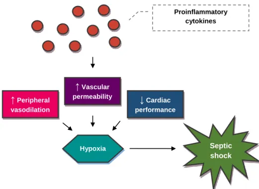
As the syndrome progresses, widespread microvascular thrombosis develops, further hindering blood flow and tissue oxygenation.81,131,132 Ultimately, this hemodynamic instability evolves into myocardial depression, followed by cardiovascular collapse, the establishment of the hypodynamic phase of SIRS, and the development of septic shock (Figure 3).51,133 Systemic oxygen delivery becomes insufficient to meet the demands of the tissues and generalised tissue hypoxia occurs, leading to the increased production of lactate due to anaerobic cellular respiration.134,135 The resulting tissue hypoxia is a consequence of generalised inflammation, and it may also further amplify the inflammatory response by inducing the production of additional proinflammatory cytokines.136
Thus, septic shock is a complex type of shock that not only includes elements of distributive shock due to increased vascular permeability but also of hypovolemic and cardiogenic shock as a result of peripheral vasodilation and reduced cardiac output. 81
Figure 3. The pathophysiology of septic shock (original figure). ↑ Peripheral vasodilation ↓ Cardiac performance ↑ Vascular permeability Hypoxia Proinflammatory cytokines Septic shock
15
1.2.2.5.
T
HE MULTIPLE ORGAN DYSFUNCTIONSYNDROME
(MODS)
The multiple organ dysfunction syndrome (MODS) is the ultimate sequela of SIRS and represents an increased risk of death.49,132,137 In fact, Kenney et al.138 have shown that the mortality rate of canine patients with sepsis suffers an increase for each additional dysfunctional organ system. MODS is characterised by the need of intervention to maintain homeostasis, which would be otherwise accomplished by adequate organ function.5
When organ dysfunction is the outcome of a systemic inflammatory reaction, the resulting phenomenon is classified as secondary MODS for the reason that its development is a consequence of the host’s response to an insult. However, MODS can also be the direct result of the damage caused by the insult itself. In this case, the syndrome is identified as primary MODS, and it tends to unfold rather quickly.5 For example, a patient that has been hit by a moving vehicle may quickly develop acute lung injury as a result of traumatic pulmonary contusion. If this is not the case, the inflammatory reaction caused by the incident itself may become excessive and cause damage to the lungs, as well as to other organs.137
The pathogenesis of secondary MODS is not entirely understood, but there appear to be many contributing factors to the development of organ failure.139,140 The hemodynamic changes resulting from the dysregulated inflammatory response in SIRS play a major role in the promotion of organ damage.82,137,140 Diminished tissue perfusion as a result of microvascular dysfunction and thrombosis leads to tissue hypoxia and cell death, which added to the increased apoptosis observed in SIRS, results in both organ damage and the release of additional DAMPs that perpetuate the inflammatory process.116,132,139,141,142 Neutrophils that are recruited and activated during SIRS also contribute to the development of organ damage, not only by secreting additional inflammatory mediators that potentiate the inflammatory response but also by causing local tissue damage through the release of ROS and proteolytic enzymes.82,140,142
16
Mitochondrial dysfunction is considered to be highly involved in the pathogenesis of MODS.143,144 Generalised tissue hypoxia resulting from an exaggerated inflammatory response may compromise the mitochondrial function of generating adenosine triphosphate (ATP). Furthermore, the excessive amount of NO and ROS in circulation can cause direct damage to mitochondrial structures, such as the lipid membrane, and suppress mitochondrial respiration and ATP synthesis.145 Low levels of triiodothyronine (T3) resulting from thyroid dysfunction in critical illness are also believed to have an adverse impact on mitochondrial activity.145,146 Cell death occurs in the absence of ATP and with it the eventual alteration of organ function.144,145 Interestingly, the mitochondrial dysfunction caused by ROS seems to trigger the production of additional ROS by the mitochondria themselves, further amplifying the oxidative damage caused.147 Additionally, damaging the mitochondria perpetuates the inflammatory response due to mitochondrial DNA being released and acting as a DAMP.84,148
Clinical signs of dysfunction of the gastrointestinal tract (GIT) include changes in appetite such as hyporexia and anorexia, as well as vomiting, gastric ulceration, and diarrhoea.137,149 However, a dysfunctional GIT may also contribute to MODS through the
phenomenon of bacterial translocation.150–152 Bacterial translocation is the passage of bacteria or antigenic macromolecules from the GIT to normally sterile tissues and organs, through the intestinal mucosal barrier.153,154 Reduced oxygen delivery to the GIT, once again as a result of the hemodynamic changes induced by SIRS, culminates in intestinal ischemia, epithelial cell injury and apoptosis, and increased intestinal permeability.155–157 Additionally, hypoperfusion of the GIT results in reduced intestinal motility that promotes bacterial overgrowth.158,159 Furthermore, the absence of luminal nutrients due to undernutrition in critically ill patients further compromises the functional and structural integrity of the intestinal epithelium.156,160 Both the dysfunction of the intestinal barrier and bacterial overgrowth, as well as the presence of a dysfunctional immune system, favour bacterial translocation.152–155,161
Early theories regarding bacterial translocation suggested that bacteria would reach the systemic circulation solely via the portal vein. However, this hypothesis was eventually rejected as new conflicting data emerged.162–164 It is currently believed that according to the “gut-lymph hypothesis”, the translocating bacteria and bacterial products are exposed
17
to intestinal immune cells and stimulate the release of inflammatory mediators.151,157,164 Although the majority of bacteria suffer phagocytosis and contribute to this local inflammatory response, a small number of translocated bacteria survive and become trapped in the intestinal lymph nodes, where additional inflammatory reactions are induced.161,165 Surviving bacteria, cell wall fragments and protein components of the dead bacteria, and cytokines and chemokines generated in the GIT then travel through the mesenteric lymphatics to the cisterna chyli and are released into systemic circulation via the thoracic duct. These products initially reach the pulmonary circulation and activate the alveolar macrophages. The end result of this process is the development of acute lung injury, along with the intensification of systemic proinflammatory activity and MODS.151,153
Interestingly, the lungs are common targets of organ damage in patients with MODS.137 Pulmonary damage as a result of a deleterious inflammatory response often leads to the development of acute lung injury, followed by its most severe presentation, the acute respiratory distress syndrome (ARDS).166 The process through which systemic
inflammation promotes ARDS involves the infiltration of activated neutrophils into the pulmonary interstitium and alveolus, epithelial and endothelial cell damage and apoptosis, and increased microvascular permeability, followed by pulmonary oedema, atelectasis and interstitial fibrosis.111,139,140,167 Clinical signs of this pulmonary dysfunction may include respiratory distress, tachypnea, progressive hypoxemia, and cyanosis.166
Dysfunction of the liver may also be observed in patients with MODS. Hepatic injury contributes to the establishment of hypoglycaemia as a result of reduced gluconeogenesis and glycogenolysis. Protein synthesis, along with lactate and amino acid clearance, also become decreased following hepatic dysfunction.168 Furthermore, activated Kupffer cells are responsible for producing a variety of inflammatory mediators that end up contributing to the local and systemic inflammatory responses.140,168 The main manifestations of hepatic dysfunction tend to be hyperbilirubinemia, as a result of intrahepatic cholestasis, and elevated levels of serum aminotransferases.137,168
Cardiac dysfunction in MODS is often present in the form of myocardial depression.139 The mechanism that leads to the development of myocardial depression is incompletely
18
understood and appears to be multifactorial.140,169 In addition to the previously mentioned harmful effects of systemic inflammation, cardiac dysfunction might be associated with alterations in calcium physiology, sympathetic overstimulation, and the presence of circulating myocardial depressant substances that are yet to be fully identified.137,169–171 Manifestations of cardiac dysfunction may include hypotension despite fluid resuscitation, presence of arrhythmias, and tachycardia.137,140,169 Cats may also uniquely display bradycardia, which is thought to be the consequence of increased vagal tone or cytokine-associated myocardial depression.58
The main phenomenon contributing to the development of acute kidney injury (AKI) and subsequent kidney dysfunction in MODS seems to be the increased epithelial cell apoptosis induced by inflammatory cytokines, whereas renal epithelium necrosis as a result of renal hypoperfusion appears to be less common.84,116,140 Not only does renal dysfunction promote an increase in serum creatinine concentration values but it may also contribute to the development of neurologic dysfunction.172 The process behind the dysfunction of the CNS is rather complex and involves the activation of cerebral endothelial cells and consequent alteration of the blood-brain barrier. The disruption of the blood-brain barrier causes the release of a variety of mediators into the brain that contributes to the activation of microglial cells, which are the local immune cells. These are then responsible for releasing proinflammatory mediators such as cytokines, NO, and ROS which cause local injury and perpetuate the dysfunction of the blood-brain barrier.173 Encephalopathy and peripheral neuropathy are the repercussions of CNS damage in MODS, as well as the deterioration of the mental statuses of the affected patients.137,167 Another sequela of SIRS is the occurrence of critical illness-related corticosteroid insufficiency (CIRCI) due to the dysfunction of the HPA axis and subsequent adrenal insufficiency.126,174–176 The HPA axis is activated in response to the stress caused by the systemic inflammatory insult.177 Activation of the HPA axis ultimately leads to increased cortisol release from the adrenal cortex.176 This increase in cortisol production is important in the organism’s adaptation to illness and the magnitude of its release tends to be proportional to the severity of stress.146,176,178 Cortisol contributes to the maintenance of adequate perfusion to the vital organs by aiding in the modulation of the immune response and in the preservation of vascular reactivity to circulating
19
catecholamines.135,146,176 However, this response weakens as SIRS progresses, resulting in reduced adrenal function and the establishment of CIRCI.126,179 CIRCI represents the inadequacy in corticosteroid activity for the severity of a patient’s illness, and it can be the result of adrenal failure or tissue resistance to corticosteroids.176,180,181 Even though CIRCI tends to disappear with the resolution of SIRS, it is possible that some patients develop long-term adrenal insufficiency due to structural damage to the adrenal glands as a result of haemorrhage and ischemia.146,180 CIRCI can lead to further hemodynamic instability along with persistent hypotension.41,146
1.3.
C
OAGULOPATHY IN SEPSIS
Sepsis is associated with haemostatic abnormalities resulting from the dysfunctional activation of blood coagulation throughout the process of systemic inflammation.83,140,182 The promotion of clotting observed in SIRS results in coagulation abnormalities that range from subclinical clot formation to widespread microvascular thrombosis and haemorrhage which are typical of disseminated intravascular coagulation (DIC).183,184 The coagulation disorders that accompany sepsis are major contributors to the development of MODS and are thus associated with increased mortality.83,132,137,140,185 The systemic inflammatory response present in SIRS is responsible for inducing dysfunctional coagulation through three primary mechanisms: increased activation of blood coagulation, impairment of anticoagulant mechanisms and suppression of fibrinolysis.128,182,185,186
1.3.1.
F
ROM SYSTEMIC INFLAMMATION TO THEACTIVATION OF BLOOD COAGULATION
Coagulation used to be traditionally described through a cascade model involving independent intrinsic and extrinsic pathways. At the present time, however, this classification is deemed outdated, and a newer cell-based model is considered to offer a
20
better description of the coagulation process. This contemporary model describes coagulation through three different phases: initiation, amplification, and propagation.187–
191
Tissue factor (TF) is a 47 kDa transmembrane glycoprotein whose expression plays a central role in the activation of blood coagulation in sepsis.132,192 The disruption of vascular integrity caused by inflammation leads to the exposure of TF in cells which are not in circulation or direct contact with blood.186,193 Furthermore, cytokines released throughout the systemic inflammatory process, such as TNFα, IL-1, and IL-6, are responsible for inducing endothelial, immune, and various other cell types to express TF.132,186,194–196
Once exposed to the bloodstream, TF binds to circulating coagulation factor VII (FVII), also known as proconvertin, converting it to its active form (FVIIa) and generating an active TF-FVIIa complex.191,193,197 This complex is then responsible for activating factor IX (FIX), also called Christmas factor, to FIXa, and factor X (FX), also known as Stuart-Prower factor, to FXa.190,191,198 FIXa also further activates FX by interacting with factor
VIII (FVIII), also named antihaemophilic factor A, in its active form (FVIIIa).191,197 In turn, FXa forms a complex with factor V (FV), or proaccelerin, in its active form (FVa). The formed complex is then responsible for inducing the cleaving of prothrombin to thrombin.189,191,193,197 The aforementioned process represents the initiation phase of coagulation.187,189–191
FXa is capable of generating a small amount of thrombin by itself, which in turn is responsible for activating FV and FVIII and subsequently bolstering further thrombin production.190,191,199 Initially generated thrombin activates nearby platelets, which are essential in the amplification of the coagulation process.190,193,199,200 During the inflammatory response, exposed collagen as well as circulating endotoxin and proinflammatory mediators, such as platelet-activating factor, may also activate platelets.186,187,190,193,200,201 The activation of a platelet leads to the expression of P-selectin on its membrane. Similarly, activated endothelial cells also express P-selectin. P-selectin is a glycoprotein that mediates the adherence of platelets to endothelial cells and leukocytes, which helps to localise thrombus formation. Additionally, these interactions lead to further NF-kB activation and monocyte TF expression.186,189,193,201 Activated
21
platelets and endothelial cells also release a glycoprotein called von Willebrand factor (vWF) which enhances both platelet aggregation and adherence to the site of injury.77,189 As the platelet aggregate grows, a temporary platelet plug is formed.191,200 Once this localised plug is established, the activated platelets augment thrombin generation by providing a procoagulant phospholipid surface on which thrombin can convert FV to FVa and FVIII, which is initially bound to vWF, to FVIIIa.190,191,193,201 Calcium acts as a cofactor in many interactions throughout the coagulation process by facilitating coagulation factor assembly on phospholipid membranes, such as those of activated platelets.189,191,197,199,202,203
The activation of platelets and generation of FVa and FVIIIa represent the amplification phase of the coagulation process, whereas the resulting increased thrombin generation represents the propagation phase (Figure 4).187,189–191
Figure 4. The current concept of coagulation in sepsis (original figure). Tissue factor
forms a complex with factor VIIa (FVIIa) that ultimately leads to the generation of trace amounts of thrombin. The generated thrombin then activates factor V (FV) and factor VIII (FVIII) on the membrane of activated platelets, which results in a substantial increase in thrombin production.190,191,197 Tissue Factor FVII TF-FVIIa FIXa FVIIa FX FXa FIX FVIIIa FVa Prothrombin Thrombin FV FVIII
22
The propagation phase results in the generation of a burst of thrombin that causes the conversion of fibrinogen to fibrin.187,189 Thrombin additionally activates factor XIII (FXIII), also known as fibrin-stabilising factor, whose function is to cross-link the fibrin now incorporated in the platelet plug, granting it enhanced strength and stability. Furthermore, thrombin activates the thrombin-activatable fibrinolysis inhibitor (TAFI), an enzyme that helps prevent the fibrinolysis of the newly formed thrombus.187,191
1.3.2.
I
MPAIRMENT OF ANTICOAGULANT MECHANISMSPhysiological anticoagulant pathways exist to prevent blood coagulation from becoming excessively activated. During systemic inflammation, however, these mechanisms may become suppressed. There are three main antithrombotic mechanisms through which procoagulant activity is regulated. These include the anticoagulant activity of the tissue factor pathway inhibitor (TFPI), protein C, and antithrombin (AT).73,188,193,204
The majority of TFPI is bound to the microvascular endothelium. Smaller amounts of this glycoprotein can also be found in circulation, either bound to plasma lipoproteins or in free form, and within the cytoplasm of platelets.203,205–207 TFPI is released in response to thrombin and other stimulants. Interestingly, heparin is a potent inducer of TFPI release.205,207,208 TFPI inhibits the production of thrombin by binding to and inactivating FXa and the TF-FVIIa complex.83,207,209,210 In sepsis, the production of TF that accompanies the systemic inflammatory response appears to overwhelm the generation of TFPI, thus promoting a procoagulant state.132,206,211–213 Furthermore, an enzyme called neutrophil elastase, which is released by activated neutrophils during inflammation, is responsible for causing the proteolysis of TFPI, preventing it from inactivating FXa and the TF-FVIIa complex.212,214,215 Studies with animal models have shown that both the administration of TFPI and the inhibition of neutrophil elastase, in sepsis, were associated with improved survival.216,217
Protein C is a circulating glycoprotein which is activated by thrombin. Once activated, protein C degrades FVa and FVIIIa, limiting further thrombin generation.189,191,193,203,218 Additionally, thrombin complexes with a transmembrane receptor present on endothelial
23
cells named thrombomodulin. The creation of this complex enhances protein C activation which leads to a substantial increase in the generation of activated protein C (aPC).189,191,203,219 Protein C activation is further amplified by the presence of another receptor found on the membrane of endothelial cells, the endothelial protein C receptor (EPCR), that binds to it and optimally presents it to the complex formed between thrombin and thrombomodulin.193,203 The combination of protein C consumption and reduced production due to organ dysfunction, namely the dysfunction of the liver, where it is synthesised, is likely the reason why protein C levels become reduced in septic patients, contributing to the development of a procoagulant state and increased mortality.203,220–223 Protein S, another glycoprotein which acts as a cofactor to aPC in the inactivation of FVa and FVIIIa, may also contribute to the development of a procoagulant state by becoming reduced in a similar fashion.182,191,203,224 The anticoagulant capability of Protein S is not confined to its interaction with protein C, as it is also responsible for enhancing the interaction between TFPI and FXa and inhibiting the complex formed between FVa and FXa.191,203,225 Moreover, endotoxin, IL-1, and TNFα are all responsible
for inhibiting the expression of thrombomodulin and EPCR.184,222,226–228
Thrombomodulin activity is further impaired by neutrophil elastase which cleaves it from the endothelial cell membrane, generating a less active form of the receptor.184,185,222,228,229 AT is another circulating glycoprotein with anticoagulant properties mainly due to its ability to bind to and inhibit thrombin, as well as other coagulation factors such as FIXa and FXa.191,222,230–232 The presence of heparin highly improves the inhibitory ability of AT. However, physiological circulating levels of heparin are not high enough to significantly contribute to the activation of AT.191,231,233 Thus, in the absence of heparin, AT is activated by endogenous glycosaminoglycans, such as heparan sulphate, expressed on the surface of endothelial cells.224,232–235 In sepsis, AT levels are considerably reduced due to its consumption caused by continued thrombin generation. Reduced synthesis and degradation by neutrophil elastase also contribute to the depletion of AT during severe inflammation.182,193,203,222,223 Furthermore, proinflammatory cytokines released during the inflammatory response suppress the production of glycosaminoglycans on the endothelial surface, subsequently impairing AT function.184,193,224,235
24
1.3.3.
S
UPPRESSION OF FIBRINOLYSISFibrinolysis exists as a parallel mechanism through which haemostasis is regulated.188,191,235,236 For fibrinolysis to occur, plasminogen must be converted to plasmin. Plasminogen is primarily synthesised in the liver and requires posterior activation to plasmin to perform its fibrinolytic function.203,236,237 Plasminogen is usually activated once incorporated into the clot, which is only possible due to its affinity for fibrin.203,238 Following its conversion, plasmin causes the proteolysis of fibrin, dissolving the fibrin clot into fibrin degradation products, which are cleared by the liver.191,238 Fibrin itself enhances plasminogen activation.190,239
The main plasminogen activating enzymes include the tissue plasminogen activator (TPA) and the urokinase-type plasminogen activator (UPA).188,203,236,239 TPA is the most important plasminogen activator. It is synthesised by endothelial cells and released both constitutively and as a response to a variety of triggers including cell injury and thrombin stimulation.188,190,191,236,238,239 TPA requires the presence of fibrin to adequately activate plasminogen.190,239,240 In comparison, UPA appears to play a minor role in the conversion
of plasminogen to plasmin. It can, however, be produced by a larger number of cells, including monocytes, endothelial cells, and epithelial cells, and is released in response to cell activation by endotoxin and inflammatory cytokines.241,242 Unlike TPA, UPA binds to specific cell surface receptors named urokinase-type plasminogen activator receptors (UPARs), and not fibrin, to activate plasminogen.203,236,240
Fibrinolysis is limited by the activity of the previously mentioned TAFI and by plasminogen activator inhibitors (PAIs), both of which are suppressed by aPC. While TAFI reduces the rate of fibrinolysis by protecting fibrin from the breakdown caused by plasmin, PAIs prevent the activation of plasminogen by irreversibly inhibiting both TPA and UPA.191,203,204,235,243 The main PAI is the plasminogen activator inhibitor type 1 (PAI-1), which is produced by a miscellany of cells including platelets, leukocytes, and endothelial cells.203,219,236,239 Fibrinolysis is further suppressed by circulating plasmin inhibitors, such as alpha-2-antiplasmin and alpha-2-macroglobulin.191,203,219,238