The genetics of canine periodontal disease
–
A candidate gene approach
Tese de Doutoramento em Ciências Veterinárias
Carlos Manuel Ramos Albuquerque
Orientadores:
Professor Doutor Carlos Alberto Antunes Viegas
Professora Doutora Estela Maria Bastos Martins de Almeida
The genetics of canine periodontal disease
–
A candidate gene approach
Tese de Doutoramento em Ciências Veterinárias
Carlos Manuel Ramos Albuquerque
Orientadores:
Professor Doutor Carlos Alberto Antunes Viegas, Universidade de Trás-os-Montes e Alto Douro
Professora Doutora Estela Maria Bastos Almeida, Universidade de Trás-os-Montes e Alto Douro
Composição do Júri:
Professor Doutor Vicente de Seixas e Sousa, Universidade de Trás-os-Montes e Alto Douro
Professora Doutora Aura Antunes Colaço, Universidade de Trás-os-Montes e Alto Douro
Professor Doutor Augusto de Matos, Instituto de Ciências Biomédicas Abel Salazar
Professor Doutor Fernando Guerra, Faculdade de Medicina – Universidade de Coimbra
Professor Doutor Victor Alves, Faculdade de Medicina Veterinária – Universidade Técnica de Lisboa
Professor Doutor Carlos Alberto Antunes Viegas, Universidade de Trás-os-Montes e Alto Douro
Professora Doutora Estela Maria Bastos Almeida, Universidade de Trás-os-Montes e Alto Douro
The research described in this Thesis was financially supported by the Portuguese Foundation for Science and Technology (FCT) under the Carlos Manuel Ramos Albuquerque’s PhD scholarship (SFRH/BD/44490/2008).
I declare that the contents of this Thesis are my own work and that they have not been presented to any University other than the University of Trás-os-Montes and Alto Douro.
To my Family and Friends
“The important thing in science is not so much to obtain new facts as to discover new ways of thinking about them.” (Sir William Bragg)
No final de cada etapa devemos estar orgulhosos do trabalho desenvolvido, principalmente quando reconhecemos nele todo o nosso empenho e dedicação. A escrita de uma tese pocura demonstrar e materializar o trabalho que foi desenvolvido, mas um doutoramento e tudo aquilo que um período de dedicação de vários anos encerra, ultrapassam grandemente o que fica aqui registado. O caminho percorrido, as dificuldades e a forma como foram enfrentadas e ultrapassadas com o contributo de várias pessoas tem que ser valorizado e meditado para que juntamente com o cientista cresça também o homem. Uma carreira académica deve ser isso mesmo, um contínuo crescimento científico e humano. Assim, esta pequena secção de agradecimentos representa essa vertente tão importante e para a qual contribuíram as pessoas que foram e são parte integrante deste caminho que continuaremos a percorrer juntos.
Em primeiro lugar, agradeço à Universidade de Trás-os-Montes e Alto Douro (UTAD), na pessoa do Magnífico Reitor Professor Doutor António Fontainhas Fernandes, por autorizar e criar condições para o desenvolvimento do trabalho.
Agradeço aos meus orientadores, Professor Carlos Viegas e Professora Estela Bastos que aceitaram percorrer comigo este caminho partilhando o seu entusiasmo e suportando as minhas dificuldades. Mantendo o papel de orientadores, souberam ser parceiros e amigos. O meu sincero obrigado pelos muitos ensinamentos, pela enorme compreensão com algumas das minhas decisões e por serem um grande exemplo humano.
Agradeço à Professora Maria dos Anjos Pires pela sua colaboração no trabalho e principalmente pela forma como se disponibilizou sempre que precisei. Agradeço também à Professora Isabel Dias por toda a colaboração e amizade. No núcleo de Desporto da UTAD quero agradecer ao Professor José Carlos Leitão e o António Cortinhas que, de uma forma incansável, me ajudaram a superar muitos dos obstáculos estatísticos. Ao Professor José Luís Castrillo e ao Professor Ivo Gut agradeço pela valiosa colaboração.
Ao Francisco Morinha poderia agradecer tudo aquilo que me ensinou e que me permitiu “sobreviver” num laboratório de genética molecular, o inesgotável tempo e a paciência que
me dedicou; mas isso seria um agradecimento demasiado curto. Agradeço simplesmente ter-te conhecido e ao teu enorme exemplo de genuíno entusiasmo e dedicação à ciência e a tua gigante dimensão humana.
À Joana Magalhães tenho que agradecer ter-me mostrado que existem pessoas genuinamente boas, com um caráter e um altruísmo infinitos. Teria muitas coisas a agradecer-te, por isso: Joaninha, obrigado por tudo!
Ao João Requicha nem me atrevo a destacar quaisquer agradecimentos. Já partilhamos o mesmo caminho há mais de dez anos e há um momento nas amizades a partir do qual não é necessário pedir quaisquer ajudas nem faz sentido agradecer gestos. Tudo isso acontece de forma natural simplesmente porque tem que ser assim, e isso é a verdadeira amizade!
Aos colegas de laboratório, principalmente à Leonor Pereira, à Sónia Gomes, à Ana Pereira e à Vanessa Rodrigues, por me terem recebido tão bem e pela boa disposição constante. Também agradeço a toda a equipa da Hospital Veterinário da UTAD por toda a ajuda e colaboração. Assim como ao Cantinho dos Animais Abandonados de Viseu e à sua grande líder, Dona Ana, por terem colaborado connosco e acima de tudo pelo magnífico trabalho que desenvolvem na defesa dos animais.
À Joana Valente, ao Luís Sousa, ao Maurino, à Luísa Viterbo, à Cristina Sousa e à Ana Jacinta por terem sido durante estes anos a minha família vila-realense. Cada um à sua maneira, vocês foram realmente importantes neste percurso. Obrigado!
À minha mãe e minha irmã por serem o meu grande suporte e que, de uma forma totalmente incondicional, sempre me apoiaram e apoiam, mesmo quando o caminho se torna tortuoso e as ausências são prolongadas. Não sou talhado para demonstrar afetos, mas vocês sabem que eles existem e que são enormes. Este agradecimento é também extensível ao Zé Manel e à Tia Fernanda.
Por fim, queria dedicar este trabalho ao meu pai, simplesmente porque sei o enorme orgulho que sentiria ao ver-me chegar aqui!
The genetics of canine periodontal disease – A candidate gene approach ________________________________________
Periodontal disease (PD) refers to a group of inflammatory diseases caused by bacterial plaque in the periodontium and ranges from an early stage (gingivitis) to an advanced stage (periodontitis). It is a multifactorial disease that results from the interaction of the host defence mechanisms with the plaque microorganisms. PD has an enormous impact on human medicine and veterinary medicine due to its high prevalence as well as its local and systemic implications. Dog model has been extensively used in oral disease research, contributing significantly to the current understanding of periodontology. The most important clinical aspects of canine PD were considered in this work and the various animal models were examined with emphasis on the role of the dog as the most useful model for understanding human PD and to develop new therapeutic and preventive measures.
In recent decades, it has been consolidated the idea of the genetics influence in PD by controlling the inflammatory process severity and the therapy responses. Various single nucleotide polymorphisms have been identified as risk factors, mainly in genes responsible for molecules involved in immunoregulation and/or metabolism, but many questions still remain. In canine PD, this is a completely unexplored issue but a highly relevant and promising research field namely because the strong similarity between canine and human disease provides the possibility to share the knowledge attained from one species to another, with mutual benefits.
Following a comparative genomics approach to identify the most promising candidate genes, the main goal of this work was to contribute for a better characterization of canine PD, particularly in terms of genetic basis. Five candidate genes (IL1A, IL1B, IL10, IL6 and LTF) encoding molecules with recognized relevant role in the PD pathogenesis (interleukin-1α, interleukin-1β, interleukin-10, interleukin-6 and lactotransferrin, respectively) were selected and case-control studies were delineated, in which a molecular analysis of each gene was performed to identify genetic variations and to evaluate its possible association with PD. It
was hypothesized that in canine PD, similar to human PD, a disfunction or a dysregulated production of these molecules resulting from genetic variations can be part of the explanation for the differences in disease susceptibility between individuals.
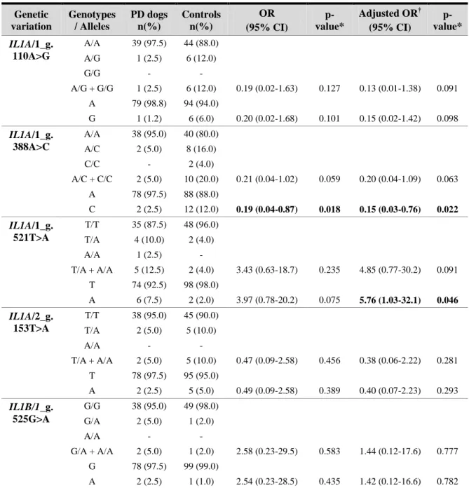
A total of twenty-six genetic variations were identified and analyzed, eight in the IL1A and IL1B genes, seven in the IL10 gene, three in the IL6 gene, and eight in the LTF gene. The IL1A/1_g.388A>C and IL1A/1_g.521T>A variations showed statistically significant differences between groups [adjusted OR (95%CI): 0.15 (0.03-0.76), p=0.022; 5.76 (1.03-32.1), p=0.046, respectively], meaning that, in the studied population, the IL1A/1_g.388C allele is associated with a decreased PD risk, whereas the IL1A/1_g.521A allele is associated with an increased risk. Regarding all the others variations, no statistically significant differences were detected, but the IL1A/2_g.515G>T, IL10/2_g.285G>A, IL6/2_g.105G>A, LTF/3_g.411C>T, LTF/3_g.420G>A and LTF/3_g.482G>A variations resulted in a change of encoded amino acid, which may alter protein structure and function, as demonstrated by different bioinformatics tools.
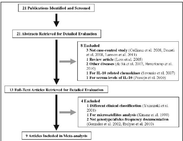
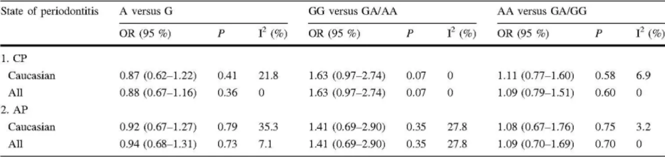
Before the molecular analysis of the IL10 gene, two additional studies were delineated. Considering that no clear consensus has been reached about the association of IL10 polymorphisms and human PD, a meta-analysis of all available studies was performed. It was found statistically significant association of IL10-819(-824)C>T and IL10-592(-597)C>A polymorphisms, with IL10-819(-824)T and -592(-597)A alleles conferring a relative increased risk for chronic periodontits in Caucasians. Additionally, a study to evaluate the levels of interleukin-10 in plasma of dogs with periodontitis was delineated assessing a possible correlation between these levels and periodontal condition, being found lower levels in the periodontitis group comparing with the control group.
The outcome from this work suggests that dog IL1A, IL1B, IL10, IL6 and LTF genes, as occurs in the human orthologous genes, are highly polymorphic with genetic variants that may be important in PD susceptibility. The results obtained for the IL1A are particularly relevant, but this is the first work in this issue and further studies are essential to reinforce these findings and to clarify its biological importance; as well as other studies with different candidate genes. But, it is undeniable that advances in this area are fundamental to understand properly the complex causal pathways of PD and to improve the clinical management of PD,
these objectives, which may lead to great benefits in human and veterinary periodontology, adding important knowledge to design new preventive and therapeutic strategies, and ultimately to improve health in both humans and dogs.
Keywords: Periodontal disease; Animal models; Dog; Genetic variations; Single nucleotide polymorphism, Genetic susceptibility; Clinical genomics
A genética na doença periodontal canina – Estudo de genes candidatos ________________________________________
A doença periodontal (DP) constitui o grupo de doenças inflamatórias causadas pela placa bacteriana no periodonto, incluindo o espectro de doença desde estádios precoces (gengivite) até estádios avançados (periodontite). É uma doença multifatorial resultante da interação dos mecanismos de defesa do hospedeiro com os microrganismos da placa. A DP tem um enorme impacto na medicina humana e na medicina veterinária, pela sua elevada prevalência e pelas suas implicações locais e sistémicas. O modelo cão tem sido extensivamente usado na investigação das doenças orais, contribuindo significativamente para o conhecimento atual em periodontologia. Os aspetos clínicos mais importantes da DP canina são apresentados neste trabalho e são explorados os diferentes modelos animais, com particular relevância para o papel do cão como o modelo mais útil para estudar a DP e o desenvolvimento de novas medidas profiláticas e terapêuticas.
Nas recentes décadas tem sido consolidada a ideia da influência genética na DP, controlando a gravidade do processo inflamatório e a resposta terapêutica. Vários polimorfismos de nucleótidos simples têm sido identificados como fatores de risco, nomeadamente em genes responsáveis por moléculas envolvidas na imunoregulação e/ou metabolismo, mas muitas dúvidas ainda persistem. Na DP canina este é um assunto completamente inexplorado, mas um campo de investigação muito relevante e promissor considerando a elevada similaridade da doença no cão e no homem, o que permite a partilha de conhecimento entre espécies com benefícios mútuos.
Seguindo uma abordagem de genómica comparativa para selecionar os genes candidatos mais promissores, o principal objetivo deste trabalho foi contribuir para uma melhor caracterização da DP no cão, principalmente da sua base genética. Foram selecionados cinco genes (IL1A, IL1B, IL10, IL6 e LTF) que codificam moléculas com um papel relevante na patogenia da DP (interleucina-1α, interleucina-1β, interleucina-10, interleucina-6 e lactotransferrina, respetivamente) e foram delineados estudos caso-controlo, nos quais foi realizada uma análise
molecular de cada gene procurando identificar variações genéticas e avaliar a sua possível associação à DP. Foi colocada a hipótese de que na DP canina, similarmente à DP do homem, uma disfunção ou produção desregulada destas moléculas, resultante de variações genéticas, poderá ser parte da explicação das diferenças de susceptibilidade entre indivíduos.
Foram identificadas vinte e seis variações genéticas, oito nos genes IL1A e IL1B, sete no gene IL10, três no gene IL6 e oito no gene LTF. As variações IL1A/1_g.388A>C e IL1A/1_g.521 T>A apresentaram diferenças estatisticamente significativas entre grupos [OR ajustada (95%CI): 0.15 (0.03-0.76), p=0.022; 5.76 (1.03-32.1), p=0.046, respetivamente], significando que, na população estudada, o alelo IL1A/1_g.388C está associado a um menor risco de DP, enquanto que o alelo IL1A/1_g.521A se associa a um risco acrescido. Relativamente às outras variações genéticas não foram encontradas diferenças estatisticamente significativas, mas as variações IL1A/2_g.515G>T, IL10/2_g.285G>A, IL6/2_g.105G>A, LTF/3_g.411C>T, LTF/3_g.420 G>A e LTF/3_g.482G>A conduzem à alteração do aminoácido codificado, o que pode alterar a estrutura e a função protéicas, como demonstrado por várias ferramentas bioinformáticas.
Previamente à análise molecular do gene IL10, foram delineados dois estudos. Pela inexistência de um consenso claro relativamente à associação de polimorfismos no gene IL10 e a DP no homem, foi realizada uma meta-análise de todos os estudos disponíveis. Foi encontrada associação estatisticamente significativa para os polimorfismos IL10-819(-824)C>T e IL10-592(-597)C>A, em que os alelos IL10-819(-824)T e -592(-597)A conferem um acréscimo relativo do risco para periodontite crónica em Caucasianos. Adicionalmente foi delineado um estudo para avaliar os níveis plasmáticos de interleucina-10 em cães com periodontite, investigando a possível correlação destes níveis com a saúde periodontal, e os níveis plasmáticos medidos foram mais baixos no grupo de doentes comparativamente com o grupo controlo.
Os resultados deste trabalho sugerem que os genes caninos IL1A, IL1B, IL10, IL6 e LTF, tal como acontece nos genes ortólogos na espécie humana, são altamente polimórficos e com algumas variações genéticas que podem ser importantes na susceptibilidade para a DP. Os resultados obtidos para o gene IL1A são particularmente relevantes, mas sendo este o primeiro trabalho nesta temática, estudos futuros são essenciais para os reforçar e clarificar a sua
complexas vias causais da DP e melhorar o seu acompanhamento clínico, particularmente com o desenvolvimento de novas estratégias de avaliação de risco. A abordagem de genes candidatos, suportada em ferramentas de genómica comparativa, é um caminho de investigação promissor para atingir estes objetivos, que podem resultar em enormes benefícios na periodontologia humana e veterinária, principalmente por acrescentarem conhecimento para delinear novas estratégias preventivas e terapêuticas, e em última análise, melhorar a saúde de ambas as espécies.
Palavras-chave: Doença periodontal; Modelos animais; Cão; Variações genéticas; Polimorfismos de nucleótidos simples, Susceptibilidade genética; Genómica clínica
Acknowledgements / Agradecimentos ... vii
Abstract ... ix
Resumo ... xiii
Table of Contents ... xvii
Index of Tables ... xxi
Index of Figures ... xxiii
Abbreviations List ... xxv
List of Publications / Submitted manuscripts ... xxix
Chapter I – General Introduction ... 1
I.1.CANINE PERIODONTITIS:THE DOG AS AN IMPORTANT MODEL FOR PERIODONTAL STUDIES ... 3
I.1.1. Periodontal disease in the dog ... 3
I.1.1.1. Aetiology and pathogenesis ... 3
I.1.1.2. Clinical presentation ... 4
I.1.1.3. Treatment options ... 8
I.1.2. Research in periodontology ... 9
I.1.2.1. Animal models used in periodontal disease research ... 9
I.1.2.1.1. Non-human primate models ... 9
I.1.2.1.2. Rat and mouse models ... 10
I.1.2.1.3. Hamster models (Mesocricetus auratus) ... 10
I.1.2.1.4. Rabbit models (Oryctolagus cuniculus) ... 10
I.1.2.1.5. Miniature pig models (Sus scrofa domesticus) ... 11
I.1.2.1.6. Domestic ferret models (Mustela putorius furo) ... 11
I.1.2.1.7. Domestic sheep models (Ovis aries) ... 11
I.1.3. The dog as a promising model for gaining new insights into periodontology ... 14
I.2.CLINICAL GENOMICS OF PERIODONTAL DISEASE – CANDIDATE GENES APPROACH ... 17
I.2.1. Interleukin-1 ... 19
I.2.2. Interleukin-10 ... 20
I.2.3. Interleukin-6 ... 22
I.2.4. Lactotransferrin ... 22
I.3.OBJECTIVES ... 23
Chapter II – Materials and Methodologies ... 27
II.1.STUDY POPULATION –CASE AND CONTROL SELECTION ... 29
II.1.1. Odonto-stomatological evaluation ... 29
II.2.WHOLE-BLOOD COLLECTION, STORAGE AND PROCESSING ... 31
II.3.DNA EXTRACTION AND PURIFICATION ... 31
II.3.1. DNA QuickGene whole blood kit S (DB-S) ... 31
II.4.DNA QUANTIFICATION ... 32
II.4.1. Quantification by spectrophotometry ... 32
II.5.IN SILICO ANALYSIS –TARGET REGIONS SELECTION AND PRIMERS DESIGN... 33
II.5.1. IL1A and IL1B genes fragments selection ... 33
II.5.2. IL10 gene fragments selection ... 33
II.5.3. IL6 gene fragments selection ... 34
II.5.4. LTF gene fragments selection ... 34
II.5.5. Primers design ... 34
II.6.POLYMERASE CHAIN REACTION CONDITIONS ... 35
II.6.1. IL1A and IL1B genes ... 36
II.6.2. IL10 gene ... 37
II.7.AGAROSE GEL ELECTROPHORESIS ... 37
II.8.PURIFICATION OF PCR PRODUCTS ... 38
II.9.DNA SEQUENCING ... 39
II.10.ENZYME-LINKED IMMUNOSORBENT ASSAY ... 39
II.11.META-ANALYSIS ... 40
II.11.1. Search strategy ... 40
II.11.2. Inclusion criteria ... 41
II.11.3. Data extraction ... 41
II.12.STATISTICAL ANALYSIS ... 42
II.12.1. Candidate genes (IL1A, IL1B, IL10, IL6, LTF) analysis ... 42
II.12.2. Plasma IL-10 levels analysis ... 42
II.12.3. Meta-analysis ... 43
Chapter III – Results ... 45
III.1.IL1A AND IL1B GENETIC VARIATIONS ... 47
III.2.IL10 GENE ANALYSIS ... 51
III.2.1. Meta-analysis ... 51
III.2.1.1. IL10-1082(-1087)G>A polymorphism and periodontitis ... 53
III.2.1.2. IL10-819(-824)C>T polymorphism and periodontitis ... 55
III.2.1.3. IL10-592(-597)C>A polymorphism and periodontitis ... 56
III.2.2. Plasma IL-10 levels ... 57
III.2.3. IL10 genetic variations ... 59
III.3.IL6 GENETIC VARIATIONS ... 62
Chapter IV – Discussion ... 69
IV.1.IL1A AND IL1B GENETIC VARIATIONS ... 71
IV.2.IL10 GENE ANALYSIS ... 75
IV.2.1. Meta-analysis... 75 IV.2.2. Plasma IL-10 levels ... 80 IV.2.3. IL10 genetic variations ... 83
IV.3.IL6 GENETIC VARIATIONS ... 87
IV.4.LTF GENETIC VARIATIONS ... 91
Chapter V – Final conclusions ... 95
Chapter VI – References ... 101
Chapter I – General Introduction ... 1
I.1.CANINE PERIODONTITIS:THE DOG AS AN IMPORTANT MODEL FOR PERIODONTAL STUDIES ... 3
Table 1.1 – Periodontal disease classification ... 6 Table 1.2 – Animal models of periodontal disease ... 13
Chapter II – Materials and Methodologies ... 27
II.1.STUDY POPULATION –CASE AND CONTROL SELECTION ... 29
Table 2.1 – Main characteristics of the population under study ... 30
II.5.IN SILICO ANALYSIS –TARGET REGIONS SELECTION AND PRIMERS DESIGN ... 33
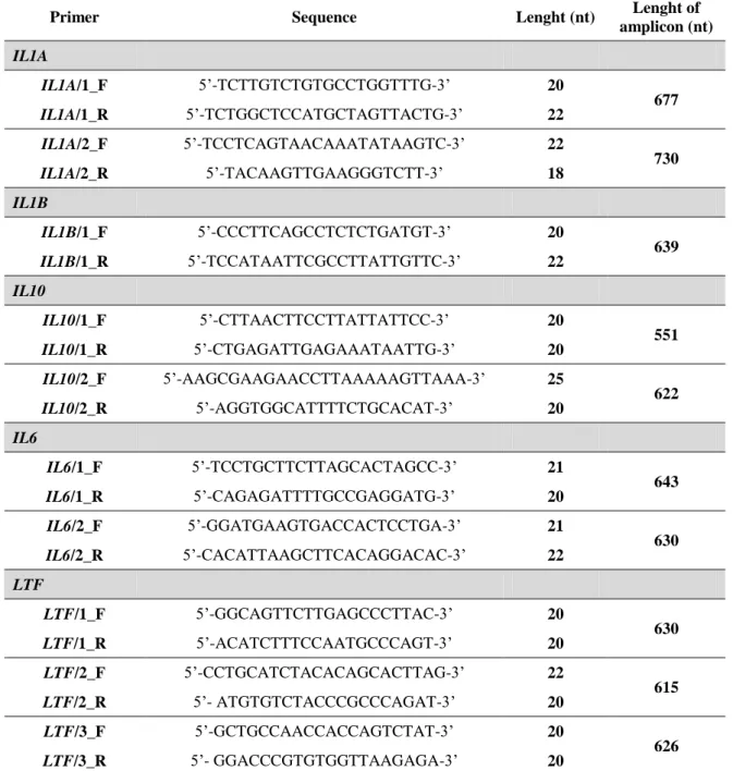
Table 2.2 – Primers used in the studies on the scope of this Thesis ... 35
Chapter III – Results ... 45
III.1.IL1A AND IL1B GENETIC VARATIONS ... 47
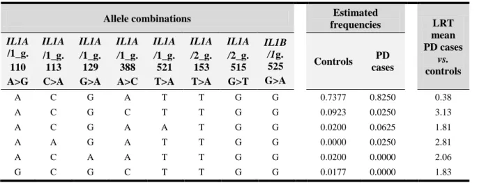
Table 3.1 – Genetic variations identitied in IL1A and IL1B genes ... 47 Table 3.2 – Genotype and allele frequencies of the IL1A and IL1B genes variations ... 50 Table 3.3 – IL1 haplotype estimated frequencies ... 51
III.2.IL10 GENE ANALYSIS ... 51
Table 3.4 – List and characteristics of all eligible studies ... 53 Table 3.5 – Overall risk estimates of the IL10-1082(-1087)G>A polymorphism ... 54 Table 3.6 – Subgroup analysis of the IL10-1082(-1087)G>A polymorphism ... 54 Table 3.7 – Overall risk estimates of the IL10-819(-824)C>T polymorphism ... 55 Table 3.8 – Subgroup analysis of the IL10-819(-824)C>T polymorphism ... 56 Table 3.9 – Overall risk estimates of the IL10-592(-597)C>A polymorphism ... 56 Table 3.10 – Subgroup analysis of the IL10-592(-597)C>A polymorphism ... 57
Table 3.11 – Plasma IL-10 concentrations ... 58 Table 3.12 – Genetic variations identified in IL10 gene ... 59 Table 3.13 – Genotype and allele frequencies of the IL10 genetic variations ... 61 Table 3.14 – IL10 haplotype estimated frequencies ... 62
III.3.IL6 GENETIC VARIATIONS ... 62
Table 3.15 – Genetic variations identified in IL6 gene ... 63 Table 3.16 – Genotype and allele frequencies of the IL6 genetic variations ... 64 Table 3.17 – IL6 haplotype estimated frequencies ... 65
III.4.LTF GENETIC VARIATIONS ... 65
Table 3.18 – Genetic variations identified in LTF gene ... 65 Table 3.19 – Genotype and allele frequencies of the LTF genetic variations ... 67 Table 3.20 – LTF haplotype estimated frequencies ... 68
Chapter I – General Introduction ... 1
I.1.CANINE PERIODONTITIS:THE DOG AS AN IMPORTANT MODEL FOR PERIODONTAL STUDIES ... 3
Figure 1.1 – Periodontal disease ... 5 Figure 1.2 – Periodontium at different stages of health/disease ... 7 Figure 1.3 – Complex multifactorial nature of periodontal disease ... 15 Figure 1.4 – Strategies of risk assessment in periodontal disease ... 16
I.2.CLINICAL GENOMICS OF PERIODONTAL DISEASE – CANDIDATE GENES APPROACH ... 17
Figure 1.5 – Single nucleotide polymorphism (SNP) ... 18
Chapter III – Results ... 45
III.1.IL1A AND IL1B GENETIC VARATIONS ... 47
Figure 3.1 – Nucleotide sequence of the IL1A gene fragments ... 48 Figure 3.2 – Nucleotide sequence of the IL1B gene fragment ... 49
III.2.IL10 GENE ANALYSIS ... 51
Figure 3.3 – Flow diagram of search strategy and study selection ... 52 Figure 3.4 – Plasma IL-10 concentrations... 58 Figure 3.5 – Nucleotide sequence of the IL10 gene fragments ... 60
III.3.IL6 GENETIC VARIATIONS ... 62
Figure 3.6 – Nucleotide sequence of the IL6 gene fragments ... 63
III.4.LTF GENETIC VARIATIONS ... 65
Figure 3.7 – Nucleotide sequence of the LTF gene fragments ... 66
Figure 4.1 – The predicted tertiary structures of IL-1α protein... 74 Figure 4.2 – Interactions of the canine IL-1α and IL-1β proteins ... 75
IV.2.IL10 GENE ANALYSIS ... 75
Figure 4.3 – Analysis of IL-10 amino acid sequences ... 85 Figure 4.4 – Interactions of the canine IL-10 protein ... 86
IV.3.IL6 GENETIC VARIATIONS ... 87 Figure 4.5 – Analysis of IL-6 amino acid sequences and protein structure prediction ... 89 Figure 4.6 – Interactions of the canine IL-6 protein ... 90
IV.4.LTF GENETIC VARIATIONS ... 91
A
A – Adenine
AP – Aggressive periodontitis
Arg-R – Arginine
Asp-D – Aspartic acid
AVDC – American Veterinary Dentistry College B bp – Base pairs C C – Cytosine CI – Confidence interval CP – Chronic periodontitis D del – Deletion
DNA – Deoxyribonucleic acid
dNTPs – Deoxyribonucleotides
E
EDTA – Ethylenediaminetetraacetic acid
ELISA – Enzyme-linked immunosorbent assay
G
G – Guanine
Gln-Q – Glutamine
Glu-E – Glutamic acid
Gly-G – Glycine
H
HCl – Hydrochloric acid
HWE – Hardy-Weinberg equilibrium
I
IL1A – Gene coding for interleukin-1 alpha
IL1B – Gene coding for interleukin-1 beta
IL10 – Gene coding for interleukin-10
IL6 – Gene coding for interleukin-6
IL-1 – Interleukin-1
IL-1α – Interleukin-1 alpha
IL-1β – Interleukin-1 beta
IL-10 – Interleukin-10
IL-6 – Interleukin-6
I2 – Inconsistency index
L
LTF – Lactotransferrin
LTF – Gene coding for lactotransferrin
LD – Linkage disequilibrium
Leu-L – Leucine
LLD – Low limit of detection
LRT – Likelihood-ratio test Lys-K – Lysine M MgCl2 – Magnesium chloride mL - Millilitres mM – Millimolar
mRNA – Messenger RNA
N
NCBI – National Center for Biotechnology Information
ND – Non-detectable
ng – Nanogram
O
OD – Optical density
ORs – Odds ratios
P
pg – Picogram
PolyPhen-2 – Polymorphism Phenotyping v2
Pro-P – Proline
PROVEAN – Protein Variation Effect Analyzer
R
RNA – Ribonucleic acid
Rpm – Revolutions per minute
S
SIFT – Sorting Intolerant from Tolerant
SNP – Single nucleotide polymorphism
SPSS – Statistical Package for Social Sciences
STRING – Search Tool for the Retrieval of Interacting Genes/Proteins
T – Thymine
TBE – Tris-borate-EDTA
U
UTR – Untranslated region
UK – United Kingdom
US – United States of America
V Val-V – Valine vs. – Versus μL – Microlitre χ2 – Chi-square test
The work performed under the scope of this PhD Thesis resulted in the publications / submitted manuscripts listed below.
International Refereed Journals
Morinha F, Albuquerque C, Requicha J, Dias I, Leitão J, Gut I, Guedes-Pinto H, Viegas C, Bastos E (2011) Detection and characterization of interleukin-6 gene variants in Canis familiaris: association studies with periodontal disease. Gene 485(2), 139-145.
Albuquerque C, Morinha F, Requicha J, Martins T, Dias I, Guedes-Pinto H, Bastos E, Viegas C (2012) Canine periodontitis: the dog as an important model for periodontal studies. The Veterinary Journal 191(3), 299-305.
Morinha F, Albuquerque C, Requicha J, Dias I, Leitão J, Gut I, Guedes-Pinto H, Viegas C, Bastos E (2012) Analysis of new lactotransferrin gene variants in a case– control study related to periodontal disease in dog. Molecular Biology Reports 39(4), 4673-4681.
Albuquerque CM, Cortinhas AJ, Morinha FJ, Leitão JC, Viegas CA, Bastos EM (2012) Association of the IL-10 polymorphisms and periodontitis: a meta-analysis. Molecular Biology Reports 39(10), 9319-9329.
Albuquerque C, Morinha F, Requicha J, Dias I, Guedes-Pinto H, Viegas C, Bastos E. (2014) A case-control study between interleukin-10 gene variants and periodontal disease in dogs. Gene 539(1), 75-81.
Albuquerque C, Morinha F, Requicha J, Dias I, Guedes-Pinto H, Bastos E, Viegas C. Variants in the interleukin-1 alpha and beta genes and the risk for periodontal disease in dogs. (Submitted Manuscript)
Albuquerque C, Pires M, Morinha F, Magalhães J, Requicha J, Dias I, Guedes-Pinto H, Bastos E, Viegas C. Plasma interleukin-10 levels in dogs with periodontitis: a case-control study. (Submitted Manuscript)
Non-indexed International Refereed Journal
Albuquerque C, Morinha F, Dias I, Bastos E, Viegas C (2013) Genómica clínica de la enfermedad periodontal. Selecciones Veterinarias – Argentina 21(2):18-22.
Communications in National and International Conferences
Oral communications
Morinha F, Albuquerque C, Requicha J, Dias I, Guedes-Pinto H, Bastos E, Viegas C. Doença periodontal canina - análise do gene da interleucina-6. XVIII Congresso Nacional da Associação Portuguesa de Médicos Veterinários Especialistas em Animais de Companhia - Grupo de Interesse Especial em Estomatologia. Lisbon, Portugal, 29-31 May 2009.
Albuquerque C, Morinha F, Requicha JF, Dias I, Guedes-Pinto H, Bastos E, Viegas C. Susceptibilidad para la enfermedad periodontal – polimorfismos genéticos. Título de Odontología y Cirugía Maxilofacial de la Universidad Complutense de Madrid – Facultad de Veterinaria de la Universidad Complutense de Madrid. Madrid, Spain, 10 June 2009.
Albuquerque C, Morinha F, Requicha J, Dias I, Guedes-Pinto H, Bastos E, Viegas C. Doença periodontal canina – fundamentos genéticos. VI Congresso Ordem Médicos Veterinários. Lisbon, Portugal, 3-6 October 2009.
analysis of new genetic variations in LTF gene. 19th European Congress of Veterinary Dentistry. Nice, France, 23-25 September 2010.
Morinha F, Albuquerque C, Requicha J, Dias I, Pereira A, Leitão J, Guedes-Pinto H, Viegas C, Bastos E. Genetic variations in IL6 and LTF genes: association studies with periodontal disease. 2nd Meeting of the Institute for Biotechnology and Bioengineering. Braga, Portugal, 23-24 October 2010.
Albuquerque C, Morinha F, Bastos E, Viegas C. Susceptibilidade Genética em Periodontologia. I Congresso Ibérico de Medicina Estomatológico-dentária Veterinária. Vila Real, Portugal, 4-6 December 2010.
Morinha F, Albuquerque C, Requicha J, Dias I, Leitão J, Guedes-Pinto H, Viegas C, Bastos E. Estudo de associação de novas variações nos genes IL6 e LTF com a doença periodontal no cão. I Congresso Ibérico de Medicina Estomatológico-dentária Veterinária. Vila Real, Portugal, 4-6 December 2010.
Albuquerque C, Morinha F, Requicha J, Dias I, Guedes-Pinto H, Bastos E, Viegas C. Genética Clínica: Novo paradigma na abordagem da doença periodontal do cão. Autores: III Encontro de Formação OMV/XIII Congresso de Medicina Veterinária em Língua Portuguesa, Lisbon, Portugal, 17-18 November 2012.
Albuquerque C, Pires M, Morinha F, Magalhães M, Requicha J, Dias I, Guedes-Pinto H, Bastos E, Viegas C. Plasma interleukin-10 levels in dogs with periodontitis: a case-control study. 22nd European Congress of Veterinary Dentistry/12th World Veterinary Dental Congress. Prague, Czech Republic, 23-26 May 2013.
Albuquerque C, Morinha F, Requicha J, Dias I, Guedes-Pinto H, Bastos E, Viegas C. Analysis of interleukin-10 gene variants in dogs with periodontal disease. 101st FDI Annual World Dental Congress. Istanbul, Turkey, 28-31 August 2013.
Poster presentations
Albuquerque CM, Requicha JF, Sousa CM, Dias MI, Viegas CA. Higiene profissional da cavidade oral. V Congresso Hospital Veterinário Montenegro - Congresso de Gastroenterologia, Sta. Maria da Feira, Portugal, 17-18 January 2009.
Morinha F, Albuquerque C, Requicha J, Dias I, Guedes-Pinto H, Bastos E, Viegas C. Dog periodontitis - interleukin-6 gene analysis. XXXIV Jornadas Portuguesas de Genética, Lisbon, Portugal, 28-30 April 2009.
Morinha F, Albuquerque C, Requicha J, Dias I, Pereira A, Leitão J, Guedes-Pinto H, Viegas C, Bastos E. Periodontal Disease in dog: study of new genetic variations in two candidate genes (IL6 and LTF). II Jornadas Nacionais de Genética e Biotecnologia. Vila Real, Portugal, 14-15 May 2010.
Morinha F, Albuquerque C, Requicha J, Dias I, Pereira A, Leitão J, Guedes-Pinto H, Viegas C, Bastos E. Genetic Characterization of Periodontal Disease in Dog: Molecular Analysis of IL6 Gene. XXXV Jornadas Portuguesas de Genética. Braga, Portugal, 31 May – 2 June 2010.
Albuquerque C, Morinha F, Requicha J, Martins T, Dias I, Guedes-Pinto H, Bastos E, Viegas C. Canine periodontitis: The dog as an important model for periodontal studies. I Simpósio Inter-Universitário de Investigação em Medicina Dentária. Faculty of Medicine – University of Coimbra, Portugal, 9 March 2013.
Chapter I
General Introduction
I.1. Canine periodontitis: The dog as an important model for periodontal studies
Periodontal disease (PD) refers to a group of inflammatory diseases caused by bacterial plaque in the periodontium (Pihlstrom et al., 2005; Niemiec, 2012). The periodontium contains the supporting structure of the teeth and includes the gingiva, alveolar bone, periodontal ligament and cementum (West-Hyde and Floyd, 1995). In humans, there is limited information regarding PD epidemiology (Dye, 2012), but a recent US survey showed that almost 50% of adults have periodontitis (Eke et al., 2012). In veterinary medicine, PD is the most prevalent disease in domestic carnivores and is found in approximately 80% of dogs aged 2 years or older (Niemiec, 2008a).
PD is the most frequent cause of tooth loss in adult dogs and humans and is associated with serious systemic health concerns (West-Hyde and Floyd, 1995; Papapanou and Lindhe, 2003; Kim et al., 2006). Human PD has been associated with a higher risk of preterm delivery, low birth weight, diabetes, osteoarticular and cardiovascular diseases (Kuo et al., 2008; Cullinan and Seymour, 2013). In dogs, PD has been linked with renal, hepatic and cardiac disorders (Pavlica et al., 2008; Glickman et al., 2009; Glickman et al., 2011) although there is no conclusive proof of a direct link between PD and systemic disorders (Peddle et al., 2009a,b).
I.1.1. Periodontal disease in the dog
PD has a significant impact upon small animal practice due to its high prevalence (Harvey, 1998). Epidemiological studies indicate increased prevalence with age (Kortegaard et al., 2008) and higher occurrence in small or toy breeds (Harvey et al., 1994).
I.1.1.1. Aetiology and pathogenesis
The aetiology of PD is multifactorial, as microbiologic, behavioural, environmental, systemic, and genetic factors contribute to its susceptibility and clinical expression (Van Dyke and Dave, 2005). Although dental plaque is the primary cause of PD, several additional factors contribute to dental plaque accumulation (e.g. teeth overcrowding, malocclusions, soft foods,
and absence of oral hygiene) or to decreased resistance to infection (e.g. metabolic disease, nutritional disturbances, and immunodeficiency) (Harvey, 1998; Tatakis and Kumar, 2005).
Plaque is a microbial biofilm, a well-organized community of cooperating microorganisms (e.g. Actinomyces spp., Streptococcus spp., early colonizers) on the teeth surface, embedded in a matrix of polymers of bacterial and salivary origin (West-Hyde and Floyd, 1995). In the subgingival plaque, which forms within the gingival sulcus, the microenvironment changes to facultative anaerobic with an increase in the number of Gram-negative, motile, anaerobic bacteria (e.g. Aggregatibacter actinomycetemcomitans, Porphyromonas spp., Prevotella spp., Tannerella forsythia, and Treponema spp.), resulting in the onset of periodontal inflammation (American Academy of Periodontology, 1999; Hardham et al., 2005). Additionally, cytomegalovirus and other herpesvirus form a pathogenic consortium with subgingival bacteria, playing an important role in this process (Contreras et al., 2014, Nemec et al., 2014). Plaque accumulation leads to gingivitis, but the shift to periodontitis depends on both host factors and the presence of periodontal pathogens, with tissue damage resulting by host inflammatory reactions, rather than by toxic products from these microorganisms (Tatakis and Kumar, 2005; Gorrel and Nind, 2008).
Bacterial components and products promote the chemotactic attraction of neutrophils and vasodilatation, as well as the activation of host systems, such as the complement and kinin systems and the arachidonic acid pathways (Kinane et al., 2003). Additionally, cellular components, including monocytes/macrophages and fibroblasts, are stimulated by viruses and by bacterial components, such as lipopolysaccharides, to produce cytokines. These cytokines stimulate inflammatory responses and catabolic processes, such as bone resorption and collagen destruction via the matrix metalloproteinases (Ishikawa, 2007; Contreras et al., 2014). This complex immune-inflammatory response results in severe destruction of the periodontium (Teng, 2006), so the emphasis of ongoing investigations is on host/dental plaque interactions (Hasturk et al., 2007; Amano, 2010; Kinane et al., 2011).
I.1.1.2. Clinical presentation
PD is not a single disease, but rather a group of diseases with similar patterns and symptoms affecting the periodontium (Kinane et al., 2005; Pihlstrom et al., 2005). The most obvious
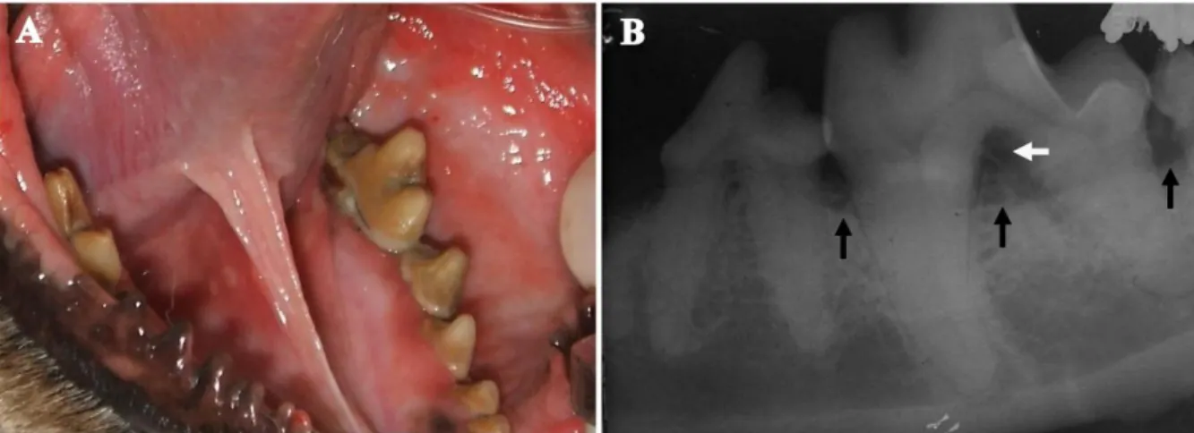
clinical sign (and the one that most often produces complaints from owners) is halitosis. Other clinical signs include ptyalism, anorexia, behaviour alterations, altered gingival colour, gingival bleeding, tooth mobility, periodontal and periapical abscesses, nasal discharge, sneezing, osteomyelitis, contact ulcers, intranasal dental migration, oronasal and oroantral fistulas (Wiggs and Lobprise, 1997; Kesel, 2000). A diagnosis of PD relies on interpreting clinical signs, full-mouth examination (including periodontal probing), and intraoral radiographic exams (Figure 1.1) (Klein, 2008).
Figure 1.1 – Periodontal disease. (A) Clinical signs; photograph of the mandibular left arcade of a dog
showing significant calculus accumulation, severe stomatitis, and gingival inflammation. (B) Radiographic
signs; intraoral parallel-angle dental radiograph of a mandibular left first molar in a dog. Note the horizontal
bone loss indicative of periodontitis (black arrows) and the evidence of furcation involvement (white arrow).
The healthy non-pigmented canine gingiva is coral pink with a smooth and regular texture, and its gingival margin is knife-edged (Figure 1.2 A) (West-Hyde and Floyd, 1995). Clinically, the attachment loss measurement represents the most important aspect of evaluating PD, but other indices adequately adapted from human periodontology, such as gingival index, radiographic index, plaque index, calculus index and sulcus bleeding index, are used to quantify the extent of inflammation and disease (Wiggs and Lobprise, 1997; Gorrel and Nind, 2008). Periodontal probing stills a key element in the diagnosis (normal probing depth should be ≤3mm in most dogs), but the clinical attachment loss converted into a percentage is more useful than expressed in millimetre increments due it is highly variable according to animal size (Wiggs and Lobprise, 1997).
Accordingly, PD is commonly referred to as gingivitis and periodontitis and can be classified in four stages [PD 1 (gingivitis), PD 2 (early periodontitis), PD 3 (moderate periodontitis), and PD 4 (advanced periodontitis)] based on the veterinary PD index system, which quantifies the periodontal attachment loss making use of probing and radiographic examinations (Table 1.1) (Wolf et al., 2005; AVDC, 2009).
Table 1.1 – Periodontal disease classification (Wolf et al., 2005; AVDC, 2009). Normal (PD 0) No gingival inflammation or periodontitis clinically evident.
Clinically normal
Stage 1 (PD 1) Gingivitis only without attachment loss. The height and architecture of the alveolar margin are normal.
Gingivitis
Stage 2 (PD 2) Less than 25% of attachment loss or at most, there is a stage 1 furcation involvement in multirooted teeth. There are early radiologic signs of periodontitis. The loss of periodontal attachment is less than 25% as measured either by probing of the clinical attachment level, or radiographic determination of the distance of the alveolar margin from the cemento-enamel junction relative to the length of the root.
Early periodontitis
Stage 3 (PD 3) 25-50% of attachment loss as measured either by probing of the clinical attachment level, radiographic determination of the distance of the alveolar margin from the cemento-enamel junction relative to the length of the root, or there is a stage 2 furcation involvement in multirooted teeth.
Moderate periodontitis
Stage 4 (PD 4) More than 50% of attachment loss as measured either by probing of the clinical attachment level, or radiographic determination of the distance of the alveolar margin from the cemento-enamel junction relative to the length of the root, or there is a stage 3 furcation involvement in multirooted teeth.
Advanced periodontitis
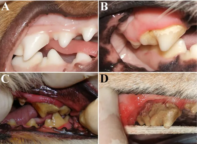
The PD 1 stage is characterized by different substages scored from mild gingivitis, in which only a slight gingival margin erythema is seen (Figure 1.2 B), through moderate gingivitis, when the gingiva is swollen and bleeding on probing (Figure 1.2 C), to severe gingivitis, in which the gingiva is inflamed, hyperplasic or retracted, with obvious erythema and oedema and with spontaneous bleeding (Wiggs and Lobprise, 1997). The periodontitis stage commences with attachment loss and is characterized by several alterations in periodontal tissues such as apical migration of the junctional epithelium with formation of periodontal
pockets, gingival recession (Figure 1.2 D) and alveolar bone resorption (Niemiec, 2008a). The PD 2 stage is characterized by signs of deepening sulcus (usually <5mm) and radiologic signs revealing up to 25% attachment loss. The PD 3 stage is present when probing (usually <7mm) and radiographic examination reveal 25–50% attachment loss around a root. The PD 4 stage occurs when the measurement of sulcus depth (usually ≥7mm) and radiological signs show an attachment loss of >50% (Wiggs and Lobprise, 1997; Wolf et al., 2005; AVDC, 2009).
Figure 1.2 – Periodontium at different stages of health/disease. (A) Healthy gingiva; photograph of the
maxillary and mandibular right arcades of a dog showing normal gingival tissues. (B) Mild gingivitis; photograph of the maxillary right fourth premolar of a dog showing a slight gingival margin erythema and mild calculus on the teeth. (C) Moderate gingivitis; photograph of the maxillary left fourth premolar of a dog showing a significant erythema and edema to the gingival, as well as increasing calculus accumulation on the tooth. (D) Advanced periodontitis; photograph of the maxillary right fourth premolar of a dog showing an obvious gingival recession, severe gingival inflammation and calculus accumulation.
Gingivitis is a reversible process when recognized early and properly treated. Otherwise, gingivitis can evolve into periodontitis, which is irreversible and leads to tooth mobility and
exfoliation (Gorrel and Nind, 2008; Niemiec, 2008a). It is interesting to note that not all animals with untreated gingivitis evolve into periodontitis, emphasizing the multifactorial character of PD and the importance of several susceptibility factors (Wiggs and Lobprise, 1997).
I.1.1.3. Treatment options
The careful plaque and calculus removal from the tooth crown, gingival sulcus and root surfaces is essential for the prevention and control of PD. Plaque removal can be accomplished by a combination of home care procedures that include mechanical and chemical plaque reduction techniques (e.g. tooth brushing with dentifrices, topical application of chemical plaque retardants such as 0.12–0.2% chlorhexidine gluconate), dietary manipulation (special diet and chew toys) and regular professional periodontal therapy (Niemiec, 2008b).
Non-surgical periodontal therapy is always the first line treatment and involves scaling (a combination of hand scaling, mechanical scaling, polishing and sulcular lavage) and closed root debridement under general anaesthesia. With progressive PD, advanced periodontal surgery becomes necessary. To treat patients with deep pockets and bone loss, mucogingival surgery (flap exposure) and open curettage are required (Niemiec, 2008b).
Clindamycin hydrochloride, amoxicillin/clavulanate and metronidazole seem to be particularly effective antimicrobials. They may be used for a week before periodontal treatment, prior to anaesthesia, postoperatively for 7–10 days, and as intermittent therapy in selected patients (impaired host defences or failure of conventional root debridement) (Lobprise, 2007; Niemiec, 2008b). Locally delivered antimicrobials (perioceutics), such as doxycycline gel, can be applied to teeth that have been cleaned and polished (Niemiec, 2008b). Nevertheless, the long-term usage of antimicrobials in the management of PD cannot be encouraged due to unproved benefits and possible side effects.
New therapies have started to be used with the aim of inducing periodontal regeneration and include: soft tissue grafts, bone replacement grafts, root biomodifications, enamel matrix derivatives, use of bioactive products such as bone morphogenic protein, guided tissue
regeneration, and combinations thereof (Greenwell, 2001). The final modality for PD therapy is currently tooth extraction (Niemiec, 2008b).
I.1.2. Research in periodontology
I.1.2.1. Animal models used in periodontal disease research
Animal models have been extensively used in oral disease research, particularly in PD, which has resulted in enormous advances in our understanding of aetiology, pathogenesis, prevention and treatment (Weinberg and Bral, 1999; Dannan and Alkattan, 2008).
An optimal animal model of PD needs to be standardized, reproducible, and to share some characteristics with humans, such as periodontal anatomy, aetiology, pathophysiology, disease course, and clinical outcome. Other attractive attributes include availability and simplicity of handling. The most commonly used models are dogs and non-human primates, although other animals (rats, mice, hamsters, rabbits, miniature pigs, ferrets, and sheep) have also been used (Weinberg and Bral, 1999). Table 1.2 summarizes the advantages and disadvantages of different animal models in periodontal research.
I.1.2.1.1. Non-human primate models
Non-human primates have been used in PD research in several studies because of their anatomical, immunological and microbiological similarity to the human oral cavity and periodontium. Other advantages are the natural occurrence of PD and their phylogenetic similarity to humans. These animals are considered the models closest to humans in terms of PD (Miller et al., 1995; Weinberg and Bral, 1999).
Many studies have been carried out related to periodontal healing, filling with biomaterials, guided tissue regeneration, enamel matrix derivatives and implant surgery (Caton et al., 1994; Fritz et al., 1997). Nevertheless, these models have important limitations. For example, naturally occurring periodontitis appears later in life, the lesions are asymmetrical, and the teeth and pocket depths are usually much smaller than in humans. Research access to these
animals is hindered by high costs, ethical considerations, difficulty in handling, aggressiveness, high susceptibility to infections and systemic illness, and the possibility of infectious agent transmission from and to these animals (Weinberg and Bral, 1999). Squirrel monkeys and marmosets are small in size and relatively easy to handle, but present a very limited inflammatory infiltrate, making them inappropriate models for studying the pathogenesis of PD (Schectman et al., 1972; Adams et al., 1981; Struillou et al., 2010).
I.1.2.1.2. Rat and mouse models
Rats and mice have been used because of their small size, low cost, prompt availability, ease of handling and housing, and our detailed knowledge of genetics. Additionally, they present some anatomical and histological similarities with the human periodontium and PD (Genco et al., 1998). However, there are significant differences in oral cavity size, dental anatomy, oral microbiota, inflammatory processes, and PD lesions (Genco et al., 1998; Weinberg and Bral, 1999). The use of gnotobiotic or germ-free rats/mice allows for the study of the individual effect of a particular bacterium without the interference of other microorganisms, whereas the use of knockout mouse models facilitates the exploration of new concepts regarding the pathogenesis of PD (Sasaki et al., 2004; Fine, 2009). Rats can also be used in periodontal tissue regeneration and bone healing studies (Struillou et al., 2010).
I.1.2.1.3. Hamster models (Mesocricetus auratus)
Hamsters have been used mainly in bacteriological studies (Hojo et al., 2008). The gilded hamster is also an interesting model for any immunological research (Struillou et al., 2010). Nevertheless, PD in hamsters is quite similar to that in rats, and therefore presents the same limitations (Weinberg and Bral, 1999). The golden Syrian hamster is the most commonly used strain (Struillou et al., 2010).
I.1.2.1.4. Rabbit models (Oryctolagus cuniculus)
Rabbits have been used in periodontal tissue regeneration studies for the testing of biomaterials (El-Bokle et al., 1993) or for evaluating the treatment of peri-implantitis (Struillou et al., 2010). Rabbits do not exhibit the spontaneous form of PD, so the disease has
to be induced. Moreover, this is a poorly standardized model with respect to relevant aspects of PD pathogenesis (Hasturk et al., 2007).
I.1.2.1.5. Miniature pig models (Sus scrofa domesticus)
Miniature pigs are described as useful animal models in dental and orofacial research, including PD (Lang et al., 1998; Wang et al., 2007). The animals are very similar to humans in terms of oral and maxillofacial anatomy and inflammatory response. Miniature pigs develop spontaneous PD with high prevalence at a young age. These traits, coupled with other scientific, economic and ethical factors, lead to a high usage of these animals in biomedical studies (Lang et al., 1998; Wang et al., 2007). The model has been used to test the regenerative capacity of periodontal tissues (Lang et al., 1998), the effects of dental lasers on periodontal healing, and dental implant surgery (Singh et al., 1993).
I.1.2.1.6. Domestic ferret models (Mustela putorius furo)
The domestic ferret was suggested as an animal model, mainly due to its PD similarities with human PD (Fischer and Klinge, 1994). The animals appear to be good alternatives to dogs and primates in the ligature-induced periodontitis model (Harper et al., 1990; Fischer and Klinge, 1994). Additionally, ferrets are an interesting experimental model for the study of calculus (Harper et al., 1990). Further studies are needed to confirm the use of this animal as appropriate for PD research (Weinberg and Bral, 1999).
I.1.2.1.7. Domestic sheep models (Ovis aries)
Sheep present a natural form of periodontitis called ‘broken mouth’ (Duncan et al., 2003). The periodontium, oral microbiota associated with PD and the bone metabolism in sheep are similar to those of humans (Genco et al., 1998). The publications using this model describe it as a suitable model for training surgical methods and for guided tissue regeneration research (Danesh-Meyer et al., 1997; Al-Qareer et al., 2004). However, its size, cost and handling demands, as well as the challenging diagnosis of PD as a result of poor access to posterior teeth, are disadvantages (Genco et al., 1998; Duncan et al., 2003).
I.1.2.2. Advantages of the canine model
The dog has several attractive attributes that make it an important model for the study of PD (Gad, 1968; Hamp and Lindberg, 1977). There are certain aspects of the disease for which the applicability of the dog is widely accepted and other aspects (as yet poorly researched) where the dog has promising potential as a model for the study of PD (Genco et al., 1998; Weinberg and Bral, 1999). PD in dogs is a naturally occurring disease and as such is more likely to mimic the pathophysiological mechanisms of human PD (Berglundh et al., 1991; Klein, 2008). Histological traits of the normal and diseased periodontium are similar in humans and dogs (Hamp and Lindberg, 1977). Moreover, there is a very high prevalence of PD in the canine population (Sorensen et al., 1980; Lund et al., 1999). The Beagle dog already presents high prevalence of PD at 2 years of age, but the major disease burden is carried by only a few individuals (Kortegaard et al., 2008). Therefore, in periodontal research, in parallel with naturally occurring PD, it is possible to induce experimental periodontal defects by placing silk bindings around the teeth for a period of 4–6 months or to use surgically created lesions (Struillou et al., 2010).
The canine periodontal anatomy, the aetiology of PD and its physiological mechanisms are well described because of the extensive use of dogs in periodontological research and in clinical and epidemiological research in veterinary medicine (Wikesjö et al., 1994; Koo et al., 2004; Kortegaard et al., 2008). Several comparative studies have shown that the factors involved in canine PD, including bacterial plaque, are similar to those of humans (Gad, 1968; Hamp and Lindberg, 1977). Nevertheless, despite many similar organisms in dog and human dental plaque, there are also important differences. Porphyromonas spp. are black pigmented anaerobic bacteria which have a central role both in human and canine PD. While humans are known to have Porphyromonas gingivalis, a catalase-negative bacterium, dogs apparently hold other catalase-positive P. gingivalis-like organisms (e.g. P. canoris, P. salivosa, P. denticanis, and P. gulae) (Genco et al., 1998; Hardham et al., 2005; Tatakis and Kumar, 2005). There is currently no consensual model for identifying critical microorganisms in the aetiology of PD but natural occurrence models are good candidates for reproducing the polymicrobial and chronic nature of disease (Sanz and Quirynen, 2005).
Table 1.2 – Animal models of periodontal disease. The comparison is made based on general characteristics with major implications for the ease/difficulty in PD research and based
on similarity with the disease in humans.
Model
General characteristics Similarity with human PD
Size Cost Handling Availability Dietary
habitsa Anatomy a Spontaneous PDa Histopathology a Oral Microbiotaa Well-characterized animal model Dog ++ - ++ - ++ + ++ + + ++ Non-human primates ++ -- -- -- ++ ++ + + + + Rat; mouse -- ++ ++ ++ -- -- - - - + Hamster -- ++ ++ ++ -- -- - - - -- Rabbit + + ++ ++ -- - - -- Miniature pig ++ - + -- ++ ++ ++ + + + Ferret - + + + -- - + + + - Sheep + - -- - -- + ++ + + -
I.1.2.3. Past and present contributions of dogs in periodontal research
The dog has been one of the most widely used animal models in periodontology research (Sorensen et al., 1980; Dannan and Alkattan, 2008), especially for the study of the histopathological aspects of the disease and for developing new therapeutic procedures (Hamp and Lindberg, 1977; Berglundh et al., 1991).
In 1899, Talbot pointed out the need for an animal model to study PD and noted the high prevalence and multi-stage characteristics of PD in dogs (Talbot, 1899). Since then, the dog has been used to study several PD aspects, including prevalence, aetiological factors and clinical and histological aspects (Gad, 1968; Lindhe et al., 1975; Sorensen et al., 1980).
The development of new treatment strategies, particularly periodontal reconstructive therapies, has always been dependent on animal models (Wikesjö et al., 1994; Polimeni et al., 2009). Histometric analysis is essential in order to evaluate the biological potential and application of those candidate therapies prior to clinical translation (Koo et al., 2004). Several studies have used the Beagle dog to evaluate the regenerative potential of periodontal tissue through the use of different techniques (Wikesjö and Nilvéus, 1991; Karring et al., 1993).
Wikesjö et al. (1991, 1994) developed and characterized a surgical protocol for the creation of a periodontal critical size defect in the mandibular premolar region of Beagles to evaluate the security and efficacy of several methodologies and devices proposed in periodontal reconstructive therapies. This is known as ‘periodontal supra-alveolar defect’ and is a widely accepted model for periodontal regenerative studies (Wikesjö and Selvig, 1999; Koo et al., 2004).
I.1.3. The dog as a promising model for gaining new insights into periodontology
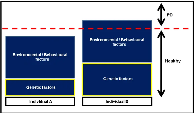
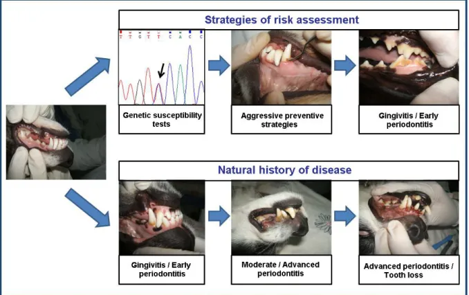
An emerging new medical concept is the establishment of susceptibility profiles (Kinane et al., 2005). There are many risk factors associated with the prevalence and severity of PD, which may be both modifiable and non-modifiable (Figure 1.3). Modifiable risk factors are usually environmental or behavioural (e.g. nutrition, oral hygiene, chewing habits) (Harvey, 1998; Gorrel, 2004), whereas non-modifiable risk factors (e.g. genetic factors) are usually
intrinsic to the individual, and thus not easily changed (Van Dyke and Dave, 2005). The early identification of non-modifiable risk factors may signal more predisposed individuals, set up personalized therapies and create more effective preventive strategies (Yoshie et al., 2007) (Figure 1.4).
Figure 1.3 – Complex multifactorial nature of periodontal disease. Both genetic susceptibility and
environmental/behavioural factors are implicated in the PD aetiology. For the same level of environmental/behavioural aggression, an individual with higher genetic susceptibility (individual B) will develop earlier and more severe PD than a less susceptible individual (individual A).
In the recent decades, clinical and scientific data have indicated that there is a significant genetic influence on PD which seems to control the severity of the inflammatory process and the therapy responses (Nares, 2003; Yoshie et al., 2007; Laine et al., 2012). A study using human monozygotic twins estimated that chronic periodontitis in adults reveals a 50% hereditability rate, suggesting that about half of the variation of the disease within a population is due to genetic factors (Michalowicz et al., 2000). To the best of our knowledge, there are no previous studies evaluating genetic predisposition in canine PD. Even so, by examining the similarities of PD in humans and dogs, it is plausible to speculate that genetics will be one of the factors that partly justify the difference in susceptibility/resistance to
disease amongst dogs. Knowledge of PD genetics is still very limited due to its polygenic nature and environmental and behavioural interactions. This knowledge will open the door to new diagnostic and therapeutic strategies (Nares, 2003; Kinane et al., 2005).
Figure 1.4 – Strategies of risk assessment in periodontal disease. The early identification of genetc risk
factors may signal predisposed individuals, set up more aggressive preventive strategies avoiding disease progression to advanced and irreversible stages.
The large number of diseases with breed predisposition, some of them similar to human counterparts (Wayne and Ostrander, 2007), makes the dog a promising clinical model (Tsai et al., 2007). Moreover, dogs are physiologically quite close to man with marked similarities in the form and function of several tissues and organs (Tsai et al., 2007). Another advantage is that dogs live in close proximity to people, so allowing research to be undertaken in controlled laboratory settings and also environmental and epidemiological trials (Olson, 2007).
The canine genetics research community has recently made significant strides, producing dense linkage and radiation hybrid maps, oligo-based microarrays, single nucleotide
polymorphism arrays, and, most importantly, the sequence of the canine genome at 7.6X coverage (Tsai et al., 2007). Consequently, a comparative genomic approach (whereby disease-carrying genes can be identified in dog diseases and then mapped onto the human genome) is now recognized as a valid method and such studies are increasing in popularity. Genome-wide canine SNP arrays have been developed, and increasing success in using these arrays to map disease loci in dogs is emerging, which is highly relevant, not only for canine health, but also as comparative models for analogous human conditions (Ke et al., 2010). It is expected that over the coming years work on the dog genome will help increase our understanding of the genetic cause of many diseases shared by humans and dogs (Mellersh, 2008).
I.2. Clinical genomics of PD – candidate genes approach
Clinical genomics refers to the use of information from genomes and their derivatives to guide medical decision making, namely to make individualized risk predictions and to guide in prophylactic or treatment decisions. This approach is a key component of the personalized medicine which represents an emerging new model for health care (Guttmacher and Collins, 2002; Ginsburg and Willard, 2009).
Genetic association studies are essential for identifying the complex disease genetic contributors, and there are two main approaches, one based on candidate genes, the other based on analysing the entire genome (genome-wide scanning). Each strategy has specific advantages and disadvantages (Amos et al., 2010). Candidate gene approach has been proven to be extremely powerful for studying the genetic basis of complex diseases, being a much more effective and economical method (Tabor et al., 2002; Zhu and Zhao, 2007). Nevertheless, in order to apply this method it is necessary to have a previous knowledge about the biology or physiopathology of the disease under investigation, because candidate genes must be genes with known biological function regulating the disease process (Zhu and Zhao, 2007; Amos et al., 2010). The combination with a comparative genomics strategy is extremely useful because this method uses a cross-species approach to select the most suitable candidate genes, which may be functionally conserved or structurally homologous genes identified from other related species (Zhu and Zhao, 2007). This kind of study approach was
arthritis and other joint diseases (Ollier et al., 2001; Short et al., 2007; Clements et al., 2010). The applicability of this strategy to select candidate genes for canine PD study is suitable because various homologous genes associated with PD susceptibility have already been confirmed in humans (Zhang et al., 2011).
Genetic research on human PD is primarily focused on single nucleotide polymorphisms (SNPs) in genes responsible for molecules with a role in immunoregulation and/or the metabolism such as cytokines, cell surface receptors, chemokines and enzymes (Yoshie et al., 2007; Zhang et al., 2011). SNPs (Figure 1.5) are single-nucleotide substitutions of one base for another [replaces between adenine (A), cytosine (C), guanine (G) and thymine (T)], which by definition must be present in at least 1% of the population. These polymorphisms are the most frequent type of genetic variation and are currently refered as important biological markers or directly associated with complex diseases susceptibility (Wang et al., 1998).
Figure 1.5 – Single nucleotide polymorphism (SNP). An example of sequencing chromatograms showing a
SNP [both homozygous and heterozygous are represented (AA, TT and AT)]. A SNP is a single nucleotide variation at a specific location in the genome.
![Figure 1.5 – Single nucleotide polymorphism (SNP). An example of sequencing chromatograms showing a SNP [both homozygous and heterozygous are represented (AA, TT and AT)]](https://thumb-eu.123doks.com/thumbv2/123dok_br/16119873.1108808/52.892.182.709.562.1067/figure-nucleotide-polymorphism-sequencing-chromatograms-homozygous-heterozygous-represented.webp)