Universidade de Aveiro Ano 2016
Departamento de Biologia
Inês Mendes Sardo
Avaliação de Potenciais Agentes Anti-tumorais
para Terapia Fotodinâmica
Assessment of Potential Anti-tumour Agents for
Photodynamic Therapy
DECLARAÇÃO
Declaro que este relatório é integralmente da minha autoria, estando devidamente referenciadas as fontes e obras consultadas, bem como identificadas de modo claro as citações dessas obras. Não contém, por isso, qualquer tipo de plágio quer de textos publicados, qualquer que seja o meio dessa publicação, incluindo meios eletrónicos, quer de trabalhos académicos.
Universidade de Aveiro Ano 2016
Departamento de Biologia
Inês Mendes Sardo
Avaliação de Potenciais Agentes Anti-tumorais para
Terapia Fotodinâmica
Assessment of Potential Anti-tumour Agents for
Photodynamic Therapy
Dissertação apresentada à Universidade de Aveiro para cumprimento dos requisitos necessários à obtenção do grau de Mestre em Biologia Molecular e Celular, realizada sob a orientação científica da Doutora Maria Amparo Ferreira Faustino e da Doutora Margarida Sâncio da Cruz Fardilha, respetivamente, Professora auxiliar no Departamento de Química da Universidade de Aveiro e Professora auxiliar do Departamento de Ciências Médicas da Universidade de Aveiro.
Dedico esta dissertação às pessoas mais importantes da minha vida Pelo que me ensinaram e transmitiram
Pelo apoio incondicional e incessante Pelo que sou
o júri
presidente Prof. Doutora Maria de Lourdes Pereira
Professora Associada com Agregação do Departamento de Biologia da Universidade de Aveiro
Prof. Doutora Maria do Amparo Ferreira Faustino
Professora Auxiliar do Departamento de Química da Universidade de Aveiro
Prof. Doutor Bruno Miguel Rodrigues das Neves
Professor Auxiliar do Centro de Neurociências e Biologia Celular e da Faculdade de Farmácia da Universidade de Coimbra
agradecimentos Quero deixar este espaço dedicado a todos aqueles que, direta ou
indiretamente, contribuíram paara que esta dissertação fosse realizada. Não vai ser possível nomear todos, no entanto há alguns a quem não posso deixar de manifestar o meu profundo reconhecimento e sincero agradecimento.
À Professora Doutora Amparo Faustino, expresso o meu profundo agradecimento pela orientação e apoio incondicional que em muito elevaram os meus conhecimentos científicos e que me fez crescer em vários aspetos, não apenas académicos. Obrigada pela oportunidade de me deixar realizar este trabalho.
À Professora Doutora Margarida Fardilha, expresso o meu sincero agradecimento pela orientação e pela oportunidade de me integrar no seu Grupo de Investigação. Obrigada também pelo profissionalismo, compreensão e disponibilidade que sempre mostrou para comigo.
Não posso deixar de agradecer à Juliana Felqueiras por tudo, mas mesmo tudo o que fez por mim. É inexplicável e compreendo que um simpes “obrigada” não chegue. Portanto, e como prometido, escrevo o poema (não da minha autoria) mas (penso) que se adequa.
“Ninguém escapa ao sonho de voar, de ultrapassar os limites do espaço onde nasceu, de ver novos lugares e novas gentes. Mas saber ver em cada coisa, em cada pessoa, aquele algo que a define como especial, um objeto singular, um amigo – é fundamental. Navegar é preciso, reconhecer o valor das coisas e das pessoas, é mais preciso ainda!”
Antoine de Saint-Exupéry
À Magda Henriques, quero agradecer, por todos os momentos passados no laboratório e pela paciência que teve comigo. À Rita Almeida, sou eternamente grata, pelo caminho que levamos juntas, tanto académico como pessoal, há mais de oito anos.
Não posso também deixar de agradecer à minha família. Aos meus pais Ricardo e Milú, ao meu primo Diogo, à minha tia Paula e às minhas avós e avô.
Para finalizar e não menos importantes quero agradecer aos meus amigos pelo apoio e pela amizade, que sem saberem são um pilar na minha vida. Um grande obrigado ao Tiago, à Carolina, à Diana e ao Micael, à Ana e ao Lourenço ao Marco, ao Nuno e ao Creoulo. Obrigada também aos meus colegas de Mestrado que se tornaram mais do que isso: Zé, André, Mónica e Emilie.
palavras-chave Cancro da Próstata, Fotossensibilizadores, Porfirinas, Terapia Fotodinâmica.
resumo Atualmente, um dos cancros mais incidentes e mortíferos, no homem, é o cancro da próstata. O tratamento utilizado para este tipo de cancro é eficaz e com elevadas taxas de sucesso quando este é detetado precocemente. Contudo, quando a sua deteção ocorre num estadio mais avançado, as opções de tratamento existentes apresentam efeitos secundários que devem ser tidos em conta na decisão terapêutica. A terapia fotodinâmica (PDT) é uma metodologia emergente no tratamento de diversas doenças oncológicas, entre outras aplicações, que se baseia na utilização combinada de uma molécula fotossensibilizável, luz e oxigénio que, quando atuam em conjunto, são capazes de gerar espécies reativas de oxigénio citotóxicas no tecido alvo.
Reconhecendo esta potencialidade, este estudo pretendeu avaliar o potencial fotodinâmico de quatro novos fotossensibilizadores de tipo porfirínico (PS4a, PS4b, PS5a e PS5b). Os derivados testados apresentam na sua estrutura, para além do macrociclo porfirínico, uma unidade uracilo fundido com unidades glicosidicas ligada ao macrociclo na posição beta-pirrólica. Os derivados PSXa e PSXb apresentam, respetivamente, unidades de xilose e galactose quer protegidas (PS4) quer desprotegidas (PS5). A atividade citotóxica destes novos derivados foi testada em células isoladas de um carcinoma da próstata (PC3) e numa linhagem celular prostática não tumoral (PNT-2). Este estudo, in
vitro, foi feito tanto na presença como na ausência de luz.
Os resultados obtidos indicam diferentes perfis de citotoxicidade para os compostos analisados para ambas as linhas celulares. Assim, de todos os compostos testados, o PS4b foi aquele que apresentou fototoxicidade nas células tumorais da próstata não afetando as células não tumorais nas concentrações estudadas. Os restantes derivados ou apresentam citotoxicidade e fototoxicidade para ambas as linhas celulares testadas (PS4a e PS5a) ou são particularmente tóxicos para as células não tumorais (PS5b). Assim sendo, podemos concluir pela avaliação da citotoxicidade e fototoxicidade do derivado PS4b que este apresenta propriedades biológicas promissoras revelando-se um potencial candidato a ser utilizado como agente fotossensibilizador para tratamento por PDT do cancro da próstata, se utilizado nas condições adequadas.
keywords Photodynamic therapy, Photosensitizers, Porphyrins, Prostate cancer.
abstract Currently, one of the most incident and deadly cancers, in men, is prostate cancer. The treatment used to this type of cancer is efficient and with high success rates when early detected. However, when the detection occurs in a more advanced stage, the existing treatment options have side effects that must be considered in the therapeutic decision. Photodynamic therapy (PDT) is an emerging approach in the treatment of various oncological diseases, and other applications, based on the combined use of a photosensitizer molecule, light and oxygen which, when acting together, are able to generate cytotoxic reactive oxygen species (ROS) in the target tissue.
Recognizing this potential, this study aimed to evaluate the photodynamic potential of four new porphyrinic type photosensitizers (PS4a, PS4b, PS5a and PS5b). The tested derivatives are porphyrins bearing in its beta-pyrrolic positions fused uracils containing a sugar moiety. The derivatives PSXa (xylose)and PSXb (galactose) contain their sugar units protected (PS4) or unprotected (PS5). The cytotoxic activity of these new derivatives was tested in isolated cells from prostate carcinoma (PC3) and from a non-tumour prostate cell line (PNT-2). This study, in
vitro, has been done both in the presence and absence of light.
The results point out different cytotoxicity profiles for the tested compounds for both cell lines. Thus, the PS4b showed phototoxicity in prostate tumour cells without affecting non-tumour cells, at the studied concentrations. The remaining derivatives or possessed cytotoxicity and phototoxicity for both cell lines tested (PS4a and PS5a) or were particularly toxic to non-tumour cells (PS5b). Therefore, we can conclude by the cytotoxicity evaluation and phototoxicity that PS4b shows promising biological properties revealing to be a potential candidate to be used as a photosensitizer for the prostate cancer PDT treatment, if used under the right conditions.
CONTENTS
List of Figures ... xxi
List of Tables ... xxv
List of Abbreviations, symbols and acronyms ... xxvii
1. Introduction ... 1
1.1 General overview on cancer ... 3
1.2 Prostate cancer ... 3
1.2.1 Risk factors ... 4
1.2.2 Prostate carcinogenesis ... 5
1.2.3 Diagnosis and staging ... 6
1.2.4 Treatment ... 7
1.3 Photodynamic Therapy ... 7
1.3.1 Photophysical and photochemical properties ... 8
1.3.1.1 Light ... 10
1.3.1.2 Photosensitizers ... 10
1.3.2 Tumour destruction... 11
1.3.2.1 Direct lethal effects on tumour cells ... 11
1.3.2.2 Vascular events ... 12
1.3.2.3 Immune system ... 12
1.4 Photodynamic therapy and prostate cancer ... 13
1.5 Objective of this thesis ... 14
2. Materials and Methods ... 15
2.1 Cell culture reagents ... 17
2.2 Human prostate cells maintenance ... 17
2.4 Light source for photodynamic therapy... 19
2.5 Optimization of cell-related parameters ... 20
2.6 Cytotoxicity assay... 20
2.7 Photodynamic therapy Assay ... 21
2.8 Statistical analysis... 21
3. Results ... 23
3.1 Optimization of the experimental conditions ... 25
3.2 Preliminary cytotoxicity assay... 26
3.3 Photodynamic therapy ... 29
3.3.1 Effect of the photosensitizers in PNT-2 cell line ... 30
3.3.2 Effect of the photosensitizers in PC3 cell line ... 33
4. Discussion ... 37
5. Conclusion and Future perspectives ... 43
6. References ... 47
LIST OF FIGURES
Figure 1- Estimated incidence (left) and estimated mortality (right) of prostate cancer in men in Portugal, in 2012. Taken from [3], [4]. ... 4 Figure 2- Photosensitization process and Type I and Type II reactions. ... 9 Figure 3- Chemical structures of the four new photosensitizers used in the present study and the correspondent molecular weight. ... 18 Figure 4- Growth curve of PNT-2 and PC3 cell lines determined using the Alamar Blue (AB) reagent. PNT-2 (A) and PC3 (B) cells were plated at various densities (500, 1 250, 2 500, 5 000, 10 000, 20 000 and 40 000 cells) in a 96-well plate and the growth was assessed after 1, 2, 4, 6, 8 and 24 h of incubation. Each experimental condition was performed once in duplicate using AB assay. ... 26 Figure 5 - Effect of PS4a (top image) and PS4b (bottom image) on the viability of PNT-2 (graphs A and C, respectively) and PC3 (graphs B and D, respectively) cell lines in light-restricted conditions. Cells were incubated with PS4a and PS4b at different concentrations (1.00x10⁻⁴ M, 1.00x10⁻⁵ M, 1.00x10⁻⁶ M and 1.00x10⁻⁷ M) along time (2, 4, 6, 8 and 24 h). Cells and cells treated with 1% DMSO were used as controls. Cell viability was assessed using the Alamar Blue (AB) reagent. Data are presented as the mean value referent to one experiment performed with two replicates for each condition. ... 27 Figure 6 - Effect of PS5a (top image) and PS5b (bottom image) on the viability of PNT-2 (graphs A and C, respectively) and PC3 (graphs B and D, respectively) cell lines in light-restricted conditions. Cells were incubated with PS5a and PS5b at different concentrations (1.00x10⁻⁴ M, 1.00x10⁻⁵ M, 1.00x10⁻⁶ M and 1.00x10⁻⁷ M) along time (2, 4, 6, 8 and 24 h). Cells and cells treated with 1% DMSO were used as controls. Cell viability was assessed using the Alamar Blue (AB) reagent. Data are referent to one experiment performed with two replicates for each condition. ... 28 Figure 7- Cytotoxic and photocytotoxic effects of 1% DMSO in PNT-2 (graphs A and B, respectively) and PC3 (graphs C and D, respectively) cell lines. Cells were treated with 1% DMSO or left untreated and incubated in light-restricted conditions for 24 h. After this incubation period, one of the cell plates was exposed to red light (λ= 630 nm ± 20 nm) with an irradiance of 1.28 mW.cm-2 during 20 min, while the other remained in light-restricted conditions. Both plates were further incubated for (24 h). Cytotoxicity and phototoxicity were then assessed using the Alamar Blue reagent. Data are presented as the mean value ± S.D. of three independent experiments, each with three replicates of each condition. Non-parametric tests for comparing independent samples were used to assess the statistical significance of the results (P-value<0.05). ... 30 Figure 8- Percentage of AB reduced by concentration (1.00x10⁻⁴ M, 1.00x10⁻⁵ M, 1.00x10⁻⁶ M) with treatment (PS4a, PS4b, PS5a and PS5b) in PNT-2 cell line, in the absence of light. Cytotoxicity was assessed by AB assay 24 h after supposed PDT. The percentage of cytotoxicity was calculated relatively to control cells (cells incubated in RPMI with 1% DMSO). Data are the mean value ± S.D. of three independent experiments performed in triplicates. Non-parametric test for comparing two independent samples. *- difference is significant at the P-value<0.05. ... 31
Figure 9- Percentage of AB reduced by concentration (1.00x10⁻⁴ M, 1.00x10⁻⁵ M, 1.00x10⁻⁶ M) with treatment (PS4a, PS4b, PS5a and PS5b) in PNT-2 cell line, after PDT. Photocytotoxicity was assessed by AB assay 24 h after PDT. The percentage of photocytotoxicity was calculated relatively to control cells (cells incubated in RPMI with 1% DMSO). Data are the mean value ± S.D. of three independent experiments performed in triplicates. Non-parametric test for comparing two independent samples. *- difference is significant at the P-value<0.05. ... 32 Figure 10- Percentage of AB reduced by concentration (1.00x10⁻⁴ M, 1.00x10⁻⁵ M, 1.00x10⁻⁶ M) with treatment (PS4a, PS4b, PS5a and PS5b) in PC3 cell line, in the absence of light. Cytotoxicity was assessed by AB assay 24 h after supposed PDT. The percentage of cytotoxicity was calculated relatively to control cells (cells incubated in RPMI with 1% DMSO). Data are the mean value ± S.D. of three independent experiments performed in triplicates. Non-parametric test for comparing two independent samples. *- difference is significant at the P-value<0.05. ... 33 Figure 11- Percentage of AB reduced by concentration (1.00x10⁻⁴ M, 1.00x10⁻⁵ M, 1.00x10⁻⁶ M) with treatment (PS4a, PS4b, PS5a and PS5b) in PC3 cell line, after PDT. Photocytotoxicity was assessed by AB assay 24 h after PDT. The percentage of photocytotoxicity was calculated relatively to control cells (cells incubated in RPMI with 1% DMSO). Data are the mean value ± S.D. of three independent experiments performed in triplicates. Non-parametric test for comparing two independent samples. *- difference is significant at the P-value<0.05. ... 34
LIST OF TABLES
Table 1- Photodynamic therapy advantages over traditional treatments. ... 8 Table 2- Requirements for the ideal photosensitizer to be used in cancer treatment. ... 11 Table 3- Photophysical properties of the photosensitizers PS4a, PS4b, PS5a and PS5b. ... 19
LIST
OF
ABBREVIATIONS,
SYMBOLS
AND
ACRONYMS
1
O2 – Singlet oxygen 1
PS* - Photosensitizer in excited singlet state
3
O2 – Triplet oxygen; molecular oxygen in ground state 3
PS* - Photosensitizer in excited triplet state AB – Alamar Blue
ALA or 5-ALA - δ-Aminolevulinic acid or 5-Aminolevulinic acid DMF – N,N-Dimethylformamide
DMSO – Dimethyl Sulfoxide DPBF – 1,3-diphenylisobenzofuran DRE – Digital rectal examination EDA - Exploratory data analysis FBS – Fetal Bovine Serum FI - Fluorescent Intensity
G-CSF – Granulocyte colony stimulating factor H2O2 - Hydrogen peroxide
H-ALA – Hexaminolevulinate
HIFU – High-intensity focused ultrasound HpD – Hematoporphyrin derivatives HPV – Human papilloma virus IL – Interleukin
LEDs – Light emitting diodes M-ALA - Methyl aminolevulinate MLu or LuTex - Motexafin lutetium
mTHPC – 5,10,15,20-Tetrakis (3-HydroxyPhenyl) Chlorin O2 – Molecular Oxygen
O2─● – Superoxide radical
P – P-value
PDT – Photodynamic Therapy
PIA - Proliferative inflammatory atrophy PIN – Prostatic intraepithelial neoplasia PPIX - Protoporphyrin IX
PS – Photosensitizer
PS─● or PS+● – Radical photosensitizer PSA – Prostate specific antigen
QOPNA – Organic Chemistry, Natural Products and Food Stuffs research unit ROS – Reactive oxygen species
RPMI – Roswell Park Memorial Institute medium S.D. – Standard Deviation
S0 – Photosensitizer molecule in the ground state
SnET2 – Etiopurpurin dichloride
TB – Trypan Blue
TNF – Tumour necrosis factor
TNM system - T – tumour, N – lymph node, M – metastases system TRUS – Transrectal ultrasonography
WST – Tookad
εox – Oxidized constant
1. Introduction
1.1 General overview on cancer
Cancer is an emerging public health problem in several parts of the world independently if it is a developed or developing country [1], [2]. It is described by abnormal cells that can divide without control and can invade tissue and organs of the body. The incidence of this disease is likely to increase due to growth and population aging and also lifestyle behaviors such as smoking, poor diet, physical inactivity and reproductive changes [2].
In European Union, in 2012, were registered over one million (1 417 495) incident cancer cases (all sites but non-melanoma skin) and less than one million (707 755) cancer deaths [3], [4]. This disease has several psychycological repercussions including fear, pain, depression or even anxiety. Along the years, advances in Technology and in the diagnostic allow the early detection of the disease. The cancer treatment comprises the conventional methods: chemotherapy, surgery and radiotherapy that have some severe adverse effects.
Photodynamic therapy (PDT) has emerged as a promising treatment for numerous diseases, including cancer, with less adverse effects than the ones presented by conventional treatment [5], [6]. This technique uses a photoactive agent, in cancer cells that, followed with light irradiation, allows the trigger of chemical and biochemical reactions leading to cell death.
1.2 Prostate cancer
Prostate cancer (PCa) is still one of the most incident and deathly cancers worldwide. According to the GLOBOCAN project (from the International Agency for Research on Cancer), PCa is the second most common cancer in men, with nearly 1.1 million men diagnosed worldwide in 2012, and the fifth leading cause of death from cancer in men, accounting for 6.6% of the total men deaths. In Europe, PCa was the most common cancer in men, accounting for 343 174 diagnoses, in 2012, and it was responsible 71 033 deaths, being classified as the second leading cause of death from cancer in men. In Portugal, the scenario is similar (Figure 1) with PCa being the most common cancer in men in 2012, as well. With thereabout 23.25% (Age Standardized
1. Introduction
Rate (European) per 100 000) and was the fourth leading cause of death with 11.06% total deaths of PCa [3], [4].
Figure 1- Estimated incidence (left) and estimated mortality (right) of prostate cancer in men in Portugal, in 2012. Taken from [3], [4].
Since the main risk factor for PCa is age, these numbers are expected to increase due to population aging [1], [2].
1.2.1 Risk factors
Several aspects of PCa development and progression remain to be elucidated. However, epidemiological evidences sustain that advanced age, black ethnicity and family history of PCa are amongst the most well-established risk factors for the disease [7], [8]. While about 30% of men between 30 and 40 years old were detected with small prostatic carcinomas, this percentage duplicates when considering men between 60 and 70 years old [9], [10]. Due to the population aging, PCa incidence is expected to increase in the next years. On the other hand, differences in health care systems, financial barriers, socio-economic status or even fear of both diagnosis and treatment of the disease might explain, at least in part, racial differences. Moreover, since cancer is considered a genetic disorder, ethnical genetic and epigenetic alterations might also explain why African-American men have increased cell proliferation and metastasis, and consequently, more aggressive PCa compared to European-American men [11], [12]. Regarding the family history, it is estimated that inheritance is responsible for about 10% of PCa cases. Men with first-degree relatives (it can be a brother, father or son) with PCa have at least the risk doubled in comparison to general population. In contrast, distant relative showed only marginally increased risk [11], [13].
There are also some findings that indicate that environmental and lifestyle factors may affect the risk and progression of PCa, such as body size, eating habits,
1. Introduction
hypertension, sexual behavior, sexually transmitted diseases (in particular gonorrhea, syphilis, Trichomonas vaginalis) and smoking (more associated with PCa mortality instead of incidence). With an inverse association with PCa are metabolic syndrome (dyslipidemia, body mass index, low HDL cholesterol and high triglycerides), diabetes, physical activity, aspirin and nonsteroidal anti-inflammatory drugs. However, environmental and lifestyle factors are still debatable [7], [8], [10], [14]–[16].
1.2.2 Prostate carcinogenesis
There are four stages and processes underlying PCa initiation and development: Proliferative inflammatory atrophy (PIA), Prostatic intraepithelial neoplasia (PIN), localized PCa and metastatic prostate cancer. PIA englobes simple atrophy and postatriphic hyperplasia associated with variable amounts of inflammatory cells and stromal fibrosis. PIN is composed by cells of prostate adenocarcinoma occurring within pre-existing prostatic acini/ducts. The high-grade PIN have cells where nuclei are enlarged and hyperchromatic and often have prominent nucleoli unlike low-grade PIN which most of the cells lacked prominent nucleoli [17]. The cellular and molecular events that cause PCa remain unknown but it is well known that age is the most significant risk factor for PCa development. Androgens and reactive oxygen species (ROS) are also responsible for prostate carcinogenesis because ROS cause oxidative damage in epithelial cells that can react with other cellular components initiating a free radical chain reaction favoring progression of prostate carcinogenic process. On the other hand the prostate gland depend on the androgen signaling for growth and androgen suppression may lead to an oxidative status increasing the risk of carcinogenesis [18]. Another factor is multiple genetic changes such as activation of dominantly acting oncogenes that promote cell proliferation and loss of tumour suppressor genes that negatively regulate cell proliferation [19]. Inflammation have a potential role in prostatic carcinogenesis and tumour progression. Recent studies have suggested that inflammation around prostate cancer is associated with worst case scenario and chronic inflammation in benign tissue was predictive of a higher-grade disease [20], [21]. Some sexual diseases such as human papilloma virus (HPV) (an oncogenic virus) have influence on prostate carcinogenesis and can be independent of inflammation [22].
1. Introduction
1.2.3 Diagnosis and staging
Currently, there are three main methods for PCa screening: measurement of blood prostate specific antigen (PSA) levels, digital rectal examination (DRE) and transrectal ultrasonography (TRUS) [23]. PSA is a glycoprotein produced by normal epithelial cells of the prostate gland and was one of the first biomarkers to be established when its synthesis was found to be increased in malignant cells and, consequently, its levels become elevated in the blood. However, the use of PSA in PCa screening is controversial because this molecule is specific for prostate, but not for cancer; so, many other diseases (e.g. benign prostate hyperplasia, prostatitis and other urological problems) can increase the PSA levels [24], [25]. DRE may detect cancers in the peripheral zone, independently of PSA levels, while TRUS – an invasive technique that is helpful in guided biopsies – is poorly specific for PCa, but highly sensitive [26]–[30]. The use of these three exams in combination is more reliable than the use of only one of them; however, the ultimate diagnosis is only possible by the histopathological analysis of prostate biopsies [31]–[38].
When the disease is diagnosed, an accurate staging is essential to select the most suitable treatment, as well as to infer about the prognosis and to predict tumour behavior [39]. In 1992, the TNM system was introduced (T – tumour, N – lymph node, M – metastases) to determine the stage of cancers [40] and nowadays the TNM system is updated. On the other hand, the standard grading of PCa is performed according to the Gleason score that predicts cancer behavior. This pattern is graded on a score from 1 (least aggressive) to 5 (most aggressive) on the largest available specimen, adding the most and second most common patterns with Gleason scores ranging from 2 to 10. The Gleason system has significant deficiencies that can have an impact on patient care. For example: a Gleason score 7 can represent mostly well-differentiated cancer with a lesser component of more poorly differentiated cancer (Gleason 3+4) or mostly poorly differentiated cancer with a smaller component of well-differentiated cancer (Gleason 4+3) and this two are prognostically very different [41], [42].
1. Introduction
1.2.4 Treatment
When detected at early stages, PCa can be eradicated with high rates of success by surgical removal. Besides radical prostatectomy, other treatment strategies are available and include hormonal therapy, radiotherapy, brachytherapy, cryosurgery and high-intensity focused ultrasound (HIFU). All these options, however, present important side effects that should be taken into account during treatment decision-making. The most well-established treatment for various stages of PCa is hormonal therapy which reduces serum testosterone levels, however in some cases PCa progress and evolve to an androgen-independent state where this therapy is worthless [18]. Radical surgery and radiotherapy are the most efficient treatments to localized PCa they are commonly associated with urinary, sexual and bowel dysfunctions. Radiotherapy has a high risk of biochemical relapse and radiation could lead to the development of radioresistant tumours with local and systemic progression. Radiation can also have side effects, such as severe damage of epithelial surfaces, infertility and swelling of soft tissues. Brachytherapy is, in general, prescribed to patients with lower risk PCa and the side effect is associated with urinary domain (incontinence) [43]–[45]. Cryosurgery ablation of the prostate and HIFU had emerged as alternative therapies for patients in whom radical surgery is not suitable [46], but the use of sophisticated and expensive equipment’s makes the procedure too expensive to be routinely applied in the majority of the countries. Moreover, the use of these two techniques have some disadvantages, such as the requirement of preoperative hormonotherapy or biopsy proven recurrence and modification of prostate morphology, turning the follow-up more complex [47]. HIFU can also cause retrograde ejaculation [48].
1.3 Photodynamic Therapy
Photodynamic therapy (PDT) is a minimally invasive method that involves the administration of a photoactivated drug [photosensitizer (PS)], its selective accumulation in the target tissue, followed by light exposure of the injured area, thereby resulting in structural and functional damage of the target tissue [49]. Hence, PDT is based on photosensitization – the process that occurs when a PS absorbs light. PS is the designation given to molecules that need light (usually 400-800 nm) to be activated to
1. Introduction
originate a biochemical process [50]. Despite the photophysical and photochemical properties of PDT are not completely understood, the following subsection summarize what is known about the subject. PDT has several advantages over other traditional therapies that are summarized in Table 1 [45].
Table 1- Photodynamic therapy advantages over traditional treatments [45].
Applied to places where surgery cannot be performed;
Patients vulnerable to surgery, chemotherapy or radiation can be treated with this modality;
Current treatments can cause side effects (nausea and vomiting and in some cases depress the immune system);
Photosensitizers alone do not exhibit any organ cytoxicity;
The presence of photosensitizers in liver and kidney do not damage these organs;
Used as primary or adjunctive treatment for solid cancers of the bladder, esophagus, head and neck, brain, lung, prostate, intraperitoneal cavity, breast and skin.
1.3.1 Photophysical and photochemical properties
When the PS molecule in its ground state (S0) is irradiated with light of appropriate
wavelength absorb it, change its energetic level to a higher energy state ─ the excited singlet state (1PS*), and becomes unstable. As a consequence, it tends to release the excess of energy in three ways: heat release, emission of light (fluorescence emission) or by conversion to an intermediate energy state (triplet state ─ 3PS*). In this new energetic condition, the PS can return to the ground state by two pathways, which are classified as type I and type II mechanisms (Figure 2), and both can produce reactive oxygens spcies (ROS) responsible for the photodynamic action. In type I mechanism, the excited PS (3PS*) transfer or accept directly an electron from a substrate present in the system. In the presence of molecular oxygen (O2), the radical PS
─●
or PS+● formed from the former reaction, can transfer the acquired electron to O2 generating, for
instance, the superoxide radical (O2 ─●
) and the PS molecule returns to S0. Other ROS
can be formed under this pathway (H2O2, HO ●
) which are responsible for cellular damage and oxidative stress. In type II reaction, the 3PS* transfers its energy directly to the molecular oxygen in ground state (3O2), resulting in the excited singlet oxygen
1. Introduction
molecule (1O2) and the PS in S0 [51], [52]. The 1O2 is extremely oxidant species and
although its very short half-life (~ 4.2 µs in water), is able to oxidize the cellular targets in a radius of 20 nm [53], [54].
Figure 2- Photosensitization process and Type I and Type II reactions.
PDT application should be preceded by optimization conditions, namely, light, PS and their interaction. This can be achieved by selecting the appropriate PS and the most adequate light wavelength for its activation. High irradiances (total energy of exposed light across the sectional area of irradiated spot) means more light photons delivered that can be responsible for the PS excitation and consequently more ROS can be generated per time unit. The used of low dose of PS, applying high light irradiances allows to achieve similar PDT responses, without undue morbidity. Another critical component, in vivo PDT, is the time that a PS takes to accumulate in tumour cells and not be found in the blood stream. Other aspect to be take into account is the possibility of PS to have fluorescence emission ability which can help either tumour visualization and delimittion or PS quantification in the target tissue; the loss of fluorescence after PDT treatment may indicate tumour ablation or the PS photodecomposition [49], [53].
1. Introduction
The presence of oxygen during the PDT treatment has a profound effect on its outcome because without oxygen no photodynamic action take place [49].
1.3.1.1 Light
There are regulatory agencies approving light sources to be commercially available as lasers or light emitting diodes (LEDs). The technological evolution has enabled that light sources could be reliably introduced into human organism by means of fiber optic cables or the LED itself with ultrasound or computed tomography-guided placement. The light selection has to take into account that each PS has a specific wavelength window and an intensity required for its successful activation. Some PSs can be activated at higher wavelengths (red region of the electromagnetic spectrum), allowing for deeper penetration into tissues, while others activate at lower wavelengths (blue region) for more superficial illumination. An example of that is Foscan® that can be activated at multiple wavelengths, from blue to green to red, allowing for more selective illumination depth based on the individual tumours depth and location [53]. Other clinical parameters to be considered are the optical properties of the target tissue, including reflection, scattering, transmission and absorption. In fact, these aspects might explain why, for example, melanoma (which is a pigment-rich tissue) is resistant to PDT. Both absorption and scattering of light by the tissue, increase as the wavelength decrease [45].
1.3.1.2 Photosensitizers
PSs agents could be either natural or synthetic molecules that after absorbing light are able to transfer part of the absorbed energy to molecular oxygen. For instance, chlorophyll derivatives (from plants and bacteria) and porphyrins are excellent PSs agents [53]. In fact, in the last three decades several PSs were found and prepared to be used in different contexts. Considering the different chemical and biological features they were classified into three generation classes: first, second and third generations (see Appendix 1: Classes of photosensitizers) [45], [49], [55]–[58].
Currently, several PS are approved to be used in the treatment of a number of diseases [59]; however, not all molecules are suitable to be use as PS in PDT for cancer
1. Introduction
treatment. In fact, there are a number of requirements when choosing the ideal PS for this purpose (Table 2) [45], [60], [61]. The PSs can be administered to patients by either oral, intravenous injection or intraperitoneal injection [45].
Table 2- Requirements for the ideal photosensitizer to be used in cancer treatment.
The ideal photosensitizer to be used in photodynamic therapy for cancer treatment should:
Exhibit none or minimal dark toxicity
Be activated at wavelengths that allow optimal tissue penetration
Be selective for tumour cells and accumulate rapidly within these cells
Be activated when desired
Generate ROS in an amount sufficient to ultimately trigger cell death
Be able to eliminate itself
Produce minimal adverse effects during and after therapy, including skin photosensitivity and pain
Be compatible with other forms of treatments to be used in combined therapies
Easily made commercially available and inexpensive
1.3.2 Tumour destruction
PDT can drive tumour destruction by three main distinct ways: (1) direct lethal effects on tumour cells; (2) destruction of tumour vasculature, which limits the blood supply to the region, or (3) enhancement of the immune response [62]–[64].
1.3.2.1 Direct lethal effects on tumour cells
Tumour destruction from PDT can occur by the death programmed pathway (apoptosis) and/or non-programmed pathway (necrosis). It is generally believed that the type of cell death is related to the doses of PDT applied.
Apoptosis is a well conserved method, characterized by chromatin condensation, cleavage of chromosomal DNA into fragments, cell shrinkage and membrane blebbing to eliminate damaged cells. Apoptotic death may be initiated by PDT with lower doses ceasing the function of cells that undergo on an orderly and programmed dissolution were organelles and plasma membrane tend to retain their structure for a long period. In this type of cell death, no inflammatory mediators are released. This process has two
1. Introduction
major pathways: receptor-mediated or extrinsic pathway and mitochondria-mediated or intrinsic pathway. The extrinsic pathway is characterized by the stimulation of tumour necrosis factor (TNF) receptors that activate caspase-8 and scaffolding proteins. The intrinsic pathway starts when mitochondrial function is disrupted and cytochrome C is released to cytosol that binds to Apaf-1 inducing oligomerization activating caspase-9. Both pathways activate the initiator caspases activating the effector caspases (caspase-3, -6 and -7) [53], [62], [63].
Taking into account that PS is inside tumour cells, when high light intensity on PS concentration is employed, the tumour cells are rapidly ablated by necrosis because cellular and sub cellular membrane destruction is rapid with cytoplasm swelling, devastation of organelles and disruption of the plasma membrane. The ablation of tumour cells can be cause by calcium and metabolic byproducts released leading to release cytokines and toxic chemicals creating lethal damage in cells nearby and in vivo inflammation [53], [62], [63], [65].
1.3.2.2 Vascular events
Endothelial cells of the vascular systems can also concentrate the PS. When PS is activated by the specific wavelength several events will occur such as disrupting the vascular walls, blood will not flow to the tumour and oxygen will become scarce. After this events necrosis is expected and platelets will be activated and will aggregate so a rapid loss of blood supply in concert with direct tumour and vascular cell lysis will be a lethal event to the tumour. If wavelength is not that high (as explain above the light penetrates at lower irradiances) the apoptosis pathways may occur leading to tumour hypoxia and destruction but without cytokine and immune activation [53].
1.3.2.3 Immune system
PDT does not cause severe negative effects on the host immune system although, under certain experimental conditions, can cause short-term and reversible immune suppression. Immune response is probably a combination of direct PDT effects on the tumour and its vasculature in combination with up regulation of the immune system. When PDT induces necrosis of tumours and their vasculature an immune cascade is also
1. Introduction
initiated and inflammatory mediators (such as cytokines, growth factors and proteins) are released. PDT can also activate the expression and production of several cytokines, such as Interleukin-1β (IL-1β), IL-2, IL-6, IL-10, TNF-α and granulocyte colony stimulating factor (G-CSF), which can play an important role in regulating host immune response involving lymphoid and non-lymphoid cells. The release of inflammatory mediators stimulates white blood cells (neutrophils) that converge on the treatment region. Macrophages phagocytize PDT damage cancer cells and some proteins from tumours to CD4 helper T-lymphocytes that activate CD8 cytotoxic T lymphocytes. This immune reaction may occur at regional and distant lymphatic tissue. Cytotoxic T cells can cause necrosis but can also induce apoptotic pathways whenever tumour cells are found, even after PDT is complete [49], [53].
1.4 Photodynamic therapy and prostate cancer
The prostate is a good target organ for PDT because prostate cancers are often locally confined and techniques already exist for the interstitial administration of radiation that are easily adapted. So, this treatment can provide a second chance for cure in cases of locally recurrent prostate cancer after prior radiation therapy in which salvage options are limited [52].
Once a PS shows efficiency in vitro studies it can be taken forward to animal studies prior to human studies. The commonly used animal model is dog because they have a similar prostate structure [66]. Although studies in dogs are in benign prostates because there is no implanted PCa model, there are already some studies with spontaneously prostate cancer [67].
In vivo studies allows to determine the efficiency, safety and reproducibility of PDT
with different PSs. There are already some studies done in this area such as 5-ALA studies in dunnings R3327 rat prostate adenocarcinoma [68], Tookad® (WST-11) [69], PhotoPoint (etiopurpurin dichloride, SnET2) [70] and Temoporfin or Foscan (mTHPC)
[71], all of them tested in dogs.
Currently, there are already some PSs being studied in humans. Windahl et al, reported for the first time in 1990 the use of PDT in 2 patients with localized prostatic cancer using HpD and Photofrin [72]. Another PS in phase I-II tested was
5,10,15,20-1. Introduction
tetrakis(3-hydroxyphenyl)chlorin (also designated under several denominations: mTHPC, Temoporfin, Foscan) that was experienced in men with organ confined prostate cancer, in 2002 in 13 patients [73] and in 2006 in 6 patients [74]. Protoporphyrin IX (PPIX) is a photosensitizer induced by the exogenous administration of 5-aminolevulinic (5-ALA) where six patients receives interstitial PDT, one patient received PDT during radical retropubic prostatectomy exposing all of the prostate gland, three patients received interstitial transurethral PDT where irradiation of the total prostate was needed and in two patients a transperineal approach was used [75]. Another trial tested 18 patients with histologically confirmed PCa suggesting that 5-ALA inducing PPIX is enhanced selectively in human PCa [76]. Other well-known PS tested is Motexafin lutetium (MLu or LuTex) that treat 17 patients who experienced locally recurrent disease [77]. Padoporfin (WST-09, Tookad) is a palladium bacteriopheophorbide PS that needs a carrier in order to be given intravenously and in this study PDT was a method of whole-prostate ablation in 28 patients with recurrent localized prostate cancer after the failure of external beam radiotherapy [78], [79]. Other trial studied a total of 24 patients with histologically proven recurrent prostate carcinoma following definitive radiotherapy [80]. All these studies in humans show side effects being the most common skin phototoxicity. To avoid this adverse effect, patients must protect from direct sunlight for 6 weeks. Other disadvantages reported in trials are sepsis, mild stress incontinence/decrease of urinary function, deterioration in erectile function, intraoperative hypotension or even tissue sloughing or rectal injury [74]–[80]. The results obtain in all this trials are results of a reduced sampling of man but there are other parameters that change such as different PSs and light doses delivery.
1.5 Objective of this thesis
The objective of this work was to validate the photodynamic potential of the four new porphyrinic PSs (PS4a, PS4b, PS5a and PS5b) against two human prostate cell lines (PNT-2 and PC-3) by verifying the cytotoxicity and phototoxicity of the conjugates in darkness and after red light activation.
2. Materials and Methods
2.1 Cell culture reagents
Roswell Park Memorial Institute (RPMI) 1640 Medium was purchase from Gibco, Invitrogen (Grand Island, New York, USA). Fetal Bovine Serum (FBS), antibiotics (Penicillin/Streptomycin mixture) and trypsin-EDTA were acquired from Hyclone (Utah, USA). Alamar Blue (AB) and Trypan Blue (TB) were obtained from ThermoFisher (Massachusetts, USA).
2.2 Human prostate cells maintenance
To test the biological potential of the compounds, two human prostate cell lines were used: PNT-2 and PC3. PNT-2 cells (kindly given by Dr. Ricardo Pérez-Tomás, University of Barcelona, Spain) are prostate pre-neoplastic epithelial cells, immortalized with SV40 genome, whereas PC-3 cells (kindly provided by Dr. Rui Medeiros, University of Porto, Portugal) are prostatic adenocarcinoma (grade IV) cells isolated from bone metastasis of a 62-years-old Causasian man.
Cell cultures were monitored by PCR to ensure they were free of mycoplasma. The cells grown as monolayers in RPMI 1640 Medium, supplemented with 10% FBS and 1% antibiotics. Cells were maintained in a MCO-170AICUV incubator (Panasonic Healthcare CO., LTD – Hamburg, Germany) at 37 ºC in a humidified 5% CO2
atmosphere. Cells were passed regularly and the medium was changed two to three times a week. Trypsin-EDTA was used to detached cells from the plate during the subculturing process.
2.3 Photosensitizers
In this study, four new porphyrinic PSs were used. PSs were synthesized by the Organic Chemistry Group (QOPNA) of the Chemistry Department, University of Aveiro (Aveiro, Portugal) and kindly provided by Dr. Maria Amparo F. Faustino. In general, the compounds, named PS4a, PS4b, PS5a and PS5b, consisted of uracil and
2. Materials and Methods
sugar units linked to the porphyrin macrocycle at the-pirrolic position. The chemical structures of the PSs tested are depicted in Figure 3.
Figure 3- Chemical structures of the four new photosensitizers used in the present study and the correspondent molecular weight.
All PSs have a porphyrinic macrocycle and an uracil unit associated to a xylose (compounds a) or a galactose (compounds b) skeleton. PS4 (PS4a and PS4b) have the hydroxyl groups protected with acetyl groups, while PS5 (PS5a and PS5b) are the sugar unprotected derivatives. When the hydroxyl groups are unprotected, the PS shows higher solubility in water and physiological media, making it more suitable for use in PDT.
The photophysical properties of the compounds were assessed by Cristina Dias during her BSc project (Biochemistry Degree, University of Aveiro, Aveiro, Portugal) and are summarized in Table 3. Briefly, these PSs exhibit an intense red color in solution and have an absorption spectra characteristic of porphyrin macrocycles: an
2. Materials and Methods
intense band at 419 nm (Soret band) and four weak bands (Q bands) with maxima at ca. 515, 552, 592 and 648 nm. All compounds exhibit intense red fluorescence emission when exicited with visible light.
The ability of the compounds PS4a, PS4b, PS5a and PS5b to generate 1O2 was also
evaluated through an indirect chemical method in which the yellow 1,3-diphenylisobenzofuran (DPBF) is oxidized by 1O2, generated in situ upon irradiation of
the solution containing the PS with red light. As a result, 1,2-dibenzoylbenzene — a colorless molecule — is produced, allowing a decay of the DPBF maximum absorption if 1O2 is present. All the compounds under evaluation demonstrated to be able to
generate 1O2. In fact, all compounds after 15 min of irradiation photodecomposed the
DPBF more than 50% which opens good forecast for these compounds to be considered good candidates for PDT.
Table 3- Photophysical properties of the photosensitizers PS4a, PS4b, PS5a and PS5b, in DMF
PS4a PS4b PS5a PS5b UV-vis Soret band 419 420 419 419 Q bands 516, 551, 592, 647 515, 552, 592, 649 514, 553, 591, 648 515, 551, 593, 647 Singlet oxygen generation 52.7% 49.4% 57.3% 60.5%
2.4 Light source for photodynamic therapy
A Back-Light LED-Setup (AG Photobiophysik, Humboldt-Universität zu Berlin, Germany), kindly provided by Dr. Beate Roeder from the Institut fur Physics of Humboldt University (Berlin, Germany), which delivers white light (400–800 nm), was used in PDT assays. The LED-Setup was covered with a red filter (λ= 630 nm ± 20 nm) and cells were expose to red light at an irradiance of 1.28 mW/cm2 for 20 min (more details in the next subtopics). The light irradiance was measured with a Power Meter Coherent FieldMaxII-Top (Coherent, Santa Clara, USA) combined with a Coherent PowerSens PS19Q (Coherent, Santa Clara, USA) energy sensor.
2. Materials and Methods
2.5 Optimization of cell-related parameters
A first preliminary experiment was performed to find the most suitable cell density to be used in the following experiments. To do that, cells were counted using Trypan Blue (TB) and plated in 96-well culture plates (100 µL per well) at different cell densities (500, 1 250, 2 500, 5 000, 10 000, 20 000 and 40 000 cells per well). Cell growth was monitored over time (1, 2, 4, 6, 8 and 24 h after platting) using the AlamarBlue (AB) reagent according to the manufacturer’s instructions.
AB is a water-soluble blue dye consisting of resazurin that is reduced to resorufin — a red and fluorescent compound — by viable cells. Viable cells continuously convert resazurin to resorufin, allowing us to determine the cell viability by direct correlation with the percentage of AB reduced. The viability alterations can be monitored by measuring the absorbance (570 and 600 nm) or the fluorescence (emission at 590 nm with excitation at 560 nm) of the growth media. In the present study, the microplate reader Tecan Infinite® 200 PRO series (Tecan, Männedorf, Switzerland) was used to perform these measurements.
The optimization experiment was performed once for both cell lines using two replicates of each condition.
2.6 Cytotoxicity assay
The cytotoxicity of each PS was determined by platting 7 500 cells per well (counted using TB) in a 96-well culture plate (100 µL per well) for 24 h at 37 ºC in a humidified 5% CO2 atmosphere. The compounds were added in different concentrations
(1.00x10⁻⁴ M, 1.00x10⁻⁵ M, 1.00x10⁻⁶ M, 1.00x10⁻⁷ M and 1.00x10⁻⁸ M) to each cell line and incubated in light-restricted conditions. Cells without treatment and cells treated in 1% DMSO (the solvent in which the compounds were prepared) were used as controls. Cell viability was analyzed 2, 4, 6, 8 and 24 h after treatment by calculating the percentage of AB reduction as described above. This experiment was performed in duplicate and using two replicates of each condition per experiment.
2. Materials and Methods
2.7 Photodynamic therapy Assay
For the phototoxicity assays, cells (7 500 cells per well, counted using TB) were placed in a 96-well culture plate for 24 h at 37 °C in a humidified 5% CO2 atmosphere.
Compounds were added at different concentrations (1.00x10⁻⁴ M, 1.00x10⁻⁵ M and 1.00x10⁻⁶ M) and incubated for 4 h. Cells without treatment and cells treated in 1% DMSO (the solvent in which the compounds were prepared) were used as controls. After that time, the 96-well culture plate was positioned on a Back-Light LED-Setup and irradiated with red light (λ= 630 nm ± 20 nm) with an irradiance of 1.28 mW.cm-2 during 20 min. After irradiation the medium was replaced by fresh complete growth medium (without compounds) and the cells were incubated in light-restricted conditions for an additional 24 h. Cell viability was assessed using AB assay as described above, but additionally to the absorbance measurements (570 and 600 nm) the fluorescence was also measured (emission at 590 nm and absorption at 560 nm) using the Tecan Infinite® 200 PRO series. A plate with exactly the same experimental conditions and handled the same way, but not irradiated was used as negative control for PDT. This experiment was performed in triplicate with three replicates of each condition being measured in each experiment.
2.8 Statistical analysis
Data from the optimization experiments and the cytotoxicity assays were analysed using Excel (version 2016) and line charts were produced to represent cell viability along time.
For the photodynamic therapy and assessement of cell viability assays, an initially exploratory data analysis (EDA) was conducted using graphical techniques (bar charts, box and scatter plots) and quantitative analysis (statistical measures and frequency tables) in order to characterize each condition, detect possible extreme outliers and measurement error. Following this first approach, and in order to identify the statistical significant alterations in the percentage of AB reduced, Mann-Whitney tests of the equality of means for independent samples (the assumptions of the test were first verified) were conducted using IBM SPSS Statistics Software 22. The significance level was set at 0.05.
3.1 Optimization of the experimental conditions
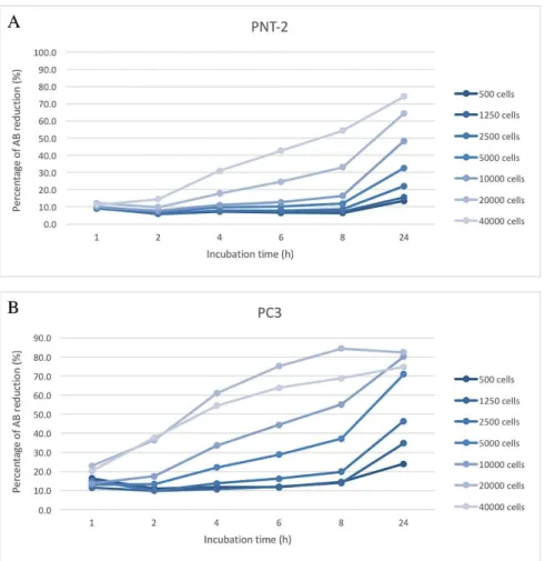
As a first approach, preliminar experiments were performed to determine the adequate cell density and incubation time for both PNT-2 and PC3 cell lines, to be used in the subsequent experiments. To that end, cells were plated at different densities and cell growth was monitorized for 24 h using the AB reagent (as described in detail in the Material and Methods section) (Figure 4). The percentage of AB reduction was calculated as follows below:
where, ελ1 and ελ2 are constants representing the molar extinction coefficient of AB
at 570 and 600 nm which represent the oxidized (εox) and reduced (εred) forms,
respectively: Aλ1 and Aλ2 represent absorbance of the test wells at 570 and 600 nm,
respectively, and A’λ1 and A’λ2 represent the absorbance of negative control (cells
treated with 1% DMSO) at 570 and 600 nm, respectively.
When performing experiments using cell cultures, the number of cells initially plated and the incubation time should be carefully choosed. Hence, since our goal was to study cell viability in response to different treatment conditions it was important to be sure that cells were in the logarithmic phase of growth when performing the assay. The graphs depicted on Figure 4 show that the growth rate of PNT-2 and PC3 cells is different: PC3 have an increased cell growth rate when compared to PNT-2. Knowing that PC3 are prostatic adenocarcinoma cells and PNT-2 are prostate pre-neoplasic epithelial cells, these results were expected.
Therefore, based on the results and the factors described above, and since it was decided to incubate the cells with the compounds for 24 h, we choosed to start the experiments with 7 500 cells per well in the following experiments. Using this cell density, we guaranteed that cells were in the logarithmic phase of growth and also that we didn’t reach the saturation point of the dye used in the assay (for the PNT-2 cell line the percentage of AB reduction was around 40% (Figure 4A) and for the PC3 cell line it was around 70% (Figure 4B)).
Figure 4- Growth curve of PNT-2 and PC3 cell lines determined using the Alamar Blue (AB) reagent. PNT-2
(A) and PC3 (B) cells were plated at various densities (500, 1 250, 2 500, 5 000, 10 000, 20 000 and 40 000 cells) in a 96-well plate and the growth was assessed after 1, 2, 4, 6, 8 and 24 h of incubation. Each experimental condition was performed once in duplicate using AB assay.
3.2 Preliminary cytotoxicity assay
One of the most important features of an ideal PS is the absence or minimal cytotoxicity under non-irradiating conditions. Therefore, before starting PDT assays, we analyzed eventual intrinsic cytototoxic effects of the four compounds in both cell lines. For that, the cell viability of PNT-2 and PC3 cells was determined after incubation with each PS (PS4a, PS4b, PS5a and PS5b), at different concentrations (1.00x10⁻⁴ M, 1.00x10⁻⁵ M, 1.00x10⁻⁶ M and 1.00x10⁻⁷ M), during 24 h, in light-restricted conditions. As controls of these experiments we used cells without any PS addiction and cells treated only with 1% DMSO – since the compounds were previously dissolved in DMSO. This DMSO control was important to understand if the alterations observed in
the cells treated with the PS were due to the DMSO or the compounds themselves. Figures 5 and Figure 6 show the effect of each compound on PNT-2 and PC3 cell viability after 2, 4, 6, 8 and 24h of PS contact.
Figure 5 - Effect of PS4a (top image) and PS4b (bottom image) on the viability of PNT-2 (graphs A and C, respectively) and PC3 (graphs B and D, respectively) cell lines in light-restricted conditions. Cells were
incubated with PS4a and PS4b at different concentrations (1.00x10⁻⁴ M, 1.00x10⁻⁵ M, 1.00x10⁻⁶ M and 1.00x10⁻⁷ M) along time (2, 4, 6, 8 and 24 h). Cells and cells treated with 1% DMSO were used as controls. Cell viability was assessed using the Alamar Blue (AB) reagent. Data are presented as the mean value referent to one experiment performed with two replicates for each condition.
The dark toxicity of PS4a and PS4b for PNT-2 and PC3 vary with the PS as it can be seen (Figure 5). We observed that PS4a have a smaller decrease in cell viability in both cell lines, namely for the higher concentrations tested, i.e., as higher is the concentration lower is the percentage of AB reduction. Instead, the PS4b have opposite results. This means that the control (cells incubated in RPMI) have lower percentage of AB reduced and PS4b seems to induce cell viability with a direct correlation of concentrations tested (this means, as higher is the concentration higher is the percentage
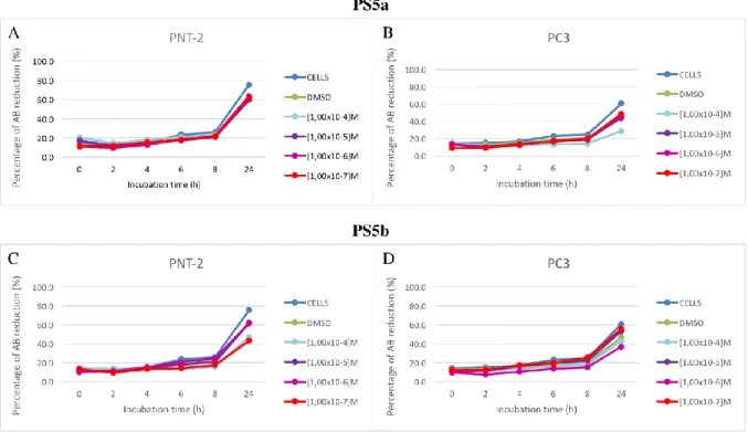
of reduction of AB). The PS5a and PS5b were also tested (see Figure 6) and the results were similar to PS4a; we observed a decrease in cellular viability in PNT-2 and PC3 cell lines when were compared the percentage of AB reduction values ones obtain for cell control with the control (cells incubated in RPMI) but there was not a correlation within and cell viability.
Figure 6 - Effect of PS5a (top image) and PS5b (bottom image) on the viability of PNT-2 (graphs A and C, respectively) and PC3 (graphs B and D, respectively) cell lines in light-restricted conditions. Cells were
incubated with PS5a and PS5b at different concentrations (1.00x10⁻⁴ M, 1.00x10⁻⁵ M, 1.00x10⁻⁶ M and 1.00x10⁻⁷ M) along time (2, 4, 6, 8 and 24 h). Cells and cells treated with 1% DMSO were used as controls. Cell viability was assessed using the Alamar Blue (AB) reagent. Data are referent to one experiment performed with two replicates for each condition.
Other interesting result is that cells treated with 1% DMSO did not seems to have a substantial difference from the control of cells only. After this, we can see, in all PSs for both cell lines, that 1% DMSO did not affect cell viability and for that this control “cells and DMSO” start’s to be our main control. This happens due to all new PSs were diluted in DMSO and in medium culture. DMSO in high percentage can kill cells but at 1% in all conditions that does not happen and do not influence cell growth and viability.
In short, Figure 5 and Figure 6 present our first approach to have a general idea about the intrinsic cytotoxicity of each compound in both cell lines. Although the results showed some cytotoxicity in light-restricted conditions, we considered that it does not have a meaningful influence on cell viability and prompted us to perform the PDT studies.
However, we excluded the lowest condition since concentrations below 10-6 are not photocytotoxic under PDT conditions used (data not shown).
3.3 Photodynamic therapy
In PDT assays, we analyzed the percentage of AB reduction after treatment with PS4a, PS4b, PS5a or PS5b, at three different concentrations (1.00x10⁻⁴ M, 1.00x10⁻⁵ M and 1.00x10⁻⁶ M). The experiments were done in 96-well plates using three replicates for each condition and three independent experiments were performed. To facilitate the comprehension of the results, they were divided by cell line.
As explained in the Materials and Methods section, the stock solution of all compounds were diluted in DMSO. Hence, we first verified the impact of the DMSO concentration used (1% DMSO) in cell viability, both in presence and absence of the light source of our PDT assays (Figure 7). All PDT results were monitorized for 24 h using the AB reagent (as described in detail in the Material and Methods section). The percentage of AB reduction was calculated as follows below:
where, FI = Fluorescent Intensity at 590 nm after sample excitation at 560 nm. The test agent was each condition used and the untreated control is cells treated with 1% DMSO, except when we analized the 1% DMSO effects on cells were untreated control was only cells.
As shown in Figure 7, the difference between the two conditions (cells and cells with 1% DMSO) were not significant and, therefore, were not thought to compromise the results (P-value<0.05). In PDT experiments both controls — cells and cells treated
with 1% DMSO — were performed, but we used the last one as our main negative control.
Figure 7- Cytotoxic and photocytotoxic effects of 1% DMSO in PNT-2 (graphs A and B, respectively) and PC3 (graphs C and D, respectively) cell lines. Cells were treated with 1% DMSO or left untreated and incubated in
light-restricted conditions for 24 h. After this incubation period, one of the cell plates was exposed to red light (λ= 630 nm
± 20 nm) with an irradiance of 1.28 mW.cm-2 during 20 min, while the other remained in light-restricted conditions. Both plates were further incubated for (24 h). Cytotoxicity and phototoxicity were then assessed using the Alamar Blue reagent. Data are presented as the mean value ± S.D. of three independent experiments, each with three replicates of each condition. Non-parametric tests for comparing independent samples were used to assess the statistical significance of the results (P-value<0.05).
3.3.1 Effect of the photosensitizers in PNT-2 cell line
PNT-2 cells were treated with PS4a, PS4b, PS5a or PS5b, at three different concentrations (1.00x10⁻⁴ M, 1.00x10⁻⁵ M, 1.00x10⁻⁶ M), for 24 h, in light-restricted conditions. The percentage of AB reduction in each condition was calculated to assess the intrinsic cytotoxic effect of each compound in PNT-2 cells (Figure 8).
Figure 8- Percentage of AB reduced by concentration (1.00x10⁻⁴ M, 1.00x10⁻⁵ M, 1.00x10⁻⁶ M) with treatment (PS4a, PS4b, PS5a and PS5b) in PNT-2 cell line, in the absence of light. Cytotoxicity was assessed by AB assay
24 h after supposed PDT. The percentage of cytotoxicity was calculated relatively to control cells (cells incubated in RPMI with 1% DMSO). Data are the mean value ± S.D. of three independent experiments performed in triplicates. Non-parametric test for comparing two independent samples. *- difference is significant at the P-value<0.05.
As shown in Figure 8, PS5a and PS5b at the highest concentration tested (1.00x10⁻⁴ M) led to a significant reduction of cell viability when compared to non-treated cells (P= 0.024 and P= 0.024, respectively). However, no significant differences were found between cell viability of cells treated with these compounds comparing to the direct control of cells treated with 1% DMSO. Concentrations tested, as well as the other PSs, did not shown to have significant impact in cell viability. As this test represents the intrinsic cytotoxicity of each PS in PNT-2 cells, because no PDT was applied, we might conclude that none of the conditions tested are cytotoxic for these cells in the dark.
The application of PDT to the same conditions led to different observations (Figure 9). At the highest concentration (1.00x10⁻⁴ M) PS5a and PS5b, show to be phototoxic when compared to non-treated cells (P= 0.024 and P= 0.024, respectively) and when
compared to direct control (P= 0.040 and P= 0.024, respectively). At 1.00x10⁻⁵ M, PS5b was the only PS that show phototoxicity (P= 0.024) when compared to non-treated cells. At 1.00x10⁻⁶ M, PS4a and PS5b presented phototoxicity when compared to non-treated cells (P= 0.024 and P= 0.024, respectively). Yet, in this two last conditions (at 1.00x10⁻⁵ M and 1.00x10⁻⁶ M), no significant differences were found for these compounds when comparing to the direct control of cells treated with 1% DMSO. As well, the rest of the concentrations tested and the other PSs, did not revealed significant impact in PNT-2 cell viability.
Figure 9- Percentage of AB reduced by concentration (1.00x10⁻⁴ M, 1.00x10⁻⁵ M, 1.00x10⁻⁶ M) with treatment (PS4a, PS4b, PS5a and PS5b) in PNT-2 cell line, after PDT. Photocytotoxicity was assessed by AB assay 24 h
after PDT. The percentage of photocytotoxicity was calculated relatively to control cells (cells incubated in RPMI with 1% DMSO). Data are the mean value ± S.D. of three independent experiments performed in triplicates. Non-parametric test for comparing two independent samples. *- difference is significant at the P-value<0.05.