Universidade de Aveiro 2017
Departamento de Química
Sofia Reis Brandão
Caracterização da dinâmica mitocondrial na
deficiência múltipla das acil-CoA desidrogenases
Characterization of mitochondrial dynamics in
multiple acyl-CoA dehydrogenase deficiency
Universidade de Aveiro 2017
Departamento de Química
Sofia Reis Brandão
Caracterização da dinâmica mitocondrial na
deficiência múltipla das acil-CoA desidrogenases
Characterization of mitochondrial dynamics in
multiple acyl-CoA dehydrogenase deficiency
Dissertação apresentada à Universidade de Aveiro para cumprimento dos requisitos necessários à obtenção do grau de Mestre em Bioquímica, ramo em Bioquímica Clínica, realizada sob a orientação científica da Doutora Rita Maria Pinho Ferreira, professora auxiliar do Departamento de Química da Universidade de Aveiro e do Doutor Hugo Daniel Carvalho de Azevedo Rocha, assistente principal da carreira dos técnicos superiores de Saúde, ramo de Genética no Instituto Nacional de Saúde Doutor Ricardo Jorge do Porto.
o júri
presidente Prof. Doutor Francisco Manuel Lemos Amado
Professor Associado com Agregação do Departamento de Química da Universidade de Aveiro
Prof. Doutor António Alexandre Moreira Ribeiro de Ascensão
Professor Auxiliar da Faculdade de Desporto da Universidade do Porto
Prof. Doutor Hugo Daniel Carvalho de Azevedo Rocha
Assistente principal da carreira dos técnicos superiores de Saúde, ramo de Genética no Instituto Nacional de Saúde Doutor Ricardo Jorge do Porto
agradecimentos Em primeiro lugar quero agradecer à minha orientadora, a Professora Rita Ferreira, por todas as sugestões e correções, pelas oportunidades que me possibilitaram aumentar o conhecimento científico e laboratorial e pelo apoio incansável ao longo deste ano. O empenho e dedicação que demonstra na vida profissional são um exemplo que espero alcançar um dia.
Ao meu co-orientador, o Doutor Hugo Rocha, agradeço pela oportunidade de aprender num ambiente exterior ao da Universidade de Aveiro, no INSA-Porto, e por todos os ensinamentos científicos e laboratoriais. Ter acompanhado o seu trabalho permitiu-me traçar novos objetivos profissionais e pessoais.
Agradeço também às técnicas dos laboratórios que frequentei, Cristina Barros e Lurdes Lopes, pela disponibilidade e paciência que mostraram sempre que lhes pedi ajuda. Um agradecimento especial à Cristina pelas palavras de carinho no final daqueles dias mais difíceis.
Ao José pela companhia no laboratório, sem dúvida conseguiu quebrar a rotina daqueles dias mais monótonos. À Rita Nogueira Ferreira agradeço a enorme paciência e disponibilidade por todos os esclarecimentos e conselhos laboratoriais. À Professora Margarida Fardilha pela facilidade na partilha de informações e receção no iBiMED e claro às suas alunas, a Magda e a Juliana, pelos conhecimentos laboratoriais que me transmitiram e pela simpatia e disponibilidade para colaborar sempre que lhes pedi ajuda. Aos amigos que me acompanharam nos momentos de desespero e euforia e fizeram com que nunca deixasse de ser quem sou. Carolina, Daniel, Joaquim, Anabela, Gonçalo, Dani, Dinês, Barbara e Inês obrigada por tornarem este ano tão especial e mais fácil. À colega de casa que se tornou numa amiga para a vida, Raquel, obrigada por me fazeres sentir a melhor pessoa do Mundo. As tuas palavras de conforto, mesmo à distância, fizeram a diferença para ignorar os dias difíceis.
Ao meu irmão e aos meus pais agradeço a paciência e carinho que tiveram sempre que fui a casa e não lhes dediquei todo o tempo porque estava ocupada com outras tarefas. Um agradecimento especial à minha mãe pelas longas horas de conversa que passámos ao telemóvel. À restante família agradeço os desafios e amizade com que desde cedo me habituaram, estou certa que me tornaram uma pessoa mais lutadora e persistente.
palavras-chave β-oxidação de ácidos gordos, rastreio neonatal, dinâmica mitocondrial, proteoma mitocondrial, doenças da β-oxidação mitocondrial de ácidos gordos, deficiência múltipla das acil-CoA desidrogenases.
resumo As doenças da β-oxidação mitocondrial dos ácidos gordos fazem parte do painel de doenças detetadas no rastreio neonatal na grande maioria dos países desenvolvidos, incluindo Portugal. A deficiência múltipla das acil-CoA desidrogenases é uma das doenças rastreadas, sendo rara e apresentando um padrão de transmissão autossómico recessivo. Esta disfunção no metabolismo dos ácidos gordos é caracterizada por fenótipos bastante distintos, sendo reconhecidas duas formas clínicas: moderada e grave. Estudos anteriores do nosso grupo de investigação revelaram que o proteoma mitocondrial de pacientes homozigotos para a mesma mutação evidencia níveis de expressão diferentes das mesmas proteínas e que os pacientes com formas moderadas da doença apresentam semelhanças e diferenças quando comparados com pacientes com formas graves. O objetivo deste estudo foi relacionar as alterações dos processos biológicos associados à homeostasia mitocondrial com a severidade da doença, associada a mutações no gene que codifica a proteína electron transfer flavoprotein dehydrogenase (ETFDH), traçando semelhanças e diferenças. No geral, o nosso estudo fornece uma perspetiva global da dinâmica mitocondrial nas duas formas da doença. Ambas as formas, moderada e grave, apresentaram biogénese mitocondrial diminuída e adaptação metabólica, suportadas por níveis baixos de peroxisome proliferator-activated receptor γ coactivator 1 alpha (PGC-1α) e de gliceraldeído-3-fosfato desidrogenase (GAPDH), respetivamente, em todos os pacientes com a doença. Os níveis de expressão de sirtuina 3 (SIRT3) e a atividade de ATP sintase foram encontrados diminuídos em quase todos os pacientes, sugerindo uma disfunção mitocondrial. Os níveis reduzidos de SIRT3 foram corroborados pela diminuição dos níveis de PGC-1α. Os níveis de expressão das outras proteínas analisadas foram diversos entre os pacientes e, portanto, sugerem que a correlação entre a severidade da doença e as adaptações mitocondriais não é transversal a todas as formas da doença. De facto, os diferentes resultados obtidos podem explicar, pelo menos em parte, a variedade de fenótipos observados em pacientes com a doença. Neste sentido, são necessários mais estudos para compreender melhor a patogénese da doença. No futuro, seria interessante analisar os efeitos da deficiência de ETFDH noutras células em vez de fibroblastos e pesquisar outros metabolitos energéticos relacionados com estes mecanismos.
keywords Fatty acid β-oxidation, newborn screening, mitochondrial dynamics, mitochondrial proteome, disorders of mitochondrial fatty acid β-oxidation, multiple acyl-CoA dehydrogenase deficiency.
abstract Mitochondrial fatty acid β-oxidation disorders are some of the many diseases detected by newborn screening in most developed countries, including Portugal. Among screened disorders, multiple acyl-CoA dehydrogenase deficiency (MADD) is a rare autosomal recessively inherited disorder that presents very distinct phenotypes, being recognized in two clinical forms: mild and severe. Previous studies from our research group showed that the mitochondrial proteome of homozygous patients having the same mutation presents different expression levels of the same proteins and that patients with mild forms of MADD share some proteins with severe forms and at the same time present distinctive expression patterns. The aim of this study was to relate the regulation of biological processes associated to mitochondrial homeostasis with the severity of MADD, due to mutations on the gene that codifies the protein electron transfer flavoprotein dehydrogenase (ETFDH), evidencing similarities and differences.
In overall our study provides a global perspective of the mitochondrial dynamics in the two forms of MADD, mild and severe. Both forms presented down-regulation of mitochondrial biogenesis and metabolic adaptation highlighted by lower levels of peroxisome proliferator-activated receptor γ
coactivator 1 alpha (PGC-1α) and glyceraldehyde-3-phosphate dehydrogenase (GAPDH), respectively, in all MADD patients. Expression levels of sirtuin 3 (SIRT3) and the activity of ATP synthase were found decreased in almost all patients, suggesting mitochondrial dysfunction. Down-regulation of SIRT3 was corroborated by decreased levels of PGC-1α. The expression levels of the other proteins analyzed were diverse among MADD patients and thus highlighting no straight full correlation between disease severity and mitochondrial adaptations. In fact, the different results obtained may explain, at least in part, the variety of phenotypes observed in MADD patients. So, more studies are needed to better understand MADD pathogenesis. In the future, it would be interesting to analyze the effects of ETFDH deficiency in other cell types rather than fibroblasts and search for other energetic metabolites related with these mechanisms.
Table of contents
Figure Index ... iii
Table Index ... vii
Abbreviations ... viii
1. Introduction ... 1
1.1. Mitochondrial fatty acid β-oxidation ... 4
1.2. Disorders of mitochondrial fatty acid β-oxidation ... 8
1.2.1. Multiple acyl-CoA dehydrogenase deficiency ... 11
1.2.1.1. Relation between genotype and phenotype in MADD ... 12
1.3. The role of mitochondria on the regulation of cell homeostasis in FAOD ... 15
1.4. The potential contribution of mitochondrial biogenesis to FAOD ... 20
2. Aims ... 25
3. Materials and Methods ... 29
3.1. Experimental design ... 31
3.2. Samples characterization ... 31
3.3. Cell culture and cell extracts preparation ... 32
3.4. Total protein quantification ... 32
3.5. Protein precipitation ... 33
3.6. SDS-PAGE and Western blotting ... 33
3.7. Determination of the content of carbonylated proteins by Slot-blot ... 34
3.8. Immunocytochemistry ... 35
3.9. Spectrophotometric activity assays ... 36
3.9.1. ATP synthase activity ... 36
3.9.2. Citrate synthase activity ... 37
4. Results... 39
4.1. Analysis of ETFDH expression ... 41
4.2. Effect of MADD on cells’ metabolic status ... 42
4.3. Effect of MADD on oxidative stress ... 43
4.4. Effect of MADD on mitochondrial biogenesis and mitophagy ... 44
4.5. Effect of MADD on apoptosis ... 47
5. Discussion ... 49
6. Conclusion and Future perspectives ... 59
7. References ... 63
Figure Index
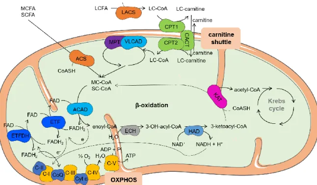
Figure 1. Schematic representation of mitochondrial fatty acid β-oxidation and its
association with Krebs cycle and OXPHOS. Figure made with Servier Medical Art. Abbreviations: ACAD: acyl-CoA dehydrogenases, acetyl-CoA: acetyl-coenzyme A, ACS: acyl-CoA synthetase, ADP: adenosine diphosphate, ATP: adenosine triphosphate, C-I: nicotinamide adenine dinucleotide-ubiquinone oxidoreductase, C-II: succinate dehydrogenase, III: ubiquinol-cytochrome c reductase, IV: cytochrome c oxidase, C-V: ATP synthase, CACT: carnitine/acylcarnitine translocase, CoASH: coenzyme A, CoQ: ubiquinone, CPT1: carnitine palmitoyltransferase 1, CPT2: carnitine palmitoyltransferase 2, Cyt c: cytochrome c, e-: electrons, ECH: 2-enoyl-CoA hydratases, ETF: electron transfer flavoprotein, ETFDH: electron transfer flavoprotein dehydrogenase, FAD: flavin adenine dinucleotide, FADH2: reduced form of FAD, HAD: 3-hydroxyacyl-CoA dehydrogenases, H2O: water, KAT: 3-ketoacyl-CoA thiolases, LACS: long-chain acyl-CoA synthetase, LC-carnitine: long-chain fatty acylcarnitine ester, LC-CoA: long-chain fatty acyl-CoA ester, LCFA: long-chain fatty acids, MC-CoA: medium-chain fatty acyl-CoA ester, MCFA: medium-chain fatty acids, MTP: mitochondrial trifunctional protein, NAD+: nicotinamide adenine dinucleotide, NADH: reduced form of NAD+, O2: oxygen, OH: hydroxy, OXPHOS: oxidative phosphorylation, Pi: inorganic phosphate, SC-CoA: short-chain fatty acyl-CoA ester, SCFA: short-chain fatty acids, VLCAD: very long-chain acyl-CoA dehydrogenase. ... 5
Figure 2. Extrinsic and intrinsic apoptotic pathways. The extrinsic pathway is initiated by
binding of ligands to their respective DR, which leads to the formation of DISC and activates Casp-8 and this directly activates Casp-3, executing apoptosis. Casp-8 also activates Bid, starting the intrinsic pathway, which interacts with mitochondrial pro-apoptotic proteins Bax and Bak, leading to the release of several pro-pro-apoptotic proteins, which triggers caspase-dependent (black arrows) or caspase-independent (blue arrows) cytosolic signaling events. Cyt c binds to Apaf-1 to form the apoptosome that recruits and activates Casp-9 which activates Casp-3. Caspase activation is improved by Smac/Diablo and Omi/HtrA2 since they inhibit IAPs. AIF and EndoG bind with DNA, inducing DNA fragmentation, via caspase-independent pathway. The intrinsic apoptotic pathway can also
be stimulated by ROS. Figure made with Servier Medical Art. Abbreviations: AIF: apoptosis-inducing factor, Apaf-1: apoptotic protease-activating factor-1, Bak: bcl-2 antagonist killer 1, Bax: bcl-2 associated X protein, Bid: BH3-only pro-apoptotic protein, Casp-: caspase, number refers to the type of caspase, Cyt c: cytochrome c, DISC: death-inducing signaling complex, DNA: deoxyribonucleic acid, DR: death receptors, EndoG: endonuclease G, IAPs: inhibitor of apoptosis proteins, Omi/HtrA2: high-temperature requirement A2 serine protease, Smac/Diablo: second mitochondria-derived activator of caspases/direct IAP binding protein with low isoelectric point, ROS: reactive oxygen species. ... 18
Figure 3. Regulation of mitochondrial biogenesis by PGC-1α signaling. Enhanced
NAD+/NADH ratio stimulates SIRT1 which deacetylates PGC-1α, whereas the increase in AMP/ATP ratio activates AMPK which phosphorylates it. Both deacetylation (A) and phosphorylation (B) activate PGC-1α, leading to stimulation of specific nuclear factors, such as NRFs, ERRs and PPARs, which activate their target genes. NRF1 regulates mtTFA which increases mtDNA gene transcription and replication. This and both NRFs and ERRs lead to increase in OXPHOS. ERRs also stimulate FAO and PPAR which has the same effect. SIRT3, in NAD+-dependent via, stimulates OXPHOS, FAO and ROS metabolism proteins. All these processes lead to mitochondrial biogenesis. Figure made with Servier Medical Art. Abbreviations: AMP: adenosine monophosphate, AMPK: AMP-activated protein kinase, ATP: adenosine triphosphate, ERRs: oestrogen-related receptors, FAO: mitochondrial fatty acid β-oxidation, MnSOD: manganese superoxide dismutase, mtDNA: mitochondrial deoxyribonucleic acid, mtTFA: mitochondrial transcription factor A, NAD+: nicotinamide adenine dinucleotide, NADH: reduced form of NAD+, NRFs: nuclear respiratory factors, OXPHOS: oxidative phosphorylation, PGC-1α: peroxisome activated receptor γ coactivator 1 alpha, PPARs: peroxisome proliferator-activated receptors, ROS: reactive oxygen species, SIRT1: sirtuin 1, SIRT3: sirtuin 3. ... 21
Figure 4. Experimental design followed at the present work. Skin biopsy was done in all
MADD patients and controls (heathy individuals aged-matched with patients) to obtain cultured fibroblasts. These cells were then analyzed by immunoblotting to semi-quantify the amount of specific proteins, by immunocytochemistry to estimate the mitochondrial density and by specific spectrophotometric assays to measure the activity of key metabolic enzymes. ... 31
Figure 5. Expression levels of ETFDH measured by Western blotting in total homogenates
of skin fibroblasts from controls and MADD patients. MADD1 and MADD2 correspond to a mild form while MADD3 and MADD4 have severe form of MADD. Above the graph is presented a representative image of the Western blots obtained. The values (mean ± SD) are expressed in arbitrary units of optical density (OD). ** p < 0.01. ... 41
Figure 6. Effect of ETFDH deficiency on the metabolic status of skin fibroblasts’
homogenate. MADD1 and MADD2 correspond to a mild form while MADD3 and MADD4 have severe form of MADD. Expression levels of ATP synthase α subunit measured by Western blotting; above the graph is presented a representative image of the Western blots obtained (A). ATP synthase activity was spectrophotometrically measured and values are expressed in mmol Pi.min-1.mg-1 (B). Expression levels of GAPDH measured by Western blotting; above the graph is presented a representative image of the Western blots obtained (C). Ratio between GAPDH and ATP synthase α subunit (D). The values (mean ± SD) are expressed in arbitrary units of optical density (OD) for A and C. * p < 0.05; ** p < 0.01; *** p < 0.001. # The value obtained for this sample was below the detection limit so the value is hide in the graph. ... 42
Figure 7. Effect of ETFDH deficiency on protein carbonylation (A), SIRT3 (B) and
MnSOD (C) levels in total homogenates of skin fibroblasts assessed by immunoblotting. MADD1 and MADD2 correspond to a mild form while MADD3 and MADD4 have severe form of MADD. Above each graph is presented a representative image of the immunoblots obtained. The values (mean ± SD) are expressed in arbitrary units of optical density (OD). * p < 0.05; ** p < 0.01; *** p < 0.001. ... 44
Figure 8. Effect of ETFDH deficiency on mitochondrial biogenesis and mitophagy
markers assessed in total homogenates of skin fibroblasts. MADD1 and MADD2 correspond to a mild form while MADD3 and MADD4 have severe form of MADD. CS activity was spectrophotometrically measured and the values (mean ± SD) are expressed in nmol.min-1.mg-1 (A). Expression levels of mtTFA (B), PGC-1α (C) and ATG5 (D) measured by Western blotting; above each graph is presented a representative image of the Western blots obtained. The values (mean ± SD) are expressed in arbitrary units of optical density (OD) for B, C and D. * p < 0.05; ** p < 0.01; *** p < 0.001. # The value obtained for this sample was below the detection limit so the value is hide in the graph. ... 45
Figure 9. Effect of ETFDH deficiency on mitochondrial density in cultured skin
fibroblasts from MADD patients. For control, mild and severe skin fibroblasts is presented a representative image of COX IV (red, Alexa Fluor® 594) and the merge between COX IV and the cell nuclei (blue, Hoechst 33258). Negative controls are presented in Appendix section. All images were obtained with a 100x magnification in an Olympus IX-81 inverted epifluorescence microscope. The scale bar of images is 20 μm. ... 47
Figure 10. Effect of ETFDH deficiency on the content of Bax (A) and Bcl-2A1 (B), ratio
between Bax and Bcl-2A1 (C) and Casp-3 (D) measured by Western Blotting in total homogenates of skin fibroblasts. MADD1 and MADD2 correspond to a mild form while MADD3 and MADD4 have severe form of MADD. Above each graph is presented a picture of the Western blots obtained. The values (mean ± SD) are expressed in arbitrary units of optical density (OD). * p < 0.05; *** p < 0.001; **** p < 0.0001. ... 48
Figure 11. Integrated perspective of molecular mechanisms modulated by ETFDH
deficiency in mild (A) and severe (B) forms of MADD. The proteins analyzed in the present study are highlighted with the up-regulated presented in red and the down-regulated in green. Proteins with no expression variation are presented in blue and proteins differently expressed between patients are presented in orange. These proteins belong to different pathways that relate to mitochondria and many of them are specifically expressed on mitochondrion and thus these results for the different patients may explain why MADD is such a heterogeneous disorder. Figure made with Servier Medical Art. Abbreviations:
ATG5: autophagy protein 5, Bax: bcl-2 associated X protein, Bcl-2A1: bcl-2 related protein A1, Casp-3: cleaved caspase-3, CS: citrate synthase, C-V: ATP synthase, ETFDH: electron transfer flavoprotein dehydrogenase, GAPDH: glyceraldehyde-3-phosphate dehydrogenase, MnSOD: manganese superoxide dismutase, mtTFA: mitochondrial transcription factor A, PGC-1α: peroxisome proliferator-activated receptor γ coactivator 1 alpha, SIRT3: sirtuin 3, ROS: reactive oxygen species. ... 57
Figure 12. Effect of ETFDH deficiency on mitochondrial density in cultured skin
fibroblasts from MADD patients. Negative control (NC) for control, mild and severe skin fibroblasts were obtained from incubation with blocking solution instead of primary antibody (rabbit polyclonal anti-COX IV). Representative image of the merge between COX IV (red, Alexa Fluor® 594) and the cell nuclei (blue, Hoechst 33258) for NC. The absence of red (COX IV) dots compared to the merge where the primary antibody was added (Figure 9), indicates that there is specificity for the primary antibody chosen. All images were obtained with a 100x magnification in an Olympus IX-81 inverted epifluorescence microscope. The scale bar of images is 20 μm. ... 77
Table Index
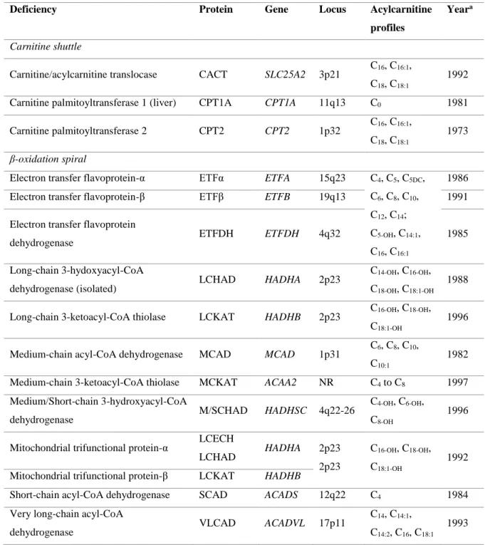
Table 1. Genetic and biochemical characteristics of the mitochondrial fatty acid
β-oxidation disorders. Gene, locus and year information was collected from Sim et al. (23) and Gregersen et al. (28) while the information for acylcarnitine profiles was adapted from Sahai et al. (1) and Sim et al. (23). Acylcarnitine profile was further completed with information from Olsen et al. (8). ... 9
Table 2. Type and number of gene mutations in MADD. Adapted from Gregersen et al.
Abbreviations
ACAD Acyl-CoA dehydrogenases
Acetyl-CoA Acetyl-coenzyme A
ACS Acyl-CoA synthetase
ADP Adenosine diphosphate
AIF Apoptosis-inducing factor
AMP Adenosine monophosphate
AMPK AMP-activated protein kinase
Apaf-1 Apoptotic protease-activating factor-1
ATG5 Autophagy protein 5
ATP Adenosine triphosphate
ATPase α ATP synthase α subunit
Bak Bcl-2 antagonist killer 1
Bax Bcl-2 associated X protein
Bcl-2A1 Bcl-2 related protein A1
Bid BH3-only pro-apoptotic protein
BSA Bovine serum albumin
C5DC Glutarylcarnitine
C-I Nicotinamide adenine dinucleotide-ubiquinone oxidoreductase
C-II Succinate dehydrogenase
C-III Ubiquinol-cytochrome c reductase
C-IV Cytochrome c oxidase
C-V ATP synthase
CACT Carnitine/acylcarnitine translocase
cAMP Cyclic adenosine monophosphate
Casp- Caspase (cysteine-aspartic protease), number refers to the type of
caspase
Cnumber Acylcarnitine species with the corresponding carbon chain length
Cnumber-OH 3-Hydroxyacylcarnitine species with the corresponding carbon chain length
CoQ Ubiquinone (Coenzyme Q10)
COX IV Subunit IV of cytochrome c oxidase
CPT Carnitine palmitoyltransferase
CS Citrate synthase
Cyt c Cytochrome c
DISC Death-inducing signaling complex
DMEM Dulbecco's Modified Eagle Medium
DNA Deoxyribonucleic acid
DNP 2,4-Dinitrophenyl
DNPH 2,4-Dinitrophenylhydrazine
DR Death receptors
Drp1 Dynamin-related protein 1
DTNB 5,5’-Dithiobis-(2-nitrobenzoate)
ECH 2-Enoyl-CoA hydratases
EMA Ethylmalonic acid
EndoG Endonuclease G
ER Endoplasmic reticulum
ERRs Oestrogen-related receptors
ESI Electrospray ionization
ETF Electron transfer flavoprotein
ETFDH ETF dehydrogenase
ETF-QO ETF-ubiquinone oxidoreductase
FA Fatty acids
FAD Flavin adenine dinucleotide
FADH2 Reduced form of FAD
FAO Mitochondrial fatty acid β-oxidation
FAOD Mitochondrial fatty acid β-oxidation disorders
FasL Fas ligand
Fis1 Mitochondrial fission 1 protein
Fzo Drosophila mitofusin gene
GA Glutaric aciduria
GPx Glutathione peroxidase
HAD 3-Hydroxyacyl-CoA dehydrogenases
HIF-1α Hypoxia-inducible factor-1α
IAPs Inhibitor of apoptosis proteins
INSA National Institute of Health Doctor Ricardo Jorge
KAT 3-Ketoacyl-CoA thiolases
LACS Long-chain acyl-CoA synthetase
LCAD Long-chain acyl-CoA dehydrogenase
LC-carnitine Long-chain fatty acylcarnitine ester
LC-CoA Long-chain fatty acyl-CoA ester
LCECH Long-chain 2-enoyl-CoA hydratase
LCFA Long-chain fatty acids
LCHAD Long-chain 3-hydroxyacyl-CoA dehydrogenase
LCKAT Long-chain 3-ketoacyl-CoA thiolase
MAD Multiple acyl-CoA dehydrogenase
MADD Multiple acyl-CoA dehydrogenase deficiency
MAPK Mitogen-activated protein kinase
MCAD Medium-chain acyl-CoA dehydrogenase
MCADD Medium-chain acyl-CoA dehydrogenase deficiency
MC-CoA Medium-chain fatty acyl-CoA ester
MCFA Medium-chain fatty acids
MCKAT Medium-chain 3-ketoacyl-CoA thiolase
Mfns Mitofusins
MnSOD Manganese superoxide dismutase
mRNA Messenger ribonucleic acid
M/SCHAD Medium/Short-chain 3-hydroxyacyl-CoA dehydrogenase
MS/MS Tandem mass spectrometry
mtDNA Mitochondrial DNA
mtGSH Mitochondrial glutathione
MTP Mitochondrial trifunctional protein
MTP18 Mitochondrial membrane protein 18
NAD+ Nicotinamide adenine dinucleotide
NADH Reduced form of NAD+
NBS Newborn screening
NC Negative control
NMD Nonsense-mediated mRNA decay
NR Not reported
NRFs Nuclear respiratory factors
OD Optical density
Omi/HtrA2 High-temperature requirement A2 serine protease
OPA1 Mitochondrial dynamin-like 120 kDa protein
OXPHOS Oxidative phosphorylation
PBS Phosphate-buffered saline
PGC-1 Peroxisome proliferator-activated receptor γ coactivator 1
Pi Inorganic phosphate
PKA Protein kinase A
PPAR Peroxisome proliferator-activated receptor
PQC Protein quality control
Prx III Peroxiredoxin mitochondrial
PTP Permeability transition pore
ROS Reactive oxygen species
RNS Reactive nitrogen species
RR-MADD Riboflavin-responsive MADD
SCAD Short-chain acyl-CoA dehydrogenase
SC-CoA Short-chain fatty acyl-CoA ester
SCECH Short-chain 2-enoyl-CoA hydratase
SCFA Short-chain fatty acids
SCHAD Short-chain 3-hydroxyacyl-CoA dehydrogenase
SD Standard deviation
SDS Sodium dodecyl sulphate
SDS-PAGE SDS-polyacrylamide gel electrophoresis
SIRT Sirtuin (silent information regulator two (Sir2) protein)
protein with low isoelectric point
TBS Tris-buffered saline
TG Triacylglycerides
TNB 2-Nitro-5-thiolbenzoate
TNF Tumor necrosis factor
TRAIL TNF-related apoptosis-inducing ligand
Trx II Thioredoxine mitochondrial
TTBS TBS with Tween 20
UCP2 Uncoupling protein 2
VLCAD Very long-chain acyl-CoA dehydrogenase
Fatty acid oxidation defects are a group of inherited metabolic disorders, affecting the enzymes involved in the oxidation of fatty acids (1,2). These disorders are usually transmitted in an autosomal recessive pattern being individually rare, but together they represent a large and important group of inherited metabolic disorders (1). Patients have heterogeneous clinical phenotypes and the same metabolic blockage may be associated to very different clinical phenotypes. The main affected organs include heart, liver and skeletal muscles (2). The mortality and morbidity rates associated with these conditions can be prevented, or at least decreased, if disorders are early recognized and treated (1). Indeed, fatty acid oxidation disorders can lead to serious health problems and even to death if not treated or when the diagnosis is done too late. The advantages of an early intervention make this group of disorders main targets of newborn screening (NBS) programs worldwide (1,3,4). The NBS aims to identify newborns with serious but treatable health diseases before the onset of symptoms and the development of irreversible damage (5,6). Since the introduction of tandem mass spectrometry (MS/MS) into screening programs in the 1990s, the number of inherited metabolic disorders screened greatly increased, including amino acid, organic acid and fatty acid metabolism disorders, since this technique allows the simultaneously determination of many analytes on the same analysis (1,5). NBS is one of the most successful public health programs initiated in the last fifty years (1,6).
Among mitochondrial fatty acid β-oxidation (FAO) disorders, multiple acyl-CoA dehydrogenase deficiency (MADD) is one of the twenty-five disorders screened by NBS in Portugal. The birth prevalence of MADD is 1:164,765 in Portugal (7). These FAO disorders (FAOD) result from defects in enzymes of mitochondrial fatty acid β-oxidation; however, in most cases the association between the phenotype and the genotype is not straightforward (2,8), which suggest that other individual cellular/molecular mechanisms might interfere with disease pathogenesis. Herein, we overview FAO metabolism, its regulation and contextualize MADD, the chosen model to study mitochondrial dynamics disturbances in FAO. We also critically analyze the potential contribution of the molecular pathways harbored in mitochondria or involved in the regulation of its functionality to the pathogenesis of FAOD, giving emphasis to MADD.
1.1. Mitochondrial fatty acid β-oxidation
The main function of mitochondrial fatty acid β-oxidation (FAO) is to generate acetyl-coenzyme A (acetyl-CoA) and reducing equivalents such as flavin adenine dinucleotide (FADH2) and nicotinamide adenine dinucleotide (NADH), especially during periods of fasting and metabolic stress. These molecules are linked to the Krebs cycle and to the oxidative phosphorylation (OXPHOS) system leading to energy production in the form of adenosine triphosphate (ATP) (9,10). Figure 1 overviews the FAO pathway from the activation of fatty acids to the generation of acetyl-CoA and its association with the Krebs cycle and OXPHOS.
OXPHOS is the key metabolic pathway in mitochondrial energy production. It requires the action of five multiheteromeric complexes located in the inner mitochondrial membrane, designated complexes I to V (11,12). Electrons donated from NADH are passed from complex I to complex III through ubiquinone (CoQ). In addition, CoQ also takes electrons from succinate via complex II and from electron transfer flavoprotein-ubiquinone oxidoreductase (ETF-QO). Finally, cytochrome c (Cyt c), an iron-containing heme protein, shuttles electrons from complex III to IV, which transfers its electrons to oxygen (O2), the final electron acceptor. During this process, the electrochemical gradient generated by the pumping of protons out of the mitochondrial inner membrane to the intermembrane space, is used by complex V in order to convert adenosine diphosphate (ADP) to ATP (12) (Figure 1).
Adipose tissue triacylglycerides (TG) are the primary source of fatty acids (FA) used for FAO. During fasting conditions TG are mobilized, transported and distributed to various tissues through bloodstream in lipoproteins (9). Once inside the cells, long-chain FA (LCFA) are activated to their coenzyme A (CoASH) esters and transported into mitochondria for subsequent β-oxidation (9,10,13). The transporter involved in the process corresponds to the “carnitine shuttle” that requires the concerted action of three proteins: carnitine palmitoyltransferase 1 (CPT1), carnitine/acylcarnitine translocase (CACT), and carnitine palmitoyltransferase 2 (CPT2). CPT1 converts acyl-CoA compounds to their acylcarnitine metabolites at the outer mitochondrial membrane. CACT transports the acylcarnitines across the inner mitochondrial membrane in exchange for free carnitine, and CPT2 re-esterifies the acylcarnitines to their acyl-CoA esters on the inner mitochondrial
diffuse freely across the mitochondrial membrane into the matrix, where they are activated to their corresponding acyl-CoA esters, which are oxidized by FAO enzymes (9,10,14). Once in the mitochondrial matrix FA are oxidized by the -oxidation system (Figure 1).
Figure 1. Schematic representation of mitochondrial fatty acid β-oxidation and its association with Krebs
cycle and OXPHOS. Figure made with Servier Medical Art. Abbreviations: ACAD: acyl-CoA dehydrogenases, acetyl-CoA: acetyl-coenzyme A, ACS: acyl-CoA synthetase, ADP: adenosine diphosphate, ATP: adenosine triphosphate, C-I: nicotinamide adenine dinucleotide-ubiquinone oxidoreductase, C-II: succinate dehydrogenase, C-III: ubiquinol-cytochrome c reductase, C-IV: cytochrome c oxidase, C-V: ATP synthase, CACT: carnitine/acylcarnitine translocase, CoASH: coenzyme A, CoQ: ubiquinone, CPT1: carnitine palmitoyltransferase 1, CPT2: carnitine palmitoyltransferase 2, Cyt c: cytochrome c, e-: electrons,
ECH: 2-enoyl-CoA hydratases, ETF: electron transfer flavoprotein, ETFDH: electron transfer flavoprotein dehydrogenase, FAD: flavin adenine dinucleotide, FADH2: reduced form of FAD, HAD: 3-hydroxyacyl-CoA
dehydrogenases, H2O: water, KAT: 3-ketoacyl-CoA thiolases, LACS: long-chain acyl-CoA synthetase,
LC-carnitine: long-chain fatty acylcarnitine ester, LC-CoA: long-chain fatty acyl-CoA ester, LCFA: long-chain fatty acids, MC-CoA: medium-chain fatty acyl-CoA ester, MCFA: medium-chain fatty acids, MTP: mitochondrial trifunctional protein, NAD+: nicotinamide adenine dinucleotide, NADH: reduced form of
NAD+, O
2: oxygen, OH: hydroxy, OXPHOS: oxidative phosphorylation, Pi: inorganic phosphate, SC-CoA:
short-chain fatty acyl-CoA ester, SCFA: short-chain fatty acids, VLCAD: very long-chain acyl-CoA dehydrogenase.
The β-oxidation cycle is characterized by four sequential steps in which an acyl-CoA ester undergoes dehydrogenation, hydration, dehydrogenation again and finally thiolytic cleavage (Figure 1). These steps are catalyzed by enzymes with overlapping chain length specificities (10,14). Beta-oxidation can also occur in peroxisomes, although it is different from mitochondrial oxidation in terms of enzymology, regulation, energy production and especially substrate specificity (15,16). Indeed, LCFA are mainly oxidized in peroxisomes and after shortening of their chain length, they are transported to mitochondria (15). Inside mitochondria, FA are metabolized by FAO enzymes and fully oxidized to carbon dioxide (CO2) and water (H2O) by Krebs cycle and OXPHOS, respectively (14). There are another types of FA oxidation such as α- and ω-oxidation, which require additional enzymes for the oxidation of branched-chain or odd-numbered FA (14,17).
The first step in β-oxidation cycle is catalyzed by at least three distinct acyl-CoA dehydrogenases (ACAD), each having preference for acyl-CoA substrates of differing chain lengths: very long-chain acyl-CoA dehydrogenase (VLCAD) acts on C12-C24 substrates, medium-chain acyl-CoA dehydrogenase (MCAD) acts on C6-C16 substrates, and short-chain acyl-CoA dehydrogenase (SCAD) acts on C4-C6 substrates (14,18). There are other two additional ACAD with less well established physiological roles: long-chain acyl-CoA dehydrogenase (LCAD) and acyl-CoA dehydrogenase 9 (19). SCAD, MCAD and LCAD are soluble, mitochondrial matrix enzymes composed of four identical subunits with a native molecular mass of 160-180 kDa, whereas VLCAD is membrane-bound and is not a tetramer but a dimer of two identical subunits of about 70 kDa. Each of these subunits carries a flavin adenine dinucleotide (FAD) noncovalently bound at the active site, supporting the activity of these enzymes in the catalysis of a FAD-linked dehydrogenation (18). Reoxidation of the reduced flavoproteins is possible by electron transfer flavoprotein (ETF), which has FAD as prosthetic group, and passes reducing equivalents to another flavoprotein called electron transfer flavoprotein-ubiquinone oxidoreductase (ETF-QO) (18,20). ETF-QO, also designated as electron transfer flavoprotein dehydrogenase (ETFDH), passes its electrons to the mitochondrial electron transport chain via CoQ (Figure 1). ETF is a dimer of two nonidentical subunits, α and β, containing one FAD molecule and is localized in the mitochondrial matrix, while ETFDH is a flavoprotein localized in the inner mitochondrial membrane with 68 kDa (9,20). Crystallographic studies support the association ACAD-ETF-ETFDH during electron and
hydrogen transfer from ETFDH to CoQ, which delivers electrons to complex III of OXPHOS (20) (Figure 1).
The second step of β-oxidation is the hydration of 2-trans-enoyl-CoAs to their corresponding 3-hydroxyacyl-CoAs catalyzed by 2-enoyl-CoA hydratases (ECH) (Figure 1). There are at least two distinct enzymes: crotonase and long-chain 2-enoyl-CoA hydratase (LCECH). The first, composed of six identical subunits, is most active for short-chain substrates, while the second, responsible for hydration of long-short-chain substrates, is part of the mitochondrial trifunctional protein (MTP) (10,14).
The third step of the β-oxidation cycle involves the dehydrogenation of 3-hydroxyacyl-CoAs to 3-oxoacyl-3-hydroxyacyl-CoAs by two distinct forms of 3-hydroxyacyl-CoA dehydrogenases (HAD) (Figure 1). Short- and medium-chain substrates are preferentially dehydrogenated by a generic short- to medium-chain hydroxyacyl-CoA dehydrogenase, which was first described as short-chain 3-hydroxyacyl-CoA dehydrogenase (SCHAD), a dimer of two identical subunits of 33 kDa each. Long-chain 3-hydroxyacyl-CoA dehydrogenase (LCHAD) is the other specific enzyme and belongs to the MTP (9,14).
In the last step of β-oxidation, 3-ketoacyl-CoAs are converted to acetyl-CoA and its corresponding acyl-CoA ester two-carbon atoms shorter by thiolytic cleavage (9,10) (Figure 1). This reaction is catalyzed by thiolases, which exist in three different isoforms. One thiolase, a homotetramer of 42 kDa subunits localized in the mitochondrial matrix, is specific for acetoacetyl-CoA and 2-methylacetoacetyl-CoA. The other thiolase, often called general thiolase or medium-chain 3-ketoacyl-CoA thiolase (MCKAT), acts on substrates ranging from C4 to C12. The third thiolase is the long-chain specific thiolase and is one of three proteins of the MTP. MTP is a heterooctamer with four α- and four β-subunits. The α-subunit carries the enzymes of the second and third steps of β-oxidation cycle (LCECH and LCHAD), whereas the β-subunit harbors the thiolase component (9,10,14).
In overall, there are at least twenty-five enzymes and specific transport proteins involved in FAO and defects in many of them are associated with human diseases (2,4,21,22).
1.2. Disorders of mitochondrial fatty acid β-oxidation
Several diseases related to defects in β-oxidation enzymes have been identified, which are generally designated as mitochondrial fatty acid β-oxidation disorders (FAOD) (2,23). FAOD are generally inherited in an autosomal recessive pattern and are individually rare, although they are collectively common (1,2). The inherited metabolic defects can be assigned to two groups: (a) those associated with the “carnitine shuttle”, for example CPT1; and (b) those of β-oxidation spiral, such as SCAD, ETF and ETFDH (23). In table 1 are represented FAOD from the two distinct groups mentioned above, their protein, gene and locus defect, the main acylcarnitines found in plasma and the year which they were first described.
Most FAOD are identified by the acylcarnitine profile analyzed by flow injection electrospray ionization (ESI) tandem mass spectrometry (MS/MS) (3,24). In fact, the acylcarnitine profile data obtained from NBS programs is crucial for the diagnosis of FAOD and consequently to estimate its birth prevalence (4). The birth prevalence is a more robust evidence of FAOD since there are significant cases of patients that only present symptoms in late infancy or adulthood. In Portugal, the overall birth prevalence of FAOD is 1:5,991 live births (7). The acylcarnitine profile found in patients with CPT1 and CPT2/CACT deficiencies is very distinct from those observed for ETF and ETFDH deficiencies (Table 1). The CPT1 deficiency is associated with elevated levels of free carnitine (C0) in plasma and absence of long-chain acylcarnitines due to its inability to produce these metabolites (2). In opposite, patients with CPT2/CACT deficiencies present elevated levels of long-chain acylcarnitines (C16, C16:1, C18, C18:1) since they are formed but are not correctly transported through the mitochondrial membrane. Deficiencies in ETF and ETFDH proteins are associated with a varied acylcarnitine profile, ranging from short-chain (C4) to long-chain (C14, C16) acylcarnitines, since they compromise the function of the different chain-specific acyl-CoA dehydrogenases (1,23). However, when the defect is in a specific acyl-CoA dehydrogenase (SCAD or VLCAD deficiency) the acylcarnitine profile observed is representative of its chain specific substrates (Table 1).
Table 1. Genetic and biochemical characteristics of the mitochondrial fatty acid β-oxidation disorders. Gene,
locus and year information was collected from Sim et al. (23) and Gregersen et al. (28) while the information for acylcarnitine profiles was adapted from Sahai et al. (1) and Sim et al. (23). Acylcarnitine profile was further completed with information from Olsen et al. (8).
Deficiency Protein Gene Locus Acylcarnitine
profiles
Yeara
Carnitine shuttle
Carnitine/acylcarnitine translocase CACT SLC25A2 3p21 C16, C16:1, C18, C18:1
1992
Carnitine palmitoyltransferase 1 (liver) CPT1A CPT1A 11q13 C0 1981
Carnitine palmitoyltransferase 2 CPT2 CPT2 1p32 C16, C16:1, C18, C18:1
1973
β-oxidation spiral
Electron transfer flavoprotein-α ETFα ETFA 15q23 C4, C5, C5DC,
C6, C8, C10,
C12, C14;
C5-OH, C14:1,
C16, C16:1
1986 Electron transfer flavoprotein-β ETFβ ETFB 19q13 1991 Electron transfer flavoprotein
dehydrogenase ETFDH ETFDH 4q32 1985
Long-chain 3-hydoxyacyl-CoA
dehydrogenase (isolated) LCHAD HADHA 2p23
C14-OH, C16-OH,
C18-OH, C18:1-OH
1988
Long-chain 3-ketoacyl-CoA thiolase LCKAT HADHB 2p23 C16-OH, C18-OH, C18:1-OH
1996
Medium-chain acyl-CoA dehydrogenase MCAD MCAD 1p31 C6, C8, C10, C10:1
1982
Medium-chain 3-ketoacyl-CoA thiolase MCKAT ACAA2 NR C4 to C8 1997
Medium/Short-chain 3-hydroxyacyl-CoA
dehydrogenase M/SCHAD HADHSC 4q22-26
C4-OH, C6-OH,
C8-OH
1996
Mitochondrial trifunctional protein-α LCECH
LCHAD HADHA 2p23 2p23
C16-OH, C18-OH,
C18:1-OH
1992 Mitochondrial trifunctional protein-β LCKAT HADHB
Short-chain acyl-CoA dehydrogenase SCAD ACADS 12q22 C4 1984
Very long-chain acyl-CoA
dehydrogenase VLCAD ACADVL 17p11
C14, C14:1,
C14:2, C16, C18:1
1993
a: Year of the first defect described. Abbreviations: C5DC: glutarylcarnitine, Cnumber: acylcarnitine species with
the corresponding carbon chain length, Cnumber-OH: 3-hydroxyacylcarnitine species with the corresponding
The symptoms of FAOD are diverse according to the type of defective enzyme being associated with reduced production of energy-yielding substrates (acetyl-CoA and ketone bodies) and accumulation of free fatty acids and toxic acyl-CoA intermediates and their respective acylcarnitines and acylglycines in tissues (2,22,25). The main organs affected are skeletal muscle, heart and liver since they are highly reliable on FAO to obtain energy. Consequently, the principal symptoms include fasting hypoglycemia, rhabdomyolysis, cardiomyopathy or hepatic dysfunction (2,22). However, hypoketotic hypoglycemia is the hallmark, being present in almost all disorders due to shortage of acetyl-CoA and ATP which lead to sustain glucose and ketone bodies formation (2,21,22,25). In general, acute symptoms are precipitated by infections, fasting, prolonged exercise and stress because these conditions require energy from FAO metabolism resulting in increased levels of accumulated metabolites (2,25).
Some FAOD, including CPT2 deficiency, VLCAD deficiency (VLCADD) and multiple acyl-CoA dehydrogenase deficiency (MADD), have showed a certain degree of correlation between genotype and phenotype, whereas for MCAD deficiency (MCADD), which is the most incident FAOD in Portugal with a birth prevalence of 1:7,973 (7), the phenotype-phenotype correlation is poor (26–28). In fact, the clinical heterogeneous phenotype-phenotypes observed in FAOD result from the interaction among the genetic mutations, their interactions with other genes and with environmental factors. Consequently, understand the molecular mechanisms underlying these interactions is crucial to better elucidate the pathophysiology of FAOD (27,28).
1.2.1. Multiple acyl-CoA dehydrogenase deficiency
Multiple acyl-CoA dehydrogenase deficiency (MADD) is a rare disorder of fatty acid, amino acid and choline metabolism inherited in an autosomal recessive pattern (29,30). The clinical signs observed are due to defects in electron transfer flavoprotein (ETF) or electron transfer flavoprotein dehydrogenase (ETFDH) proteins, which leads to secondary defects of FAD-dependent enzymes such as acyl-CoA dehydrogenases (29,31). These two proteins, ETF and ETFDH, are essential factors for the flavoproteins to deliver electrons to ubiquinone (CoQ) and therefore they make the interplay between β-oxidation and OXPHOS. The recommended long-term treatment of MADD includes riboflavin, carnitine or glycine, and a diet restricted in fat and protein and high in carbohydrate. In addition to these, fasting avoidance is also recommended (32,33).
In Portugal, the prevalence of MADD is 1:164,765 according to the report of the Portuguese Newborn Screening Program (7). MADD was firstly described in 1976 by Przyrembel and co-workers (34). The main findings retrieved from patient’s analysis were the excretion of glutarate, isovalerate, severe untreatable hypoglycemia, elevation of long-chain fatty acids in serum and fatty infiltration of the liver. These evidences do not correlate to those described in the cases of glutaric aciduria type I (GA I) (35,36); however, as the main biochemical abnormality was the excess of glutaric acid, the authors proposed the designation of glutaric aciduria type II (GA II). This denomination distinguishes it from GA I, which is due to isolated glutaryl-CoA dehydrogenase deficiency (35,36). Actually the term MADD is more used because it describes more precisely the metabolic deficiency, although GAII is still used (37).
MADD is generally characterized by hypoketotic hypoglycemia and by accumulation and excretion of abnormal amounts of organic acids derived from the substrates of acyl-CoA dehydrogenases (1,23,29). In addition to the acylcarnitine profile reported by Sahai et
al.(1) and Sim et al. (23), MADD is also characterized by elevated concentrations of C16,
C16:1, C14:1, C5-OH, and ethylmalonic acid (EMA) (8) (Table 1). This acylcarnitine pattern, ranging from short- to long-chain, support that almost all acyl-CoA dehydrogenases are malfunctional. ETF is a dimer of two nonidentical subunits, α and β, derived from ETFA (locus 15q23) and ETFB (locus 19q13) genes, respectively. ETFDH is codified by locus 4q32 from ETFDH gene (23) (Table 1). Thus, mutations on ETFA, ETFB and ETFDH genes decrease the in vivo activity for all the acyl-CoA dehydrogenases. Indeed, the
ETF/ETFDH complex is essential for the acyl-CoA dehydrogenases functionality since it accept electrons and protons and transfer them to CoQ (31,37,38). The polymorphism α-T171, which is responsible for a significant decrease of thermal stability in the ETFA gene was reported as significantly overrepresented in VLCADD patients with mild childhood presentation (38). Therefore, the residual levels of VLCAD enzyme activity observed in these patients support the importance of the ETF/ETFDH proteins in FAO metabolism and consequently gives evidence of protein-protein interactions as a possible mechanism underlying the pathophysiology of FAOD.
1.2.1.1. Relation between genotype and phenotype in MADD
Patients with MADD have been classified in three distinct clinical forms (22,33,39). In the neonatal-onset form (type I), the patients are often premature and characterized by dysmorphic features including high forehead, hypoplastic midface, wide-open anterior fontanel, abnormal genitalia and renal cysts. In addition, they can present hypotonia, hepatomegaly, hypoketotic hypoglycemia, metabolic acidosis and excretion of large amounts of abnormal fatty acid and organic acid metabolites (22). Generally, most of these patients die within the first week of life. In type II, the clinical presentation is most likely type I, but without the congenital abnormalities (39). The patients’ phenotype of mild- or late-onset forms (type III) is extremely variable, ranging from recurrent episodes of lethargy, vomiting, hypoglycemia, metabolic acidosis and hepatomegaly often triggered by fever, infection or fasting to progressive lipid storage myopathy in adulthood or even asymptomatic cases (22,33,39).
Riboflavin-responsive multiple acyl-CoA dehydrogenase deficiency (RR-MADD) is a variant of MAD defects, firstly described in 1982 (40). In the originally studied patient, the urine organic acid and plasma acylcarnitine profile were abnormal and similar to those with other forms of multiple acyl-CoA dehydrogenation defects, and normalized after riboflavin treatment (40). Riboflavin is the precursor of FAD, which is a cofactor for many enzymes such as acyl-CoA dehydrogenases, ETFDH and other mitochondrial enzymes, acting as a chaperone too, being essential for the folding and stability of these
(42) harbored pathogenic variations in the ETFDH gene. These variations may affect the biogenesis, stability and activity of the ETFDH protein (42). Indeed, patients with RR-MADD showed milder folding defects of ETFDH variants, with a significant increase in protein stability and activity after riboflavin treatment (43). It is not clear whether lack of ETFDH or aberrant accumulated ETFDH protein affects FAD functionality or its precursors (42,44). When mutations in ETFDH, ETFA or ETFB genes are not present, the possibility of defects on riboflavin transporters should be investigated. In fact, these defects lead to cellular riboflavin deficiency, which biochemical and clinical abnormalities may mimic MADD and is possible that some patients with genetic riboflavin transporter defects have been misdiagnosed with MADD (33,41,45). Since the pathogenic variations are generated by single nucleotide substitutions and localized near the CoQ or FAD binding domains, patients suspected of RR-MAD should be treated with both riboflavin and CoQ (37).
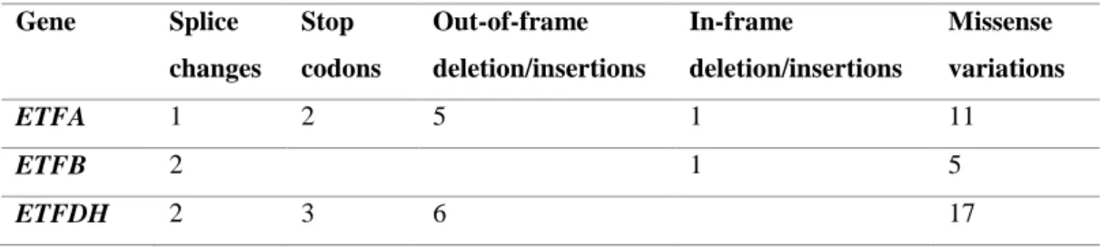
For the three clinical types of MADD it has been described a certain degree of correlation between genotype and phenotype (26,46). Indeed, nonsense, frameshift and splice junction mutations that may lead to messenger ribonucleic acid (mRNA) degradation by the nonsense-mediated mRNA decay (NMD) system or by other processes result in complete absence of functional protein are associated with severe, in many cases fatal, clinical disease (severe MADD). On opposition, missense mutations and small in-frame deletions and insertions are usually associated with milder clinical presentations (mild MADD) (37,47). Commonly, homozygosity for null ETF or ETFDH mutations is associated with the type I/II (26,39). The late-onset patients carry at least one missense variation, especially in ETFDH gene, while ETFA or ETFB mutations are less common in these individuals. Interestingly, almost patients with late-onset MADD are clearly responsive to riboflavin, since they usually present a ETFDH missense mutation which is more common associated with RR-MADD phenotypes (33). In table 2 are presented the number of these mutations type for each gene associated with MADD.
Table 2. Type and number of gene mutations in MADD. Adapted from Gregersen et al. (47). Gene Splice changes Stop codons Out-of-frame deletion/insertions In-frame deletion/insertions Missense variations ETFA 1 2 5 1 11 ETFB 2 1 5 ETFDH 2 3 6 17
Indeed, a higher number of missense variations has been identified in ETFDH gene compared to the other genes (Table 2). According to this, it would be expected milder phenotypes related to ETFDH mutations. However, severe forms for this gene have been reported (48,49). The genotype-phenotype correlation in MADD is poor due to other factors that may modulate the clinical phenotype. Exogenous factors such as febrile infections, cellular temperature, a restricted diet and other physiologic stressors, in addition of the disease-causing mutation, contribute to heterogeneous phenotypes (26,33).
ETF deficiency, due to changes of one of its subunits, results in substrate blockage, and depending on the nature of the causing mutations may lead to the accumulation of misfolded, inactive conformations of ETF proteins (26). The mitochondrial functionality is consequently disturbed, resulting in energy deficiency, accumulation of hydrogens and reducing equivalents, increase in ROS production and oxidative stress (46). When monomeric ETFDH is missing or present but defective, the effects are similar to those in the ETF deficiency (26). So, deficiencies in both ETF and ETFDH lead to the accumulation and excretion of metabolites that reflect functional deficiency of ETF/ETFDH-dependent enzymes, damaging and inactivating them, disabling the shuttle of reducing equivalents and resulting in increased ROS (37,42).
Interestingly, the increase of ROS levels, produced directly by electron leakage from misfolding variant ETFDH proteins or as result of mitochondrial dysfunction, triggers activation of cellular defense signaling pathways to facilitate cellular adaption to mutation (50). It was reported that mild misfolding of ETFDH enzymes, with impaired CoQ binding, results in increased content of superoxide radical anion (O2•−), due to the disturbed electron transfer pathway from the ETFDH to CoQ. (43). Indeed, several studies associate these biochemical alterations with increased oxidative stress (26,42,43,46,48). Therefore,
dysregulated, leading to multi-organ failure and the observed clinical phenotypes (50). Thus, the study of the molecular pathways harbored in mitochondria or involved in the regulation of its functionally is crucial to understand the pathogenesis of MADD and therefore in FAOD, since mitochondrial dysfunction have been related with these disorders (27,46,51).
1.3. The role of mitochondria on the regulation of cell homeostasis in
FAOD
Mitochondria are intracellular double membrane-bound structures ubiquitous in eukaryotes and essential in the regulation of cellular homeostasis and survival (25,52,53). Their primary function is to provide energy substrates being considered the powerhouses of the cell. Furthermore, they have an important role in the regulation of apoptosis and redox homeostasis (50,54,55). Dysregulation of the metabolic pathways harbored in mitochondria can lead to mitochondrial disorders, such as FAOD. In parallel to metabolic disturbances, broad alterations in mitochondrial functionality have been reported in FAOD (8,27,46,50,51). In fact, it is accepted that the accumulated acylcarnitines and their derivatives due to defects in FAO enzymes disturb the mitochondrial homoeostasis (25).
Mitochondria are major sources of reactive oxygen species (ROS), which include free radicals such as hydroxyl radical (HO•), peroxyl radical (RO2•) and alkoxyl radical (RO•) as well as nonradical species (hydrogen peroxide: H2O2) (8,56). Superoxide radical anion (O2•−), which results from the partial reduction of O2, and thereby ROS are mainly generated at complexes I and III of OXPHOS, being a side product of respiration (57,58). Other local of ROS production inside mitochondria is the ETF complex, by ETF and ETFDH electron leakage (58,59), which is of particular relevance in FAOD (8,59). Superoxide radical anion may react with nitric oxide (NO), produced within mitochondria, forming the radical peroxynitrite (ONOO•−). These two radicals are designated as reactive nitrogen species (RNS) (57).
ROS and RNS trigger antioxidant and protein quality control (PQC) systems to regulate their intracellular levels and maintain cellular homeostasis (55,60). Inside mitochondria, the antioxidant systems involve the action of manganese superoxide dismutase (MnSOD), peroxiredoxin (Prx III), thioredoxine (Trx II) and glutathione peroxidase (GPx), and also of mitochondrial glutathione (mtGSH). MnSOD converts O2•− to H2O2, and this molecule can be reduced to HO• or be detoxified to H2O by GPx and Prx III (55,57,60). Mitochondrial GSH is crucial in mitochondria homeostasis, regulating ROS generation and survival pathways. Indeed, decreased levels of mtGSH are associated with loss of mitochondrial membrane potential and release of cytochrome c (Cyt c) from mitochondria (61).
On the other hand, when the ROS and RNS levels exceed the threshold necessary for maintaining the healthy state, mitochondria and other cellular components are damaged, which triggers cellular dysfunction (57). Indeed, ROS and RNS produced at mitochondria oxidize the biomolecules nearby including mitochondrial deoxyribonucleic acid (mtDNA), phospholipids and proteins. These molecules are particularly susceptible to oxidative damage, and their function can be altered leading to mitochondrial dysfunction and, eventually to cell dysfunction and death if the damage is not repaired. Consequently, more ROS and RNS are generated and pathological processes may start due to cell dysfunction and death, in a vicious cycle (50,55,57,60). FAO-related inherited gene defects result in the accumulation of substrates and misfolded proteins and consequently in chronic oxidative stress (8). In fact, increased levels of ROS and elevated expression levels of antioxidant enzymes, such as MnSOD and peroxiredoxin-6, were reported among FAOD (8,48,62).
When mitochondria are damaged and dysfunctional, mitophagy, mitoptosis and autophagy must be activated to eliminate damaged organelles to avoid cellular dysfunction (53,63). When these mechanisms are not correctly working or are overwhelmed, cell death pathways should be activated (8,52,60). Indeed, mitochondrial damage-induced autophagy leads to preferential degradation of impaired mitochondria in a process designated mitophagy (52). This process consists on opening of permeability transition pore (PTP) in individual mitochondria, leading to depolarization of them, which are sequestered to form autophagosomes (64,65). Then, autophagosomes fuse with lysosomes to create autolysosomes, where mitochondrial digestion and degradation take place (52,66). So, autophagy can be seen as a mitochondrial quality control process, since it prevents
proliferation of defective mitochondria (52,63). Indeed, increased mitophagy was reported in RR-MADD, evidenced that mitochondria damaged by elevated ROS were eliminated by this mechanism in those patients (67). However, more studies focused on the contribution of mitophagy to the pathogenesis of FAOD are needed.
Mitoptosis is a more robust mechanism of mitochondria elimination that also protects cells from the damage caused by malfunctioning mitochondria (68,69). This process starts with fission of mitochondrial filaments into small spherical structures, leading to fragmentation of mitochondria (68). This step has been reported in apoptosis, suggesting that mitoptosis may initiate cell death when the damage of mitochondria is irreversible (70). After this, clusters of mitochondria are formed in the perinuclear region depending on cytoskeletal elements including microtubules, actin microfilaments and intermediate filaments. The clusters are occluded by a membrane to form the mitoptotic bodies, where mitochondrial decomposition takes place. The final step is protrusion of the mitoptotic bodies from the cell, releasing them into the extracellular medium (68,69). The mitoptotic program could be induced by several factors such as ATP depletion, block of respiration or depolarization of mitochondrial membrane, although overproduction of ROS has been reported as the main factor that triggers mitoptosis. ROS seem to induce PTP opening as observed in autophagy. Indeed, mitoptosis is not independent of mitophagy since autophagosomes and lysosomes are probably involved in the intermediate steps of mitoptotic body formation (68). When certain mitochondria become ROS overproducers, mitophagy is the preferential mitoptotic mechanism chosen (52,69). However, when ROS overproduction occurs in the entire mitochondrial population, mitoptosis looks like a more adequate mitoptotic mechanism (68).
When cells cannot be saved by the mitophagy or mitoptosis mechanisms, apoptosis takes over (8,52,60). The main mediators of apoptosis are caspases (cysteine-aspartic proteases), which are proteolytic enzymes that cleave inactive precursors to their active form (71,72). Caspases are a family of enzymes containing more than ten proteins divided into initiator (caspase-8 and -9) and executioner (caspase-3 and -7) ones. The initiator caspases, activated by cellular specific proteins, cleave executioner caspases which cleave protein substrates, being the executors of apoptosis (71). There are two main pathways the extrinsic and the intrinsic ones that leads to cell apoptosis (72). A schematic representation
of these apoptotic pathways is presented in figure 2, giving special attention of mitochondria-induced effectors.
Figure 2. Extrinsic and intrinsic apoptotic pathways. The extrinsic pathway is initiated by binding of ligands
to their respective DR, which leads to the formation of DISC and activates Casp-8 and this directly activates Casp-3, executing apoptosis. Casp-8 also activates Bid, starting the intrinsic pathway, which interacts with mitochondrial pro-apoptotic proteins Bax and Bak, leading to the release of several pro-apoptotic proteins, which triggers caspase-dependent (black arrows) or caspase-independent (blue arrows) cytosolic signaling events. Cyt c binds to Apaf-1 to form the apoptosome that recruits and activates Casp-9 which activates Casp-3. Caspase activation is improved by Smac/Diablo and Omi/HtrA2 since they inhibit IAPs. AIF and EndoG bind with DNA, inducing DNA fragmentation, via caspase-independent pathway. The intrinsic apoptotic pathway can also be stimulated by ROS. Figure made with Servier Medical Art. Abbreviations: AIF: apoptosis-inducing factor, Apaf-1: apoptotic protease-activating factor-1, Bak: bcl-2 antagonist killer 1, Bax: bcl-2 associated X protein, Bid: BH3-only pro-apoptotic protein, Casp-: caspase, number refers to the type of caspase, Cyt c: cytochrome c, DISC: death-inducing signaling complex, DNA: deoxyribonucleic acid, DR: death receptors, EndoG: endonuclease G, IAPs: inhibitor of apoptosis proteins, Omi/HtrA2: high-temperature requirement A2 serine protease, Smac/Diablo: second mitochondria-derived activator of caspases/direct IAP binding protein with low isoelectric point, ROS: reactive oxygen species.
The extrinsic pathway, also called the death receptor pathway, is initiated by binding of ligands to their respective death receptors (DR), which activates downstream signaling and leads to the formation of the death-inducing signaling complex (DISC) that culminates in the activation of caspase-8 (Figure 2). These ligands belong to the tumor necrosis factor (TNF) superfamily of cytokines and include TNFα, Fas ligand (FasL), and TNF-related apoptosis-inducing ligand (TRAIL). Activation of caspase-8 may activates directly
caspase-3 or, when caspase-8 activation is low, mediates caspase-3 activation through a process involving mitochondria (55,60,72) (Figure 2).
The second activation corresponds to the intrinsic pathway of apoptosis also designated as mitochondria-mediated pathway. Indeed, activated caspase-8 cleaves BH3-only pro-apoptotic protein (Bid), which interacts with pro-pro-apoptotic proteins bcl-2 associated X protein (Bax) and bcl-2 antagonist killer 1 (Bak), inducing the permeabilization of the outer mitochondrial membrane and the release of pro-apoptotic proteins from mitochondria (72,73) (Figure 2). ROS, particularly H2O2, induce mitochondrial translocation of Bax and Bak and release of Cyt c. The more Cyt c is released from mitochondria, the more ROS are produced due to OXPHOS impairment (74). Other released proteins are second mitochondria-derived activator of caspases/direct IAP binding protein with low isoelectric point (Smac/Diablo) and apoptosis-inducing factor (AIF), which triggers caspase-dependent or caspase-incaspase-dependent cytosolic signaling events (73,75). Within the cytosol, Cyt c binds to apoptotic protease-activating factor-1 (Apaf-1) and ATP to form the apoptosome complex which recruits and activates the initiator procaspase-9. The activated caspase-9 activates the effector caspases-3 and -7, which execute the final steps of apoptosis (71,72). Caspase activation is improved by Smac/Diablo and high-temperature requirement A2 serine protease (Omi/HtrA2) which are crucial mitochondrial proteins for enhanced caspase activation since they antagonize the inhibitory effects of inhibitor of apoptosis proteins (IAPs) (55,60,72). The mitochondrial proteins AIF and endonuclease G (EndoG) mediate a caspase-independent apoptotic pathway by binding with deoxyribonucleic acid (DNA) and thus inducing nuclear chromatin condensation and DNA fragmentation (75,76) (Figure 2). In addition to caspase-8, the intrinsic apoptotic pathway can also be stimulated by ROS and mtDNA damage, which mediate the permeabilization of the mitochondrial outer membrane and the release of pro-apoptotic proteins (55,60) (Figure 2). In the set of FAOD there are some studies that report an overexpression of apoptotic proteins (48,49,62), including Smac/Diablo and Girdin (48) or Bax (62). So, it seems that the intrinsic pathway of apoptosis contributes to the pathogenesis of FAOD, including MADD.