Ana Raquel Pinho Freitas
ECOLOGY AND EVOLUTION OF ANTIMICROBIAL RESISTANCE
IN ENTEROCOCCUS: A MULTILAYERED MOLECULAR
APPROACH WITH EMPHASIS IN THE PLASMID DIVERSITY
Tese do 3.º Ciclo de Estudos Conducente ao Grau de Doutoramento em Ciências
Farmacêuticas na Especialidade de Microbiologia
Tutors:
Doutora María Teresa Coque Servicio de Microbiología,
Instituto Ramón y Cajal de Investigación Sanitaria (IRYCIS), Madrid, Spain
Professora Doutora Luísa Vieira Peixe Faculdade de Farmácia
Universidade do Porto, Portugal
April 2011
DE ACORDO COM A LEGISLAÇÃO EM VIGOR, NÃO É PERMITIDA A REPRODUÇÃO DE QUALQUER PARTE DESTA TESE.
in the plasmid diversity
This work was financially supported by research grants from:
• Fundação para a Ciência e a Tecnologia, Ministério da Ciência, Tecnologia e Ensino Superior of Portugal
(fellowship SFRH/BD/24604/2005)
(Research Scientific Projects POCI/AMB/61814/2004 and POCI/SAL/61385/2004) • European Union. Sixth Framework Programme – Approaches to control
multi-resistant enterococci (ACE-LSHE-2007-037410)
• Ministry of Science and Innovation from Spain (PI 07/1441, PS09/02381)
• Ministerio de Educación y Ciencia of Spain, Programa Acciones Integradas Hispano-Portuguesas (H2004-0092)
Ao Bruno Aos meus pais e ao meu irmão
Às minhas orientadoras, Dra. Teresa Coque e Prof. Doutora Luísa Peixe, que me iniciaram nesta viagem cientifica, quero expressar o meu sincero e profundo agradecimento pela oportunidade de integrar a vossa equipa de trabalho quando as solicitei e a confiança depositada desde o início. Pela orientação cientifica, pela revisão critica e aprendizagem constante, pela disponibilidade e amizade incansáveis, bem como pelas palavras de reconhecimento carinhosamente dirigidas, fonte de renovado estímulo para a concretização desta tese. Muito obrigada por esta oportunidade que seguramente marcará o meu percurso profissional e pessoal.
À Prof. Doutora São José Nascimento, pela oportunidade de realizar este trabalho no Laboratório de Microbiologia da Faculdade de Farmácia da Universidade do Porto.
A todos os Prof. Doutores do Serviço de Microbiologia, quero agradecer a simpatia com que me receberam e a disponibilidade demonstrada.
À Cristina Costa e ao Nuno Oliveira, colaboradores essenciais no dia a dia do Laboratório de Microbiologia, agradeço o apoio e incentivo ao longo deste percurso. Obrigada pela companhia, pelo bom humor e pela prontidão em ajudar!
À companheira de equipa no mundo dos coconuts, Carla Novais, quero registar e agradecer de forma especial todo o apoio demonstrado desde o primeiro dia. Pela disponibilidade, pelo que aprendi durante a nossa convivência, pelos desabafos nos momentos mais difíceis, mas também pelos muitos bons momentos e risadas dos últimos anos. Obrigada por estares presente em fases cruciais deste trabalho.
À Patrícia Antunes, Sandra Quinteira e Filipa Grosso, companheiras assíduas dos últimos anos, agradeço o carinho com que me receberam, a partilha de conhecimentos, a amizade e incentivo sempre presentes dentro e fora do laboratório, as animadas aventuras dos congressos internacionais! Ficarão para sempre na minha memória os bons momentos que passamos.
Ao Jorge Oliveira, pela ajuda crucial em momentos importantes, pela paciência infinita em ensinar os avanços da informática que tento acompanhar, pela amizade e apoio constantes.
À Elisabete Machado, que sempre me apoiou nesta caminhada com palavras de alento e motivação mesmo à distancia.
Ao Dr. Fernando Baquero, por me ter recebido sempre de forma amistosa nas muitas estadias no Serviço de Microbiologia do Hospital Ramón y Cajal de Madrid, pela oportunidade de integrar a sua equipa de investigação nomeadamente em projectos internacionais, pelo incentivo e apoio em diferente etapas. Será sempre para mim uma referência tanto a nível profissional como pessoal.
Ao Prof. Doutor Rafael Cantón e a todos os membros do Serviço de Microbiologia do Hospital Ramón y Cajal, agradeço a forma carinhosa com que sempre me receberam e o apoio constante.
A todos os companheiros das aventuras internacionais, em especial à Tânia, Aida, Arancha, Carmen, Maria, Marta, Elia e Manu, que sempre me fizeram sentir em casa. À Ana Sofia Tedim e Patricia Ruiz-Garbajosa, pela colaboração activa em muitos trabalhos, pelo apoio e entreajuda constantes. Uma menção especial à amiga de longa data Ângela Novais que foi essencial na minha integração no laboratório mas também no quotidiano das estadias em Madrid, obrigada pelos bons momentos que aí passamos, pelo apoio e amizade inesgotáveis!
A investigadores de outros países com os quais convivi em reuniões de trabalho, Vicky Francia, Guido Werner, Jenny Laverde, Rob Willems, Lars Jensen, Anette Hammerum e Camila Lester, obrigada pela oportunidade de partilhar conhecimentos científicos e pela simpatia com que sempre me trataram.
A todos os alunos de Mestrado Integrado em Ciências Farmacêuticas da Faculdade de Farmácia do Porto, Rosa Silva, Rosa Correia, Márcia Monteiro, João Pires e Ana Cunha, que comigo colaboraram.
A todos os familiares e amigos (os mesmos de sempre e de longos anos) que me apoiaram e compreenderam a minha ausência em momentos importantes.
À Fundação para a Ciência e Tecnologia, agradeço o apoio financeiro concedido ao longo de quatro anos de investigação bem como ajudas de custo para estadias e congressos no estrangeiro.
permitiu iniciar e encerrar este projecto. Não há palavras para expressar o apoio incondicional que esteve sempre presente e que de forma discreta, mas decisiva, contribuiu para este sucesso. Ainda à Mariana que já mostra vida dentro de mim e que me alentou nesta recta final. Para vocês, o meu mais sincero e profundo obrigada.
Um trabalho de investigação nunca é um trabalho solitário, mas sim a confluência do esforço e dedicação de muitos.
A todos que de alguma forma contribuíram para este projecto nos últimos anos, um muito OBRIGADA!
!
!
!
!
!
!
!
!
!
!
!
!
!
!
!
!
!
!
!
!
!
!
!
!
!
!
“Como todas as coisas contribuíram para o seu crescimento, você deveria incluir todas as coisasem seu agradecimento.”
The genus Enterococcus constitutes one of the best examples of bacterial adaptation to different environments due to the abilityto survive in harsh conditions and their intrinsic resistance to different antibiotics, biocides and metals. Enterococci are well recognized commensals and opportunistic pathogens of humans and animals since the early 1900’s. However, nosocomial enterococcal infections have increasingly been reported during the last 30y with these microorganisms becoming one of the most common pathogens causing severe nosocomial infections worldwide. This situation has been facilitated by the acquisition of resistance to all antibiotics available in the therapeutic arsenal including vancomycin, the last resort for the treatment of multiresistant Gram-positive organisms, and all new antibiotics recently licensed in the pharmaceutical market as tigecycline. The emergence of vancomycin-resistant enterococci is of particular concern because of the limited therapeutic options to treat multiresistant enterococci, and the possibility to transfer this trait to more pathogenic bacteria as Staphylococcus aureus.
The emergence and recent increase of enterococcal strains with new adaptive traits (inferred by their antibiotic resistance, colonization, or virulence phenotypes and genotypes) illustrate the role of lateral genetic transfer (LGT) in the evolution of these microorganisms. The global goal of this work was to establish the diversity and the
influence of different mobile genetic elements (MGE) belonging to the enterococcal mobilome in the transfer of adaptive traits between different hosts and in the selection of particular enterococcal lineages. With this purpose, the role of clonal expansion and
lateral genetic transfer in the dissemination of vancomycin resistance among
Enterococcus faecium and Enterococcus faecalis, recovered from hospitalized persons in
different countries of the five continents (1986-2009) and from different hosts or community environments (healthy humans, foodborne animals, distinct aquatic environments) (1995-2008) was analyzed.
The analysis of the population structure of E. faecium and E. faecalis isolates from different origins revealed an overrepresentation of particular clonal complexes (CC) in swine (E. faecium CC5), poultry (E. faecium CC9) and humans (E. faecium CC17, CC22 and CC94 and E. faecalis CC2), all of them comprising a diversity of sequence types (ST). The results highlighted the presence of different reservoirs for antibiotic-resistance. Also, the interhost transmission (swine-Human) of clones belonging to host-adapted clonal complexes of E. faecium (CC5, CC17) and E. faecalis (CC2), confirms the relevance of reverse and alternative routes for dissemination of opportunistic bacteria.
!
host (e.g. Inc18) or particular narrow host plasmids (megaplasmids and small plasmids) encoding traits associated with specialized environments could have contributed to such differences.
A detailed study of the genetic elements encoding resistance to glycopeptides showed that vanA-Tn1546-carrying plasmids are an outstanding driver of the pandemic spread of vancomycin resistance among enterococci. Transferable plasmids carrying replication, conjugation and/or maintenance systems related to plasmid families of narrow host range as RepA_N (pRUM in E. faecium and pheromone-responsive in E.
faecalis) or broad host range as Inc18 would have facilitated the successful dissemination
of Tn1546 among enterococci. Besides the host specificity of plasmids of E. faecalis and
E. faecium, the diversity and frequency of LGT/recombination events occurring within
each species provided interesting clues to understand why vanA plasmids remain confined to this genera. Of particular interest were the results related to E. faecalis. The narrow host range of pheromone plasmids and the frequent rearrangements observed within plasmids of this species might explain the maintenance of pAD1-Tn1546 within this bacterial host. The unsuccessful transference of these plasmids to Staphylococcus aureus seems to be associated with the presence/absence of the broad host conjugative module of Inc18 plasmids.
Finally, the cumulative acquisition of adaptive traits by these species-specific plasmids would have conferred an increased fitness relevant for the selection and successful expansion of certain lineages in the predominant enterococcal species.
The recovery of enterococcal isolates resistant to tigecycline, among enterococci exposed to this antibiotic, highlights the possible selection by other antibiotics in non-clinical settings as in farms for animal production.
In summary, this work illustrated the selection of epidemic clones and the diversity and influence of enterococcal mobile genetic elements in the current expansion of specific clonal lineages and high prevalence of vancomycin-resistant enterococci worldwide and in Portugal in particular. The diversity of plasmids (plasmidome) encompassing different replication, transfer and maintenance systems highlights the wide possibilities of adaptation of these microorganisms.
bacteriana a diferentes ambientes devido à sua capacidade de sobrevivência em ambientes hostis e à resistência intrínseca a diferentes antibióticos, biocidas e metais. Ainda que reconhecidas como bactérias comensais e oportunistas patogénicos de humanos e animais desde os anos 1900, as infecções hospitalares causadas por
Enterococcus têm vindo a aumentar progressivamente durante os últimos 30 anos.
Actualmente, são um dos agentes de infecções nosocomiais graves mais comuns a nível hospitalar e mundial. Tal tem sido facilitado pela aquisição de resistência a múltiplos antibióticos disponíveis no arsenal terapêutico incluindo a vancomicina, um antibiótico considerado de última escolha para o tratamento de Gram-positivos multiresistentes, e todos os novos antibióticos recentemente aprovados para uso em ambiente hospitalar como é o caso da tigeciclina. O aumento de isolados de Enterococcus faecalis e
Enterococcus faecium resistentes à vancomicina (ERV) tem suscitado particular interesse
devido à escassez de opções terapêuticas de tratamento e à possibilidade de transferência desta resistência para outras bactérias mais patogénicas como
Staphylococcus aureus.
O aumento dramático e recente de Enterococcus com novas características adaptativas (inferidas pela sua resistência aos antibióticos, colonização, ou fenótipos e genótipos de virulência) ilustra o papel da transferência genética horizontal (TGH) na evolução deste género bacteriano. O objectivo geral deste trabalho foi estabelecer a
diversidade e a influência de diferentes elementos genéticos móveis (EGM) na transferência de características adaptativas entre diferentes hospedeiros e na selecção de linhagens clonais específicas. Com este propósito, foram analisadas a importância da
expansão clonal e da TGH na disseminação da resistência à vancomicina em isolados de
E. faecium e E. faecalis obtidos de hospitais em diversos países dos cinco continentes
(1986-2009) e da comunidade (humanos saudáveis, animais de produção alimentar, ambientes aquáticos) (1995-2008).
A análise da estrutura populacional de isolados de E. faecium e E. faecalis de diferentes origens revelou a predominância de determinados complexos clonais (CC) em suínos (CC5 E. faecium), aves (CC9 E. faecium) e humanos (E. faecium CC17, CC22 e CC94 e E. faecalis CC2). Adicionalmente, a transmissão entre hospedeiros (suínos-Homem) de clones pertencentes a determinados CCs de E. faecium (CC5, CC17) e E.
faecalis (CC2) confirma a relevância de modos alternativos na disseminação de bactérias
plasmídeos de largo espectro de hospedeiro (Inc18) ou de plasmídeos de espectro reduzido (megaplasmídeos e plasmídeos muito pequenos) que conferem vantagens adaptativas (ex.: bacteriocinas, virulência) pode ter contribuído para essas diferenças.
Um estudo detalhado dos elementos genéticos de resistência aos glicopéptidos demonstrou a grande relevância dos plasmídeos que albergam o transposão Tn1546 (vanA) na disseminação pandémica da resistência à vancomicina em Enterococcus. Plasmídeos conjugativos com sistemas de replicação, conjugação e/ou manutenção relacionados com famílias de plasmídeos de espectro reduzido, como a RepA_N (pRUM em E. faecium e plasmídeos de resposta a feromonas em E. faecalis), ou de espectro alargado, como os Inc18, deverá ter facilitado a rápida disseminação de Tn1546 em
Enterococcus. Para além da especificidade do conteúdo plasmídico de E. faecalis e E. faecium, a diversidade e a frequência de eventos de TGH que ocorre em ambas as
espécies, indiciou sobre o facto de a resistência aos glicopéptidos permanecer confinada a este género. De particular relevância foram os resultados relacionados com os isolados de E. faecalis. Os plasmídeos de resposta a feromonas (PRF) de espectro reduzido de hospedeiro assim como os frequentes eventos genéticos observados entre plasmídeos desta espécie pode explicar a manutenção de PRF-Tn1546 nesta espécie bacteriana. A transferência destes plasmídeos para Staphylococcus aureus parece estar associada à presença do módulo de conjugação de plasmídeos de largo espectro do tipo Inc18.
Finalmente, a aquisição e acumulação de características adaptativas por estes plasmídeos específicos de espécies pode ter conferido um fitness acrescido, relevante na selecção e expansão dramática de certas linhagens das espécies de Enterococcus mais importantes.
A detecção da resistência à tigeciclina, em isolados de Enterococcus não expostos a este antibiótico, indica a sua possível selecção por outros antibióticos usados em ambientes não hospitalares, como os locais de produção animal.
Em resumo, este trabalho permitiu demonstrar a selecção de clones epidémicos, a diversidade de EGM e a sua influência na actual expansão de determinadas linhagens clonais assim como na elevada prevalência de ERV em diferentes países e, em particular, em Portugal. A diversidade plasmídica demonstra ainda as amplas possibilidades de adaptação destes microorganismos.
Palavras-chave: Enterococcus, plasmídeos, resistência à vancomicina, transferência genética horizontal, Tn1546
CHAPTER 1 – INTRODUCTION 27 !
1.1. The genus Enterococcus spp. 29
1.1.1. Taxonomy and historical perspective 29
1.1.2. Physiology of enterococci 30
1.1.3. Resistance to compounds with antibacterial activity (antibiotics, biocides,
heavy metals) 31
1.1.3.1. Intrinsic resistance 32
1.1.3.2. Acquired resistance 34
1.1.4. Clinical enterococcal infections 38
1.1.5. Treatment 39
!
1.2. Ecology and population structure of Enterococcus 40
1.2.1. Ecology 40
1.2.2. Population structure 41
1.2.2.1. Enterococcus faecium 41
1.2.2.2. Enterococcus faecalis 43
!
1.3. Mobile genetic elements in Enterococcus 44
1.3.1. Lateral genetic transfer 44
1.3.2. Transposable elements 46
1.3.2.1. Insertion Sequences 46
1.3.2.2. Composite transposons 48
1.3.2.3. Tn3 derivatives 50
1.3.2.4. Conjugative transposons/Integrative Conjugative Elements 51
1.3.3. Plasmids 57
1.3.3.1. Rolling-circle replicating plasmids 58
1.3.3.2. Theta-replicating plasmids 64
a) Small theta-replicating plasmids 64
b) Inc18 family 66 c) RepA_N family 66 d) pMG1-like family 68 e) Megaplasmids 69
!
!
!
2.2. Outline of the thesis 75
CHAPTER 3 – RESULTS (manuscripts) 79
3.1. Insigths on the population structure of Enterococcus faecium and
Enterococcus faecalis 81
3.1.1. Dispersion of multidrug-resistant Enterococcus faecium isolates belonging to
major clonal complexes in different Portuguese settings 83
!
3.1.2. Clonal expansion within clonal complex 2 and spread of vancomycin-resistant plasmids among different genetic lineages of Enterococcus faecalis from Portugal 91
!
3.1.3. Human and swine hosts share vancomycin-resistant Enterococcus faecium CC17, CC5, and Enterococcus faecalis CC2 clonal clusters harboring Tn1546 on
indistinguishable plasmids 101
3.2. Analysis of the plasmidome of enterococci from different environments 3.2.1. Analysis of plasmids responsible for virulence/epidemicity 109
3.2.1.1. Global spread of the colonization-virulence hylEfm gene in megaplasmids of
CC17 Enterococcus faecium polyclonal sub-cluster 111
3.2.1.2. A multiresistance megaplasmid pLG1 bearing a hylEfm genomic island in
hospital Enterococcus faecium isolates 119
3.2.2. Analysis of plasmid and transposons in antibiotic resistant enterococci from
humans and animals 131
3.2.2.1. The spread of vancomycin-resistant Enterococcus faecium (1986-2009): a
historic view from the plasmid side 133
3.2.2.2. Influence of pAD1 and Inc18 plasmids in the worldwide spread of
vancomycin-resistant Enterococcus faecalis outbreak strains (1987-2005) 171 3.2.2.3. Spread and persistence of Inc18::Tn1546 plasmids among enterococci
from poultry food products 189
!
3.2.2.4. Spread and diversification of pRUM and Inc18-like plasmids carrying Tn1546 among CC17 Enterococcus faecium from hospitals in Portugal
3.2.2.6. Diversity of Tn1546 and its role in the dissemination of
vancomycin-resistant enterococci in Portugal 231
3.3. Integration of all data and multilayered analysis of the relative contribution of epidemic clones and mobile genetic elements in the expansion of specific clonal
lineages 241
3.3.1. Mobile genetic elements and lateral genetic transfer in Enterococci 243 3.4. Screening of the in vitro activity of the new molecule tigecycline against
Enterococcus spp. from different ecological niches in Portugal 285 3.4.1. Non-susceptibility to tigecycline in enterococci from hospitalized patients, food
products and community sources 286
!
CHAPTER 4 – CONCLUSIONS 299
CHAPTER 5 – REFERENCES 303
CHAPTER 6 – APPENDIX 335
!
6.1. Material and Methods 337
6.1.1. Bacterial isolates 337
6.1.1.1. Enterococci isolates from humans 337
6.1.1.2. Enterococci isolates from swine and piggeries 338
6.1.1.3. Enterococci isolates from poultry 338
6.1.1.4. Enterococci isolates from the environment 339
6.1.2. Media, sample processing and growth conditions 339
6.1.3. Identification of Enterococcus 340
6.1.4. Antimicrobial susceptibility tests 340
6.1.4.1. The disk diffusion test 341
6.1.4.2. The agar dilution method 341
6.1.4.3. The Epsilon-test (E-test) 342
6.1.5. Molecular techniques / DNA methodology 342
6.1.5.1. DNA extraction 342
a) Genomic DNA 342
a) Electric continuous-field agarose gel electrophoresis 347
b) Pulsed-field gel electrophoresis 347
c) DNA visualization 348
6.1.5.5. Identification and typing 348
a) Pulsed-field gel electrophoresis 348
b) Multilocus Sequence Typing (MLST) 349
c) DNA transfer and hybridization 351
d) Detection and location of resistance/virulence genes 353
6.1.5.6. Bioinformatic analysis 354
6.1.6. Conjugation assays 354
6.1.7. Analysis of mobile genetic elements 355
6.1.7.1. Insertion sequences 355
6.1.7.2. Transposons 355
a) Tn1546 (typing of vanA transposons) 355
b) Tn1549/Tn5382 (typing of vanB transposons) 355
6.1.7.3. Plasmids 356
a) Plasmid extraction 356
b) Plasmid number and size 356
c) Plasmid content 356
- Rep initiator proteins 356
- Relaxases 356
- Toxin-Antitoxin systems 357
- Bacteriocins 357
- Virulence 357
d) RFLP profiling 357
6.2. Reagents, solutions and equipment 373
6.2.1. Reagents 373
6.2.2. Solutions 374
6.2.3. Equipment 376
6.3. Protocols 377
1.1.1. Figure 1. Phylogenetic dendrogram of Gram-positive genera by 16S rRNA
sequencing . . . 30
!
! 1.1.3.2. Figure 2. Chronologic representation of first descriptions of acquired resistance to different antibiotic families in enterococci . . . 34
!
! 1.1.3.2. Figure 3. Alignment of vancomycin resistance gene clusters of vanA, vanB, vanD, vanF and vanM . . . 37
!
1.2.1. Figure 4. Phylogenetic tree of human intestinal 16S rDNA sequences . . . 40
!
1.3.1. Figure 5. Highways of obligate gene transfer within and among phyla and divisions of prokaryotes . . . 45
!
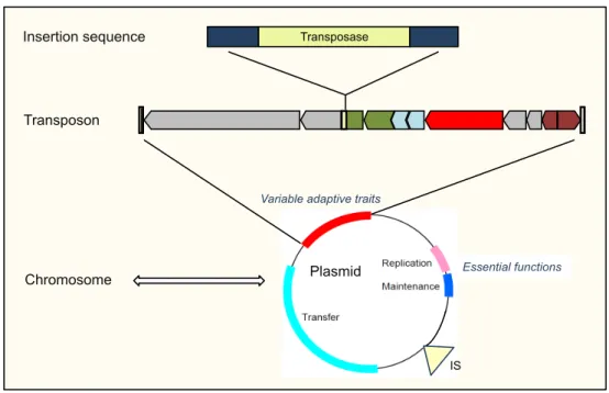
1.3.1. Figure 6. Organization of the mobile genome pool . . . 46
!
1.3.2.1. Figure 7. Organization of a typical insertion sequence . . . 46
!
! 1.3.2.2. Figure 8. General organization of a composite transposon . . . 49
!
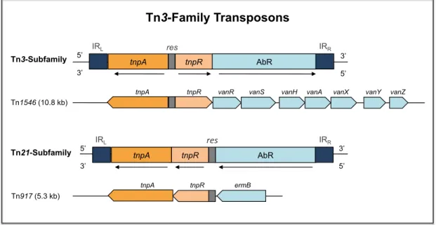
1.3.2.3. Figure 9. General organization of Tn3-family transposons . . . 50
!
1.3.2.4. Figure 10. Structure of integrative conjugative elements . . . 52
!
1.3.3. Figure 11. Hierarchal composition of MGEs . . . 57
!
3.2.2.1. Figure 1. Geographic distribution of European vanA and vanB Enterococcus faecium isolates . . . 155
!
! 3.2.2.1. Figure 2. Geographic distribution of worldwide vanA and vanB Enterococcus faecium isolates . . . 155
!
! 3.2.2.1. Figure 3. Plasmid number of CC17 and non-CC17 E. faecium isolates . . . 166
!
! 3.2.2.1. Figure 4. Plasmid content of CC17 and non-CC17 E. faecium isolates . . . 167
!
! 3.2.2.1. Figure 5. Computer analysis of Cla-I digested plasmid DNA of vancomycin-resistant VanA-type E. faecium isolates . . . 168
!
! 3.2.2.1. Figure 6. In silico analysis of enterococcal sequenced plasmids . . . 169
!
!
3.2.2.3. Figure 1. Genetic map of Tn1546 variants found in this study . . . 199
!
!
3.2.2.3. Figure 2. Rep/Mob/TA gene content of vanA strains from samples of retail poultry and healthy humans recovered in Portugal during 1999-2004. . . 200
!
!
3.2.2.4. Figure 1. Number and size of plasmids among VRE isolates analyzed . . . 216
!
!
3.2.2.4. Figure 2. Plasmid content of 75 vanA isolates recovered from Portuguese hospitals and aquatic environments during 1996-2008 . . . 216
!
!
3.2.2.4. Figure 3. Restriction fragment length polymorphism patterns of plasmid G after digestion with ClaI and EcoRI restriction enzymes . . . 217
!
!
3.4.1. Figure 1. SmaI digested genomic DNA of tetracycline resistant Enterococcus
faecalis from different sources and hybridization with a tetM probe . . . 297
!
!
6.1.5.1. Figure 1. Schematic representation of plasmid DNA electrophoresis . . . 344
!
6.1.5.5. Figure 2. PFGE of SmaI digested genomic DNA of enterococcal isolates recovered from human and swine of different countries . . . 349!
6.3. Figure 3. Gel electrophoresis of plasmid DNA from VRE isolates obtained byKado & Liu . . . 377 6.3. Figure 4. Gel electrophoresis of plasmid DNA from VRE isolates obtained by a modification of the Birnboim and Doly method . . . 378
!
!6.3. Figure 5. Gel electrophoresis of plasmid DNA from VRE isolates extracted with different methods . . . 379
!
6.3. Figure 6. S1-PFGE of VRE isolates . . . 380!
6.3. Figure 7. Gel electrophoresis of EcoRI-digested vanA plasmids obtained from1.1.3.2. Table 2. Characteristics of acquired van gene clusters in enterococci . . . 37 1.3.2.1. Table 3. Insertion sequences found among enterococcal mobile genetic
elements . . . 48
!
1.3.2.2. Table 4. Characteristics of composite transposons identified in Enterococcus . 49!
1.3.2.4. Table 5. Conjugative transposons identified in enterococci . . . 55 1.3.3.1. Table 6. Diversity, epidemiological background and main genetic characteristics of plasmids identified among Enterococcus . . . 60 1.3.3.2. Table 7. Characteristics of bacteriocins previously associated with small theta-replicating plasmids in enterococci . . . .65 3.2.2.1. Table 1. Epidemiological background of VRE/VSE isolates . . . 156 3.2.2.1. Table 2. Tn1546 diversity of vanA E. faecium isolates . . . 158
!
3.2.2.1. Table 3. Diversity of vanB transposons among E. faecium isolates . . . 160
!
3.2.2.1. Table 4. Diversity of plasmid modules among VRE . . . 161
!
3.2.2.1. Table 5. Diversity of plasmids among vanA and vanB E. faecium isolates . . . 163
!
3.2.2.1. Table 6. Diversity of plasmids among VSE isolates from Spain . . . 165 3.2.2.1. Table S1. Main plasmid types representative of plasmid families found among
Enterococcus . . . 170
!
!
3.2.2.2. Table 1. Epidemiological and genetic backgrounds of vancomycin-resistant E. faecalis causing outbreaks in different countries during 1987-2005 . . . .185 3.2.2.2. Table 2. Plasmid characterization of representative vanA E. faecalis isolated from European and American hospitals . . . 186 3.2.2.4. Table 1. Plasmid content of vanA E. faecium isolates from hospitals and aquatic environments of Portugal . . . 214
3.3.1. Table 1. Insertion sequences found among enterococcal mobile genetic
elements . . . 273
!
!
3.3.1. Table 2. Enterococcal transposons . . . 274
!
!
3.3.1. Table 3. Enterococcal plasmid types . . . 274
!
!
3.3.1. Table 4. Characteristics of bacteriocins previously associated with small theta-replicating plasmids in enterococci . . . 280 !
3.4.1. Table 1. In vitro activity of tigecycline against enterococci isolated from different species and sources . . . 291 !
3.4.1. Table 2. Features of Enterococcus isolates non-susceptible to tigecycline . . . . 292
!
!
6.1.5.5. Table 1. Features, advantages and drawbacks of molecular methods largely used in the typing of Enterococcus spp. . . 350 !
6.1.7.3. Table 2. Strains used as recipients in conjugation or controls for PCR and hybridization . . . 358 6.1.7.3. Table 3. Oligonucleotides and PCR conditions used for the characterization of Enterococcus isolates . . . 359
!
!
!
!
!
!
!
!
!
!
!
!
!
1.2.2. Box 1. Population structure glossary . . . 42
!
6.2.1. Box 1. PCR reagents . . . 373!
6.2.1. Box 2. Restriction enzymes . . . 373!
6.2.1. Box 3. DNA molecular weight markers . . . 374!
6.2.2. Box 4. Solutions for DNA extraction . . . 374!
6.2.2. Box 5. Solutions for PCR, DNA separation and detection . . . 375!
6.2.2. Box 6. Solutions for hybridization . . . 376!
6.2.3. Box 7. Equipment . . . 376!
AFLP Amplified fragment length polymorphism AMEs Aminoglycoside-modifying enzymes
BHI Brain Heart Infusion
CC Clonal complex
CCC Covalently Closed Circular
CLSI Clinical and Laboratory Standard Institute
CRISPR Clustered Regularly Interspaced Short Palindromic Repeats
DNA Deoxyribonucleic Acid
dNTP Deoxynucleoside triphosphates
EUCAST European Committee on Antimicrobial Susceptibility Testing
GEC Genetic exchange community
GI Genomic Island
ICE Integrative Conjugative Element IS Insertion Sequence
LGT Lateral Genetic Transfer
MBC Minimum bactericidal concentration
MH Mueller-Hinton
MIC Minimum Inhibitory Concentration
MGE Mobile Genetic Element
MLST Multi-Locus Sequence Typing
MRSA Methicillin-resistant Staphylococcus aureus
MSCRAMM Microbial surface component-recognizing adhesive matrix molecules NCBI National Center for Biotechnology Information
OC Open Circular
PAI Pathogenicity island
PBP Penicillin Binding Protein
PCR Polymerase Chain Reaction
PFGE Pulsed-Field Gel Electrophoresis
RAPD Rapid Amplification of Polymorphic DNA RCR Rolling-circle replicating
Rel Relaxase
Rep Replication-initiator protein
RFLP Restriction Fragment Length Polymorphism RNA Ribonucleic Acid
ST Sequence type
TA Toxin-Antitoxin
Tn / CTn Transposon / Conjugative Transposon VRE Vancomycin-resistant enterococci
VREfs Vancomycin-resistant Enterococcus faecalis VREfm Vancomycin-resistant Enterococcus faecium VRSA Vancomycin-resistant Staphylococcus aureus
It is the theory which decided what can be observed.
Albert Einstein
!"#$%&'(#)%"*
1.1.
The genus Enterococcus spp.
1.1.1. Taxonomy and Historical Perspective
Enterococcus is a genus of Gram positive cocci that fall within the phylum Firmicutes,
class Bacilli, order Lactobacillales, family Enterococcaceae.
The term “entèrocoque” was firstly used by Thiercelin in 1899 to describe the intestinal origin of bacteria in pairs and short chains recovered from human faeces (117). In the same year, MacCallum and Hastings reported an organism obtained from a case of endocarditis which they called Micrococcus zymogenes. The term “micrococcus” was by that time used to refer to a very hard bacterium isolated from different environments (252). The name Streptococcus faecalis was firstly used in 1906 also from a patient with endocarditis, and in the subsequent years other Streptococcus species (S. faecium, S.
durans, S. avium) were described as differing in the fermentation patterns of S. faecalis. In
1919, Sherman proposed a classification scheme which separated streptococci into nonenterococcal streptococci (pyogenic, viridans, lactic) and “the enterococcal group”, based on the growth at different temperatures and pH conditions (379). This classification’s scheme also correlated with the serological Lancefield scheme and in the mid-1930s, enterococci were classified as Group D Streptococcus. It was only in 1984 that all Group D Streptococcus, with exception of S. bovis, was considered genetically different from the Streptococcus, and Enterococcus was globally considered a separate genus. Studies based on DNA-DNA hybridization, 16S rRNA gene sequencing (Figure 1), whole-cell protein analysis, and conventional phenotypic tests allowed the establishment of many enterococci species (293, 366, 409).
Over forty Enterococcus species have been described to date (www.bacterio.cict.fr/e/enterococcus.html), of which the most common species associated with the human host are Enterococcus faecium and Enterococcus faecalis. E. faecium and E. faecalis are genetically not closely related as demonstrated by 16S rRNA and atpA housekeeping gene (!-subunit of ATP synthase) sequences (301).
Figure 1. Phylogenetic dendrogram of Gram-positive genera by 16S rRNA sequencing (adapted from 213).
1.1.2. Physiology of Enterococci
Enterococci are non-spore-forming, facultative anaerobes able to survive in a wide range of environmental conditions, both at extreme temperatures (10-45ºC) and pH (4.8-9.6) conditions; they grow in concentrations of sodium chloride as high as 6.5% and hydrolyze esculin in the presence of 40% bile salts (117, 409).
Enterococci have metabolic features allowing them to grow within diverse environments such as gastrointestinal compartments of different mammal’s intestine, reptils and birds and a diversity of environmental sources (e.g. food, water, inanimate surfaces). They are extraordinary tolerant to oxidative stress, azide, detergents, heavy metals, and ethanol as well as prolonged desiccation. In addition, they are able to catabolize a variety of energy sources that include complex carbohydrates, glycerol, lactate, malate, citrate, the diamino acids arginine and agmatine, and many !-keto acids.
E. faecalis contain abundant surface membrane proteins able to degrade mucin which
can be advantageous by providing additional sugar for growth and by facilitating bacterial adhesion (27, 182). Enterococci produce bacteriocin peptides and some are used as starter or protection cultures including probiotics in food and feed supplements (129, 302). All these metabolic and physiologic features enhance enterococcal competitiveness in diverse environments. However, there are physiologic differences between enterococcal species which might contribute to the adaptation to different hosts and environments (182, 229).
Different factors can mediate the establishment, the colonization and further clinical infection by E. faecalis and E. faecium. They include microbial surface component-recognizing adhesive matrix molecules (MSCRAMM) and cell-wall-anchored LPxTG
surface proteins, and the ability to form biofilms; biofilm production seems to be greater among E. faecalis species due to the high number of surface-exposed proteins (145, 173, 363, 377). Numerous proteins with MSCRAMM-like structure and several genes or gene clusters involved in biofilm formation, are increasingly described, some of which are especially abundant in clinical isolates from dominant E. faecium and E. faecalis clonal complexes. Virulence determinants of E. faecalis include a bile salt hydrolase, the cytolysin toxin, gelE within the fsr cluster (coding for gelatinase), capsular polysaccharides, glycolipids, the collagen-binding Ace (adhesin of collagen of E. faecalis),
esp (coding for an enterococcal surface protein), the ebp cluster (coding for a sortase
associated with surface pili formation), the epa gene cluster (encoding the polysacharide Epa), atn (coding for an autolysin), bopD (sugar-binding transcriptional regulator), bee (coding for a cell-anchored protein), and the LPxTG surface protein aggregation substance. E. faecium associated with human infections seem to be particularly enriched in putative virulence genes encoding a glycoside hydrolase (hylEfm), Esp, PilA- and
PilB-type pili, and several cell wall-anchored LPxTG surface proteins including Acm (adhesin of collagen from E. faecium) and Scm (second collagen adhesin of E. faecium). Enterococci also possess environment-sensing systems consisting of a large repertoire of two-component signal transduction systems, some of which are regulated by quorum-sensing mechanisms (e.g. the fsrABCD system related to biofilm formation), extra-cytoplasmic function sigma factors (ECF "), and transcriptional regulators that modulate gene expression in response to environmental changes and control diverse bacterial physiological processes (71).
Understanding the factors involved in the commensal relationship between bacteria and the host is crucial to understand the different colonization ability and host immune responses between different enterococcal species as is in the development of new and valuable therapeutic strategies to prevent human infections caused by enterococci.
1.1.3. Resistance to compounds with antibacterial activity (antibiotics, biocides, heavy metals)
Enterococci show resistance to antibiotics, heavy metals, antiseptics, disinfectants, ethidium bromide and ultraviolet radiation (35, 67, 233, 285, 303). Such resistances may be associated with multidrug efflux pumps located in the chromosome (often coding for resistance to different compounds) or different genetic traits located on mobile genetic elements (MGEs) as plasmids or conjugative transposons (often coding for resistance to a single compound or family of a given compound). Efflux pumps, permeability changes and transferable mosaic genetic platforms can encode resistance to biocides/heavy
metals/UVR/EtBr and antibiotics and therefore, the selective pressure exerted by the first ones may favour the expression and co-dissemination of resistance to different antibiotics (277, 278, 356).
Enterococci show intrinsic and acquired resistance against antibiotics of different families. While intrinsic resistance is an inherent property of a given species and thus is chromosomally encoded, acquired resistance results from a mutation or acquisition of foreign DNA (293). The compounds used to treat or prevent the appearance of bacteria can be classified according to their effects on the bacterial growth in bacteriostatic, those that prevent the growth by keeping bacteria at the stationary phase, and bactericidal, those that kills bacteria. However, some broad classes of antibacterial agents considered bactericidal can exhibit bacteriostatic activity in vitro and vice-versa (320). In addition, bacteria can develop tolerance to a given antibiotic when they are inhibited but not killed by concentrations manyfold higher than the MIC (minimum inhibitory concentration). By definition, tolerance involves a reduction in the rate of antibiotic-induced killing of a whole bacterial population which evade only the killing action of an antibiotic. Virtually, all studies on this phenomenon have been restricted to tolerance to #-lactam antibiotics or other cell wall inhibitors. In addition to genotypic tolerance, which is a property restricted to some bacterial mutants that specifically evade only the killing effect of #-lactam antibiotics without increase in the MIC, every bacterial cell can evade the killing effect of #-lactam antibiotics under certain physiological conditions, the phenotypic tolerance, as is the most universal example of nongrowing (dormant) or slowly growing bacteria. In vitro phenotypic
tolerance has been defined as a minimum bactericidal concentration (MBC) that is 32 times the MIC (293, 320, 432).
1.1.3.1. Intrinsic resistance in enterococci includes resistance to #-lactams, aminoglycosides, and trimethoprim-sulfamethoxazole. Additional resistances to lincosamides and vancomycin are characteristics of specific enterococcal species (231, 293, 294).
Enterococci show intrinsic resistance to most #-lactam antibiotics (MIC of 1 to 8
µg/mL) due to the low affinity of specific cell wall proteins to bind penicillin. Such proteins required for the synthesis of the enterococcal cell wall are the target for beta-lactam compounds and are known as penicillin-binding proteins (PBPs). More specifically, intrinsic resistance of E. faecium and E. hirae has been attributed to the presence of
PBP5, which is highly expressed and has marked lower affinity for #-lactams than the
other PBPs (MIC of 16 to 64 µg/mL). The level of resistance varies among #-lactams, with
by carbapenems and cephalosporins which are not used in the clinical practice for the treatment of enterococcal infections. In addition, enterococci are typically tolerant to all #-lactams, and in vitro MBCs of penicillins and cephalosporins are usually >100 µg/mL (231, 293).
Enterococci exhibit intrinsic low level resistance to all aminoglycosides (MIC of 8-64
µg/mL) conferred by the low uptake or permeability of these agents. In E. faecium,
intrinsic resistance to aminoglycosides is conferred by the chromosomal genes efmM, encoding the E. faecium methyltransferase, and the aac(6’)-Ii that encodes a aminoglycoside 6’-N-aminoglycoside acetyltransferase (139, 293). However, the aminoglycoside poor active transport, which is due to poor membrane energization, is
enhanced when enterococci are exposed to #-lactams or any antibiotic that act against
the cell wall.When antibiotics which act by different mechanisms, such as cell membrane
disruption (#-lactams) and inhibition of ribosomal protein synthesis (aminoglycosides), the
disruption of the bacterial cell wall permits the invasion and enhanced uptake of the aminoglycoside resulting in a synergistic and bactericidal effect (231, 283). Serious enterococcal infections like endocarditis and meningitis require bactericidal therapy and
this effect must be achieved by the synergistic combination of a cell wall active agent
(penicillin or vancomycin) with an aminoglycoside(7, 293).
Enterococci appear susceptible in vitro to trimethoprim-sulfamethoxazole
(TMP/SMX) when tested in appropriate conditions (media devoided of thymidine which reverses the action of TMP/SMX), but this combination should not be considered bactericidal in vivo as demonstrated by clinical failures during treatment with this antibiotic. This discrepancy may be due to the fact that enterococci can use exogenous folates in biological fluids and escape the inhibition of folate synthesis produced by the antibiotic. However, the reversal of this antifolate effect seems to be more important for E. faecium than for E. faecalis (231, 293). Resistance to trimethoprim species-specific of E.
faecalis is due to the enzyme dihydrofolate reductase (DHFR) codified by drfE (MIC 16 to
512 µg/mL) (74).
E. faecalis, E. avium, E. gallinarum, and E. casseliflavus exhibit low level resistance
(MIC of 12.5-100 µg/mL) to lincosamides (clindamycin and lincomycin) and
streptogramins A-type due to the putative efflux of these antibiotics. All E. faecalis are
intrinsically resistant to streptogramin A compounds due to the species specific gene lsa
(“lincosamide and streptogramin A resistance”) which confers resistance to both
clindamycin and Quinupristin-Dalfopristin (MIC of 4-32 µg/mL) (293, 384).
E. gallinarum, E. casseliflavus and E. flavescens are intrinsically resistant to
glycopeptides due to the chromosomal genes vanC1, vanC2 and vanC3, respectively, that produce a pentapeptidoglycan precursor terminating in D-Ser instead of
D-Ala-D-Ala. This low-level resistance to glycopeptides may be inducible or constitutive (MIC of
vancomycin 2-32 µg/mL and MIC of teicoplanin 0.5-1 µg/mL) (79, 293).
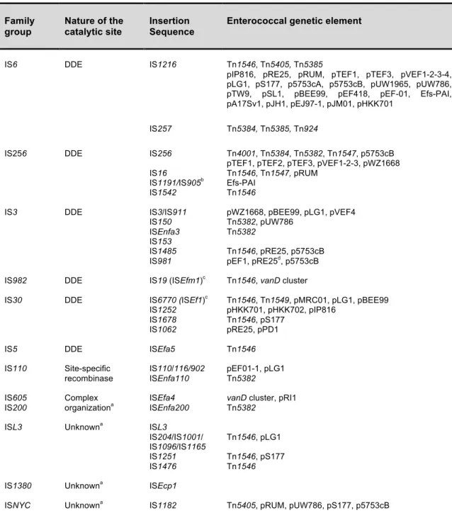
1.1.3.2. Acquired resistance has been successively observed in enterococci over the past three decades and includes different classes of antibiotics such as phenicols, tetracyclines, macrolides, lincosamides, streptogramins, aminoglycosides, #-lactams, glycopeptides, and fluoroquinolones (200, 231, 293) (Figure 2).
Figure 2. Chronologic representation of first descriptions of acquired resistance to different antibiotic families in enterococci. Enterococcal species where resistance first was reported appear in red for E.
faecalis, in blue for E. faecium, and in black for both E. faecalis and E. faecium.
HLR, high-level resistance; PBPs, penicillin-binding proteins.
The first report of transferable resistance in enterococci in 1964 showed the transfer of chloramphenicol resistance between E. faecalis strains, which is generally mediated by a chloramphenicol acetyltransferase enzyme (CAT; MIC of >16 µg/mL) (324, 334). An active efflux pump has also been described in four E. faecalis and one E. faecium strains (251).
Acquired resistance to tetracyclines (MIC of >8 µg/mL) and macrolides (MIC of $8
µg/mL) was detected in clinical E. faecalis strains in early 1970’s (11).
Two major mechanisms of tetracycline resistance are described in enterococci: i) active efflux of the antibiotic (tetK, tetL); and ii) ribosomal protection (tetM, tetO, tetT,
tetS). The mechanism of action linked to tetU from E. faecium remains unknown (58, 279).
Acquired resistance to macrolides, lincosamides and streptogramines (MLS phenotype) in enterococci can be achieved by: i) modification of the target action by a rRNA methylase (ermA, ermB, ermC, ermF, ermT); ii) inactivating enzymes such as lyases (vgbA) and transferases (lnuB, vatB, vatD, vatE); or iii) antibiotic active efflux through ATP binding transporters (msrA, msrC, msrD, lsaA, vgaB) or major facilitators (mefA) (350) (http://faculty.washington.edu/marilynr/). !"#$%!!!!!!!!!!!"#&"!!!!!!!!!!!!"#&#!!!!!!!!!"#'"!!!!!"#'(!!!!!!!"#'$!!!!!!!"#'&!!!!!!!"##%!!!!!!!)**"!!!!!!)**+!!!!!!)**&! HLR Aminoglycosides High concentrations of !-lactams (bla) High concentrations of !-lactams (PBPs) Glycopeptides Quinupristin-Dalfopristin (Streptogramins) Linezolid (Oxazolidinones) Daptomycin (Lipopetides) Tigecycline (Glycylcyclines) Chloramphenicol (Phenicols) Tetracycline (Tetracyclines) Erythromycin (Macrolides) Quinolones
Enterococci can acquire high-level resistance (HLR) to aminoglycosides by three mechanisms: i) modification of the ribosomal target; ii) alteration of antibiotic transport (expulsion by efflux pumps or decreased cell membrane permeability); and iii) antibiotic modification by aminoglycoside-modifying enzymes (AMEs) (200, 293). The first two are due to chromosomal mutations whereas the last, generally mediated by MGEs, confers the highest levels of resistance: MIC of $500 µg/mL for gentamicin, MIC of $1000 µg/mL for streptomycin, and MIC of $2000 µg/mL for kanamycin. Acquired HLR to aminoglycosides makes bacteria resistant to the synergism of an aminoglycoside with a cell active agent as #-lactams and the bactericidal treatment of serious infections compromised. Of note, some AMEs acting in particular aminoglycosides do not confer HLR in the host (MICs generally of 64 to 256 µg/mL) but can prevent synergism and even yield antagonism between aminoglycosides and cell wall active agents (231).
AMEs are the most frequent mechanism of resistance to aminoglycosides. They can be acetyltransferases (AACs), phosphotransferases (APHs), or nucleotidyltransferases (ANTs) depending if the antibiotic inactivation occurs through N-acetylation, O-phosphorylation or O-nucleotidylation, respectively. They vary in their aminoglycoside’s spectrum of action which often overlap (Table 1).
Table 1. Aminoglycoside-modifying enzymes described in enterococci (adapted from 231).
Enzyme group Enzyme Phenotyped Enterococcus range of species
Acetyltransferases AAC(6’)-Iea KAN, TOB, AMI, NEO,
FOR
E. faecalis, E. faecium, E. casseliflavus, E. gallinarum
AAC(6’)-Iib KAN, TOB, AMI, NEO E. faecium
AAC(6’)-Im KAN, TOB, AMI, DIB, NET E. faecium
Phosphotransferases APH(3’)-III KAN, AMI, ISE, NEO, BUT, RIB, LIV
E. faecalis, E. faecium, E. casseliflavus, E. gallinarum
APH(2’’)-Iaa,c GEN, NET, SIS, KAN,
TOB, AMI, ISE, NEO, BUT, RIB, LIV
E. faecalis, E. faecium, E. casseliflavus, E. gallinarum
APH(2’’)-Ibc GEN, NET, KAN, TOB,
DIB, AMI, ISE
E. faecium, E. durans
APH(2’’)-Icc GEN, NET, KAN, TOB E. faecalis, E. faecium, E. gallinarum
APH(2’’)-Idc GEN, NET, KAN, TOB E. faecalis, E. faecium, E. casseliflavus
Nucleotidyltransferases ANT(3’)-Ia STR E. faecalis
ANT(6)-Ia STR E. faecalis
ANT(9)-Ia SPE E. faecalis, E. durans
ANT(9)-Ib SPE E. faecalis
ANT(4’)-Ia KAN, TOB, AMI, NEO, DIB E. faecalis, E. gallinarum
aPart of the bifunctional enzyme AAC(6’)-APH(2’’) which confers HLR to all available aminoglycosides
except streptomycin. It is widespread in enterococci from both animal and human origins and accounts for more than 90% of clinical enterococci that have HLR to gentamicin (usually MIC of $2000 µg/mL).
bIntrinsic resistance in E. faecium.
cAll these APH enzymes confer HLR to gentamicin, often with intermediated MIC values of <500 µg/mL but
with the synergy between ampicillin and gentamicin supressed.
dAMI, amikacin; BUT, butirosin; DIB, dibekacin; FOR, fortimicin; GEN, gentamicin; ISE, isepamycin; KAN,
kanamycin; LIV, lividomycin; NEO, neomycin; NET, netilmicin; RIB, ribostamycin; SIS, sisomicin; SPE, spectinomycin; STR, streptomycin; TOB, tobramycin.
Acquired resistance to #-lactams can be due to the production of a #-lactamase (E.
faecalis) or modifications of PBPs that include the mutation, hyperproduction or
transferability of pbp genes associated to composite genetic platforms (E. faecalis, E.
faecium). The production of #-lactamase confers resistance to penicillin, aminopenicillins
(ampicillin) and ureido-penicillins (piperacillin) and low level of resistance to imipenem. The enzyme is inhibited by clavulanic acid, sulbactam and tazobactam. The enterococcal #-lactamase was firstly identified in 1981 and although has been detected in different countries of different continents, its prevalence remains low (292).
HLR to #-lactams (MIC of $32-$256 µg/mL) in most clinical isolates of E. faecium has been associated with mutations or overproduction of pbp5 with increased low affinity for penicillins, which has proved to be transferable in some instances (294, 340). This HLR of
E. faecium may be an extreme example of intrinsic resistance associated with low affinity
PBPs or may represent acquired resistance. Alterations in different PBPs can also confer resistance to #-lactams in E. faecalis. They include mutations in the pbp4 gene, apparently increased production of PBP 5 and decreased penicillin binding of PBPs 1 and 6 (52, 315).
Acquired resistance to glycopeptides (vancomycin and teicoplanin) was detected in clinical isolates of E. faecium and E. faecalis collected in Europe in 1986 and the USA in 1988, respectively (232, 357, 437). The mechanism of resistance involves modification of the target site via a switch in the cell wall composition from pentapeptide ending in D-Ala:D-Ala to other aminoacid termination, resulting in peptidoglycan precursors with low affinity for these antibiotics.
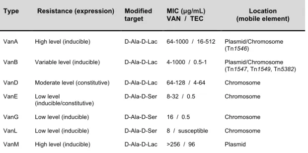
To date, seven types of acquired glycopeptide resistance (VanA/B/D/E/G/L/M) have been described based not only in the genetic level (gene similarity and synteny) (Figure 3) but also in the phenotype they confer (33, 326, 464, 475). The van gene cluster vanF has been described in a biopesticide, Paenibacilluspopilliae, but has not yet been found in
enterococci (322). The different clusters or types of vancomycin resistance are designated according to the name of the ligase gene, which encodes either a Ala:Lac or a D-Ala:D-Ser ligase for the synthesis of peptidoglycan precursors with low affinity for glycopeptides (Table 2). The mechanism of resistance is complex but in all cases, the genetic element consists on a cluster of 5-7 genes encoding similar enzymatic functions. The vanA, vanB, vanD and vanM gene clusters contain genes for a two-component
regulatory system (vanR and vanS), three resistance genes (vanH, encoding dehydrogenase; vanA, vanB, vanD or vanM, encoding ligase; vanX, encoding DD-dipeptidase); an accessory gene (vanY); and the vanZ gene,which is present in the vanA gene cluster, whereas the vanWgene is found only in the vanB operon (Figure 3). The
(D,D-dipeptidase-D,D-carboxypeptidase), vanT (serine racemase; two vanTm and vanTr in the case of vanL),vanR (response regulator), and vanS (sensor histidine kinase). The vanG
operon begins with a three-component regulatorysystem, vanU (transcriptional regulator),
vanR, vanS, vanY (D,D-carboxypeptidase), vanW (unknown function), and then vanG,
vanXY, and vanT (78, 79).
Figure 3. Alignment of vancomycin resistance gene clusters of vanA, vanB, vanD, vanF and vanM (adapted from 475). The percent amino acid identities with vanRM, vanSM, vanYM, vanHM, vanM, and vanXM products are shown below the respective genes.
Acquired glycopeptide resistance is mainly due to VanA and VanB phenotypes which are the most frequently reported in clinical isolates. The vanA gene cluster mediates high-level inducible resistance to both vancomycin and teicoplanin, while vanB causes variable levels of resistance to vancomycin but none to teicoplanin, being resistance only inducible
by vancomycin (464). The genotypes VanA and VanB are generally associated with
mobile genetic elements (transposons and/or plasmids), which are described in sections 1.3.2.2.-1.3.2.4. and 1.3.3. of this chapter.
Table 2. Characteristics of acquired van gene clusters in enterococci.
Type Resistance (expression) Modified
target
MIC (µg/mL)
VAN / TEC
Location (mobile element)
VanA High level (inducible) D-Ala-D-Lac 64-1000 / 16-512 Plasmid/Chromosome (Tn1546)
VanB Variable level (inducible) D-Ala-D-Lac 4-1000 / 0.5-1 Plasmid/Chromosome (Tn1547, Tn1549, Tn5382) VanD Moderate level (constitutive) D-Ala-D-Lac 64-128 / 4-64 Chromosome
VanE Low level
(inducible/constitutive)
D-Ala-D-Ser 8-32 / 0.5 Chromosome VanG Low level (inducible) D-Ala-D-Ser 16 / 0.5 Chromosome VanL Low level (inducible) D-Ala-D-Ser 8 / susceptible Chromosome VanM High level (inducible) D-Ala-D-Lac >256 / 96 Plasmid
Acquired high-level resistance to quinolones is associated with amino acid changes in the “quinolone-resistance determining region” (QRDR) of the genes encoding topoisomerase IV (parC, parE) and DNA gyrase (gyrA, gyrB), which are essential for replication. The ParC mutation 80!Ile alone or combined with the GyrA mutation Ser-83!Tyr confers high-level ciprofloxacin resistance (MIC of 64 to >128 µg/mL) (200, 438). Resistance by active efflux of quinolones or increased expressionof endogenous efflux pumps has also been suggested for E. faecalis (194, 233).
Linezolid (a synthetic oxazolidinone) shows good activity against most clinical isolates of enterococci and it is restricted to the treatment of multidrug resistant isolates. However, resistance to this antimicrobial agent due to mutations of the 23S rRNA modifying the target interaction site has been reported (7, 119).
Daptomycin (a cyclic lipopeptide) is active against most multidrug resistant enterococci including vancomycin-resistant enterococci, being the only antibiotic with in
vitro bactericidal activity against VRE (vancomycin-resistant enterococci). Rare
occurrences of isolates resistant to this antibiotic have been reported (MIC of >4 µg/mL), but the mechanisms underlying the reduced susceptibility to daptomycin were not investigated (7, 48).
Tigecycline (a glycylcycline) is used to treat a broad spectrum of multidrug resistant isolates. Only an E. faecalis resistant to tigecycline (MIC of 2 µg/mL) has been recently described although the exact mechanism of resistance was not elucidated (458).
1.1.4. Clinical enterococcal infections
Enterococci have been recognized as a cause of endocarditis and urinary tract infections since the early 1900’s, and have rapidly emerged as one of the most common nosocomial pathogens since the 1980s. They may also be responsible for other severe healthcare-associated infections that includes intra-abdominal, pelvic and soft tissue infections, and bacteremia. Rare infections caused by enterococci include meningitis, hematogenous osteomyelitis, septic arthritis, and pneumonia (260, 293, 294, 403). As opportunistic pathogens, they are often isolated from elderly and debilitated patients who have received multiple courses of antibiotics and/or have been hospitalized for long periods or time (162, 287, 293, 294).
Last reports provided by the National Nosocomial Infections Surveillance System (NNISS) and the National Healthcare Safety Network (NHSN) in the United States, from the European Antimicrobial Resistance Surveillance Network (EARS-Net), and from the SENTRY program show that rates of serious enterococcal infections remained relatively stable through the last years (22, 174, 403) (www.rivm.nl/earss). However, a progressive
inversion of the ratio of E. faecalis and E. faecium causing disease (from 10:1 before 1990, to 3:1 in the late 1990s, and currently 1.5:1) has been reported (174, 431). This coincided with an increasing trend towards multiantibiotic-resistant E. faecium, for which ampicillin and vancomycin resistance are much more prevalent than in E. faecalis recovered from clinical infections (57, 102, 152, 265, 291, 431, 471). Also a higher mortality rate has been associated with E. faecium than with E. faecalis independent of the antibiotic profile (244, 305). Human infections caused by E. durans, E. avium, E.
gallinarum, E. casseliflavus, E. hirae, E. muntdii, and E. raffinosus have been rarely
documented (69, 165, 202, 293, 360, 406). Multidrug-resistant enterococci are a major cause of mortality and morbidity in hospitalized patients worldwide posing increased therapeutic and economic challenges to healthcare institutions.
1.1.5. Treatment
Ampicillin and penicillin remain the antibiotics of choice for susceptible isolates but tetracyclines, chloramphenicol and quinolones are alternatives against these strains. Fosfomycin and nitrofurantoin are restricted to treat urinary tract infections.
A bactericidal effect is required for severe enterococcal infections which is achieved by the combination of a cell active agent (#-lactam, vancomycin) and an aminoglycoside as stated above (section 1.1.3.1; 293). Nonetheless, the treatment of ampicillin- and vancomycin-resistant enterococcal infections is nowadays compromised with the increasing acquisition and accumulation of different mechanisms of resistance to these agents.
New treatment options have been introduced in the therapeutic arsenal in the last years. Several combination therapies using different antibiotics have been recently used in the treatment of enterococcal infections, though the frequent lack of clinical experience, the increased rates of resistance to some compounds, and their variable activity depending on the strain, demand their use for multidrug resistant enterococci. The determination of the bactericidal effect by time-kill curves might be needed for selection of the most appropriate therapeutic regimen when options are limited (7).
1.2. Ecology and population structure of Enterococcus
1.2.1. EcologyEnterococci are a normal inhabitant of the intestinal tract in most mammals and birds, and they can be also recovered from reptiles, insects, soil, plants, food and water (3).
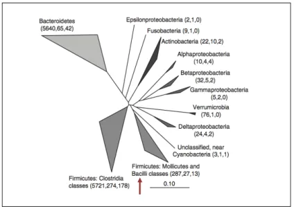
The healthy human intestinal microbiota are dominated (>90%) by species from the phyla Firmicutes and Bacteroidetes (Figure 4; 112, 264). The concentration of enterococci in faeces is about 6-8 log10 cells/g of faeces, which is significantly lower than that for the
most abundant orders of bacteria in faeces such as the Clostridiales (Firmicutes),
Bifidobacteriales (Actinobacteria) and Bacteroidales (Bacteroidetes) (9-10 log10 cells/g
feces). However, E. faecium and E. faecalis were found to be one of the predominant members of the small intestine (jejunum and ileum) (167).
Figure 4. Phylogenetic tree of human intestinal 16S rRNA sequences (adapted from 264). The arrow indicates the clade that includes
Enterococcus.
The genus Enterococcus comprises several species some of which showing adaptation to different host or environmental conditions. While E. faecium and E. faecalis are widely distributed, yellow-pigmented species E. mundtii, E. casseliflavus or E.
sulfureus are associated with plants and E. columbae and E. asinii are apparently
associated with pigeons and donkeys, respectively. Variations in the distribution of widespread enterococcal species varies depending on the hosts (E. faecalis predominates in cats, dogs, calves and pigs, while E. faecium are more abundant among poultry), and the age (E. cecorum is particularly associated with older poultry), or the intestinal compartments (E. faecium predominates in the caecum and E. faecalis in the recto-sigmoid colon of human intestine) within a given host (3, 167, 213). In addition to E.
species recovered from farm animals and pets; and E. durans from dairy products (fermented food products as cheeses and milk), fish, olives, vegetables, raw minced beef and pork (129, 213, 313). A variable occurrence of different enterococci species has been described among wild animals (250, 267, 328, 329). Enterococcus raffinosus has only been detected in pathological materials and its habitat is unknown (406).
E. faecium and E. faecalis are abundant in the small intestine, whereas E. avium and E. durans only occasionally colonize the human intestine (30, 273, 374, 406). Enterococci
may also colonize the oropharynx, mouth, vagina, perineal region and soft tissue wounds of asymptomatic humans, where E. faecalis is generally more abundant than E. faecium (56, 293).
1.2.2. Population structure
The geographic and ecologic expansion of different clones or clonal complexes of enterococci from different origins have been documented in different studies. The diversity and evolution of the main enterococci species found in humans is greatly influenced by homologous recombination related with the acquisition of different genetic traits. Main definitions related with the study of population structure are included in Box 1 (for reviews see 122, 157, 391, 392, 436).
1.2.2.1. Enterococcus faecium
Different studies have demonstrated that E. faecium species comprises different ecovars o genogroups showing common genotypic and phenotypic features (177, 442, 466, 469). Multilocus sequence typing (MLST) of a high number of isolates from different origins, hosts and geographical areas has shown that E. faecium has a highly recombinant population structure that comprises a large number of sequence types (STs) at linkage equilibrium with overrepresentation of specific clonal complexes (CCs).
The first population structure analysis of E. faecium performed with isolates from different origins and using the techniques amplified fragment length polymorphism (AFLP) and ribotyping, grouped isolates in different genogroups A (non-hospitalized persons and pigs), B (poultry), C (hospitalized patients - designated by that time as “lineage C1” - and pets), and D (veal calves) (26, 31, 38, 41, 76, 196, 197, 442, 466).