DOI: http://dx.doi.org/10.18363/rbo.v77.2020.e1758
1
Rev. Bras. Odontol. 2020;77:e1758
Literature Review/Endodontics
Adhesion Capacity of Bioceramic and Resin-Based
Root Canal Sealer to Root Dentin:
An Integrative Review
Carolyne de Pinho Quintão,1 Sabrina Teixeira Pinto Costa,1 Mariane Floriano Lopes Santos Lacerda,2 Caroline Felipe Magalhães Girelli,1 Carolina Oliveira
de Lima3
1Brazilian Dental Association, Section Governador Valadares, MG, Brazil
2Department of Dentistry, School of Dentistry, Juiz de Fora Federal University, Governador Valadares, MG, Brazil
3Department of Integrated Clinical Procedures (Operative Dentistry), School of Dentistry, State University of Rio de Janeiro (UERJ), Rio de Janeiro, RJ, Brazil • Conflicts of interest: none declared.
AbstrAct
Objective: to compare the adhesion capacity of the bioceramic EndoSequence BC sealer and the AH Plus sealer through an integrative review. Material and Methods: the Medline/PubMed, Scopus, Web of Science and Virtual Health Library (VHL) online databases were used for the literature review. Eligibility criteria comprised articles available in full on the researched databases, in English, and content addressing Endosequence BC sealer adhesion compared to AH Plus sealer. Results: a total of 45 articles were found. After duplicate removal, 22 articles were selected. After reading the abstracts, full texts and applying the inclusion criteria, eight articles in total were included in the present study. Concerning the adhesion capacity of the tested cements, the AH Plus was reported as presenting better adhesion than the BC sealer in three articles, and less adhesion in two articles. Similar adherence strength was observed between groups in three studies. Conclusions: based on the included studies, the AH Plus displays greater bond strength when compared to the BC Sealer.
Keywords: Bond strength; Epoxy resin-based root canal sealer; Tricalcium silicate.
Introduction
T
he success of endodontic therapy depends on the cleaning, shaping and filling of the root canal system (RCS) to prevent the penetration or proliferation of microorganisms into the periradicular tissue.1,2 To this end, RCS filling materials should adapt to the dentin walls, in order to avoid infiltration throughout the canal and the apical region,3,4,5 during mechanical stresses due to masticatory function and restorative and surgical procedures, ensuring that the seal is maintained.6As gutta-percha does not display the ability to adhere to root dentin, the most widely applied method for RCS obturation is its use associated with endodontic sealers.7 The AH Plus sealer (Dentsply Maillefer, Tulsa, OK, USA) consists in an epoxy resin8,9 and has been considered the gold standard material for RCS filling, due to its low solubility, good dimensional stability, good adaptation and adhesion to dentinal walls.10
Clinical studies, however, have demonstrated that no filling technique is able to prevent certain canal areas displaying a lack of filling material2 especially regarding oval canals, which are prevalent in up to 50% of cases, depending on the dental group.11 Thus, bioactive endodontic sealers have been developed to improve the quality of root canal filling, since they are formed by nanospheric particles that allow for a sealer flow through canal irregularities and dentinal tubules12 while also establishing a chemical connection between dentin and the filling materials.13
In fact, these sealers are extremely biocompatible (non-toxic) and chemically stable, undergoing an expansion process (0.002) during the setting time, instead of contraction. Another advantage inherent to these materials during the hardening process is their ability to form hydroxyapatite.14,15 An example is the EndoSequence BC Sealer (BC Sealer) (EndoSequence, Brassler, Savannah, GA, USA) which consists of a pre-manipulated bioceramic sealer with excellent flowability and dimensional stability.7,12 Its particles are so small - less than 2 µm - that its can be used with a 0.012 capillary tip, thus reaching areas not filled by other cements.13 In addition, it has an alkaline pH, resulting in antibacterial activity and adequate biocompatibility,16 avoiding inflammatory reactions in the case of overfilling.13 Previous studies have also shown that the BC sealer displays radiopacity and adhesion to the dentinal walls 3,7,17,18 and this requirement has been considered a parameter to evaluate the effectiveness of root canal filling.5,19, 20.
For this reason, the present study aimed to compare the adhesion capacity of the bioceramic Endosequence BC sealer and the AH plus – resin based sealer, through an integrative review.
Material and Methods
This study selected the integrative review method to achieve the proposed objective of comparing the adhesion capacity of the BC sealer and the AH plus. The question that supported the scientific evidence collection was: which
endodontic cement displays better adhesion capacity, the bioceramic EndoSequence BC sealer or the resinous AH plus?
The Medline/PubMed (database developed by the National Center for Biotechnology Information National Library of Medicine), Scopus, Web of Science and Virtual Health Library - VHL (Lilacs and BBO) online databases were used. The search strategy comprised the following: ((((((“shear strength”[MeSH Terms]) OR “push out”[Title/Abstract]) OR “bond strength”[Title/Abstract])) AND (((“bioceramic sealer”[Title/Abstract]) OR EndoSequence OR “BC sealer “)) AND ((((“epoxy resin-based root canal sealer”[MeSH Terms]) OR “epoxy resin-based root canal sealer”[Title/Abstract]) OR “AH-plus”[Title/Abstract]) OR “AH plus”)). A manual search by four reviewers was also performed, after reading the titles, abstracts and keywords. Articles were read in full when the information contained in these topics was insufficient. The references of possible studies to be used, as well as a cross-search of the authors’ database, served as a guide for the selection of other relevant articles.
The eligibility criteria comprised the following: articles available in full in the researched database, with contents addressing the adhesion of BC Sealer compared to the AH
Plus. Laboratory studies in artificial tooth simulators or developed in primary teeth and written in a language other than English were excluded. Articles involving the adhesion ability of endodontic filling materials using different types of bioceramic or resin sealers or that addressed the use of sealers in root canal retreatments, were also eliminated.
After reading the selected articles, the information was collected and typed into a database containing the following variables: Article author, year, objective, methodology and conclusion.
Results
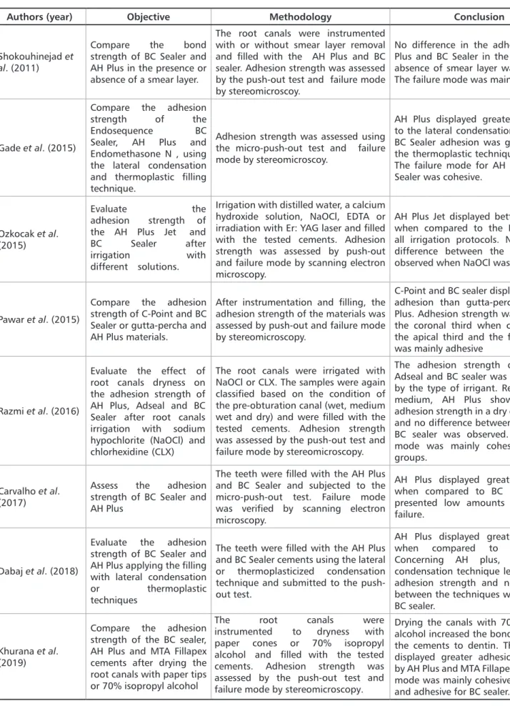
A total of 45 articles were found. After removing duplicates, 22 articles were selected (Figure 1). After reading the abstracts, full texts and applying the inclusion criteria, a total of eight articles were included in the present study (Table 1).
Of the selected articles, all were published in dental journals, between 2011 and 2017. Analyzing the adhesion capacity of the tested sealers, the AH Plus was reported as displaying better adhesion than the BC sealer in three articles and less adhesion in two selected articles. Similar adherence strength was observed between the groups in three studies.
3
Rev. Bras. Odontol. 2020;77:e1758
Authors (year) Objective Methodology Conclusion
Shokouhinejad et
al. (2011)
Compare the bond strength of BC Sealer and AH Plus in the presence or absence of a smear layer.
The root canals were instrumented with or without smear layer removal and filled with the AH Plus and BC sealer. Adhesion strength was assessed by the push-out test and failure mode by stereomicroscoy.
No difference in the adhesion of AH Plus and BC Sealer in the presence or absence of smear layer was observed. The failure mode was mainly cohesive.
Gade et al. (2015)
Compare the adhesion strength of the Endosequence BC Sealer, AH Plus and Endomethasone N , using the lateral condensation and thermoplastic filling technique.
Adhesion strength was assessed using the micro-push-out test and failure mode by stereomicroscoy.
AH Plus displayed greater adherence to the lateral condensation technique. BC Sealer adhesion was greater when the thermoplastic technique was used. The failure mode for AH Plus and BC Sealer was cohesive.
Ozkocak et al. (2015)
Evaluate the adhesion strength of the AH Plus Jet and BC Sealer after irrigation with different solutions.
Irrigation with distilled water, a calcium hydroxide solution, NaOCl, EDTA or irradiation with Er: YAG laser and filled with the tested cements. Adhesion strength was assessed by push-out and failure mode by scanning electron microscopy.
AH Plus Jet displayed better adhesion when compared to the BC sealer in all irrigation protocols. No statistical difference between the sealers was observed when NaOCl was used.
Pawar et al. (2015)
Compare the adhesion strength of C-Point and BC Sealer or gutta-percha and AH Plus materials.
After instrumentation and filling, the adhesion strength of the materials was assessed by push-out and failure mode by stereomicroscopy.
C-Point and BC sealer displayed greater adhesion than gutta-percha and AH Plus. Adhesion strength was greater in the coronal third when compared to the apical third and the failure mode was mainly adhesive
Razmi et al. (2016)
Evaluate the effect of root canals dryness on the adhesion strength of AH Plus, Adseal and BC Sealer after root canals irrigation with sodium hypochlorite (NaOCl) and chlorhexidine (CLX)
The root canals were irrigated with NaOCl or CLX. The samples were again classified based on the condition of the pre-obturation canal (wet, medium wet and dry) and were filled with the tested cements. Adhesion strength was assessed by the push-out test and failure mode by stereomicroscopy.
The adhesion strength of AH Plus, Adseal and BC sealer was not affected by the type of irrigant. Regarding the medium, AH Plus showed greater adhesion strength in a dry environment and no difference between Adseal and BC sealer was observed. The failure mode was mainly cohesive for the groups.
Carvalho et al. (2017)
Assess the adhesion strength of BC Sealer and AH Plus
The teeth were filled with the AH Plus and BC Sealer and subjected to the micro-push-out test. Failure mode was verified by scanning electron microscopy.
AH Plus displayed greater adhesion when compared to BC Sealer. Both presented low amounts of adhesive failure.
Dabaj et al. (2018)
Evaluate the adhesion strength of BC Sealer and AH Plus applying the filling with lateral condensation
or thermoplastic techniques
The teeth were filled with the AH Plus and BC Sealer cements using the lateral or thermoplasticized condensation technique and submitted to the push-out test.
AH Plus displayed greater adhesion when compared to BC Sealer. Concerning AH plus, the lateral condensation technique led to greater adhesion strength and no difference between the techniques was noted for BC sealer.
Khurana et al. (2019)
Compare the adhesion strength of the BC sealer, AH Plus and MTA Fillapex cements after drying the root canals with paper tips or 70% isopropyl alcohol
The root canals were instrumented to dryness with paper cones or 70% isopropyl alcohol and filled with the tested cements. Adhesion strength was assessed by the push-out test and failure mode by stereomicroscopy.
Drying the canals with 70% isopropyl alcohol increased the bond strength of the cements to dentin. The BC sealer displayed greater adhesion, followed by AH Plus and MTA Fillapex. The failure mode was mainly cohesive for AH Plus and adhesive for BC sealer.
intracanal medication prior to filling. The high surface tension generated by this medication explains the difficulty of endodontic sealers in penetrating the dentinal tubules and, consequently, adhering to dentin.3,26
In relation to the filling technique, the AH Plus displayed better adhesion when applying the lateral condensation technique when compared to the thermoplastic technique.23, 24 This is due to the accelerated polymerization of the resin sealer when the thermoplastic technique is used, which results in a decreased flow and lower bond strength of AH Plus to dentin.27
Several methods are suggested in order to assess the bond strength of sealers to dentin, such as shear strength, tensile strength and tensile tests (push-out).28 In the present review, all included studies used the push-out test. This test has been considered the most effective method for assessing adhesion strength since, although it does not demonstrate the actual clinical performance of the material, it provides substantial information when comparing different endodontic sealers or obturation techniques.6,29 In addition, the push-out test more accurately reflects the fractures that occur in endodontic sealers.30
Regarding failure modes (cohesive, mixed and adhesive), the mixed and cohesive types were the most frequent for failure modes for the AH Plus,12,22,24 and the cohesive type was more frequent for the BC Sealer,12,22 which indicates the good adhesion of the studied cements to the dentinal wall.
Failure modes were assessed under a stereomicroscope
12,18,22,24,25 and by Scanning Electron Microscopy (SEM).3,5 The
SEM evaluation is the most adequate methodology, since it permits not only for the visualization of the penetration of endodontic sealers, but also the presence of the smear layer in the dentinal tubules, allowing for correlations between
these variables and adhesion failure.5
SEM micrographs also indicated the presence of mineral precipitates such as calcium and phosphorus after 30 days of BC Sealer incubation, suggesting cement bioactivity.5 Bioceramic sealers consists of calcium silica which, in the presence of water from the dentinal tubules, hydrates and produces calcium silicate gel and calcium hydroxide. The calcium hydroxide, in turn, reacts with calcium phosphate to produce hydroxyapatite and water.31 In addition, the calcium hydroxide formed in this reaction results in a highly alkaline pH medium (pH=12.8), which can influence not only the repair, minimizing inflammatory reactions, but also promoting an antimicrobial environment, which is partly responsible for the destruction of most bacteria.32 Thus, the BC Sealer may be considered not only biocompatible, but also bioactive.12
Discussion
Endodontic sealers must fill the root canal space and be able to adhere to the root canal wall and gutta-percha,3 to allow for empty space elimination and avoid fluid and microorganism percolation through the apical region,5,21 while also remaining in place under displacement forces, such as stresses caused by teeth function.12
Although the AH Plus sealer is considered the gold standard material for filling root canals, as it has displays physical and chemical properties,5,22 bioceramic sealers, such as the BC Sealer, are proposed as useful in increasing the flow through canal irregularities and towards the dentinal tubules, as they are formed by nanospheric particles.12
In view of the above, the present study compared the adhesion capacity of the BC sealer and AH Plus sealer through an integrative review and, according to table 1, different results were found concerning the adhesion strength of both to root dentin.
Some studies reported that BC Sealer adhesion strength was lower when compared to AH Plus.3,5,23 This fact can be explained by the use of ethylenediaminetetraacetic acid (EDTA), before filling the root canals. The chelating action of EDTA removes the inorganic portion of the smear layer, which is composed of calcium ions, which may interfere with the BC sealer adhesion strength, whose mechanism of action depends on binding to mineralized dentin tissues. 3,5 However, a previous study reported that the presence of the smear layer did not affect the adhesion strength of either the BC Sealer or the AH Plus sealers.12
Another interfering factor may be the absence of moisture in the dentinal tubules during the experiments.3,5 Bioceramic sealers were produced to use water, inherent to the dentinal tubules, to conduct the hydration reaction that promotes its hardening.13 Thus, adverse effects of directly drying the canal may include interference with the cement seal.
Still in this regard, the BC sealer demonstrated greater adhesion strength when compared to AH Plus in the presence of moisture.18,22 This finding is probably due to the presence of calcium phosphate in the BC sealer , which facilitates the reaction with calcium hydroxide after activation by the moisture that remains inside the dentinal tubules. This moisture then induces the formation of hydroxyapatite, characterizing bioactivity, thus resulting in greater dentin adhesion.24 Accordingly, a previous study observed that canal drying with paper points leads to worse adhesion results of the tested sealers when compared to the use of 70% isopropyl alcohol.25
The adhesion strength of the AH Plus also decreased in samples in which calcium hydroxide was used as an
5
Rev. Bras. Odontol. 2020;77:e1758 Based on the studies included in the present integrative
review, the AH Plus displays greater bond strength when compared to the BC Sealer. However, the presence or absence of the smear layer, canal humidity, use of intracanal medication and different root canal filling techniques, present the potential to interfere with cement
adhesion to the dentinal wall, and should be carefully taken into account before choosing the endodontic sealer.
Acknowledgements
The authors deny any conflicts of interest related to this study.
root fillings made with C-Point and BC sealer versus gutta-percha and AH Plus after the instrumentation of oval canals with the Self-Adjusting File versus Wa-veOne. Int Endod J. 2016;49(4):374-81.
19. Neelakantan P, Subbarao C, Subbarao CV, De-Deus G, Zehnder M. The im-pact of root dentine conditioning on sealing ability and push-out bond strength of an epoxy resin root canal sealer. Int Endod J. 2011;44(6):491-8.
20. Yazdi KA, Bolhari B, Sabetmoghaddam T, Meraji N, Kharazifard MJ. Effect of Blood Exposure on Push-Out Bond Strength of Four Calcium Silicate Based Cements. Iran Endod J. 2017;12(2):196-200.
21. Vilas-Boas DA, Grazziotin-Soares R, Ardenghi DM, Bauer J, Souza PO, Can-deiro GTM, et al. Effect of different endodontic sealers and time of cementation on push-out bond strength of fiber posts. Clin Oral Investig. 2018;22(3):1403–9. 22. Razmi H, Bolhari B, Dashti NK, Fazlyab M. The Effect of Canal Dryness on Bond Strength of Bioceramic and Epoxy-resin Sealers after Irrigation with Sodi-um Hypochlorite or Chlorhexidine. Iran Endod J. 2016;11(2):129-33.
23. Dabaj P, Kalender A, Unverdi Eldeniz A. Push-Out bond strength and SEM evaluation in roots filled with two different techniques using new and conven-tional sealers. Mater. 2018;11(9):1620.
24. Gade VJ, Belsare LD, Patil S, Bhede R, Gade JR. Evaluation of push-out bond strength of Endosequence BC sealer with lateral condensation and thermoplasti-cized technique: An in vitro study. J Conserv Dent. 2015;18(2):124-7.
25. Khurana N, Chourasia HR, Singh G, Mansoori K, Nigam AS, Jangra B. Effect of Drying Protocols on the Bond Strength of Bioceramic, MTA and Resin-based Sealer Obturated Teeth. Int J Clin Pediatr Dent. 2019;12(1):33–6
26. Ghabraei S, Bolhari B, Yaghoobnejad F, Meraji N. Effect of Intra-Canal Cal-cium Hydroxide Remnants on the Push- Out Bond Strength of Two Endodontic Sealers. Iran Endod J. 2017;12(2):168-72.
27. Carneiro SM, Sousa-Neto MD, Rached FA Jr, Miranda CE, Silva SR, Sil-va-Sousa YT. Push-out strength of root fillings with or without thermomechani-cal compaction. Int Endod J. 2012;45(9):821–8.
28. Gogos C., Economides N, Stavrianos C, Kolokouris, I, Kokorikos I. Ad-hesion of a new methacrylate resin-based sealer to human dentin. J. Endod. 2004;30(4):238–40.
29. Pane ES, Palamara JEA., Messer HH. Critical evaluation of the push-out test for root canal filling materials. J. Endod. 2013;39(5):669–73.
30. Goracci C, Tavares AU, Fabianelli A, Monticelli F, Raffaelli O, Cardoso PC
et al. The adhesion between fiber posts and root canal walls: comparison
be-tween microtensile and push-out bond strength measurements. Eur J Oral Sci. 2004;112(4):353–61.
31. Damas BA, Wheater MA, Bringas JS, Hoen MM. Cytotoxicity comparison of mineral trioxide aggregates and EndoSequence bioceramic root repair materials. J Endod. 2011;37(6):372–5.
32. Tomson PL, Grover LM, Lumley PJ, Sloan AJ, Smith AJ, Cooper PR. Disso-lution of bio-active dentine matrix components by mineral trioxide aggregate. J Dent. 2007;35(8):636-42.
References
1. Jainaen A, Palamara JEA, Messer HH. Push-out bond strengths of the dentine– sealer interface with and without a main cone. Int Endod J. 2007;40(11):882–90. 2. Madhuri GV, Varri S, Bolla N, Mandava P, Akkala LS, Shaik J. Comparison of bond strength of different endodontic sealers to root dentin: An in vitro push-out test. J Conserv Dent. 2016;19(5):461-4
3. Ozkocak I, Sonat B. Evaluation of effects on the adhesion of various root canal sealers after Er:YAG laser and irrigants are used on the dentin surface. J Endod. 2015;41(8):1331–6.
4. Carvalho CN, Martinelli JR, Bauer J, Haapasalo M, Shen Y, Bradaschia-Correa V, et al. Micropush-out dentine bond strength of a new gutta-percha and niobium phosphate glass composite. Int Endod J. 2014;48(5):451–9.
5. Carvalho CN, Grazziotin-Soares R, Candeiro GTM, Martinez LG, Souza JP, Oliveira PS, et al. Micro Push-out bond strength and bioactivity analysis of a bioceramic root canal sealer. Iran Endod J. 2017;12(3):343-8.
6. Gurgel-Filho ED, Leite FM, Lima JB, Montenegro JPC, Saavedra F, Silva EJ NL. Comparative evaluation of push-out bond strength of a MTA-based root canal sealer. Braz J Oral Sci. 2014;13(2):114-7.
7. DeLong C, He J, Woodmansey KF. The effect of obturation technique on the push-out bond strength of calcium silicate sealers. J Endod. 2015;41(3):385-8. 8. Ungor M, Onay EO, Orucoglu H: Push-out bond strengths: the Epiphany-Resi-lon endodontic obturation system compared with different pairings of Epiphany, Resilon, AH Plus and gutta-percha. Int Endod J. 2006;39(8):643–7.
9. Patil AS, Dodwad KP, Patil AA. An in vitro comparison of bond strengths of Gutta-percha/AH Plus, Resilon/ Epiphany self-etch and EndoREZobturation system to intraradicular dentin using a pushout test design. J Conserv Dent. 2013;16: 238–42.
10. Dias KC, Soares CJ, Steier L, Versiani MA, Rached-Júnior FJA, Pécora JD, et
al. Influence of drying protocol with isopropyl alcohol on the bond strength of
resin-based sealers to the root dentin. Int Endod J. 2014;40(9):1454-8.
11. Jou Y-T, Karabuchak B, Levin J, et al. Endodontic working width: current concepts and techniques. Dent Clin North Am. 2004;48:323-35.
12. Shokouhinejad N, Gorjestani H, Nasseh AA, Hoseini A, Mohammadi M, Shamshiri AR. Push-out bond strength of gutta-percha with a new bioceramic sealer in the presence or absence of smear layer. Aust Endod J. 2011;39:102-6. 13. Koch KA, Brave DG. Bioceramics, part I: the clinician’s viewpoint. Dent To-day. 2012;31(1):130-5
14. Koch K. Bioceramic technologya: game changer in endodontic obturation. NJAGD Wisdom. 2009;6:8-11.
15. Nasseh A. The rise of bioceramics. Endod Pract. 2009;2:17-22.
16. Candeiro GTM, Correia FC, Duarte MA, Duarte MAH, Ribeiro-Siqueira DC, Gavini G. Evaluation of radiopacity, pH, release of calcium ions, and flow of a bioceramic root canal sealer J Endod. 2012;38(6):842-5.
17. Prati C, Gandolfi MG. Calcium silicate bioactive cements: Biological perspec-tives and clinical applications. Dent Mater. 2015;31(4):351-70.
18. Pawar AM, Pawar S, Kfir A, Pawar M, Kokate S. Push-out bond strength of
Submitted: 02/03/2020 / Accepted for publication: 03/28/2020 Corresponding author
Carolina Oliveira de Lima
E-mail: c.oliveiradelima@yahoo.com.br
Mini Curriculum and Author’s Contribution
1. Carolyne de Pinho Quintão – DDS. Contribution: Contribuited to effective scientific and intellectual participation for the study. ORCID: 0000-0001-9453-5564 2. Sabrina Teixeira Pinto Costa – DDS. Contribution: Contribuited to effective scientific and intellectual participation for the study. ORCID: 0000-0002-0649-3024 3. Mariane F. L. Santos Lacerda – DDS;PhD. Contribution: Contributed to the idea, interpretation of data and wrote the manuscript. ORCID: 0000-0002-1534-0150 4. Caroline Felipe Magalhães Girelli – DDS;MSc. Contribution: Contributed to critical review of the work. ORCID: 0000-0001-9424-904X