296
Rev. odonto ciênc. 2010;25(3):296-299Received: Jan 29, 2010 Accepted: April 19, 2010
Conflict of Interest Statement: The authors state that there are no financial and personal conflicts of interest that could have inappropriately influenced their work.
Copyright: © 2010 Steier et al.; licensee EDIPUCRS. This is an Open Access article distributed under the terms of the Creative Commons Attribution-Noncommercial-No Derivative Works 3.0 Unported License.
Original Article
Comparison of the interface dentin-endodontic
sealer using two SEM magnifications
Comparação da interface dentina-cimento endodôntico
usando dois aumentos de MEV
Liviu Steier a
Jose Antonio Poli de Figueiredo b Sema Belli c
aWarwick Medical School, Coventry, England,
United Kingdom
bPontifical Catholic University of Rio Grande do
Sul, Porto Alegre, RS, Brazil
c Department of Endodontics, Faculty of Dentistry,
Selcuk University, Konya, Turkey
Correspondence:
José Antonio Poli de Figueiredo Post-Graduate Program in Dentistry Pontifical Catholic University of Rio Grande do Sul – PUCRS Av. Ipiranga 6681 Prédio 6 sala 507 Porto Alegre, RS – Brazil
90619-900
E-mail: [email protected]
Abstract
Purpose: The aim of this in vitro study was to compare the interface dentin-sealer of two sealers (RealSeal and AH Plus) using two magnifications under Scanning Electron Microscope (SEM).
Methods: The coronal two thirds of eight extracted molars were removed and the dentin surfaces were grounded with the sequential use of #180 and #320 SIC paper under water cooling. The samples were divided into two groups of four in accordance to the sealer to be used. The dentin surfaces were treated with 17% EDTA followed by 5.25% NaOCl. Cylinders from polyethylene tubes were applied to the dentin surfaces and filled with freshly prepared sealers. Following 2h setting and storage at 37ºC at 100% humidity for a week, the teeth were cut perpendicularly to allow the visualization of the area containing the hole, thus the interface dentin-sealer. SEM analysis was done at three areas along the interface, at 150x and1000x.
Results: The repeated measures ANOVA showed no significant differences between the magnifications. AH-Plus displayed significantly more gaps in the interface dentin-sealer than RealSeal. (p=0.002). The increase in magnification, from 150x to 1000x, did not allow any additional identification of gaps.
Conclusion: RealSeal produced less gaps than AH Plus. It could be assumed that 150x is good enough to show defects in the interface dentin-sealer.
Key words: Sealer; dentin; SEM analysis
Resumo
Objetivo: Este estudo teve por objetivo comparar a interface dentina-cimento endodôntico de dois cimentos (RealSeal e AH Plus) usando dois aumentos de Microscopia Eletrônica de Varredura (MEV).
Metodologia: Os dois terços coronários de oito molares extraídos foram removidos e as superficies dentinárias foram desgastadas com o uso sequencial de lixas abrasivas 180 e 320, sob refrigeração a água. Os espécimes foram divididos em dois grupos com quatro espécimes cada de acordo com o cimento endodôntico a ser usado e as superfícies dentinárias foram tratadas com EDTA a 17%, sendo seguido por NaOCl a 5,25%. Cilindros obtidos a partir de tubos de polietileno foram colocados sobre as superfícies dentinárias e preenchidos com os cimentos preparados. Após 2 h de presa de armazenamento a 370 C em ambiente com 100%
de umidade por uma semana, os dentes foram seccionados perpendicularmente para permitir a visualização da área contendo o orifício, ou seja, a interface dentina-cimento. A análise por MEV foi realizada em três áreas ao longo da interface com aumentos de 150x e 1000x. Resultados: O teste de ANOVA para medidas repetidas não mostrou diferenças significativas entre os aumentos de MEV. O cimento AH-Plus mostrou significantemente mais gaps na interface dentina-cimento que o material RealSeal. (p=0,002). A mudança de aumento de MEV de 150x para 1000x não resultou em identificação maior de gaps.
Conclusão: O cimento RealSeal produziu menos gaps que o cimento AH Plus. Pode-se concluir que o aumento de 150x é suficiente para mostrar defeitos na interface dentina-cimento endodôntico.
Rev. odonto ciênc. 2010;25(3):296-299
297
Steier et al.
Introduction
The search for adhesive properties on root canal sealers has
long been subject of interest for the ield of Endodontology.
The method of obturation of the root canals by using
gutta-percha is widely accepted. Gutta-gutta-percha does not bond to
root dentin therefore must be used with a sealer cement (1)
and a sealer must adhere irmly to both root canal wall and the
core material to improve sealing ability of the illing (2-4).
Resilon has been proposed to form a bond to the dentin wall
and the core material (monoblock concept) reducing the
interface sealer-core and preventing microbial leakage (5).
Methacrylate resin-based sealer of this system (Epiphany
or RealSeal; Pentron, Wallingford, CT) is reported to be
able to irmly adhere to the Resilon core material (6). The
literature shows conlicting results regarding sealing ability
and micro-leakage (6-9) and a debate of high level has been
installed amongst researchers about the state of the art of this
novel technique and what can be achieved as compared to the
current gutta-percha and sealer illing techniques (9-11).
Tay et al. (7) have evaluated the ultrastructure of the apical
seal in root canals illed with Resilon and AH Plus using
environmental SEM and Transmission Electron Microscope
(TEM). They found that a hermetic seal could not be
achieved as both materials had gap-free and gap-containing
regions. A qualitative approach was used in their study. To
date, comparative studies using quantitative analysis of the
interface dentine-sealer are not available with RealSeal.
The magniication is also a variable that may inluence the
results, but this has not been assessed either.
The aim of this study was to compare RealSeal and AH-Plus
in the dentine interface using two SEM magniications. The
null hypothesis was that magniication would not interfere
with the results, and that the illing material did not interfere
with the presence of gaps.
Methodology
Eight human third molar teeth were scaled to remove all
adhering soft tissue and debris, washed under running tap
water, placed in distilled water, and refrigerated at 4°C.
The coronal two-thirds were removed with a low speed
diamond saw (Isomet, Buehler Ltd., Lake Bluff, NY) and
the exposed dentin surfaces were used. Two cm in diameter
and 2.5 cm deep plastic cylindrical rings were illed with
freshly mixed autoplymerizing polymethyl methacrylate
(PMMA-Meliodent, H.Kulzer, Berkshire) and the teeth were
embedded keeping the dentin surfaces exposed. After the
PMMA had set, dentin surfaces were grounded on a
water-irrigated grinding wheel (Buehler Ltd.) with the sequential
use of #180 and #320 SiC paper. The smear layer of the
exposed dentin surfaces was removed by rinsing for three
minutes with 17% EDTA and followed by 5.25% NaOCl. The
teeth were then divided into two groups of four specimens
each.
Polyethylene tubes were cut to form 3 mm high cylinders
and these cylinders were used to apply the sealers on to the
dentin. The sealers (RealSeal and AH Plus) were mixed
according to the manufacturer’s instructions, at room
temperature at 23°C and a humidity of 37%. To eliminate
air bubbles, the polyethylene cylinders were illed from
the bottom with a plastic instrument and then vibrated for
15 s. The tested sealers were allowed to bench set for 2 h
and then stored at 100% humidity at 37°C for a period of
1 week. A chisel was used to cut parallel to the dentinal
surface, perpendicular to the cylinder, contacting the sealer
cylinder at its interface with this surface. This allowed the
visualization of the interface dentin-sealer.
The specimens were ixed in stubs, dehydrated in an
ascending sequence of alcohols (70, 90 and 99.96%, for
5 h in each), attached to the stubs with the interface
dentin-sealer upwards, and sputtered (Balzers, Liechtensten) with
gold palladium, to a thickness of 150 Å. Scanning electron
microscopy (SEM) was conducted using a PhilipsXL 20
(Eindhmoven, Netherlands) microscope, operating at 15 kV.
SEM analysis was done at three equidistant areas randomly
selected along the interface, at 150x and 1000x. The observer
was blinded to the sealer under analysis. Each area was given
a score, from 1 to 10, in accordance of the percentage of
contact area with visible gap: score 1: 0 - 10%; 2: 11-20%;
3: 21-30%; 4: 31-40%; 5: 41-50%; 6: 51-60%; 7: 61-70%;
8: 71-80%; 9: 81-90%; 10: 90-100%.
A repeated measures ANOVA was performed, looking at the
inluence of the magniication (150x vs 1000x), the sealer
(RealSeal vs AH Plus) and the interaction between them.
Results
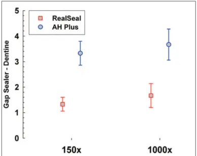
The results are summarized in
Table 1
and Figure 1. The
average gap scores for RealSeal were 1.33 ± 0.27 at 150x
magniication and 1.67 ± 0.47 at 1000x. The gap scores
for AH Plus were 3.33 ± 0.47 at 150x and 3.67 ± 0.61 at
1000x.
Fig. 1. Average scores of the dentin-sealer gaps, considering the
298
Rev. odonto ciênc. 2010;25(3):296-299 Interface dentin-sealerThere were no statistically signiicant differences between
150x and 1000x magniications (
P
=0.15). The sealers
displayed signiicantly different gap sizes, being AH Plus
gaps bigger than RealSeal (
P
=0.002) (Fig. 2 and Fig. 3).
Discussion
Adhesion to root dentin has been subject to several
debatable issues. Leakage studies using different methods
show conlicting results, some unfavorable (12-15) and
some favorable (16-18). Together with the question of the
usefulness of leakage studies, tooth resilience to adapt to the
physiological load thus providing a dynamic status of the
interface dentin-sealer, should be taken into consideration.
Some studies propose
in vivo
models, using experimental
animals (19). The effect over time also comes into the
equation, and yet so far there is no prompt answer as to the
best test model.
This study aimed to look at the interface dentin-sealer
using SEM. Other studies have also used this method (2).
However, the use of pre-deined magniications (150x and
1000x) looking at equidistant areas along the interface and
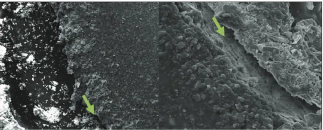
Fig. 2.
Interface dentin-sealer AH Plus. Left: 150x; Right: 1000x (SEM).
Fig. 3.
Interface dentin-sealer RealSeal. Left: 150x; Right: 1000x (SEM).
attributing scores is an innovative aspect of this method.
We believe this provides a less subjective approach to the
observations, which were blinded. Finding the interface
dentin-sealer proved straightforward as the delimitation of
the area of contact allowed an easy localization even in the
absence of gaps.
The increase in magniication, from 150x to 1000x, did
not allow any additional identiication of gaps. It could be
assumed that 150x is good enough to show defects in the
interface dentin-sealer.
RealSeal produced less gaps than AH Plus. There should
be caution as to the clinical relevance of this inding, as the
samples were not subjected to aging or moisture, being done
in ideal conditions. However, it does allow the assumption
that some of the advocated properties of Resilon were
Rev. odonto ciênc. 2010;25(3):296-299
299
Steier et al.
References
Skinner RL, Himel VT. The sealing ability of injection-molded 1.
thermoplasticized gutta-percha with and without the use of sealers. J Endod 1987;13:315-7.
Saleh IM, Ruyter IE, Haapsalo MP, Orstavik D. Adhesion of 2.
endodontic sealers: scanning electron microscopy and energy disperse spectroscopy. J Endod 2003; 29:595-601.
Orstavik D, Eriksen HM, beyer-Olsen EM. Adhesive properties and 3.
leakage of root canal sealers in vitro. Int Endod J 1983;16:59-63. Saunders EM, Saunders WP, Rashid MY. The effect of post space 4.
preparation on the apical seal of root fillings using chemically adhesive materials. Int Endod J 1991;24:51-7.
Shipper G, Trope M. In vitro microbial leakage of endodontically 5.
treated teeth using new and standard obturation techniques. J Endod 2004;30:154-8.
Shipper G, Ørstavik D, Teixeira FB, Trope M. An evaluation of 6.
microbial leakage in roots filled with a thermoplastic synthetic polymer-based root canal filling material (Resilon). J Endod 2004;30:342-7.
Tay FR, Loushine RJ, Weller RN, Kimbrough WF, Pashley DH, Mak 7.
Y-F. Ultrastructural evaluation of the apical seal in roots filled with a polycaprolactone-based root canal filling material. J Endod 2005;31:514-9.
JR, Wedding Brown JJ, CE, Legan Moore K, Vail MM. An in vitro 8.
comparison of microleakage between Resilon and Gutta-Percha with a fluid filtration model. J Endod 2007;33:1447-9.
Paqué F, Sirtes G. Apical sealing ability of Resilon/Epiphany versus 9.
gutta-percha/AH Plus: immediate and 16-months leakage. Int Endod J 2007;40:722-9.
Trope M. Letters to the editor. J Endod 2006;32:85-6. 10.
Tay FR. Reply. J Endod 2006;32:85-6. 11.
Shemesh H, Wu MK, Wesselink PR. Leakage along apical root fillings 12.
with and without smear layer using two different leakage models: a two-month longitudinal ex vivo study. Int Endod 2006;39:968-76. Pitout E, Oberholzer TG, Blignaut E, Molepo J. Coronal leakage 13.
of teeth root-filled with gutta-percha or Resilon root canal filling material. J Endod 2006;32:879-81.
De Deus G, Audi C, Murad C, Fidel S, Fidel RA. Sealing ability of 14.
oval-shaped canals filled using the System B heat source with either gutta-percha or Resilon: an ex vivo study using a polymicrobial leakage model. Oral Surg Oral Med Oral Pathol Oral Radiol Endod 2007;104:e114-9.
De-Deus G, Namen F, Galan J, Jr. Reduced long-term sealing 15.
ability of adhesive root-fillings after water-storage stress. J Endod 2008;34:322-5.
Shin S-J, Jee S-W, Song J-S, Jung I-Y, Cha J-H, Kim E.
16. Comparison
of regrowth of Enterococcus faecalis in dentinal tubules after sealing with gutta-percha or Resilon. J Endod 2008; 34:445-8.
Zmener O, Pameiher CH, Serrano SA, Vidueira M, Macchi RL. 17.
Significance of moist root canal dentin with the use of methacrylate-based endodontic sealers: an in vitro coronal dye leakage study. J Endod 2008;34:76-9.
Wedding JR, Brown CE, Legan JJ, Moore BK, Vail MM. An in vitro 18.
comparison of microleakage between Resilon and gutta-percha with a fluid filtration model. J Endod 2007;33:1447-9.
Kopper PMP, Figueiredo JAP, Della Bona A, Vanni JR, Bier CA, 19.