IN ST IT U T O D E C IÊ N C IA S B IO M ÉD IC A S A B EL S A LA Z A R
Joana
Isabel
Castro
. Determining the Pathogenic Potential of Commensal
and Clinical
Gardnerella
vaginalis
Isolate
s
Determining the Pathogenic Potential of Commensal and Clinica
l
Gardnerella
vaginalis
Isolates
Joana I
sabel R
eis C
astro
D
.ICBAS
2018
CIÊNCIAS BIOMÉDICASDetermining the Pathogenic
Potential of Commensal and Clinical
Gardnerella vaginalis Isolates
Joana Isabel Castro
D
Tese de Candidatura ao grau de Doutor em Ciências Biomédicas submetida ao Instituto de Ciências Biomédicas Abel Salazar da Universidade do Porto.
Orientador – Doutor Nuno Cerca Categoria – Investigador Principal
Afiliação – Centro de Engenharia Biológica, LIBRO – Laboratório de Investigação em Biofilmes Rosário Oliveira, Universidade do Minho, Campus de Gualtar, 4710-057 Braga, Portugal
Coorientador – Doutora Kimberly Kay Jefferson Categoria – Professora Associada
Afiliação – Department of Microbiology and Immunology, Virginia Commonwealth University, Richmond, VA, 23298-0678c, USA
iii
“The important thing is not to stop questioning.
Curiosity has its own reason for existing.”
Albert Einstein
The work presented in this thesis was funded by the Portuguese Foundation for
Science and Technology (FCT) under the scope of the strategic funding of
UID/BIO/04469/2013 unit and COMPETE 2020 (POCI-01-0145-FEDER-006684), the
project
RECI/BBB-EBI/0179/2012
(FCOMP-01-0124-FEDER-027462)
and
BioTecNorte operation (NORTE-01-0145-FEDER-000004) funded by European
Regional Development Fund under the scope of Norte2020 – Programa Operacional
Regional do Norte. JC acknowledges the FCT individual grant with reference
SFRH/BD/93963/2013.
v
Acknowledgments
Um guerreiro da luz nunca esquece a gratidão. Durante a luta, foi ajudado pelos anjos; as forças celestiais colocaram cada coisa em seu lugar, e permitiram que ele pudesse dar o melhor de si. (…) Sua gratidão, porém, não se limita ao mundo espiritual; ele jamais esquece os amigos, porque o sangue deles se misturou ao seu no campo de batalha.
Paulo Coelho, em O Manual do Guerreiro da Luz
Ao meu orientador, Doutor Nuno Cerca, pela orientação, partilha de conhecimentos, por todas as oportunidades, pela confiança e amizade ao longo destes anos.
I would like to acknowledge my co-supervisor, Doctor Kimberly Jefferson, who kindly welcomed me in her lab at the Virginia Commonwealth University and by the guidance, knowledge transfer and assistance in writing reports, papers and also in the thesis.
A todos os meus colegas e amigos do “grupo NC”, pelo bom ambiente de trabalho, pelo apoio e amizade. Um especial agradecimento para a Ângela França, pela colaboração no estudo do transcriptoma da Gardnerella vaginalis (capítulo 5).
Gostaria também de agradecer aos antigos membros do “grupo NC”, em especial à Tatiana Cereja, Patrícia Alves e Cármen Sousa, pelo suporte técnico na caracterização fenotípica dos vários isolados de G. vaginalis (capítulo 3) e à Daniela Machado pela colaboração no trabalho dos biofilmes mistos (capítulo 7).
Não posso deixar de referir o Centro de Engenharia Biológica (que tem sido uma segunda casa nos últimos anos) e todos os que dele fazem parte.
A todos os outros meus amigos e em especial à Célia Fortuna, Lília Marques, Liliana Maia, Madalena Maia, Marino Maciel, Natália Martins, Paulo Silva, e Sara Pimenta pela amizade sincera. Terrie Scherer, I will never forget your kindness and friendship.
Não poderia de deixar de fazer um agradecimento especial aos meus Pais, Irmão e Avós por serem o meu porto de abrigo, pelos valores transmitidos, pelos meios que sempre me proporcionaram, pela força e pela coragem que sempre me incutiram para seguir em frente e realizar este sonho.
E por fim, a ti, Pedro Silva quero agradecer-te por todos os teus gestos de amor, que se refletiram na infinita paciência, compreensão e apoio moral que sempre me mostraste em todos os momentos.
vii
Scientific outputs
Under the terms of the Decree of Law 74/2006, article 34º, point 1, published in Diário da República, 1ª série, nº60, in 24 of March 2006, altereded by the Decree of Law 11/2013, published in Diário da República, 1ª série, nº151, in 7 of August 2013, that proceeds the 3rd
alteration of Decree of Law 74/2006, the author hereby declared that has actively participated in the design and technical execution of the work, interpretation of the results and manuscript preparation of the original articles included in this thesis. Under the terms of the referred Decreto-Lei, the author hereby declared that the following original articles/communications were prepared in the scope of this thesis.
Papers in peer reviewed journals
Castro J, França A, Bradwell KR, Serrano MG, Jefferson KK, Cerca N (2017). Comparative transcriptomic analysis of Gardnerella vaginalis biofilms versus planktonic cultures using RNA-seq. NPJ Biofilms Microbiomes 3:3 (doi: 10.1038/s41522-017-0012-7). Castro J, Machado D, Cerca N (2016). Escherichia coli and Enterococcus faecalis are able to
incorporate and enhance a pre-formed Gardnerella vaginalis biofilm. Pathogens and Disease 74:3 (doi: 10.1093/femspd/ftw007).
Castro J, Cerca N (2015). BV and non-BV associated Gardnerella vaginalis establish similar synergistic interactions with other BV-associated microorganisms in dual-species biofilms. Anaerobe 36:56-9 (doi: 10.1016/j.anaerobe.2015.10.008).
Castro J, Alves P, Sousa C, Cereija TB, França A, Jefferson KK, Cerca N (2015). Using an in-vitro biofilm model to assess the virulence potential of bacterial vaginosis or non-bacterial vaginosis Gardnerella vaginalis isolates. Scientific Reports 5:11640 (doi: 10.1038/srep11640).
Book chapter
Castro J, Machado D, Cerca N (2015), “Gardnerella vaginalis gene expression in biofilms” in Impact of biofilms in health: a transcriptomics perspective (Ed. Cerca N) 227-243, Universidade do Minho – DEB, Braga, Portugal, ISBN: 978-989-97478-6-9.
Oral presentations
Castro J, Cerca N* (2018). The microbiome during bacterial vaginosis: who is cooperating with Gardnerella vaginalis in enhancing crucial gene expression in a dual-species
viii
biofilm model? IUSTI, 2018 Word + European Congress, 27th – 30th June, Dublin, Ireland (*presenting author).
Castro J*, Cerca N (2018). Bacterial cooperation in Bacterial Vaginosis: are social interactions relevant to enhance Gardnerella vaginalis virulence? First Meeting of the PhD in Biomedical Sciences, Instituto de Ciências Biomédicas Abel Salazar, 7th May, Porto, Portugal (*presenting author).
Castro J, Machado D, Cerca N* (2017). Can cooperation within the vaginal microbiome lead to the development of bacterial vaginosis? NZ Microbiological Society (NZMS) Conference, 20th – 23rd November, Auckland, New Zealand (*presenting author). Castro J*, França A, Bradwell KR, Serrano MG, Jefferson KK, Cerca N (2016). What happens
to Gardnerella vaginalis when growing as a biofilm: a comparative transcriptomic analysis by RNA-seq. Biofilms 7 - Microbial Works of Art. No. SPM: 3, ISBN: 978-989-97478-7-6, 26th – 28th June, Porto, Portugal (*presenting author).
Poster presentations
Castro J, Cerca N* (2018). Can the differential response to innate immune components by commensal and clinical Gardnerella vaginalis isolates be the key leading to the development of bacterial vaginosis? IUSTI, 2018 Word + European Congress, 27th – 30th June, Dublin, Ireland (*presenting author).
Castro J*, França A, Bradwell KR, Serrano MG, Jefferson KK, Cerca N (2016). The bacterial vaginosis riddle: how transcriptomics can highlight key physiological adaptations of G. vaginalis when growing as biofilms? The New Microbiology - EMBO|FEBS Lecture course, 24th August – 1st September, Spetses, Greece (*presenting author).
Castro J*, Machado D, Cerca N (2015). Assessing synergistic interaction between Gardnerella vaginalis and other urogenital pathogens. Eurobiofilms 2015: Fourth European Congress on Microbial Biofilms, 23rd – 26th June, Brno, Czech Republic (*presenting author).
ix
Determinação do potencial patogénico de isolados clínicos e comensais
de Gardnerella vaginalis
Sumário
No último meio século, a vaginose bacteriana (VB) tem sido um tema controverso na microbiologia médica. Curiosamente, apesar de todo interesse e investigação no campo da VB, o agente etiológico ainda não foi definitivamente identificado. Os primeiros estudos realizados nesta área sugeriram que o agente infecioso causador da VB era a Gardnerella vaginalis. No entanto, de acordo com dados posteriores descobriu-se que G. vaginalis também estava presente em mulheres saudáveis. Tais descobertas levantaram dúvidas acerca do papel desta bactéria como agente etiológico na VB. Além disso, existem evidências que G. vaginalis não é capaz de causar VB de forma consistente. É de salientar, que outras espécies bacterianas também têm sido, comumente, associadas à VB. Tal facto, levou à postulação da teoria polimicrobiana para o desenvolvimento desta infeção. No entanto, dados epidemiológicos subsequentes também revelaram inconsistências com esta última teoria. Recentemente, surgiram as primeiras descrições dos biofilmes polimicrobianos de VB. Interessantemente, a G. vaginalis parece constituir a maior parte da biomassa desses biofilmes, que apresentam maior tolerância a estímulos externos. Estas descobertas levaram à formulação de uma nova hipótese, que sugere que variantes de estirpes de G. vaginalis com capacidade de induzir um biofilme podem, de facto, ser o agente causador de VB.
Assim, numa tentativa de compreender as diferenças entre estirpes de G. vaginalis isoladas de mulheres com vaginose bacteriana (VB) versus mulheres com flora normal saudável (VB), comparou-se o potencial de virulência de 7 estirpes de G. vaginalis VB e 7 estirpes não-VB. Para esse efeito foram analisadas várias características fenotípicas, nomeadamente: capacidade de formação de biofilme, adesão inicial a células humanas, interações ecológicas com bactérias endógenas com potencial benéfico, atividade citotóxica e adaptação fisiológica ao ambiente vaginal que contém fatores de proteção solúveis, como moléculas responsáveis pela imunidade inata. Notavelmente, os nossos resultados revelaram que as estirpes isoladas de mulheres com VB foram mais virulentas do que as estirpes que colonizaram as mulheres saudáveis. É de notar que apenas as estirpes de G. vaginalis associadas a VB foram capazes de destacar, em grande número, os lactobacilos endógenos previamente aderidos a uma monocamada de células epiteliais. Tais evidências sugerem que este parece ser o fator responsável pelo início do desenvolvimento da VB. No entanto, apesar de todas as diferenças entre os dois grupos de G. vaginalis, os nossos resultados demonstraram que o fator chave
x
na diferenciação de estirpes isoladas de mulheres com VB e de mulheres saudáveis não está relacionado com uma melhor adaptação destas aos componentes imunes do hospedeiro. Posteriormente, numa tentativa de estudar outros fatores de virulência da G. vaginalis, realizou-se uma análise do transcriptoma, por sequenciação do ácido ribonucleico, de uma estirpe proveniente de uma mulher com VB. Com base nesta análise, constatou-se que G. vaginalis altera o seu perfil transcriptómico quando se apresenta sob a forma de biofilme. Este fenótipo corresponde a um estado fisiológico que pode promover a natureza crónica e recorrente da VB. É de salientar, que essas alterações no perfil transcriptómico da G. vaginalis são, provavelmente, importantes para a persistência do biofilme e, consequentemente, para a virulência dessa bactéria.
Por último, analisou-se de que forma outras espécies associadas à VB poderiam influenciar o desenvolvimento do biofilme de G. vaginalis. Para isso, inicialmente, determinou-se se as estirpes de G. vaginalis isoladas de mulheres com VB apresentariam alguma vantagem sobre os isolados não-VB, quando outras espécies bacterianas são associadas a um biofilme pré-estabelecido de G. vaginalis. Os nossos resultados apontaram que a principal diferença no potencial de virulência entre os dois grupos de G. vaginalis parece não estar relacionada com a maturação do biofilme. Posteriormente, foram investigadas as interações ecológicas entre uma estirpe de G. vaginalis isolada de uma mulher com VB e outras espécies bacterianas, também associadas à VB, e que tinham apresentado previamente um sinergismo com um biofilme pré-estabelecido de G. vaginalis. Curiosamente, este estudo revelou que as interações ecológicas foram muito específicas para cada consórcio bacteriano, confirmando que nem todos os colonizadores secundários contribuíram para o aumento da patogénese da VB, com base nos níveis de transcrição de genes de virulência da G. vaginalis. Em suma, este estudo lançou uma nova luz relativamente ao papel de várias espécies bacterianas associadas à VB no desenvolvimento do biofilme de VB, podendo estas modular de forma diferente os fatores de virulência da G. vaginalis. O trabalho desenvolvido nesta tese permitiu retirar novas ilações acerca da virulência de G. vaginalis e da etiologia da VB e pode, em última instância, ajudar a delinear novas estratégias de prevenção da VB, bem como reduzir as taxas de recorrência que lhe estão associadas.
xi
Determining the pathogenic potential of commensal and clinical
Gardnerella vaginalis isolates
Abstract
In the past half century, bacterial vaginosis (BV) has been a controversial topic in medical microbiology because, despite interest and investigation, the etiological agent has not yet been definitively identified. Earlier advances suggested Gardnerella vaginalis as the infectious causative agent of BV but soon after it was found that G. vaginalis was also present in healthy women, and this cast doubts of its role as the etiological agent in BV. Furthermore, G. vaginalis was not able to cause BV consistently. Importantly, other bacterial species started to be commonly associated with BV, and this raises the theory of the multi-species infection. However, subsequent epidemiological data also revealed inconsistencies with this latter theory. Recently, the first descriptions of multi-species biofilm communities were described in BV. Interestingly, G. vaginalis appears to account for most of the biomass of BV biofilms. Further studies demonstrated that G. vaginalis biofilm cells presented higher tolerance to external stresses. These findings derived a new hypothesis, where strain variants of G. vaginalis strains could induce a biofilm and be, in fact, the causative agent of BV, owing to its higher virulence potential.
In an effort to better understand the differences between G. vaginalis isolated from women with bacterial vaginosis (BV) versus normal healthy flora (non-BV), we compared the virulence potential of 7 non-BV and 7 BV associated G. vaginalis isolates by scrutinizing its phenotypic features, namely: biofilm forming-capacity, initial adhesion to human cells, ecological interactions with endogenous beneficial bacteria, cytotoxic activity, and the physiological adaptation to vaginal niche which contains soluble innate immune molecules. Remarkably, our results revealed that strains from BV women were more virulent than strains colonizing healthy women. Notably, we demonstrated that only BV associated G. vaginalis strains were able to dramatically displace pre-coated vaginal protective lactobacilli and we hypothesize this to be a trigger for BV development. However, despite all the differences between both G. vaginalis groups, our results suggested that a better adaptation to the host immune components is not a key factor differentiating between isolates from women with BV and from healthy women. We also conducted a transcriptomic analysis by RNA-sequencing, in which we showed that a BV associated G. vaginalis changes its transcriptomic profile when growing as a biofilm, resulted in a distinct physiologic status that may promote the chronic and recurrent nature of BV. These changes are likely important for biofilm persistence and, consequently, for the
xii
virulence of this bacterium, suggesting that biofilms indeed play a key role in BV development. This phenotype may contribute towards the chronic and recurrent nature of BV.
We also addressed how other BV associated species could be contributing to the development of multi-species biofilms. For that, we first determined whether BV associated G. vaginalis presented any advantage over non-BV isolates in biofilm enhancement by other BV associated bacterial species, using an in vitro dual-species biofilm formation model. However, our findings pointed out that the key difference in virulence potential between the two G. vaginalis groups seems not be related with biofilm maturation. Furthermore, we also investigated the ecological interactions between a BV associated G. vaginalis strain with other BV associated bacteria that had previously indicated synergism with a pre-formed G. vaginalis biofilm. Interestingly, this study revealed that ecological interactions were very specific to each consortium, confirming that not all BV-secondary bacteria are able to enhance the BV pathogenesis by influencing the transcriptomic profile of key virulence genes of G. vaginalis. Finally, our study casts a new light on how BV associated species can modulate the virulence aspects of G. vaginalis, contributing to a better understanding of the development of BV associated biofilms. Together, these new findings about the virulence traits of G. vaginalis and the etiology of BV could ultimately help to shape new strategies for BV prevention and reduction of BV rates.
xiii
Aos meus pais. Ao meu irmão.
E a ti, Pedro.
xv
Table of content
Abbreviation list ... xix
Index of Figures ... xxi
Index of Tables ... xxv
CHAPTER 1 Introduction ... 1
1.1 Background ... 3
1.2 Research questions ... 4
1.3 Hypothesis and aims ... 4
1.3.1 Hypothesis ... 4 1.3.2 Aims ... 4 1.4 Significance ... 6 1.5 Thesis outline ... 6 1.6 References ... 9 CHAPTER 2 Literature review ...11
2.1 The vaginal ecosystem ...13
2.2 Vaginal innate immunity ...14
2.2.1 Defensin ...15
2.2.2 Elafin and secretory leukocyte protease inhibitor ...15
2.2.3 Cathelicidin LL37, lactoferrin, and lysozyme ...15
2.3 Bacterial vaginosis ...16
2.3.1 Clinical features and diagnosis ...17
2.3.2 Epidemiology ...18
2.3.3 Etiology ...20
2.3.4 Anaerobes involved in BV...21
2.3.5 BV associated biofilm as the key step on BV establishment ...22
2.3.6 Treatment of BV ...24
2.4 Gardnerella vaginalis ...26
2.4.1 The dilemma of vaginal colonization by G. vaginalis in healthy women ...27
2.5 References ...36
CHAPTER 3 Using an in-vitro biofilm model to assess the virulence potential of Bacterial Vaginosis or non-Bacterial vaginosis Gardnerella vaginalis isolates ...49
xvi
3.1 Brief introduction ...51
3.2 Material and methods ...51
3.2.1 Subject selection and sample collection ...51
3.2.2 Bacterial isolation and identification ...51
3.2.3 Initial adhesion to epithelial cells and cytotoxicity assays...52
3.2.4 Quantification of biofilm formation ...52
3.2.5 Antibiotic susceptibility ...53
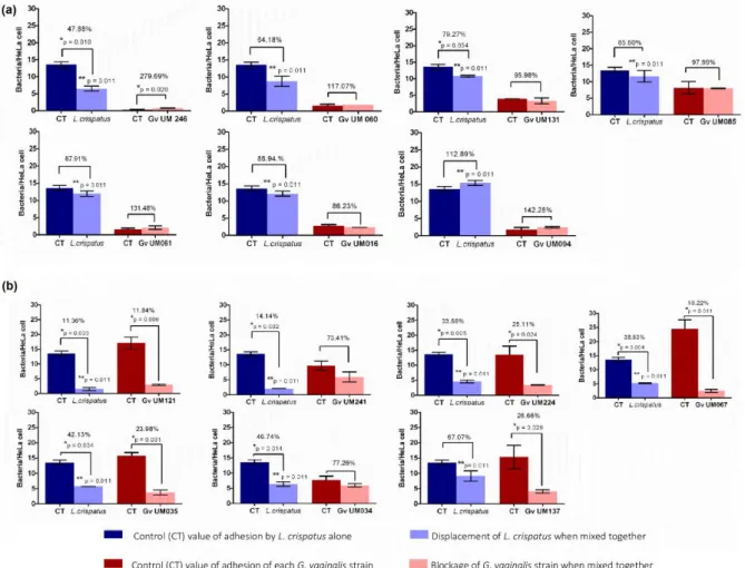
3.2.6 G. vaginalis ability to induce displacement of lactobacilli pre-adhered to epithelial cells ...53
3.2.7 PCR detection of virulence genes ...53
3.2.8 Gene expression quantification...54
3.2.9 Statistical analysis ...55
3.3 Results ...55
3.3.1 Initial adhesion to human cervical HeLa cells and cytotoxic effect ...55
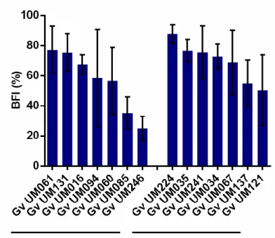
3.3.2 Biofilm formation ...56
3.3.3 Antimicrobial susceptibility ...58
3.3.4 G. vaginalis ability to induce displacement of lactobacilli pre-adhered to epithelial cells ...59
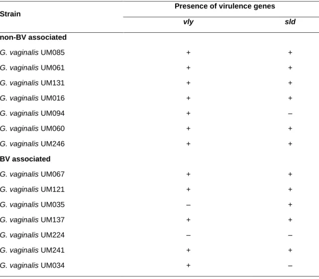
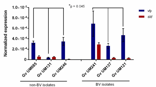
3.3.5 Presence and expression of virulence genes...60
3.4 Discussion ...62
3.5 References ...65
3.6 Supplementary data ...68
CHAPTER 4 Comparative analysis of the influence of host innate immune components in bacterial vaginosis and non-bacterial vaginosis Gardnerella vaginalis isolates ...71
4.1 Brief introduction ...73
4.2 Materials and methods ...74
4.2.1 Bacterial strains and culture conditions ...74
4.2.2 G. vaginalis clade-specific PCR assays ...74
4.2.3 Minimal inhibitory concentration...75
4.2.4 Adhesion assays ...75
4.2.5 Biofilm formation and quantification ...76
4.2.6 Study of growth kinetics ...76
4.2.7 Statistical analysis ...76
4.3 Results ...77
xvii
4.3.2 Susceptibility to antimicrobial peptides ...78
4.3.3 Initial adhesion to human cervical HeLa cells ...79
4.3.4 Influence of innate molecules on biofilm formation...80
4.3.5 Effects of LYS, LF and HBD2 on planktonic growth of G. vaginalis ...81
4.4 Discussion ...82
4.5 References ...84
CHAPTER 5 Comparative transcriptomic analysis of Gardnerella vaginalis biofilms versus planktonic cultures using RNA-seq ...89
5.1 Brief introduction ...91
5.2 Materials and methods ...91
5.2.1 Bacterial strains ...91
5.2.2 Planktonic growth ...91
5.2.3 Biofilm formation ...92
5.2.4 RNA extraction ...92
5.2.5 cDNA library preparation and sequencing ...92
5.2.6 RNA-sequencing data analysis ...93
5.2.7 Biological interactions ...93
5.2.8 Quantitative PCR ...93
5.3 Results ...95
5.3.1 Transcriptome analysis ...95
5.3.2 Enrichment analysis of genes with increased and decreased transcription ...96
5.3.3 Cluster analysis ...97
5.3.4 The top 10 most significantly down or upregulated genes in biofilms ...99
5.3.5 Upregulation of the transcription of potential virulence genes in G. vaginalis biofilms ... 102
5.3.6 Differential expression of vaginolysin in BV associated G. vaginalis biofilms ... 103
5.4 Discussion ... 104
5.5 References ... 105
5.6 Supplementary data ... 108
CHAPTER 6 BV and non-BV associated Gardnerella vaginalis establish similar synergistic interactions with other BV associated microorganisms in dual-species biofilms ... 113
6.1 Brief introduction ... 115
xviii
6.2.1 Bacterial strains and culture conditions ... 115
6.2.2 Dual-species biofilm formation and quantification ... 117
6.2.3 Statistical analysis ... 117
6.3 Results ... 117
6.4 Discussion ... 118
6.5 References ... 120
CHAPTER 7 Unveiling Gardnerella vaginalis role in Bacterial vaginosis (BV) polymicrobial biofilms: the impact of other vaginal pathogens living as neighbors ... 121
7.1 Brief introduction ... 123
7.2 Material and methods ... 123
7.2.1 Bacterial strains and culture conditions ... 123
7.2.2 Coaggregation assays ... 124
7.2.3 Biofilm formation ... 124
7.2.4 PNA FISH hybridization and DAPI staining ... 125
7.2.5 Confocal laser scanning microscopy analysis of biofilm bacterial distribution ... 125
7.2.6 Gene expression quantification... 125
7.2.7 Statistical analysis ... 126
7.3 Results ... 127
7.3.1 Co-aggregation between G. vaginalis & other BV associated isolates ... 127
7.3.2 In vitro PNA Gard162 probe specificity ... 127
7.3.3 Quantification of bacterial populations in dual-species biofilms by PNA FISH ... 128
7.3.4 Analysis of dual-species biofilms by scanning and confocal microscopy ... 131
7.3.5 Expression of critical genes related with G. vaginalis virulence can be altered in dual-species biofilms ... 133
7.4 Discussion ... 136
7.5 References ... 140
CHAPTER 8 Concluding remarks and future work ... 145
8.1 Concluding remarks ... 147
8.2 Study limitations ... 149
8.3 Future perspectives ... 150
xix
Abbreviation list
AMPs – antimicrobial peptides
ARDRA – amplified ribosomal DNA restriction analysis BFI – biofilm formation index
BHI – brain heart infusion bp – base pair
BV – bacterial vaginosis
BVAB – BV associated bacteria cDNA – complementary DNA CFU – colony-forming units
CLSM – confocal laser scanning microscopy CV – crystal violet
DAPI – 4’-6-Diamidino-2-phenylindole DNA – deoxyribonucleic acid
FDR – false discovery rate
FISH – fluorescence in situ hybridization FRT – female reproductive tract
FW – forward GO – gene ontology Gv – Gardnerella vaginalis HBD – human β-defenin HBD2 – human β-defenin 2 HD – human defensin
HNP – human neutrophil peptide
KEEG – Kyoto Encyclopedia of Genes and Genomes LB – luria broth
xx
LF – lactoferrin LYS – lysozyme
mGTS – medium simulating genital tract secretions MIC – minimum inhibitory concentration
MRS – de man-rogosa and sharpe agar OD – optical density
PBS – phosphate buffered saline PCR – polymerase chain reaction
PFGE – Pulsed-field Gel Electrophoresis PNA – peptide nucleic acid
qPCR – quantitative PCR
RAPD – random amplification of polymorphic DNA RNA – ribonucleic acid
RNA-seq – RNA-sequecing
RPKM – reads per kilobase per million RQI – RNA quality indicator
rRNA – ribosomal RNA Rv – reverse
sBHI – supplemented brain heart infusion SD – standard deviation
SEM – standard error of mean
SLPI – secretory leukocyte protease inhibitor
STRING – Search Tool to the Retrieval of Interacting Genes/proteins
TLRs – Toll-like receptors tRNA – transfer RNA TSB – tryptic soy broth
xxi
Index of Figures
CHAPTER 1
Figure 1.1 Thesis outline ... 8
CHAPTER 2
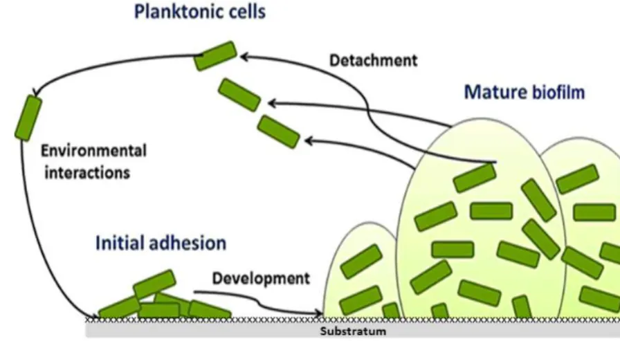
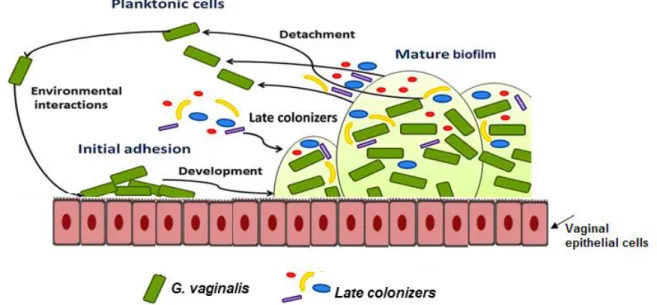
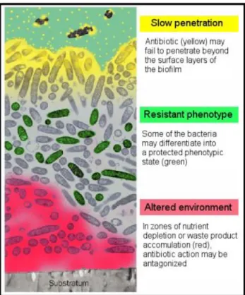
Figure 2.1 Gram-staining vaginal smears illustrate the vaginal microflora ...18 Figure 2.2 Conceptual mono-species model of the biofilm formation ...22 Figure 2.3 Conceptual multi-species model of the BV-biofilm formation ...23 Figure 2.4 Some of the most discussed hypothesis for biofilm resistance to antibiotics ...26 Figure 2.5 Scanning electron micrograph of G. vaginalis ...27 Figure 2.6 Association between data provided by the whole-sequencing genome, clade-specific system and ARDRA genotyping ...33
CHAPTER 3
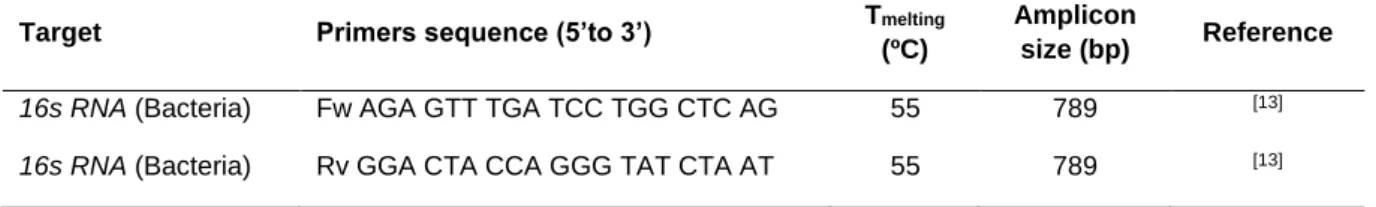
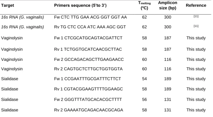
Figure 3.1 Initial adhesion of non-BV and BV G. vaginalis isolates to HeLa cells ...55 Figure 3.2 Cytotoxicity score of non-BV and BV G. vaginalis isolates ...56 Figure 3.3 Intrinsic ability of non-BV and BV G. vaginalis isolates to form biofilms ...58 Figure 3.4 Influence of L. crispatus on G. vaginalis initial adhesion to HeLa cells ...60 Figure 3.5 Expression of vaginolysin (vly) and sialidase (sld) by G. vaginalis isolates ...62 Supplementary Figure S3.1 The absence of vly gene in G. vaginalis UM035 (2) and UM224 (3) strains was confirmed by PCR its flanking regions………...69
CHAPTER 4
Figure 4.1 Comparison of the bacterial adhesion ability to HeLa cells in presence of LYS, LF or HBD2 using 2 different models ...79 Figure 4.2 Initial adhesion of non-BV and BV isolates to HeLa cells in presence of physiological vaginal concentrations of LYS, LF or HBD2 ...80 Figure 4.3 Biofilm formation of non-BV and BV isolates in presence of physiological vaginal concentrations of LYS, LF or HBD2 ...81 Figure 4.4 Bacterial fitness profile of non-BV and BV isolates in the presence of physiological vaginal concentrations of LYS, LF or HBD2 ...82
CHAPTER 5
Figure 5.1 qPCR validation of the transcription of differentially expressed genes randomly selected ...96
xxii
Figure 5.2 KEGG pathways found significantly enriched (p < 0.05) within the genes with increased and decreased transcription in biofilm cells ...97 Figure 5.3 Gene interaction network generated using Cytoscape, showing downregulated transcripts (fold-change ≤ -2) in red and upregulated transcripts (fold-change ≥ 2) in green. 98 Figure 5.4 Clusters generated by the MCODE plugin in Cytoscape ...99 Figure 5.5 Glycolysis/gluconeogenesis pathway of G. vaginalis 409-05 by KEGG Pathway Maps ... 101 Figure 5.6 Quantification of the transcription of known virulence genes in G. vaginalis cultured under biofilm and planktonic conditions ... 103 Figure 5.7 Quantification of thiol-activated cytolysin vaginolysin (vly) transcription in G. vaginalis strains cultured under biofilm or planktonic conditions ... 103
CHAPTER 6
Figure 6.1 Synergistic, antagonistic or neutral interactions detected in dual-species biofilms in relation to single biofilms of non-BV or BV G. vaginalis isolates ... 118
CHAPTER 7
Figure 7.1 Coaggregation score of mono- or dual- bacterial species ... 127 Figure 7.2 Correlation between the PNA FISH counts and the DAPI counts for G. vaginalis at different bacterial concentrations ... 129 Figure 7.3 Biofilm formation profiles for each BV associated species consortium (107 CFU/mL
of BV associated G. vaginalis & 107 CFU/mL of other BV associated bacteria) on dual-species
biofilms ... 130 Figure 7.4 Biofilm formation profiles for each BV associated species consortium (107 CFU/mL
of BV associated G. vaginalis & 105 CFU/mL of other BV associated bacteria) on dual-species
biofilms ... 131 Figure 7.5 An exemplary data set on the organization of the dual-species BV associated biofilm for 48 hours by confocal laser scanning microscopy (CLSM) ... 132 Figure 7.6 Schematic representation of the distribution of dual-species BV associated biofilm structure from the bottom to the biofilm top ... 133 Figure 7.7 Quantification of the transcription of virulence genes, related to cytotoxicity, exfoliation of vaginal epithelium or biofilm formation, in G. vaginalis cultured under dual and mono-species biofilms ... 134 Figure 7.8 Quantification of the transcription of virulence genes, related to antimicrobial resistance, in G. vaginalis cultured under dual and mono-species biofilms ... 135 Figure 7.9 Quantification of the transcription of virulence genes, related to evasion of immune response, in G. vaginalis cultured under dual and mono-species biofilms ... 136
xxiii Figure 7.10 Hypothetical model of G. vaginalis vaginolysin (vly)-mediated cytotoxicity in different bacterial phenotypes... 138 Figure 7.11 Venn diagram summarize the transcript levels of all six potential virulence genes of G. vaginalis among 15 bacterial consortia ... 139
CHAPTER 8
xxv
Index of Tables
CHAPTER 2
Table 2.1 Scheme for grading Gram-stained vaginal contents ...17 Table 2.2 Koch’s postulates ...20 Table 2.3 Regimens for BV treatment ...24 Table 2.4 Studies of G. vaginalis differentiation using biotyping, genotyping or ecotyping approaches ...28 Table 2.5 In vitro studies of functional virulence properties of G. vaginalis strains isolated from women with BV versus women without BV ...35
CHAPTER 3
Table 3.1 Primer sequences used for by partial sequencing of 16S rRNA...52 Table 3.2 Primer sequences used for PCR and qPCR assays ...54 Table 3.3 Qualitative analysisa of biofilm formed by G. vaginalis strains in 9 different media 57
Table 3.4 Minimum inhibitory concentration (MIC) of metronidazole, tinidazole and clindamycin for planktonic cells of G. vaginalis isolates...59 Table 3.5 Detection by PCR of the vly and sld genes in G. vaginalis isolates ...61 Supplementary Table S3.1 Characterization of vaginal samples ...68 Supplementary Table S3.2 Accession code of G. vaginalis strains...68
CHAPTER 4
Table 4.1 Primer sequences used for the detection of the clade-specific group of G. vaginalis strains ...74 Table 4.2 G. vaginalis genotyping based on clade classification system ...77 Table 4.3 Minimum inhibitory concentration (MIC) of lysozyme (LYS), lactoferrin (LF), and human β-defensin 2 (HBD2) for planktonic cells of G. vaginalis isolates ...78
CHAPTER 5
Table 5.1 Primers used in qPCR experiments ...94 Table 5.2 List of the 10 genes with lowest and highest fold-change values among the differentially expressed genes in G. vaginalis cultured under biofilm versus planktonic conditions ... 100 Supplementary Table 5.1 List of genes uniquely expressed in G. vaginalis cultured under planktonic or biofilm conditions and their known functions………..108
xxvi
Supplementary Table 5.2. Differentially expressed genes encoding hypothetical proteins with significant pfam domain, including predicted localization by PSORTb and pBLAST results ... …..109
CHAPTER 6
Table 6.1 GenBank accession numbers of strains used in this study ... 116
CHAPTER 7
Table 7.1 Primers used in qPCR experiments ... 126 Table 7.2 Bacterial species used in PNA-FISH assays and their specificity with PNA Gard162 probe ... 128
CHAPTER 1
Introduction
SummaryThis chapter provides a brief outline of the thesis. The background, research questions, hypothesis, aims, and significance are presented here.
Chapter 1 • 3
1.1 Background
Bacterial vaginosis (BV), characterized by a shift of the vaginal microbiota from a Lactobacillus-dominated community to a dense biofilm containing a complex mixture of microorganisms, is an important risk factor in poor reproductive health. The high prevalence, high relapse rate, and associated complications make this disorder of paramount global importance [1,2]. Therefore, control of BV has been advocated for decreasing the prevalence of these complications, but the precise etiology remains unknown [3]. As a result, current treatment regimens and prevention strategies are inadequate. Such a lack of understanding not only inhibits our ability to effectively manage BV but also severely affects our ability to prevent its associated complications [4].
Microbiological analysis of BV has shown Gardnerella vaginalis to be the most frequent microorganism in BV, being isolated in more than 95% of cases [2]. However, there has been much debate in the literature concerning the contribution of G. vaginalis to the etiology of BV, since it is also present in a considerable proportion of healthy women [5,6]. The research group of Dr. Nuno Cerca has been involved in determining the differences between G. vaginalis and other vaginal isolates in order to explain the outcome of colonization [7-9]. A recent study [7] clearly demonstrated that G. vaginalis may be more suited as an early colonizer relative to the others BV associated anaerobes tested in the initial adhesion and that it may play a key role in the early establishment of BV biofilms. Of high importance, a study led by Dr. Kimberly Jefferson demonstrated that a non-BV isolate had fundamental genomic differences, as compared with the genome of a BV isolate of G. vaginalis [10]. This lead to the hypothesis that non-virulent G. vaginalis strains could occur in healthy women, while virulent strains could cause BV.
This study was designed to determine the presence of putative virulence markers in G. vaginalis strains isolated from Portuguese women with BV (n = 7) or without BV (n = 7). Comparison of the two sets of isolates is expected to reveal factors that may assist in the diagnosis of BV. Furthermore, we also set out to study the ecological interactions of G. vaginalis and other BV associated bacteria to understand the impact of the bacterial cooperation on G. vaginalis virulence. Together, this thesis attempts to advance our understanding of the mystery of BV pathogenesis, since this is essential to make progress in the control and prevention of this common, important condition.
4 • Chapter 1
1.2 Research questions
The following questions will be addressed in this thesis:
1. Can BV associated G. vaginalis isolates exhibit more virulence factors than non-BV isolates?
2. Can the differential response to innate immune components by non-BV and BV associated G. vaginalis isolates be key in BV development?
3. What happens to the G. vaginalis virulence profile when it is growing as a biofilm?
4. Do other BV associated species cooperate with G. vaginalis and enhance its virulence?
Answers to these research questions will provide new knowledge regarding the etiology of BV and might contribute to the design of improved treatment strategies.
1.3 Hypothesis and aims
1.3.1 Hypothesis
G. vaginalis can colonize the vaginal epithelium of both women with and without BV. This investigation tested the following hypothesis:
Clinical G. vaginalis strains are able to cause BV owing to phenotypic and genotypic adaptations that provide an ecological niche advantage over non-BV G. vaginalis.
1.3.2 Aims
In an effort to better understand the differences between commensal and clinical G. vaginalis isolates, in vitro assays will be performed in order to compare virulence properties of G. vaginalis strains isolated from Portuguese women with and without BV. This will be approached using the following sub-aims.
Chapter 1 • 5
Aim 1: To assess the possible differences in the phenotype and genotype of non-BV and BV associated G. vaginalis strains.
a) To analyse the initial adhesion of non-BV and BV associated G. vaginalis strains to a monolayer of epithelial cells and to analyse their cytotoxic effects.
b) To compare the biofilm-forming capacity between both G. vaginalis groups.
c) To investigate the antimicrobial susceptibility profile of both G. vaginalis groups and their ability to displace beneficial endogenous bacteria from the epithelial cells.
d) To analyse the expression of virulence-related genes.
e) To identify the subgroups of non-BV and BV associated G. vaginalis strains according to a clade-specific genotyping system.
Aim 2: To identify the possible differences in the physiological adaptation of non-BV and BV associated G. vaginalis strains to the innate immune system. To achieve this, we will perform a series of in vitro assays to evaluate the antimicrobial susceptibility of both G. vaginalis groups to innate molecules (lysozyme, lactoferrin, human β defensin-2); and its initial adhesion ability, the biofilm-forming capacity, as well as the planktonic growth in the presence of physiological vaginal concentrations of the innate molecules.
Aim 3: To gain insight into the role of G. vaginalis biofilms in the pathogenesis of BV, we will carry out a comparative transcriptomic analysis between planktonic and biofilm cultures, using RNA-sequencing.
Aim 4: To investigate the ecological interactions between non-BV or BV associated G. vaginalis strains with other BV associated bacterial species, using a dual-species biofilm assembly consisting of G. vaginalis and secondary BV associated species.
a) To assemble pairwise combinations between non-BV or BV associated G. vaginalis isolates and 24 other BV associated bacteria, and compare the synergistic, neutral or antagonistic interactions between the two bacteria through the quantification of total biofilm biomass.
b) To discriminate the bacterial populations of dual-species biofilms using a validated peptide nucleic acid fluorescence in situ hybridization approach. Herein and in the following points, only BV associated bacteria which present a synergistic interaction with a pre-formed G. vaginalis biofilm will be analysed.
6 • Chapter 1
c) To analyse the dual-species biofilms structures by confocal laser scanning microscopy. d) To investigate the impact of the second BV associated species on G. vaginalis pathogenicity, by analyzing the expression of genes related to cytotoxicity, biofilm formation, antimicrobial resistance and evasion of the immune system in cells from mono- and dual-species biofilms.
e) To analyse the bacterial coaggregation ability between G. vaginalis and other BV associated species.
1.4 Significance
The research question to be addressed by this thesis was the dilemma of G. vaginalis vaginal colonization in both healthy and BV women. It is noteworthy that a hallmark feature of BV is the presence of a highly structured polymicrobial biofilm primarily consisting of G. vaginalis, strongly adhered to vaginal epithelium, and a variety of other bacteria. Thus, it is essential unveiling whether non-BV and BV G. vaginalis strains interact differently with both the host, and with other BV associated bacteria to shed a new light on the development of BV. This could represent a significant advancement towards the characterization of ecological interactions and virulence factors that contribute to symptoms of BV. Furthermore, this thesis could lead to new insights into the interaction between both G. vaginalis groups and beneficial endogenous bacteria. Together, the findings on G. vaginalis virulence traits and BV etiology could lead to new strategies to BV prevention and consequently reduction of BV rates.
1.5 Thesis outline
Chapter 2 presents a literature review, providing a general outline of major aspects of BV, carefully emphasizing the composition of healthy and BV associated microflora. Furthermore, the BV associated biofilm development will be focused as the key step on BV establishment. Lastly, special emphasis will be also given to the dilemma of vaginal colonization of G. vaginalis in healthy women.
Chapters 3, 4, 5, 6, and 7 are the experiment chapters and address the four aims of this thesis. Each of the experiment chapters stands alone, providing a summary, brief introduction, materials and methods, results, and discussion.
Chapter 1 • 7
Chapter 3 is focused on the examination of phenotypic and genotypic characteristics of 14 G. vaginalis strains isolated from women with and without BV (7 of each group). G. vaginalis bacteria were isolated from Portuguese women and identified by partial sequencing of 16S rRNA coding gene and screened for a small panel of putative virulence factors. The isolates underwent analysis for initial adhesion, biofilm formation, antimicrobial susceptibility profile, presence and expression of potential virulence related-genes and the capacity of G. vaginalis to displace beneficial lactobacilli.
Chapter 4 shows how commensal and clinical isolates are adapted to the innate immune system, evaluating the initial adhesion, the biofilm-forming capacity and the bacterial fitness in presence of physiological vaginal concentrations of the innate molecules. Furthermore, this chapter provides genotyping information of the 14 G. vaginalis strains, based on a recent clade-specific system.
Chapter 5 addresses the third aim of this thesis, comparing the transcriptomic profile of G. vaginalis cultured under planktonic and biofilm conditions by RNA-sequencing. This chapter provides data regarding the upregulation of the transcription of potential virulence genes in G. vaginalis biofilms.
Chapter 6 presents the ecological interactions between non-BV or BV associated G. vaginalis strains and 24 other BV associated isolates, using an in vitro dual-species biofilm model. Chapter 7 is focused on deciphering the impact of other BV associated species on BV associated G. vaginalis virulence profile. After the first screening of ecological interactions (chapter 6), we set out to better analyse the cases of synergistic interactions between G. vaginalis and other BV associated bacterial species using the same in vitro dual-species biofilm model.
Finally, Chapter 8 presents a summary of the thesis findings, major outcomes, the significance of the findings, limitations and future directions.
8 • Chapter 1
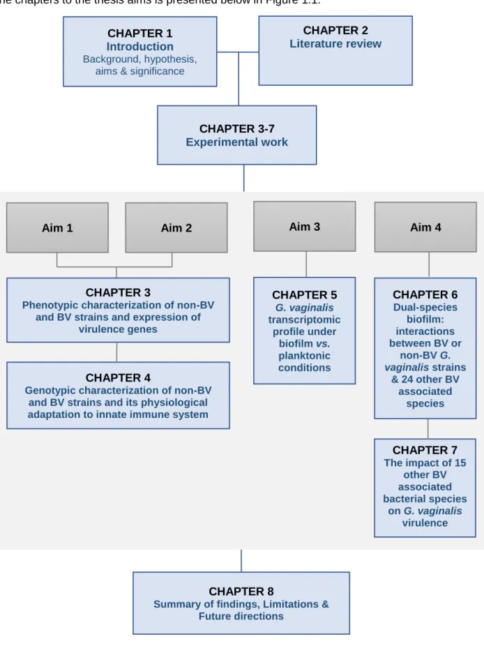
A schematic diagram which shows the general layout of this monograph and relationships of the chapters to the thesis aims is presented below in Figure 1.1.
Aim 1 Aim 2 Aim 3 Aim 4
CHAPTER 1 Introduction
Background, hypothesis, aims & significance
CHAPTER 2 Literature review
CHAPTER 3-7 Experimental work
CHAPTER 4
Genotypic characterization of non-BV and BV strains and its physiological adaptation to innate immune system
CHAPTER 5 G. vaginalis transcriptomic profile under biofilm vs. planktonic conditions CHAPTER 6 Dual-species biofilm: interactions between BV or non-BV G. vaginalis strains & 24 other BV associated species CHAPTER 7 The impact of 15 other BV associated bacterial species on G. vaginalis virulence CHAPTER 8
Summary of findings, Limitations & Future directions
CHAPTER 3
Phenotypic characterization of non-BV and BV strains and expression of
virulence genes
Chapter 1 • 9
1.6 References
[1] Bradshaw, C.S. et al. (2006). High recurrence rates of bacterial vaginosis over the course of 12 months after oral metronidazole therapy and factors associated with recurrence. J Infect Dis 193, 1478-86.
[2] Bagnall, P. and Rizzolo, D. (2017). Bacterial vaginosis: A practical review. Jaapa 30, 15-21. [3] Pirotta, M., Fethers, K.A. and Bradshaw, C.S. (2009). Bacterial vaginosis - More questions than
answers. Aust Fam Physician 38, 394-7.
[4] Schwebke, J.R., Muzny, C.A. and Josey, W.E. (2014). Role of Gardnerella vaginalis in the pathogenesis of bacterial vaginosis: a conceptual model. J Infect Dis 210, 338-43.
[5] Aroutcheva, A., Simoes, J., Behbakht, K. and Faro, S. (2001). Gardnerella vaginalis isolated from patients with bacterial vaginosis and from patients with healthy vaginal ecosystems. Clin Infect
Dis 33, 1022-27.
[6] Janulaitiene, M., Paliulyte, V., Grinceviciene, S., Zakareviciene, J., Vladisauskiene, A., Marcinkute, A. and Pleckaityte, M. (2017). Prevalence and distribution of Gardnerella vaginalis subgroups in women with and without bacterial vaginosis. BMC Infect Dis 17, 394.
[7] Machado, A., Jefferson, K.K. and Cerca, N. (2013). Interactions between Lactobacillus crispatus and bacterial vaginosis (BV)-associated bacterial species in initial attachment and biofilm formation. Int J Mol Sci 14, 12004-12.
[8] Alves, P., Castro, J., Sousa, C., Cereija, T.B. and Cerca, N. (2014). Gardnerella vaginalis outcompetes 29 other bacterial species isolated from patients with bacterial vaginosis, using in an in vitro biofilm formation model. J Infect Dis 210, 593-6.
[9] Machado, A., Salgueiro, D., Harwich, M., Jefferson, K.K. and Cerca, N. (2013). Quantitative analysis of initial adhesion of bacterial vaginosis-associated anaerobes to ME-180 cells.
Anaerobe 23,1-4.
[10] Harwich, M., Alves, J., Buck, G., Strauss, J., Patterson, J., Oki, A., Girerd, P. and Jefferson, K.
(2010). Drawing the line between commensal and pathogenic Gardnerella vaginalis through
CHAPTER 2
Literature review
SummaryThis chapter provides a general outline of major aspects of BV, carefully emphasizing the current hypothesis of the pathogenesis of BV. Furthermore, a special importance will be also given to the dilemma of vaginal colonization of G. vaginalis in healthy women.
Chapter 2 • 13
2.1 The vaginal ecosystem
The female vaginal environment is a complex and dynamic nutrient-rich milieu for microorganisms resulting in a unique microbiome [1]. The composition of the vaginal ecosystem is not static: fluctuations in relative and absolute amounts of microbial species can occur over time due to several factors including, hormonal changes [2], sexual activity [3], hygienic practices [4], and underlying health conditions [5].
Since the first microbiological study of the human vagina, published by Döderlein in 1892 [6], the vaginal microflora of healthy premenopausal women has been described as constituted predominantly by Gram-positive bacilli of the genus Lactobacillus. Traditionally, lactobacilli colonization is believed to be beneficial since it prevents other microorganisms from colonizing the vaginal tract, using several protective mechanisms [7,8]. Firstly, the majority of Lactobacillus species produce lactic acid, which contributes to the maintenance of the vaginal pH below 4.5 [9,10]. This acidic environment constitutes an efficient mechanism of protection of the vaginal epithelium since it makes the environment inhospitable to other bacteria, including pathogens [11,12]. Secondly, Lactobacillus species are also known to produce other antimicrobial compounds, including hydrogen peroxide [13,14] and target-specific bacteriocins [15,16]. Despite some studies have demonstrated that hydrogen peroxide could inhibit the colonization of pathogenic bacteria [17,18], it was shown that under normal physiological concentration no detectable effect was observed in 17 vaginal pathogens under anaerobic growth conditions [19]. The vagina is virtually an anaerobic environment wherein dissolved oxygen levels are low. Therefore, it is unlikely that significant amounts of hydrogen peroxide are produced and accumulate to a toxic level to preventing the colonization of bacteria [19]. Regarding bacteriocins, their antimicrobial activity is usually based on the permeabilization of the target membrane [20]. Thus, in the vagina, bacteriocins could play a significant role in fending off non-indigenous bacteria or pathogenic microorganisms [21,22]. In addition, vaginal lactobacilli competitively block the adhesion of pathogenic bacteria to vaginal epithelial cells [23,24].
Remarkably, advances in culture-independent approaches, such as high-throughput 16S rRNA gene sequencing, have generated renewed knowledge in the composition and abundance of vaginal bacterial species in asymptomatic reproductive-age women, showing at least five major types of vaginal microflora, known as community state types. Four of these community state types are dominated by L. crispatus, L. iners, L. gasseri and L. jensenii, and one does not contain a significant number of lactobacilli (20 – 30% of the cases) but is composed of a diverse array of facultative and strictly anaerobic microorganisms, including Atopobium, Corynebacterium, Anaerococcus, Peptoniphilus, Prevotella, Gardnerella,
14 • Chapter 2
Sneathia, Eggerthella, Mobiluncus and Finegoldia among others [1,25]. Interestingly, these differences between community state types appear to be driven by a combination of cultural, behavioural, genetic and other uncharacterized underlying factors [1,25,26]. Overall, these findings challenged the wisdom that the occurrence of the high number of lactobacilli is synonymous with “normal” or “healthy”.
While knowledge accumulated over the past few decades has provided some insights into the vaginal ecosystem, there remains a need to define and better understand factors that affect the composition and dynamics of vaginal microbiota in both health and diseases. This knowledge will facilitate the development of new strategies for disease diagnosis and personalized treatments to promote health and improve the quality of women’s lives [25].
2.2 Vaginal innate immunity
In addition to the protective effects of the beneficial endogenous vaginal microflora, the colonization of pathogenic microorganisms in the female reproductive tract (FRT) is prevented by local components of the innate and adaptive immune systems. The innate immune system constitutes the first line of response to infection and, for this reason, it has a pivotal role in the host. In the FRT, the innate immune system consists of mechanical, chemical, and cellular components. The mucus lining and epithelial cells act as a mechanical barrier. The chemical barrier can be divided into natural antimicrobial peptides (AMPs) and pattern recognition receptors, especially Toll-like receptors (TLRs) [27,28]. TLRs recognize conserved pathogen-associated molecular patterns synthesized by microorganisms including bacteria, fungi, parasites, and viruses as well as endogenous ligands associated with cell damage. Specifically, the vaginal epithelium expresses TLR2 and its partners TLR1 and TLR6, which in combination (TLR1/2 and TLR2/6), recognize lipopeptides present on both Gram-positive and Gram-negative bacteria; TLR4, which recognizes lipopolysaccharide of Gram-negative bacteria; and TLR5, which recognizes flagellin, a component of the flagellum responsible for bacterial motility. Therefore, it has been thought that the expression of TLRs on the epithelium plays an important role in antigen detection, initiation of the immune response and in the connection between innate and adaptive immunity [29].
Importantly, the synthesis of AMPs has commonly emerged as the most ancient primary mechanism of the immune system [30]. AMPs possess additional functions apart from microbicidal activity, including cell proliferation, cytokine induction, chemotaxis, and modulation of innate and adaptive immunity [27]. Major AMPs with different structural and
Chapter 2 • 15
functional characteristics include defensin, elafin, cathelicidin, secretory leukocyte protease inhibitor (SLPI), lysozyme, and lactoferrin. These factors are briefly described below:
2.2.1 Defensin
Defensins are small cationic peptides consisting of 30 – 42 amino acids and have molecular weights between 3.5 – 4.5 kDa. They are subdivided into α and β-defensins. Six α-defensins have been recognized in humans: human neutrophil peptide (HNP) 1 – 4 and human defensin 5 and 6 and are produced by neutrophil granulocytes [27]. Also, six human β-defensins, HBD1 to 6 have been identified, which are structurally similar to α-defensins. Four of them are expressed by mucosa and epithelial cells of the FRT [31-33]. Importantly, the permeabilization of target membranes is the crucial step in defensin-mediated antimicrobial activity and cytotoxicity. In bacteria, permeabilization leads to inhibition of RNA, DNA, and protein synthesis ultimately, bacterial cell death [33].
2.2.2 Elafin and secretory leukocyte protease inhibitor
Elafin and SLPI are two low-molecular-mass elastases inhibitors that are mainly synthesized by macrophages and epithelial cells [34]. It is thought that their physiological properties allow them to efficiently inhibit target enzymes, such as neutrophil elastase [35,36]. This proteolytic enzyme is capable of degrading elastin, which provides elasticity and resilience to tissues [37,38]. So, the main function of SLPI is to protect local tissue against the detrimental consequences of inflammation [37]. In addition to their antiprotease activity, both elafin and SLPI have a broad range of antibacterial activity against Gram-positive and Gram-negative species [39-41]. The antimicrobial activity is mediated via their cationic charge, which, like many cationic antimicrobial proteins, allows them to destabilize bacterial membranes [42]. It is also important to note that these proteases inhibitors are found in vaginal secretions [43,44].
2.2.3 Cathelicidin LL37, lactoferrin, and lysozyme
Another component of the FRT secretions is cathelicidin, which was named according to its ability to inhibit the protease cathepsin-L, a lysosomal endoprotease [45]. In humans, LL37 is the only cathelicidin, and it is produced by neutrophils and epithelial cells of the lower FRT [46]. It is found in vaginal fluid and cervical mucus [30,31]. Similarly, lactoferrin, an iron-binding cationic glycoprotein, is also produced by neutrophils and epithelial secretions. Lactoferrin is both anti-viral and anti-bacterial, and it effects can occur by sequestration of iron essential for microbes under acidic conditions, such as lower part of the FRT [47,48]. Furthermore, lactoferrin can also prevent the entry of bacteria or virus into the host cells in the early phase
16 • Chapter 2
of infection, either by blocking cellular receptors or by direct binding to the bacterial adhesins or virus particles [49,50]. Regarding lysozyme, it is synthesized by neutrophils, monocytes and macrophages [51]. In addition to the enzymatic lysis of peptidoglycan present on bacterial cell walls leading to the rapid killing of Gram-positive bacteria [52,53], lysozyme can also kill bacteria by a non-enzymatic mechanism, owing to its highly cationic nature, through the formation of pores on the bacterial cell membrane [54,55]. Furthermore, it blocks the human immunodeficiency virus-1 viral entry and its replication [56]. Interestingly, lysozyme displays synergism with lactoferrin, which promotes innate immune protection in the FRT [57]. Notably, endogenous AMPs can act synergistically, resulting in enhancement of their antimicrobial properties [58].
2.3 Bacterial vaginosis
Worldwide, BV is the most common gynaecological disorder among women of childbearing age, affecting ~ 29% of women in the general population and 50% of African American women [59]. Microbiologically, BV is characterized by a dramatic shift in the vaginal microflora from the dominant lactic acid and H2O2-producing lactobacilli to a polymicrobial flora, consisting of
strictly and facultatively anaerobic bacteria, where G. vaginalis plays a pivotal role [60]. Importantly, the loss of lactobacilli may be a consequence of the changes in vaginal microflora rather than to be a cause of BV, as the anaerobic vaginal environment of BV is not conducive to the lactobacilli dominance [60,61]. The hypothesis of the depletion of lactobacilli as the cause of BV has not been supported by the fact that some women maintain a “healthy” vaginal environment without lactobacilli [62]. Curiously, some strains of Atopobium spp., Leptotrichia spp. and Megasphaera spp. are reportedly capable of producing lactic acid. Therefore, the presence of non-lactobacilli vaginal microbiota and the lack of beneficial lactobacilli may not necessarily be sufficient to cause BV [3,63].
In the last years, BV has emerged as a global issue of concern due to its association with a wide array of adverse outcomes. It has been reported that BV significantly increases the risk of development of gynaecological postoperative infections [64], pelvic inflammatory disease [65], urinary tract infections [66] and infertility [67]. Moreover, BV has been also associated with adverse pregnancy outcomes such as miscarriage and recurrent pregnancy losses [68]; preterm delivery and low birth weight [69]; and increased neonatal morbidity [70]. Furthermore, BV facilitates the transmission of sexually transmitted agents including the human immunodeficiency virus [71], human papillomavirus [72], Neisseria gonorrhoeae and Chlamydia trachomatis [73].
Chapter 2 • 17
2.3.1 Clinical features and diagnosis
Over the last three decades, there has been a fascinating evolution in our understanding of BV. Causing profuse vaginal discharge and fishy vaginal odour in symptomatic women, BV has been also recognized as being asymptomatic in approximately one-half of the women who experience it [74-76]. The abnormal vaginal discharge results in part from degradation of the protective vaginal mucin gel, which is performed by mucin-degrading enzymes produced by BV associated bacteria [77]. The fishy odour is due to the volatilization of amines produced as the result of the metabolism of anaerobic bacteria [78]. In clinical settings, BV is commonly diagnosed using the Amsel criteria, which include the presence of at least three of the following precepts: (i) thin and homogenous discharge, (ii) vaginal pH over 4.5, (iii) positive “whiff test” (detection of fishy odour through the addition of 10% potassium hydroxide to vaginal fluid), (iv) and presence of clue cells on microscopic examination of vaginal fluid [79]. However, these clinical signs are not always present, making Amsel criteria somewhat subjective [80].
In an attempt to improve the accuracy in BV diagnosis, Nugent and colleagues proposed a Gram stain scoring system for examining vaginal smears [81]. This method derived from the modification of the Gram-stained protocol proposed by Spiegel et al. [82] and currently it is regarded as the gold standard for BV diagnosis. According to Nugent criteria, Gram-stained smears are used for identification, classification, and quantification of the following bacterial morphotypes: large Gram-positive bacilli (Lactobacillus spp.); small Gram-variable rods (Gardnerella and Bacteroides spp.); and curved Gram-variable rods (Mobiluncus spp.), as presented in Table 2.1.
Table 2.1 Scheme for grading Gram-stained vaginal contents Score Lactobacillus
Morphotypes
Gardnerella and Bacteroides
spp. Morphotypes Curved Gram-Variable Rods
0 4+ 0 0
1 3+ 1+ 1+ or 2+
2 2+ 2+ 3+ or 4+
3 1+ 3+
4 0 4+
Vaginal microflora diagnosis by Nugent score system
Total score a Interpretation
0 – 3 Normal vaginal microflora
4 – 6 Intermediate vaginal microflora
7 – 10 Bacterial vaginosis in vaginal microflora
a Morphotypes are scored as the average number see per oil immersion field. Quantification of each individual score: 0 for no morphotype present; 1+ for 1 morphotype present; 2+, 1 to 4 morphotypes present; 3+, 5 to 30 morphotypes present; 4+, 30 or more morphotypes present. The total score is the sum of the average classification of Lactobacillus, Gardnerella and Bacteroides, and finally Mobiluncus spp. Adapted from Nugent and colleagues [81].
18 • Chapter 2
Each morphotype is scored from 0 to 4+, regarding the number of morphotypes observed per oil immersion field. The total score is obtained by adding each individual morphotype score, ranging between 0 – 10. Thus, a score of 0 – 3 is considered normal vaginal microflora, 4 – 6 as intermediate microflora and 7 – 10 as BV (Figure 2.1). Nevertheless, Nugent score system has also some disadvantages, especially related to the inter-observer variability and it requires skilled personnel to perform it.
Figure 2.1 Gram-staining vaginal smears illustrate the vaginal microflora. (a) Normal vaginal
epithelial cells. (b) Intermediate vaginal microflora. (c) BV associated microflora, showing a vaginal clue cell, which corresponds to vaginal squamous epithelial cells coated with the G. vaginalis and other anaerobic bacteria. Adapted from Nugent and colleagues [81].
Importantly, the relationship between Gram stain score and diagnosis by the clinical criteria is imperfect. Gram stain is more sensitive, whereas the Amsel criteria can be more specific. Overall the concordance between them is of ~80% to 90% [83]. These shortcomings of standard methods make BV diagnosis a challenging task, and, therefore, alternative methods for BV diagnosis have been investigated. The molecular methodologies, such as polymerase chain reaction (PCR) [84], quantitative PCR (qPCR) [85] or fluorescence in situ hybridization (FISH) [86], have allowed the detection or even quantification of the main BV associated bacteria. In fact, they have improved our knowledge of how microbial species interact among themselves and with the human host. However, most of these alternative methods are expensive and many of them still require validation. Until then, the Amsel and Nugent criteria remain the most commonly used methods for BV diagnosis [87].
2.3.2 Epidemiology
BV status has been referred to as “one of the most prevalent enigmas in the field of medicine” [88]. Despite the high clinical importance of BV, its real prevalence is unknown since it varies according to the characteristics of the studied population [59,89]. Epidemiological studies indicated that the risk factors of BV include (i) the concurrent use of medications [76], (ii) low socioeconomic status [90], (iii) increasing age [91], (iv) cigarette smoking [92], (v) young age