The Brazilian Journal of
INFECTIOUS DISEASES
w w w . e l s e v i e r . c o m / l o c a t e / b j i d
Original article
Gardnerella vaginalis
-associated bacterial vaginosis in
Bulgarian women
Raina T. Gergova
a,b,∗, Tanya V. Strateva
a, Ivan G. Mitov
aaDepartment of Medical Microbiology, Faculty of Medicine, Medical University of Sofia, Bulgaria
bDiagnostic Medical Laboratory Medirs, Sofia, Bulgaria
a r t i c l e
i n f o
Article history:
Received 12 August 2012 Accepted 13 October 2012 Available online 19 April 2013
Keywords:
Bacterial vaginosis
Gardnerella vaginalis
Bulgarian women
a b s t r a c t
Background:Bacterial vaginosis (BV) is the most common cause of vaginal discharge in women of reproductive age. The purpose of this study was to determine the frequency of BV in Bulgarian pregnant and nonpregnant women from several age ranges and to compare three different laboratory methods forGardnerella vaginalisdetection in patents suffering from BV.
Methods:Between September 2011 and June 2012, 809 women of 16–40 years of age separated in two major groups: nonpregnant – 469 (355 with and 114 without symptoms) and pregnant – 340 (213 and 127 respectively) were enrolled for the study. The women underwent three different laboratory tests simultaneously: scoring of Gram staining of vaginal smear, culture, and polymerase chain reaction (PCR) assay forG. vaginalis.
Results:The microscopic method detected high frequency of BV in symptomatic (57%) whereas only a minority of asymptomatic subjects (14%) were detected. G. vaginalis -associated BV was diagnosed in approximately equal proportions when evaluated with PCR and microscopic method for both pregnant and nonpregnant women. The comparative anal-ysis of microscopic evaluation, culture and PCR assays demonstrated greater concurrence (about 90%) between Gram staining and PCR detection for BV, than both methods com-pared to culture. The combination of microscopy and PCR turned out to be very reliable and repeatable for detectingG. vaginalis-associated BV.
Conclusions: This is the first comparative investigation on the epidemiology ofG. vaginalis -associated BV in Bulgaria. The established highest frequency in the young Bulgarian women (21–30 years) is alarming and should be considered in prophylaxis and reproductive pro-grammes.
© 2013 Elsevier Editora Ltda. All rights reserved.
Introduction
Bacterial vaginosis (BV) is the most common cause of unpleasant vaginal odor and discharge in women of
∗ Corresponding author at:Department of Medical Microbiology, Medical University of Sofia, 2 Zdrave Street, 1431 Sofia, Bulgaria.
E-mail address:[email protected](R.T. Gergova).
reproductive age.1,2 It is induced by an imbalance in
nat-urally occurring microflora. Any change in the resident flora including reduction of lactobacilli allows for different anaer-obic bacteria to gain a foothold and multiply.3–7Nevertheless
the process is multifactorial and the initial mechanism of
replacement of normal lactobacillary flora by opportunistic pathogens in vaginal ecosystem and the role of intrinsic host factors still remains unclear, requiring more research to be conducted.5–8 The essential participants in pathological
polymicrobial associations, which could be used as markers for BV, areGardnerella vaginalis(that grows under appropri-ate microaerophilic conditions) and anaerobic Atopobium vaginae.3–8 Other microorganisms involved in BV microbiota are very diverse and include anaerobes, such as Peptostrep-tococcusspp.,Mobiluncusspp.,Prevotellaspp.,Bacteroidesspp.,
Fusobacterium spp., and facultative anaerobes.6–11 It is not
clear yet if the BV is a sexually transmitted disease, but it is more common in promiscuous women with hazardous sexual behavior (with multiple and/or new sexual partners; or with female partners, sex during menses).12–18BV can be an
independent risk factor for acquisition of any other sexually transmitted infection.1,15,17 It has also been shown to be a
cause for serious health problems as preterm birth, postpar-tum fever, development of endometritis, post-hysterectomy or postabortal sepsis, and pelvic inflammatory disease.19–21
The purpose of this study was to determine the frequency of BV and G. vaginalis-associated BV in Bulgarian pregnant and nonpregnant women from different age ranges and to compare three distinct laboratory methods for G. vaginalis
detections in patients suffering from BV.
Methods
Patients and clinical samples
From September 2011 till June 2012 we obtained vaginal samples from 568 women with evident clinical symptoms of vaginal discharge and from 241 asymptomatic women of reproductive age (range 16–40 years). No women had received antimicrobial therapy for at least a week before examination. According to the pregnancy status, subjects were divided into two groups: nonpregnant – 469 women (355 with and 114 without symptoms), and pregnant – 340 women (213 and 127 respectively).
Gram staining
From the vaginal samples we prepared smears and classified them into three major groups, using the Nugent scale (range from 0 to 10)22 and the modified scoring method with five
grades of flora described by Ison and Hay.23 The first group
was comprised of subjects with normal vaginal flora – NVF (Nugent score 0–3; Ison/Hay score 0-I). The second group – with transition between normal flora and BV – TVF (Nugent score 4–6; Ison/Hay score II), the third group was with BV (Nugent score 7–10; Ison/Hay score III). The latter group was subdi-vided in two subgroups IIIA (true BV) and IIIB – BV, more rare type that was just outside the used scoring criteria and there were no positive data from other investigations (complicated with other vaginal pathogen – single areas with polymor-phonuclear leukocytes and Trichomonas vaginalisor Candida
spp.).
The use of Amsel’s criteria was based on some clinical symptoms that could not be standardized, so we did not
include them in the assessment, but we evaluated the most important and significant laboratory indication for BV which was confirmation that more than 20% from the total cell popu-lation were clue cells in the oil immersion fields of the vaginal smear that coincides with Nugent score 7–10 and Ison/Hay score III.22–24
Culture
The samples were cultivated in aerobic conditions on non-selective sheep blood agar and MacConkey agar (for residentl microflora) and Sabouraud’s agar forCandidaspp.
For detection ofG. vaginaliswe used Columbia blood agar base with G. vaginalisSelective Supplement SR0119E, Oxoid (with gentamicin and nallidix acid) in microaerophilic atmo-sphere (5–10% CO2) at 36◦C for 48–72 h. The Gram-negative or
Gram-variable short rods, transparent colonies,-hemolytic on human blood agar, catalase-negative, Glucose, Prolin, ONPG positive, were presumptively identified as G. vaginalisusing Remel RapID NH.
The presence ofT. vaginalisin vaginal samples was detected by its morphological characteristic of microscopic strain.
DNA isolation
Total DNA from vaginal samples was isolated using the DNA-sorb-AM nucleic acid extraction kit (AmpliSens) according to the manufacturer’s guidelines.
Polymerase chain reaction (PCR) assay
A species-specific PCR assay for the detection ofG. vaginalis tar-geting the 16S rRNA gene was performed. The oligonucleotides used as primers for amplification were GV1-F (5′ -TTACTGG-TGTATCACTGTAAGG-3′) and GV3-R (5′ -CCGTCACAGGCTGA-ACAGT-3′) synthesized by Alpha DNA.25They were verified for
specificity using the BLAST program.
PCR was carried out in a total volume of 25.0 (L and the final concentration of the mix for each sample contained: 0.25 (M of each primer, 0.2 mM dNTPs, 1хReaction Buffer, 2.0 mM MgCl2
and 1.5 U Taq DNA polymerase (Prime TaqTMDNA Polymerase, GENET BIO). The DNA was amplified using the following pro-tocol: an initial denaturation (94◦C for 5 min), followed by 30 cycles of denaturation (94◦C for 45 s), annealing (60◦C for 45 s) and extension (72◦C for 45 s), with a single final extension of 7 min at 72◦C. PCR products were separated in 1% agarose gel for 45 min at 140 V, stained with ethidium bromide (0.5g/mL) and detected by UV transillumination (wavelength 312 nm). Amplified genes were identified on the basis of their expected fragment size (331 bp).
Statistical analysis
The data were analysed using Chi-square and Fisher’s exact tests. The results are expressed with calculated standard deviations (SD). We consideredpvalues of≤0.05 to indicate
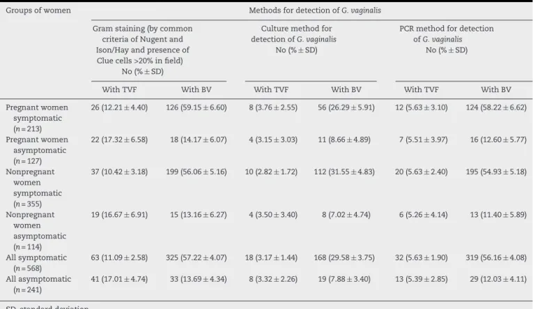
Table 1 – Concordance between the groups of women on the basis of three different laboratory methods.
Groups of women Methods for detection ofG. vaginalis
Gram staining (by common criteria of Nugent and Ison/Hay and presence of Clue cells >20% in field)
No (%±SD)
Culture method for detection ofG. vaginalis
No (%±SD)
PCR method for detection ofG. vaginalis
No (%±SD)
With TVF With BV With TVF With BV With TVF With BV
Pregnant women symptomatic (n= 213)
26 (12.21±4.40) 126 (59.15±6.60) 8 (3.76±2.55) 56 (26.29±5.91) 12 (5.63±3.10) 124 (58.22±6.62)
Pregnant women asymptomatic (n= 127)
22 (17.32±6.58) 18 (14.17±6.07) 4 (3.15±3.03) 11 (8.66±4.89) 7 (5.51±3.97) 16 (12.60±5.77)
Nonpregnant women symptomatic (n= 355)
37 (10.42±3.18) 199 (56.06±5.16) 10 (2.82±1.72) 112 (31.55±4.83) 20 (5.63±2.40) 195 (54.93±5.18)
Nonpregnant women asymptomatic (n= 114)
19 (16.67±6.91) 15 (13.16±6.27) 4 (3.50±3.40) 8 (7.02±4.74) 6 (5.26±4.14) 13 (11.40±5.89)
All symptomatic (n= 568)
63 (11.09±2.58) 325 (57.22±4.07) 18 (3.17±1.44) 168 (29.58±3.75) 32 (5.63±1.90) 319 (56.16±4.08)
All asymptomatic (n= 241)
41 (17.01±4.74) 33 (13.69±4.34) 8 (3.32±2.26) 19 (7.88±3.40) 13 (5.39±2.85) 29 (12.03±4.11)
SD, standard deviation.
Results and discussion
The data from the three procedures for BV andG. vaginalis detection in different groups of women are presented in
Table 1.
Our results from PCR assay are shown inFig. 1.
The group distributions of the additive isolates from aero-bic cultures are summarized inTable 2.
The correlations between some demographic parameters (i.e. age range) with presence ofG. vaginalisand BV are demon-strated inTable 3.
G. vaginalis was most frequently present in samples obtained from Bulgarian women in age range 21–30 years.
For achieving the goal of this investigation, the Nugent’s score22was taken as gold standard and the results from the
other methods were compared with this score. As already
Fig. 1 – Species-specific PCR assay for the detection ofG. vaginalisin vaginal samples (agarose gel-electroforesis of PCR products of the 16S rRNA gene). The product size is 331 bp. From right to left: 100 bp Ladder and 9 (+) positive samples.
published by other authors it was preferable to recommend the use of Ison/Hay method,23because in that way we could
evaluate vaginal smears in five more clearly distinguished grades with better segregation of the vaginal microflora.9,26,27
Using Gram straining in this study we detected high frequency of BV in symptomatic subjects (57.22%), in contrast to asymp-tomatic Bulgarian women (13.69%). We could also prove TVF in 11.09% of symptomatic and in 17.01% of asymptomatic subjects. Our results for TVF frequency are in unison with previously reported data in other studies from France and Australia.3,28BV frequency in the present study with
Bulgar-ian women is similar to that found in NigerBulgar-ian women, with a slightly higher rate in our population.29We have shown that
BV occurs in approximately equal proportions when eval-uated with the microscopic method for both pregnant and nonpregnant symptomatic and pregnant and nonpregnant asymptomatic women. These results are in contrast to data obtained by other authors, claiming that BV is more frequent during the pregnancy, which we could explain by separating our groups according to the symptoms, and not only by pregnancy status. So far all studies revealed different and sometimes conflicting results for BV epidemiology.1,2,9,10,27,29
We found with PCR that pregnant symptomatic patients with BV and TVF were positive forG. vaginalisin 58.22% (very similar to the 59.15% with Gram straining) and 5.63%, respec-tively. The group of nonpregnant symptomatic women had positive samples in 54.93% (56.06% using Gram straining) for BV and 5.63% for TVF (Table 1).
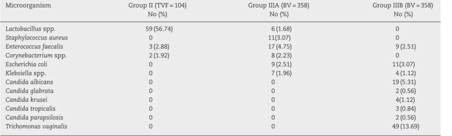
Table 2 – Isolate distribution among 104 women with diagnosis TVF and 358 women with BV according to Gram staining.a
Microorganism Group II (TVF = 104)
No (%)
Group IIIA (BV = 358) No (%)
Group IIIB (BV = 358) No (%)
Lactobacillusspp. 59 (56.74) 6 (1.68) 0
Staphylococcus aureus 0 11(3.07) 0
Enterococcus faecalis 3 (2.88) 17 (4.75) 9 (2.51)
Corynebacteriumspp. 2 (1.92) 8 (2.23) 0
Escherichia coli 0 9 (2.51) 11(3.07)
Klebsiellaspp. 0 7 (1.96) 4 (1.12)
Candida albicans 0 0 19 (5.31)
Candida glabrata 0 0 2 (0.56)
Candida krusei 0 0 4(1.12)
Candida tropicalis 0 0 3 (0.84)
Candida parapsilosis 0 0 2 (0.56)
Trichomonas vaginalis 0 0 49 (13.69)
a More than one isolate were detected in some samples.
Table 3 – Association of age range with BV according Gram staining.
Age range TVF = 104
No (%±SD)
BV = 358 No (%±SD)
16–20 28 (26.92±8.61) 29 (8.10±2.83)
21–25 19 (18.27±7.50) 101 (28.21±4.66)
26–30 21 (20.19±7.79) 96 (26.92±4.59)
31–35 20 (19.23±7.65) 70 (19.23±4.08)
36–40 18 (17.30±7.34) 62 (17.51±3.92)
SD, standard deviation.
evaluation, culture and PCR assays demonstrated greater concurrence (about 90%) between Gram staining and PCR detection for BV, than both methods compared to culture (Table 1). We found that 89% of BV and only 28% of TVF (where clue cells were significantly less than in BV) groups, according to the microscopic criteria, had positive PCR forG.vaginalis.All PCR positive results forG.vaginalishad either BV orT. vaginalis.
The combination of Gram staining and PCR methods showed very reliable and repeatable detection of BV, unlike culture, where only about 50% of PCR positive samples had evident growth on the selective agar media forG. vaginalis. PCR assay is the most sensitive method for routing outG. vaginalis(p< 0.05), but combination of this test with Gram staining for full char-acterization in the patient is needed. Gram staining is an easy, fast and affordable method that could be used, especially in low-income countries, instead of PCR, when for various rea-sons molecular detection is not possible, since the results of both techniques are very similar. The high frequency ofG. vagi-nalisdetected by PCR was evident such that this pathogen had a very important role in the aetiology of BV. The results of this study supported the data from previously reported stud-ies where 68–100% of the patients with BV were positive toG. vaginalis.3,9,27
The microbial growth on a non-selective agar media gave useful information for the presence or absence of additional microflora in the pathological process and this procedure should not be skipped, despite having low sensitivity for diagnosing BV. By using this routine method we identified that the most frequently isolated microorganisms coloniz-ing the vaginal mucosa and associated with BV, other than
G. vaginalis, were Enterococcus faecalis, Staphylococcus aureus,
Corynebacteriumspp.,Escherichia coliandKlebsiellaspp. (Table 2). Their role was unclear, as they might be of transient pres-ence or as they were detected with moderate frequency they could relate to BV aetiology or just be a co-infection. We could not find any confirmed results or evidence for their role published elsewhere. Our data showingLactobacillusspp. as the predominant bacterial genus present in the vaginal microbiota in the smears and isolates of I-st (NVF) and II-nd group (TVF) according to microscopic evaluation and culture is in line with previously published studies.7,8,24The
Gram-positives were the predominant bacterial microflora in the IIIA group in contrast to the group IIIB where prevailed some Gram-negatives species (Table 2). Some of the isolates such as
Candidaspp. were detected only among patients of group IIIB, more often in pregnant women and as initial colonization after BV (Table 2). In 13.69% of Bulgarian womenT. vaginalis was detected in association with BV (IIIB group). Similar to other reports, trichomoniasis was a frequent infection, and has to be timely diagnosed for its importance as a causative agent of sexually transmitted diseases with difficult and sometimes poor therapeutic response.18,29
whereas TVF was found in 26.92% of women in this age group. Our finding did not differ significantly from those published for Indian women, where BV prevalence in age group of 26–30 years was 23% and in 7% among the youngest group (15–20 years).30The only difference between the Indian results and
ours was in the group of 21–25 years, where BV was more frequent among Bulgarian women.
Conclusion
To our knowledge, this is the first comparative study utilizing three different laboratory methods that focuses on the epi-demiology ofG. vaginalis-associated BV in Bulgaria. Although PCR is the most sensitive method for the detection ofG. vagi-nalis, but for the full characterization of the smears the joint application of PCR and Gram staining is the best choice. An important note is that Gram staining results are compatible with PCR results, since this method is fast, easy and inexpen-sive, so that it could be used in developing countries, where and when molecular techniques are not available.
The high frequency in Bulgarian young women found in this study is alarming, since BV increases woman’s suscepti-bility to HIV, HPV and other important sexually transmitted diseases. Therefore BV has to be correctly and timely diag-nosed in order to be adequately treated.
Further investigations regarding other pathogens involved in BV such asA. vaginaeandMobiluncusspp. are warranted.
Conflict of interest
All authors declare to have no conflict of interest.
Acknowledgements
The authors would like to thank Dr. MIkana Shtereva (Medical center Nadejda), Dr. K. Tosheva (Medical center Pentagram), Dr. A. Kosturska and Dr. M. Ilieva (Medical center VIP Clinic), Dr C. Jordanova (Individual medical practice) for providing vagi-nal samples and demographic data of the patients included in this study.
r e f e r e n c e s
1. Brotman RM. Vaginal microbiome and sexually transmitted infections: an epidemiologic perspective. J Clin Invest. 2011;121:4610–7.
2. Marrazzo JM. Interpreting the epidemiology and natural history of bacterial vaginosis: are we still confused. Anaerobe. 2011;17:186–90.
3. Bradshaw CS, Tabrizi SN, Fairley CK, Morton AN, Rudland E, Garland SM. The association ofAtopobium vaginaeand
Gardnerella vaginaliswith bacterial vaginosis and recurrence after oral metronidazole therapy. J Infect Dis. 2006;194:828–36. 4. Menard JP, Fenollar F, Henry M, Bretelle F, Raoult D. Molecular
quantification ofGardnerella vaginalisandAtopobium vaginae
loads to predict bacterial vaginosis. Clin Infect Dis. 2008;47:33–43.
5. Menard JP, Mazouni C, Salem-Cherif I, et al. High vaginal concentrations ofAtopobium vaginaeandGardnerella vaginalis
in women undergoing preterm labor. Obstet Gynecol. 2010;115:134–40.
6. Ling Z, Kong J, Liu F, et al. Molecular analysis of the diversity of vaginal microbiota associated with bacterial vaginosis. BMC Genomics. 2010;11:488.
7. Redondo-Lopez V, Cook RL, Sobel JD. Emerging role of lactobacilli in the control and maintenance of the vaginal bacterial microflora. Rev Infect Dis. 1990;12:856–72.
8. Turovskiy Y, Noll SK, Chikindas ML. The aetiology of bacterial vaginosis. J Appl Microbiol. 2011;110:1105–28.
9. De Backer E, Verhelst R, Verstraelen H, et al. Quantitative determination by real-time PCR of four vaginalLactobacillus
species,Gardnerella vaginalisandAtopobium vaginaeindicates an inverse relationship betweenL gasseriandL. iners. BMC Microbiol. 2007;7:115.
10. Schwebke JR. New concepts in the etiology of bacterial vaginosis. Curr Infect Dis Rep. 2009;11:143–7.
11. Ravel J, Gajer P, Abdo Z, et al. Vaginal microbiome of reproductive-age women. Proc Natl Acad Sci U S A. 2011;108 Suppl. 1:4680–7.
12. Marrazzo JM, Thomas KK, Fiedler TL, Ringwood K, Fredricks DN. Risks for acquisition of bacterial vaginosis among women who report sex with women: a cohort study. PLoS ONE. 2010;5:e11139.
13. Fethers KA, Fairley CK, Hocking JS, Gurrin LC, Bradshaw CS. Sexual risk factors and bacterial vaginosis: a systematic review and meta-analysis. Clin Infect Dis. 2008;47:1426–35. 14. Fethers KA, Fairley CK, Morton A, et al. Early sexual
experiences and risk factors for bacterial vaginosis. J Infect Dis. 2009;200:1662–70.
15. Gallo MF, Macaluso M, Warner L, et al. Bacterial vaginosis, gonorrhea, and chlamydial infection among women
attending a sexually transmitted disease clinic: a longitudinal analysis of possible causal links. Ann Epidemiol.
2012;22:213–20.
16. Verstraelen H, Verhelst R, Vaneechoutte M, Temmerman M. The epidemiology of bacterial vaginosis in relation to sexual behaviour. BMC Infect Dis. 2010;10:81.
17. Shanmugasundaram U, Pachamuthu B, Kailapuri GM, et al. Bacterial vaginosis in female sex workers in Chennai, India. Sex Health. 2005;2:261–2.
18. Brabin L, Fairbrother E, Mandal D, et al. Biological and hormonal markers of chlamydia, human papillomavirus, and bacterial vaginosis among adolescents attending
genitourinary medicine clinics. Sex Transm Infect. 2005;81:128–32.
19. Marrazzo JM, Wiesenfeld HC, Murray PJ, et al. Risk factors for cervicitis among women with bacterial vaginosis. J Infect Dis. 2006;193:617–24.
20. Lamont RF, Sobel JD, Akins RA, et al. The vaginal microbiome: new information about genital tract flora using molecular based techniques. BJOG. 2011;118:533–49.
21. Haggerty CL, Hillier SL, Bass DC, Ness RB. Bacterial vaginosis and anaerobic bacteria are associated with endometritis. Clin Infect Dis. 2004;39:990–5.
22. Nugent RP, Krohn MA, Hillier SL. Reliability of diagnosing bacterial vaginosis is improved by a standardized method of gram stain interpretation. J Clin Microbiol. 1991;29:297–301. 23. Ison CA, Hay PE. Validation of a simplified grading of Gram
stained vaginal smears for use in genitourinary medicine clinics. Sex Transm Infect. 2002;78:413–5.
24. Amsel R, Totten PA, Spiegel CA, Chen KC, Eschenbach D, Holmes KK. Nonspecific vaginitis. Diagnostic criteria and microbial and epidemiologic associations. Am J Med. 1983;74:14–22.
26. Verhelst R, Verstraelen H, Claeys G, et al. Comparison between Gram stain and culture for the characterization of vaginal microflora: definition of a distinct grade that resembles grade I microflora and revised categorization of grade I microflora. BMC Microbiol. 2005;5:61.
27. Verhelst R, Verstraelen H, Claeys G, et al. Cloning of 16S rRNA genes amplified from normal and disturbed vaginal
microflora suggests a strong association betweenAtopobium vaginae,Gardnerella vaginalisand bacterial vaginosis. BMC Microbiol. 2004;4:16.
28. Menard JP, Mazouni C, Fenollar F, Raoult D, Boubli L, Bretelle F. Diagnostic accuracy of quantitative real-time PCR assay
versus clinical and Gram stain identification of bacterial vaginosis. Eur J Clin Microbiol Infect Dis. 2010;29:1547–52.
29. Nwadioha S, Egah D, Nwokedi E, Onwuezobe I. A study of female genital swabs in primary health care centres in Jos, Nigeria. Asian Pac J Trop Dis. 2011;1:52–4.