354
Pakistan Veterinary Journal
ISSN: 0253-8318 (PRINT), 2074-7764 (ONLINE)Accessible at: www.pvj.com.pk
Infection of Avian Pox Virus in Oriental Turtle-Doves
Kyung-Yeon Eo1, Young-Hoan Kim2, Kwang-Hyun Cho3, Jong-Sik Jang4, Tae-Hwan Kim5, Dongmi Kwak5 and Oh-Deog Kwon5*
1
Seoul Zoo, Gwacheon 427-702; 2Gyeongbuk Veterinary Service Laboratory, Daegu 702-911; 3Gyeongsangbukdo Livestock Research Institute, Yeongju 750-871; 4Department of Animal Science, Kyungpook National University, Sangju 742-711, Korea; 5College of Veterinary Medicine, Kyungpook National University, Daegu 702-701, Korea. *Corresponding author: odkwon@knu.ac.kr
A R T I C L E H I S T O R Y A B S T R A C T Received:
Revised: Accepted:
May 03, 2011 May 13, 2011 May 18, 2011 Key words:
Avian pox
Oriental Turtle-dove Pathology
Streptopelia orientalis Wild bird
Three Oriental Turtle-doves (Streptopelia orientalis) exhibiting lethargy, dyspnea, poor physical condition, and poor flight endurance, were rescued and referred to the Animal Health Center, Seoul Zoo, Korea. The doves had wart-like lesions on the legs and head. All of them died the following day after arrival, with the exception of one that survived for 6 days. Diphtheritic membranes on the tongue and oral mucosa were apparent at necropsy. Avian pox virus infection was suspected based on the proliferative skin lesions and oral diphtheritic lesions. Infection of the avian pox virus was confirmed by PCR using primers specific to the 4b core protein gene of avian pox virus. All cases were diagnosed with avian pox virus infection. This is believed to be the first description on natural infection of avian pox in Oriental Turtle-doves in Korea.
©2011 PVJ. All rights reserved To Cite This Article: Eo KY, YH Kim, KH Cho, JS Jang, TH Kim, D Kwak and OD Kwon, 2011. Infection of avian pox virus in oriental turtle-doves. Pak Vet J, 31(4): 354-356.
INTRODUCTION
Avian pox virus is a common virus affecting domestic and wild birds worldwide. Infection of avian pox has been reported in more than 60 species of birds in 20 families based on clinical signs (Samour, 2008). Pox virus infection generally manifests either cutaneously or diphtheritically, although both forms can occur in the same bird (Tripathy et al., 2000; Hukkanen et al., 2003; Khan et al., 2009). The characteristic lesions occur as wart-like proliferations on the featherless skin around the eyes, beak, nostrils, and feet in the cutaneous form and as proliferative lesions and diphtheroid membranes on the mouth or upper respiratory tract mucosa in the diphtheritic form (Tangredi, 1974; Krone et al., 2004).
Common in domestic poultry and wild birds worldwide, pox virus infection in wild birds has been reported in the USA (Tangredi, 1974), Germany (Krone et al., 2004), and Australia (Annuar, 1983), but not in Korea. This report describes severe cases of both forms of avian pox in three Oriental Turtle-doves (Streptopelia orientalis) that were referred to the Animal Health Center at the Seoul Zoo (37o25’48”N, 127o00’57”E), Korea.
Three Oriental Turtle-doves were presented to the Animal Health Center between August and November, 2010. A juvenile dove weighing 60 g and the two adult
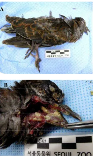
doves weighing 105 and 120 g, respectively, were in poor physical condition. The birds were rescued by local residents on the roadside having difficulty flying and brought to the Animal Health Center for further examination. The doves had wart-like lesions on the legs and head around eyelids and beaks. Multiple discrete, pale yellow to cream-colored, raised necrotic lesions were distributed irregularly across the oropharyngeal mucosa in the birds (Fig. 1). All birds died the next day after arrival, with the exception of one dove that survived for 6 days. Diphtheritic membranes on the tongue and oral mucosa were apparent at necropsy. Avian pox virus was suspected based on the clinical examination and necropsy observations.
MATERIALS AND METHODS
To confirm the diagnosis, PCR was performed on body tissue. DNA was extracted using a viral RNA extraction kit (Qiagen, USA) according to manufacturer’s instruction. PCR was performed to amplify the 4b core protein gene of avian pox virus using an AccuPower PCR Premix Kit (Bioneer, Korea). The forward (CP1, 5’-CAGCAGGTGCTAAACAACAA-3’) and reverse (CP2, 5’-CGGTAGCTTAACGCCGAATA-3’) primers specific to the 4b core protein gene of avian pox virus were used
Pak Vet J, 2011, 31(4): 354-356. 355
Fig. 1: Photograph of an Oriental Turtle-dove: A, wart-like
proliferations on the skin of the legs and head. B, multiple discrete, pale yellow to cream-colored, raised necrotic lesions was distributed irregularly across the oropharyngeal mucosa.
Fig. 2: PCR amplification with primers (CP1/CP2) specific to
the 4b core protein gene of the avian pox virus. Lane M, 100-bp DNA ladder; lane 1, rectal swab of an Oriental Turtle-dove with pox-like lesions; lane 2, tracheal swab of an Oriental Turtle-dove with pox-like lesions; lane 3, oral swab of an Oriental Turtle-dove with pox-like lesions; lane 4, oral swab of an Oriental Turtle-dove with no symptom. The DNA fragments produced were analyzed on agarose gel (1.5%) and visualized by ethidium bromide staining and UV transillumination. Selected markers (bp) are indicated on the left. The arrow on the right refers to the estimated size of the band at 576 bp.
(Weli, 2004). PCR was performed in the T-gradient thermoblock (Biometra, Germany) by the following thermal cycling: an initial hot start, 94 oC/5 min; 35 cycles of 94 oC/20 s, 55 oC/30 s and 72 oC/40 s; and a final extension, 72 oC/5 min. Sizes of PCR amplicons were
estimated on agarose gel (1.5%) electrophoresis following UV transillumination. The expected amplicon size was about 576 bp.
RESULTS AND DISCUSSION
All cases were diagnosed with avian pox virus infection (Fig. 2). Avian pox virus is a member of the family Poxviridae and the genus Avipoxvirus. While the cutaneous and diphtheritic forms are the most frequently observed, a third form, systemic pox virus, is occasionally observed (Pledger, 2005). The birds will appear weak and emaciated if the lesions are sufficiently extensive to interfere with their feeding. Some birds have labored breathing if their air passages are partially blocked. The diphtheritic, or wet, form of avian pox probably occurs more frequently in wild birds than is reported because it is less observable than the cutaneous form. Furthermore, the more severe consequences of wet pox probably cause greater morbidity and mortality (USGS, 2009). In Houbara Bustards (Chlamydotis undulata) and waterfowl, the wet and systemic forms cause multiple discrete, pale yellow to cream-colored, raised necrotic lesions irregularly distributed across the oropharyngeal mucosa (Samour, 2008). The lesions on the three Oriental Turtle-doves were characteristics of cutaneous and diphtheritic avian pox virus infections.
Avian pox occurs worldwide, but little is known about its prevalence in wild bird populations. Pox outbreaks are common in aviaries, rehabilitation centers, and other places where birds are closely confined. Birds can become carriers and spread avian pox among local populations, for example, between birds’ feeding stations and along common migratory routes (USGS, 2009). The Oriental Turtle-dove is a member of the family Columbidae, which is widely distributed throughout Asia and Europe (Wu, 2007). This wild bird is commonly observed in backyards and forests in Korea, as well as in small groups feeding in parks. This behavior increases their contact with other birds, thereby increasing the risk of acquiring infectious diseases, including avian pox (Pledger, 2005). This is the first description of natural avian pox infection in the Oriental Turtle-dove in Korea.
REFERENCES
Annuar BO, JS Mackenzie and PA Lalor, 1983. Isolation and characterization of avipoxviruses from wild birds in Western Australia. Arch Virol, 76: 217-229. Hukkanen RR, M Richardson, JC Wingfield, P Treuting
and T Brabb, 2003. Avipox sp. in a colony of gray-crowned rosy finches (Leucosticte tephrocotis). Comp Med, 53: 548-552.
Khan A, A Yousaf, MZ Khan, M Siddique, ST Gul and F Mahmood, 2009. Cutaneous form of pox infection among captive peafowl (Pavo cristatus) chicks. Avian Pathol, 38: 65-70.
Krone O, S Essbauer, G Wibbelt, G Isa, M Rudolph and RE Gough, 2004. Avipoxvirus infection in peregrine falcons (Falco peregrinus) from a reintroduction programme in Germany. Vet Rec, 154: 110-113. Pledger A, 2005. Avian pox virus infection in a mourning
dove. Can Vet J, 46: 1143–1145.
Pak Vet J, 2011, 31(4): 354-356. 356
Samour JS, 2008. Avian Medicine. 2nd Ed, Mosby, New York, USA, pp: 364-368.
Tangredi BP, 1974. Avian pox in a mourning dove. Vet Med Small Anim Clin, 69: 700-701.
Tripathy DN, WM Schnitzlein, PJ Morris, DL Janssen, JK Zuba, G. Massey and CT Atkinson, 2000. Characterization of poxviruses from forest birds in Hawaii. J Wildlife Dis, 36: 225-230.
USGS National Wildlife Health Center, 2009. Field Manual of Wildlife Disease - General Field
Procedures and Diseases of Birds pp. 163-169. (http ://www.nwhc.usgs.gov/publications/field_manual/) Weli SC, T Traavik, M Tryland, DH Coucheron and O
Nilssen, 2004. Analysis and comparison of the 4b core protein gene of avipoxviruses from wild birds: evidence for interspecies spatial phylogenetic variation. Arch Virol, 149: 2035-2046.