From the Department of Gastroenterology, Hospital das Clínicas, Faculty of Medicine, University of São Paulo.
ORIGINAL ARTICLE
HELICOBACTER PYLORI GENOTYPING FROM
POSITIVE CLOTESTS IN PATIENTS WITH DUODENAL
ULCER
Rejane Mattar and Antonio Atílio Laudanna
RHCFAP/3018
MATTAR R et al. - Helicobacter pylori genotyping from positive clotests in patients with duodenal ulcer. Rev. Hosp. Clín.
Fac. Med. S. Paulo 55(5):155-160, 2000.
Even though the seroprevalence of H. pylori may be high in the normal population, a minority develops peptic ulcer. Colonization
of the gastric mucosa by more pathogenic vacA strains of H. pylori seems to be associated with enhanced gastric inflammation and duodenal ulcer. H. pylori genotyping from positive CLOtests was developed to determine the vacA genotypes and cagA status in 40
duodenal ulcer patients and for routine use. The pathogenic s1b/ m1/ cagA genotype was the most frequently occurring strain (17/
42.5%); only two (5%) patients presented the s2/ m2 genotype, the less virulent strain. Multiple strains were also detected in 17 (42.5%) patients. Multiple strains of H. pylori colonizing the human stomach have been underestimated, because genotyping has
been performed from cultures of H. pylori. We concluded that genotyping of H. pylori from a positive CLOtest had the advantages
of reducing the number of biopsies taken during endoscopy, eliminating the step of culturing H. pylori, and assuring the presence of H. pylori in the specimen being processed.
DESCRIPTORS: Helicobacter pylori. Genotype. Duodenal ulcer. CLOtest.
Helicobacter pylori (H. pylori) are curved or spiral-shaped Gram-negative bacteria1. The organisms are found close to the mucosal surface, in inter-cellular positions or caught up in the surface mucus of the stomach2. When cultured on solid medium, the bacte-rium assumes a rod shape; spiral forms may be few or absent. After prolonged culture, coccoid forms predominate1. The colonization of the gastric mucosa by H. pylori has been associated with chronic gastritis, peptic ulcer disease, and gastric cancer3-5.
Although the seroprevalence of H. pylori may be high in the normal popu-lation, a minority develops peptic ul-cer6. Racial differences in H. pylori seroprevalence and peptic ulcer
fre-quency were observed in Singapore; Indians had a higher prevalence of H. pylori antibodies but a lower frequency of peptic ulcer than the Chinese. This finding suggested that other environ-mental or genetic factors may be in-volved in peptic ulcer disease7. Another possible explanation is the fact that some strains are more pathogenic than others6.
There are two types of H. pylori: one (toxin +) can, and the other (toxin -) cannot secrete a vacuolating cyto-toxin, which is a protein encoded by
the vacA (vacuolating cytotoxin) gene8. Toxin + strains also produce a 128 kDa protein encoded by the cagA gene (cy-totoxin-associated gene)9. Patients har-boring the toxin + strain would be prone to present gastric or duodenal ulceration and gastric cancer10-13.
strains; none of s2/ m2 strains pro-duced detectable cytotoxin activity 8,10-12. However, even though m2 cytotoxin was inactive in the in vitro HeLa cell cytotoxicity assay, the m2 cytotoxin was able to induce vacuolization in pri-mary gastric cells15.
The purpose of our study was to develop a quick technique for routine use of H. pylori genotyping from the CLOtest. H. pylori genotyping directly from gastric biopsy specimens has pre-viously been described16-19.However, the advantages of using the CLOtest for genotyping would be a reduction of the number of biopsies taken during the endoscopy and the assurance of the presenceof H. pylori in the specimen being processed for genotyping.
PATIENTS AND METHODS
Patients
Forty patients with duodenal ulcer and with positive CLOtests were se-lected for the study. All patients were positive for H. pylori, based on histo-logical findings in gastric biopsies done at the same time that the CLOtest was performed. Twenty patients were male and 20 were female, with a mean age of 48+ 13 yr.
CLOtest
The antral mucosal biopsy speci-men was inserted into a homemade urease test tube20. The urease reagent was prepared by dissolving the follow-ing in distilled water to a final volume of 100 mL: 0.010 g yeast extract, 0.0091 g KH2PO4, 0.0095 g Na2HPO4, 2 g urea, and 15 drops of phenol red 0.5%; the pH of the solution was ad-justed to 6.9. The urease reagent was sterilized by filtration, dispensed into 0.5 mL aliquots, and stored at –20ºC. If the urease enzyme of H. pylori was present in the gastric biopsy, the result-ing breakdown of urea caused the pH to rise and the color of the solution to
turn from yellow to bright magenta. The urease test tube was examined af-ter a 24 h period.
DNA extraction
After the CLOtest reading at 24 hours, the CLOtest tubes were stored at 4ºC until DNA extraction. The whole content of CLOtest including gastric biopsy was collected by centrifugation at 12 000 g for 25 min. The superna-tant was discarded, and the pellet was re-suspended in DNA extraction buffer according to Sambrook et al.21, using the phenol-chloroform method. The DNA pellet was re-suspended in 30 µL TE (10 mmol Tris-HCl pH 8.0, 1 mmol EDTA pH 8.0).
Polymerase Chain Reaction
Genomic DNA (1.2- 6.0 µg) was used as a template in a reaction volume of 50 µL, containing 20 mmol Tris-HCl (pH 8.4), 50 pmol of each primer, 200 µmol of each dNTP, and 2.5 U of Taq DNA polymerase (Gibco BRL, Gaithersburg, MD, USA). The Poly-merase Chain Reaction (PCR) was per-formed in a 2400 GeneAmp PCR sys-tem (Perkin Elmer, Branchburg, NJ, USA). Amplification was performed under the following conditions for vacA (m1, m2, s1a, s1b, s2)10 and cagA9 (Table 1): initial denaturation at 94ºC for 5 min followed by 27 cycles of denaturation at 94ºC for 30s, anneal-ing at 53ºC for 30s and extension at 72ºC for 30s. The final extension at 72ºC was for 7 min. Gene Amp lambda control reagents (Perkin Elmer, Branchburg, NJ, USA); control tem-plate lambda DNA and primers were included as positive PCR reaction in-ternal control. Negative PCR reaction internal control was performed by ex-cluding H. pylori genomic DNA in one of the PCR reaction tubes. One set of primers (P1 and P2)16 that amplifies a 26 kDa antigen gene present in all strains of H. pylori was used for the negative PCR reaction cases according
to the following conditions: initial de-naturation at 94ºC for 5 min followed by 40 cycles of denaturation at 93ºC for 1 min, annealing at 57ºC for 2 min and extension at 70ºC for 2 min. The final extension at 70ºC was for 10 min. Analysis of PCR products
Five microliters of each PCR mix-ture were separated by electrophoresis on 2% agarose (GIBCO BRL, Gaithersburg, MD, USA) gels in TAE21 (0.04 M Tris-acetate, 0.001M EDTA pH 8.0) and 0.5 µg/mL ethidium bro-mide. TAE was also used for electro-phoresis buffer. PCR mixtures and 50 bp DNA ladder (GIBCO BRL, Gaithersburg, MD, USA) were loaded into slots in 6x loading buffer (0.25% bromophenol blue, 0.25% xylene cyanol FF, and 3% glycerol in water)21.
RESULTS
The PCR technique from the posi-tive CLOtest tubes could be easily ap-plied for the characterization of H. py-lori strains. VacA and cagA genotypes of H. pylori were analyzed in 40 duodenal ulcer patients that were diag-nosed positive for H. pylori by means of histology. Three patients were not included in the study because no PCR product could be obtained, even using one set of primers (P1 and P2) that amplifies a 26kDa antigen present in all strains of H. pylori and 6 µg of ge-nomic DNA. The DNA extracted from the biopsies studied, except for the ones that had multiple strains, gave PCR products of expected sizes (Fig. 1).
1) the reduction of the number of bi-opsies taken during endoscopy; 2) the assurance of the presenceof H. pylori in the specimen being processed, and 3) the avoidance of time-consuming culturing of the strains.
The determination of the vacA genotype was possible in more than 90% of the positive CLOtests, in agree-ment with another report18, since only 3 patients were not included in this study because no PCR product could be obtained. A low density of H. py-lori on the gastric mucosa could be re-sponsible for a negative PCR, as the sensitivity of the PCR detection of H. pylori in gastric biopsies was approxi-mately of 70-100 bacterial cells16,18. One set of primers (P1 and P2)16 that amplifies a 26 kDa antigen present in all strains of H. pylori was also used to make sure that the negative PCR re-actions were due to a low density of H. pylori, instead of the genotyping tech-nique itself. Actually, typing of the vacA gene was not possible in five strains by other authors22, because of a 61-bp insertion in the signal region of two strains, and for unknown reasons in the others.
The s1b/ m1/ cagA genotype was the most frequent strain (42.5%) ob-served in these patients; a similar re-sult has already been described in an-other study of H. pylori strains from Brazil23. A high prevalence of vacA s1b alleles was also reported in South Af-rica13 and Portugal24. However, vacA Table 2- vacA and cagA status of H. pylori strains from CLOtests of 40 patients with duodenal ulcer.
Genotype status Patients (%) s1b/ m1 / cagA 17 (42.5%) s1a/ m1 / cagA 2 (5%) s1b/ m2 / cagA 2 (5%)
s2/ m2 2 (5%)
Multiple strains 17 (42.5%)
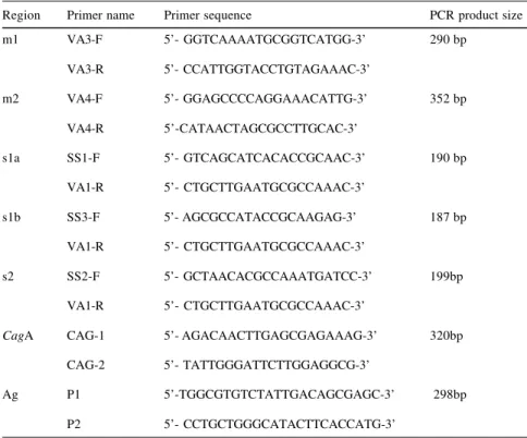
Table 1 - Primers used for genotyping H. pylori vacA alleles 10 and cagA status 9.
Region Primer name Primer sequence PCR product size
m1 VA3-F 5’- GGTCAAAATGCGGTCATGG-3’ 290 bp
VA3-R 5’- CCATTGGTACCTGTAGAAAC-3’
m2 VA4-F 5’- GGAGCCCCAGGAAACATTG-3’ 352 bp
VA4-R 5’-CATAACTAGCGCCTTGCAC-3’
s1a SS1-F 5’- GTCAGCATCACACCGCAAC-3’ 190 bp
VA1-R 5’- CTGCTTGAATGCGCCAAAC-3’
s1b SS3-F 5’- AGCGCCATACCGCAAGAG-3’ 187 bp
VA1-R 5’- CTGCTTGAATGCGCCAAAC-3’
s2 SS2-F 5’- GCTAACACGCCAAATGATCC-3’ 199bp
VA1-R 5’- CTGCTTGAATGCGCCAAAC-3’
CagA CAG-1 5’- AGACAACTTGAGCGAGAAAG-3’ 320bp
CAG-2 5’- TATTGGGATTCTTGGAGGCG-3’
Ag P1 5’-TGGCGTGTCTATTGACAGCGAGC-3’ 298bp
P2 5’- CCTGCTGGGCATACTTCACCATG-3’
Ag: DNA sequence of a species-specific protein antigen of 26kDa molecular weight that was present in all strains of H. pylori16.
Figure 1 - PCR genotyping of vacA and cagA status from one case with the s2/ m2 strain and another case with the s1b/ m1/ cagA strain. Primers described in Table 1 were used for PCR reaction.
DISCUSSION
In the present study, we described the analysis of H. pylori strains by PCR
s1a alleles were more prevalent in Ja-pan22 and in Northern and Eastern Eu-rope23.
H. pylori strains of vacA signal se-quence type s1a are associated with more gastric inflammation and duode-nal ulceration than are the s1b type. VacA s2 strains are associated with less inflammation and lower ulcer preva-lence6; in the present study, a low prevalence was also observed, as only two (5%) patients with duodenal ulcer had vacA s2/ m2 strain.
Multiple strains were detected at the same rate (42.5%) as the s1b/ m1/ cagA genotype; the clinical relevance of multiple strains in gastric biopsies should be evaluated, because the viru-lence-associated genotypes of the strains was correlated with the clinical outcome of the gastrointestinal disease in some studies10-13, but not in others5, 14, 22, 25.
The detection of a high frequency of multiple strains could be explained by the fact that genotyping was per-formed directly from gastric biopsies; other authors12,16-19 also obtained more than one strain when using the same approach. In contrast, when the step of
H. pylori culturing preceded genotyping, a single strain may have been picked up; thus, the frequency of multiple strains in the stomach might have been underestimated. According to Van Doorn et al23 and Figura et al26, purification of H. pylori strains by cul-turing from a single colony universally results in the detection of a single vacA genotype; however, when the strains used are not purified from a single colony, they may reflect the presence of multiple strains in the host’s stom-ach.
Another aspect that has to be con-sidered in the disease outcome (duode-nal or gastric ulcer, gastric cancer, and gastric mucosa-associated lymphoid tissue lymphoma) of positive H.pylori patients is the individual host’s re-sponse to the H. pylori infection. The cellular and humoral immune re-sponses that are mounted against H. pylori are vigorous; polymorpho-nuclear leukocytes and macrophages, as well as T and B lymphocytes, infil-trate the gastric mucosa, and have been shown to modify gastric acid secre-tion27. Different gastric acid responses
to H. pylori have been associated with variations in the gastritis patterns that seem to determine disease outcome28,29. Thus, the immune response of the host does not clear the infection and leaves the host prone to complications result-ing from chronic inflammation30-33.
In a mechanism known as antigenic mimicry, highly conserved immuno-genic molecules expressed by infec-tious pathogens may act as a trigger for the induction of humoral and cellular immune responses that cross-react with host cellular antigens. H. pylori seems to be very effective in inducing anti-genic mimicry; antibodies against H. pylori have been found to cross-react with both antral mucosal cells and gas-trin-producing cells. Such autoantibod-ies were detected both in human and in experimental work in rodents34.
In conclusion, genotyping from a homemade CLOtest was successfully developed for routine use in our labo-ratory. Even though more virulent strains of H. pylori were found in duodenal ulcer patients, the host im-mune responses to H. pylori should be further evaluated.
RESUMO RHCFAP/3018
MATTAR R e col. – A genotipagem do H. pylori de testes CLO positivos em pacientes com úlcera duodenal. Rev. Hosp. Clín. Fac. Med. S. Paulo 55(5):155-160, 2000. Apesar da prevalência do H. pylori na população normal ser alta, somente uma minoria desenvolve úlcera péptica. A colonização da mucosa gástrica por cepas mais patogênicas de H. pylori tem sido associada com maior inflama-ção gástrica e úlcera duodenal. A genotipagem do H. pylori de testes
CLO positivos foi estabelecida para se determinar os genótipos vacA e cagA em 40 pacientes com úlcera duodenal e para uso na rotina. O genótipo patogênico s1b/m1/cagA foi o mais freqüente (17/ 42,5%); apenas dois (5%) pacientes apresentaram o genótipo s2/m2, o que é o menos vi-rulento. Cepas múltiplas também fo-ram detectadas em 17 (42,5%) pacien-tes. Cepas múltiplas colonizando o es-tômago têm sido subestimadas, pelo fato das genotipagens serem
REFERENCES
1. MURRAY PR, ROSENTHAL KS, KOBAYASHI GS et al.- Medical Microbiology. St. Louis, Mosby, Year Book Inc, 1998. p. 254-257.
2. PRICE AB - The histological recognition of Helicobacter pylori. In: LEE A, MÉGRAUD F (Editors).- Basic Helicobacter pylori: Tech-niques For Clinical Diagnosis AndResearch. London, Saunders, 1996. p. 33-49.
3. GRAHAM DY & YAMAOKA Y - H. pylori and cagA: relationships with gastric cancer, duodenal ulcer, and reflux esophagitis and its complication. Helicobacter 1998; 3: 145-151.
4. HAMLET A, THORESON A-C, NILSSON O et al.- Duodenal
Helicobacter pylori infection differs in cagA genotype between asymptomatic subjects and patients with duodenal ulcers. Gastro-enterology 1999; 116: 259-268.
5. YAMAOKA Y, KODAMA T, KASHIMA K et al. - Antibody against
Helicobacter pylori CagA and VacA and the risk for gastric can-cer. J Clin Pathol 1999; 52: 215-218.
6. ATHERTON JC - The clinical relevance of strain types of Helicobacter pylori. Gut 1997; 40: 701-703.
7. KANG JY, YEOH KG, HO KY et al.- Racial differences in Helicobacter pylori seroprevalence in Singapore: correlation with differences in peptic ulcer frequency. J Gastroenterol Hepatol1997; 12: 655-659.
8. COVER TL, TUMMURU MKR, CAO P et al. - Divergence of genetic sequences for the vacuolating cytotoxin among Helicobacter py-lori strains. J Biol Chem1994; 269: 10566-10573.
9. COVACCI A, CENSINI S, BUGNOLI M et al.- Molecular character-ization of the 128-kDa immunodominant antigen of Helicobacter pylori associated with cytotoxicity and duodenal ulcer. Proc Natl Acad Sci (USA) 1993; 90: 5791-5795.
10. ATHERTON JC, CAO P, PEEK RM et al.- Mosaicism in vacuolating cytotoxin alleles of Helicobacter pylori. J Biol Chem 1995; 270:
17771-17777.
11. ATHERTON JC, PEEK RM, THAM KT et al.- Clinical and patho-logical importance of heterogeneity in vacA, the vacuolating cy-totoxin gene of Helicobacterpylori. Gastroenterology 1997; 112:
92-99.
12. RUDI J, KOLB C, MAIWALD M et al.- Diversity of Helicobacter pylorivacA and cagA genes and relationship to VacA and CagA protein expression, cytotoxin production, and associated diseases.
J ClinMicrobiol1998; 36: 944-948.
13. KIDD M, LASTOVICA AJ, ATHERTON JC et al. - Heterogeneity in the Helicobacter pylori vacA and cagA genes: association with gastroduodenal disease in South Africa? Gut 1999; 45: 499-502. 14. YAMAOKA Y, KODAMA T, GUTIERREZ O et al.- Relationship
be-tween Helicobacter pyloriiceA, cagA, and vacA status and clini-cal outcome: studies in four different countries. J Clin Microbiol
1999; 37: 2272-2279.
15. PAGLIACCIA C, DE BERNARD M, LUPETTI P E et al. - The m2 form of Helicobacter pylori cytotoxin has cell type-specific vacu-olating activity. Proc NatlAcad Sci (USA) 1998; 95: 10212-10217.
16. HAMMAR M, TYSZKIEWICZ T, WADSTRÖM T et al. - Rapid de-tection of Helicobacter pylori in gastric biopsy material by poly-merase chain reaction. J ClinMicrobiol 1992; 30:54-58. 17. NAVAGLIA F, BASSO D & PLEBANI M - Touchdown PCR: a rapid
method to genotype Helicobacter pylori infection. Clin Chim Acta
1997; 262: 157-160.
18. RUDI J, RUDY A, MAIWALD M et al. - Direct determination of
Helicobacter pylorivacA genotype and cagA gene in gastric bi-opsies and relationship to gastrointestinal diseases. Amer J Gastroenterol1999; 94: 1525-1531.
19. HENNIG EE, TRZECIAK L, REGULA J et al.- VacA genotyping di-rectly from gastric biopsy specimens and estimation of mixed Helicobacter pylori infections in patients with duodenal ulcer and gastritis. Scand JGastroent 1999; 34: 743-749.
20. GLUPCZYNSKI Y. - Culture of Helicobacter pylori from gastric bi-opsies and antimicrobial susceptibility testing. In: LEE A & MÉGRAUD F (Ed). Helicobacter pylori: Techniques For Clini-cal Diagnosis And Basic Research. London, Saunders, 1996. p. 17-32.
21. SAMBROOK J, FRITSCH EF & MANIATIS T - Molecular Clon-ing- A LaboratoryManual, 2nd , New York, Cold Spring Harbor
Laboratory,1989.
22. ITO Y, AZUMA T, ITO S et al. - Analysis and typing of the vacA gene from cagA-positive strains of Helicobacter pylori isolated in Ja-pan. J Clin Microbiol 1997; 35: 1710-1714.
23. VAN DOORN L-J, FIGUEIREDO C, MÉGRAUD F et al. - Geographic distribution of vacA allelic types of Helicobacter pylori. Gastro-enterology 1999; 116:823-830.
24. VAN DOORN L-J, FIGUEIREDO C, SANNA R et al.- Clinical rel-evance of the cagA, vacA, and iceA status of Helicobacter pylori.
Gastroenterology 1998; 115: 58-66.
25. PAN ZJ, VAN DER HULST RW, TYTGAT GN et al.- Relation be-tween vacA subtypes, cytotoxin activity, and disease in
Helicobacter pylori-infected patients from The Netherlands. Amer
J Gastroenterol 1999; 94: 1517-1521.
26. FIGURA N, VINDIGNI C, COVACCI A et al. - cagA positive and
negative Helicobacter pylori strains are simultaneously present in the stomach of most patients with non-ulcer dyspepsia: relevance to histological damage. Gut 1998; 42: 772-778.
27. GENTA RM - The immunobiology of Helicobacter pylori gastritis.
Semin GastrointestDis 1997; 8: 2-11.
28. GO MF – What are the host factors that place an individual at risk for
Helicobacter pylori-associated disease. Gastroenterology 1997;
113 (6 Suppl): S15-20.
29. SHIMOYAMA T & CRABTREE JE. - Bacterial factors and immune pathogenesis in Helicobacter pylori infection. Gut 1998; 43 (Suppl 1): S2-5.
30. BLANCHARD TG, CZINN SJ & NEDRUD JG- Host response and vaccine development to Helicobacter pylori infection. Curr Top
31. TELFORD JL, COVACCI A, RAPPUOLI R et al. - Immunobiology of
Helicobacterpylori infection. Curr Opin Immunol 1997; 9:
498-503.
32. CRABTREE JE - Role of cytokines in pathogenesis of Helicobacter pylori-induced mucosal damage. Dig Dis Sci 1998; 43(9 Suppl): 46S-55S.
33. ROTH KA, KAPADIA SB, MARTIN SM et al. - Cellular immune responses are essential for the development of Helicobacter felis
-associated gastric pathology. JImmunol 1999; 163: 1490-1497. 34. NEGRINI R, SAVIO A & APPELMELK BJ – Autoantibodies to
gas-tric mucosa in Helicobacter pylori infection. Helicobacter 1997;
2(Suppl 1): S13-S16.