From the Department of Orthopaedics, Hospital das Clínicas, Faculty of Medicine, University of São Paulo.
THE USE OF A MODULAR TITANIUM
ENDOPROSTHESIS IN SKELETAL
RECONSTRUCTIONS AFTER BONE TUMOR
RESECTIONS: METHOD PRESENTATION AND
ANALYSIS OF 37 CASES
Alberto Tesconi Croci, Olavo Pires de Camargo, André Mathias Baptista and Marcelo Tadeu Caiero
RHCFAP/3020
CROCI AT et al. - The use of modular titanium endoprosthesis in skeletal reconstructions after bone tumor resections: method presentation and analysis of 37 cases. Rev. Hosp. Clín. Fac. Med. S. Paulo 55(5):169-176, 2000.
We analyzed 37 patients who underwent segmental wide resection of bone tumors and reconstruction with a modular titanium endoprosthesis at the Orthopaedic Oncology Group, between 1992 and 1998. Twelve patients were male and 25 were female, with a mean age of 30 years (9 – 81). The mean follow-up was 14 months (2 – 48). The diagnoses were: osteosarcoma (14 cases), metastatic carcinoma (10), Ewing’s sarcoma (4), giant cell tumor (4), malignant fibrous histiocytoma (3), chondrosarcoma (1), and aneurysmal bone cyst (1). Eleven articulated total knee, 8 partial proximal femur with bipolar acetabulum, 8 partial proximal humerus, 3 total femur, 2 partial proximal tibia, 2 diaphyseal femur, 2 diaphyseal humerus, and 1 total proximal femur with cementless acetabulum endoprosthesis implant procedures were done. The complications related to the procedure included: infection (5 cases), dislocation (3), module loosening (1), and ulnar nerve paresthesia (1). We used the following criteria for the clinical evaluation: presence of pain, range of motion, reconstruction stability, surgical and oncologic complications, and patient acceptance. The results were good in 56.8% of the cases, regular in 32.4% and poor in 10.8%.
DESCRIPTORS: Endoprosthesis. Bone tumors. Surgical treatment. Bone neoplasms.
Nowadays in the orthopaedic field, there is much discussion about skeletal reconstructions in cases where the bone losses, particularly at the articu-lar surface, are sufficient to prevent the use of conventional prosthesis.
Replacements since 1912 involving filling the space after segmental resec-tions with bone transplants have been studied by PUTTI.
A landmark in the literature is the paper by DELITALA (1947)7, which
presented the first results with the use of endoprostheses after segmental re-sections, followed by a paper in 1956 in which the kind of material used was described.
In our country, CAMARGO (1967)1 reported on segmental
resec-tion of bone tumors and surgical skel-etal reconstruction in 81 patients, 51 of whom underwent replacements with endoprosthesis.
There has been a clear evolution in the materials since then, associated with improvements in surgical tech-niques. Despite these developments, the clinical results obtained with re-placements after segmental resections
have still had much to improve in the ensuing years.
Improved implants are needed in two orthopaedic sub-specialties: in re-vision arthroplasties, especially of the hip, and in replacements after segmen-tal resections of bone tumors.
Since there are still many problems concerning the use of homologous bone grafts, including difficulties with donators’ families, material handling, and high infrastructure costs, a rela-tively high number of endoprosthesis have been used in our country.
has tremendously increased the sur-vival for these patients. Consequently, problems have arisen related to the longer follow-up of the endoprosthesis, such as breaking and loosening of the implants, which were observed earlier with conventional prostheses (total hip and total knee prostheses).
Therefore, a greater number of re-vision procedures have been done. The physicians who perform these opera-tions are familiar with the difficulties found intraoperatively, including severe bone loss after removal of the implant and the cement.
With the objective of making these revision procedures easier, we designed a modular endoprosthesis system. Its simplicity allows the surgeon to create customized implants by combining modular elements, thereby avoiding the waiting time for conventional custom-made endoprostheses, resulting in di-minished hospitalization time and cost, which in our country is a factor to be considered.
We present an analysis of 37 pa-tients who underwent segmental resec-tions followed by replacement with the modular titanium endoprosthesis be-tween 1992 and 1998.
PATIENTS
Thirty-seven patients (25 females and 12 males) underwent segmental resection and replacement with a modular titanium endoprosthesis be-tween 1992 and 1998.
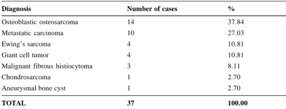
Patient ages ranged from 9 to 81 years, with a mean age of 30.05 years. All diagnoses were confirmed by bone biopsy prior to surgery and are pre-sented in Table 1.
Regarding the anatomic location of the neoplastic lesions, 4 patients had tumors in more than one site. Three patients had osteoblastic osteosar-coma—one had lesions in the distal fe-mur and proximal tibia, one had 3
le-sions in the same femur, and one had 2 lesions in the same femur. The fourth patient had metastatic carcinoma in the proximal, diaphyseal, and distal femur. Therefore, there were 44 distinct ana-tomic sites involved in the 37 patients (Fig. 1).
The follow-up ranged from 2 to 48 months, with a mean follow-up of 14.84 months.
The distribution of the cases ac-cording to the type of endoprosthesis employed is in Table 2.
SURGICAL TECHNIQUE
The surgical approach varied ac-cording to the site of the lesion and fol-lowed these principles: A) Skin and sub-cutaneous tissue incision including the biopsy scar, deepening to the tumor it-self, although not exposing it. B) Hemo-stasis with hemostats and cautery. C) Fascia incision and muscle blunt dissec-tion; there should be constant irrigation
Table 2 - Cases according to type of endoprosthesis.
Type of endoprosthesis Number of cases %
Articulated total knee 11 29.73
Partial bipolar proximal femur 8 21.62
Partial proximal humerus 8 21.62
Total femur 3 8.10
Partial proximal tibia 2 5.41
Diaphyseal femur 2 5.41
Diaphyseal humerus 2 5.41
Total hip proximal femur 1 2.70
TOTAL 37 100.00
Table 1 - Cases according to histologic diagnosis.
Diagnosis Number of cases %
Osteoblastic osteosarcoma 14 37.84
Metastatic carcinoma 10 27.03
Ewing’s sarcoma 4 10.81
Giant cell tumor 4 10.81
Malignant fibrous histiocytoma 3 8.11
Chondrosarcoma 1 2.70
Aneurysmal bone cyst 1 2.70
TOTAL 37 100.00
Figure 1 - Lesions according to anatomic
location.
WIDE RESECTION
The dissection was always per-formed in normal tissue, avoiding any contact with the tumor, allowing proxi-mal and distal safety margins of at least 1 cm around it. Adhesions between sur-gical compartments were not removed, because they may indicate tumor com-promise. The neurovascular bundle was identified in its full extent and maintained under a Penrose drain.
Tissue adhering to the tumor was resected along with it, maintaining the safety margin all around the lesion. Re-section through the tumor pseudocapsule was avoided. The dissec-tion was always through normal muscle. The osseous approach was made proximally and distally to the tumor, always with a safety margin of at least 2 cm. The surgical field was protected with sponges, and the osteotomies were carried out with an oscillating saw, always through normal bone. If necessary, an osteotome was used to finish the osteotomy. The resected tu-mor was removed from the surgical field surrounded by normal tissue.
RECONSTRUCTION
After the wide resection, the limb re-construction depended on the anatomic site of the tumor and used the endoprosthesis that will be described below.
MODULAR ENDOPROSTHESIS
The modular endoprosthesis system is composed of a simple set of instru-ments that allow a series of component combinations, and was designed for a wide variety of reconstructions. We briefly present its components.
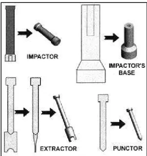
BASIC SET OF INSTRUMENTS
The basic set of instruments for the modular endoprosthesis can be seen in figures 2, 3, and 4.
Figure 4 - Basic set of instruments (acetabulum tester and acetabular augmentation clamp). Figure 3 - Basic set of instruments (impactor, impactor’s base, extractor, and punctor). Figure 2 - Flexible reamers for preparing the medullary canal.
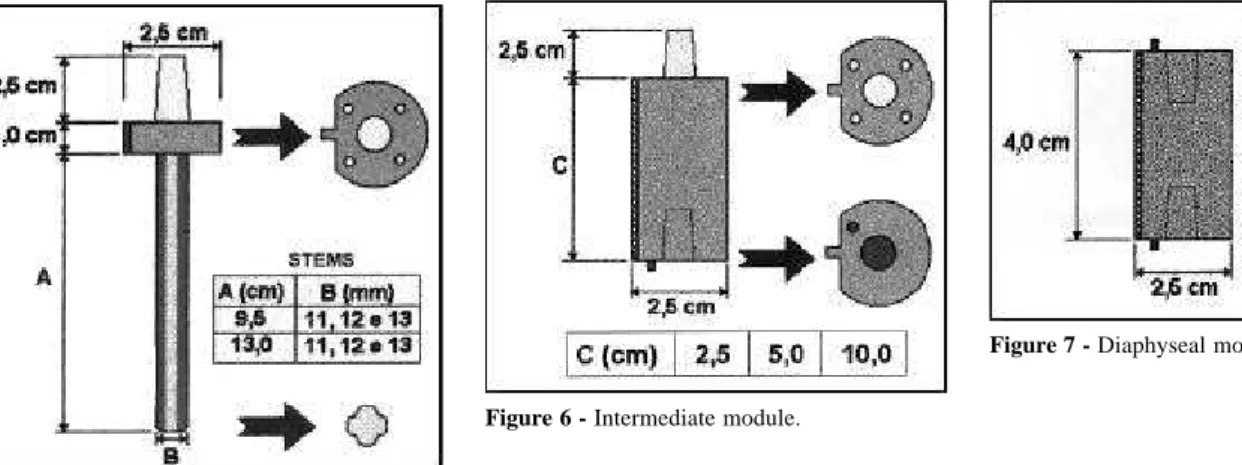
MODULES
The modules are divided into the diaphyseal base (Fig. 5),
Figure 5 - Diaphyseal base.
Figure 6 - Intermediate module.
Figure 7 - Diaphyseal module.
Figure 8 - Trochanteric module.
Figure 9 - Proximal humerus module.
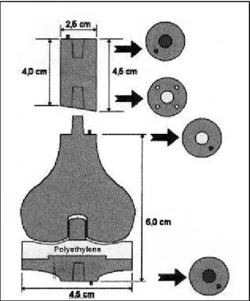
SPECIAL MODULES
The special modules, partial or to-tal, are those used in the articular re-constructions. They are composed of the test bipolar femoral heads (Fig. 10), implant bipolar femoral heads (Fig. 11), partial humeral heads (Fig. 12), articulated total knee endoprosthesis (Fig. 13), and partial proximal tibia endoprosthesis (Fig. 14).
ARTICULATED TOTAL KNEE (DIS-TAL FEMUR) ENDOPROSTHESIS
The total knee endoprosthesis is of the articulated type, with a body of ti-tanium attached to titi-tanium cylindrical pieces and to the intramedullary stem
of 11, 12, or 13 mm diameter with a metal-polyethylene articulation (Fig. 15). It is measured according to the amount of bone to be resected as de-termined in the preoperative studies.
PARTIAL PROXIMAL HUMERUS ENDOPROSTHESIS
The partial proximal humerus modular titanium endoprosthesis is at-tached by a conic fitting to small cy-lindrical pieces measuring 2.5 and 5.0 cm in length, allowing the length of the endoprosthesis to equal the length of the segment resected. The head is made of stainless steel, and the intramedul-lary stem is 8, 9, or 10 mm in diam-eter (Fig. 16).
PARTIAL PROXIMAL FEMUR ENDOPROSTHESIS
Figure 10 - Test bipolar femoral heads.
Figure 13 - Articulated total knee endoprosthesis.
Figure 12 - Partial humeral heads. Figure 11 - Implant bipolar femoral heads.
EVALUATION CRITERIA
For the results analysis, we used oncologic criteria based on the tumor recurrence, as well as clinical criteria based on presence of pain, limb func-tion, articular range of motion of the limb, infection, and patient acceptance.
The results were considered: GOOD
- No local recurrence.
- No spontaneous pain to palpation and motion.
- Functional limb, with variable de-gree of range of motion.
- No persistent local infection. - Good patient acceptance.
REGULAR
- No local recurrence.
- Mild or moderate pain with motion. - Partially functional limb, with
par-tial range of motion limitation. - No persistent local infection. - Regular patient acceptance.
POOR
- Unresectable local recurrence. - Intense pain, spontaneous or in
mo-tion.
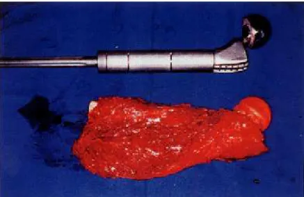
- Great articular instability, restrain-Figure 17 - Partial proximal femur endoprosthesis and proximal femur after wide resection.
Figure 16 - Titanium partial proximal humerus
endoprosthesis and proximal humerus after wide resection.
Figure 15 - Resected distal femur osteosarcoma and the articulated total knee
endoprosthesis.
neck of the prosthesis that is fitted in-ternally in the bipolar head.
PARTIAL PROXIMAL TIBIA ENDOPROSTHESIS
The proximal tibia endoprosthesis used was of the partial type; its titanium body has an intramedullary stem of 8, 9, or 10 mm diameter. Its proximal por-tion, which articulates directly with the distal femur articular surface, is made of polyethylene according to the anatomic shape of the patient’s distal femur.
TOTAL FEMUR ENDOPROSTHESIS
The total femur endoprosthesis used was partial for the hip and total for the knee. Its body is made of titanium and is modular, with cylindrical pieces of 5
or 10 cm in length attached by a conic fitting. The femoral head is of the bipo-lar type, as described for the proximal femur endoprosthesis. The distal portion is made of titanium, using a total articu-lated knee in the same fashion as for the distal femur endoprosthesis (total knee).
FEMORAL AND HUMERAL DIAPHY-SEAL ENDOPROSTHESIS
ing gait.
- Deep local infection, uncontrolled, even after surgical debridement. - Bad patient acceptance.
RESULTS AND COMPLICATIONS
According to the evaluation criteria, the results were good in 21 patients (56.8%), regular in 12 (32.4%), and poor in 4 (10.8%).
Regarding complications, 5 patients developed infection, with 2 deep infec-tions associated with dehiscence, and 3 with superficial infections, all due to Sta-phylococcus aureus. All underwent sur-gical debridement and specific intrave-nous therapy with antibiotics, 3 of them with good results and 2 with poor results. Three patients with dislocation of the proximal femur endoprosthesis under-went closed reduction under anesthesia, all with good results. One of the patients with persistent infection also presented with a module loosening; the case was already considered a poor result. Another patient presented with an intermediate module loosening, requiring a revision procedure for component exchange, with a good result. Finally, one patient devel-oped ulnar nerve paresthesia, credited to the surgical manipulation, but had full re-covery afterwards.
Two other patients developed local recurrence (both osteosarcoma) and underwent radical surgery, these cases thus being considered poor results.
DISCUSSION
Skeletal reconstructions with endoprostheses after wide resection for
bone tumors have been performed for more than a century. Reconstructions were initially used in low-grade malig-nant and metastatic lesions.
As chemotherapy progressed, mainly in primary malignant bone tu-mors such as osteosarcoma and Ewing’s sarcoma, there has been a con-siderable response in terms of tumor volume in a great percentage of the cases, thus making limb salvage pro-cedures feasible in over 50% of the cases.
Additionally, the longer survival of these patients has presented orthopedic surgeons with several orthopedic prob-lems inherent to the materials used in the implants, such as breakage and loosening of the implant, which were less frequently observed previously due to the unsuccessful oncologic treat-ment6,8,9,10.
Increasing numbers of revision pro-cedures have been made in large ortho-pedic centers, thereby increasing the number of complications, primarily in-fections. In response, simpler and longer lasting implants have been de-veloped that allow the partial exchange of the implant in case of a revision pro-cedure.
The modular endoprosthesis sys-tems used by several authors2,3,5,6,8,9,10
since 1985 have the advantage of al-lowing immediate surgery, without the delay required for custom-made endoprostheses. Additionally, use of modular systems circumvents the prob-lem of mistaken measurements for cus-tom-made implants, which leads to im-provisations resulting in more exten-sive resections, longer surgeries, and more intra- and postoperative compli-cations. Modular endoprothesis
sys-tems allow the surgeon to quickly and precisely adjust the endoprosthesis, in-dependently of the extent of the resec-tion, allowing for intraoperative changes in the surgical plan. The endoprosthesis is assembled inside the surgical suite, and additionally is lower in cost.
Another advantage of the modular systems is the possibility of expanding the endoprosthesis in skeletally imma-ture patients in order to equalize the limbs when the patient reaches skeletal maturity. It is possible to attach inter-mediate modules between the modules already in the endoprosthesis in a mi-nor surgical procedure.
The modular system that we devel-oped3 will certainly go through several
modifications as more replacements are performed and more complications from longer follow-up occur. The com-plications reported, such as dislocation of the proximal femur endoprosthesis and infection, occur in the same inci-dence as with the custom-made endoprosthesis5.
We tried to design a simple modu-lar system characterized by easy appli-cation and based on the non-modular endoprostheses, such as the bipolar femoral head (Fig. 17), and with a fit-ting system similar to that of the articu-lated Guepar endoprosthesis5,8.
RESUMO RHCFAP/3020
CROCI AT e col. - O emprego da endoprótese modular de titânio na reconstrução esquelética após ressecção de tumores ósseos. Apresentação do método e análise de 37 pacientes operados. Rev.
Hosp. Clín. Fac. Med. S. Paulo 55
(5):169-176, 2000.
Os autores analisaram 37 pacientes operados de 1992 a 1998 no Grupo de Oncologia Ortopédica, em que foi uti-lizada a endoprótese modular de titânio na reconstrução esquelética, após a ressecção segmentar de tumores ósse-os. Doze pacientes foram do sexo mas-culino e 25 do sexo feminino, sendo que a idade variou de 9 a 81 anos, com
média de 30 anos. O tempo de segui-mento variou de dois a 48 meses, com média de 14 meses. Com relação ao diagnóstico pré-operatório, este foi confirmado pela biópsia em todos os casos e teve a seguinte distribuição: osteossarcoma osteoblástico (14 ca-sos), carcinoma metastático (10), sarcoma de Ewing (4), tumor de célu-las gigantes (4), fibrohistiocitoma ma-ligno (3), condrossarcoma (1) e cisto ósseo aneurismático (1). Nestes foram realizadas onze endopróteses de joelho articulado (total), oito de fêmur proximal com quadril parcial bipolar, oito de úmero proximal parcial, três de fêmur total, duas parciais proximais da tíbia, duas diafisárias de fêmur, duas
diafisárias de úmero, e uma proximal do fêmur com quadril total e compo-nente acetabular sem cimento. As com-plicações relacionadas ao uso da endoprótese foram: infecção (5 casos), luxação (3), soltura de módulo (1) e parestesia do nervo ulnar (1). Utiliza-mos como critérios clínicos a presen-ça de dor, a mobilidade articular, a es-tabilidade da reconstrução, as compli-cações cirúrgicas e oncológicas e a aceitação do paciente. Obtivemos 56,8% de bons resultados, 32,4% de regulares e 10,8% de maus.
DESCRITORES: Endopróteses.
Tumores ósseos. Tratamento cirúr-gico. Neoplasias ósseas.
REFERENCES
1. CAMARGO FP - Ressecção segmental em tumores ósseos e reconstrução cirúrgica do esqueleto : estudo baseado em 81 casos. São Paulo, 1967. (Tese – Faculdade de Medicina, Universidade de São Paulo).
2. CAMARGO OP, OLIVEIRA NRB, CAMPOS FILHO et al. -Polyethylene endoprosthesis for the treatment of malignant bone tumors: a long term follow up study. In: LANGLAIS B (ed.): Limb
salvage major reconstruction in oncologic conditions. Berlin,
Heidelberg, Springer, 1991.
3. CAMARGO OP, CROCI AT & CAMPOS FILHO R - Nova endoprótese diafisária no tratamento das neoplasias ósseas metastáticas. Resultados preliminares. Rev Bras Ortop 1992; 27(11/12): 841-3. 4. CAMARGO OP, CROCI AT & OLIVEIRA NRB - Partial endoprosthesis of the proximal tibia for high grade malignant sarcomas and aggressive benign tumors : a report of 25 cases (1975 - 1990) with a long-term follow-up. In: LANGLAIS B - Limb Salvage. Berlim. Springer, 1993. p. 121-15. (Current Trends).
5. CAMARGO OP, CROCI AT, CAMPOS FILHO R et al. - O sistema de endoprótese modular na reconstrução após ressecção ampla de neo-plasias metastáticas e primárias. Rev Bras Ortop 1995; 30: 815-8.
6. CHAO E & SIM F - Modular Types of Endoprosthesis for Limb Salvag. In: ENNEKING, W.F.(Ed.) - Limb Salvage In Musculoskeletal
Oncology. Philadelphia, Livingstone, 1987. p.198-206.
7. DELITALA F - Costituzione di parti interme del corpo umano. Bologna, Capelli, 1956. (Endoprotesi).
8. JOHNSTON J - A modular prosthetic system for tumor surgeons. In: ENNEKING WF (Ed.) - Limb Salvage In Musculoskeletal
Oncology. Philadelphia, Livingstone, 1987. p.234-7.
9. KOTZ R, RITSCHI P, KROPEJ D et al. - Cementless Modular Prosthesis. Results and Complications. Chir Organ Mov 1990; 75(Suppl.1): 209-211.
10. MALAWER MM & MELLER I - Experience with Extracortical Fixation of Large Segmental Prosthesis and Description of a Modular Segmental Replacement System (MSRS). In: LANGLAIS B (Ed.) Limb salvage, major reconstruction in oncologic
conditions. Berlin, Heidelberg, 1991.