107
JULY-AUGUST REV. HOSP. CLÍN. FAC. MED. S.PAULO 54 (4): 107 - 110, 1999
RECTANGULAR FLAPS TECHNIQUE FOR
TREAT-MENT OF CONGENITAL HAND SYNDACTYLY
Paulo Tuma Jr., Gino Arrunategui, Alexandre Wada, Henri Friedhofer and Marcus Castro Ferreira
TUMA Jr., P. et al. – Rectangular flaps technique for treatment of congenital hand syndactyly. Rev Hosp Clín Fac Med S Paulo 54 (4): 107 - 110, 1999.
SUMMARY: The authors analysed a series of 22 patients undergoing surgical correction of congenital hand syndactyly by the rectangular flap tech-nique. Using our evaluation method, we found that good functional and aesthetic results were obtained in 77.3% of the patients, with a complication rate of 13.6%. We concluded that the rectangular flap technique has a simple design, is easily reproducible by in-training staff, has good results, and can be applied on the majority of the syndactyly cases.
DESCRIPTORS: Hand syndactyly. Surgical treatment. Rectangular flap. Congenital hand anomalies.
Congenital syndactyly is a fre-quent hand anomaly, classified as a failure of differentiation in the seg-ments. It can present by itself in soli-tary form or as a part of a more com-plex syndrome like that of Apert, Moebius, Poland, among others1.
The loss of function related to the syndactyly is usually not severe and varies according to the digital webs affected, presence or not of bone fusion and to other associated anom-alies, especially when it is part of syn-dromes2.
Hand syndactyly has two distinct forms: simple, involving fusion of skin and soft tissues, or complex, involving bone fusion or anomalies in form, size, number or position of the digits. Depending on the extension of the affected web, syndactyly may be classified in partial or complete1.
Surgical treatment aims at the restoration of an adequate interdigit space through separation of fingers using broken line incisions and enough skin cover to avoid scarring and retraction. The objective of the treatment is a functionally and aes-thetically acceptable hand, which may or may not be accomplished on the more severe cases2.
Many techniques for correction of syndactyly have been proposed by authors. The main differences between these techniques are on the geometric form of the web flap, on the incisions to separate the fingers, and the number and position of the skin grafts needed3.
Friedhofer et al.4,5 proposed in
1990 a variation on both the web flap and flaps on the fingers, using rectan-gular flaps, and used the technique for simple syndactyly.
This paper relates to the progres-sion and development of the rectan-gular flap technique previously pro-posed, presenting a further experience over the past five years in treating congenital hand syndactyly.
MATERIALS AND METHODS
Twenty-two patients presenting congenital hand syndactyly were sur-gically treated between January 1993
and January 1998 in the Division of Plastic Surgery at the Hospital das Clínicas, University of Sao Paulo School of Medicine. Thirty hands, and 37 webs were thus treated. Twelve patients were male and ten female. Age varied from eight months to 21 years old, with a median of four years and ten months.
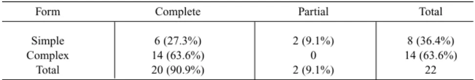
Eight were simple syndactyly (six complete and two partial). Fourteen were complex ones, and all of them complete (Table 1).
Five patients presented syndacty-ly on the right, eight on the left, and nine on both sides (Table 2). Considering the number of affected webs, 11 patients presented syn-dactyly in just one web (4 on both sides), five patients with two affected webs (all one side), two with three webs (one on both sides), and three patients with all webs affected (all both sides). One patient presented syndactyly in two webs of one hand, and three webs of the other hand (Table 3).
Regarding the treated webs, seven patients had the first, 11 the second, 12 the third, and 7 the fourth web involved (Table 4).
RHCFAP/2972
Five patients had Apert`s syndrome, one Poland`s syndrome, and one Down`s syndrome. In 15 cases, a sys-temic syndrome was not found.
SURGICAL TECHNIQUE
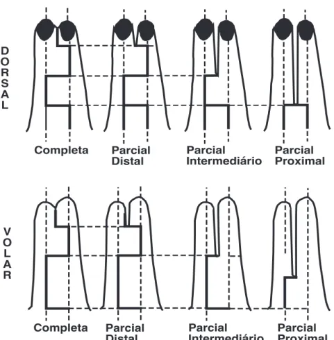
The surgical technique used in all cases was that of rectangular flaps on the fingers and another rectangular flap for the web, originated from the dorsal side of the hand (Fig 1).
Median longitudinal axes are drawn on the affected fingers in front and rear faces: I, II e I´ e II´. The segment BC is obtained on the volar side in the equiva-lent position of the high of the normal webs; the flap ABCD is thus created, with a proximal pedicle; segment A`D` is in the ideal location for positioning the new web on the volar side.
In the distal phalanx, a line passing 4 mm from the cuticular edge of the shortest finger is marked. A third transversal mark EF is made, midway from BC and GI.
The points I´G´ and F´E´ are obtained respectively from the continuation of the lines GI and EF around the fingers, when they cross the axis I´ and II´ on volar side. The lines HJ and H´J´ are over the virtual separation board of the fingers.
Consequently, three dorsal rectan-gular flaps (ABCD, CBEF and EFIH), and two volar rectangular flaps (A´D´F´E´ and F´E´G´H´) are obtained. The flaps are carefully dissected and the fingers separated, with special attention to avoid injury to the vascular and nervous pedicles.
In simple syndactylies, when the flaps are sutured in position, they almost totally cover the digit surface, except for two areas laterally to the web, which should be covered with full thickness skin grafts. In complex syn-dactylies, complementary skin grafts may be necessary for repairing the interdigital spaces. The groin was the donor area for skin mostly used.
Due to the age factor, we favored the use of suture materials with rapid absorp-tion, such as polyglycolic acid products.
Based on the described principles, alternative designs are applied to par-tial syndactylies (Fig 2).
EVALUATION OF RESULTS
The results were assessed accord-ing to the followaccord-ing criteria:
1- Size, angle and capacity to overcross the affected fingers with the normal fingers of the other hand, and the flexor-extensor function of the fin-gers.
2- The aesthetic aspect of fingers, with respect to the integrity of the dor-sal and volar flaps, the quality of the scars, and the need for complementa-ry skin grafts.
The classification is summarized in table 5.
108
REV. HOSP. CLÍN. FAC. MED. S.PAULO 54(4):107 - 110, 1999 JULY-AUGUST
Form Complete Partial Total
Simple 6 (27.3%) 2 (9.1%) 8 (36.4%)
Complex 14 (63.6%) 0 14 (63.6%)
Total 20 (90.9%) 2 (9.1%) 22
TABLE 1- Classification of syndactylies.
TABLE 2- Affected hand.
Hand Number of patients
Right 5 (22.7%)
Left 8 (36.4%)
Both 9 (40.9%)
TABLE 3- Number of affected webs.
Number of webs Patients
1 11 (50%)
2 5 (22.7%)
3 2 (9.1%)
4 4 (18.2%)
TABLE 4- Treated webs.
Web Number of treated webs
1a 7 (18.9%)
2a 11 (29.7%)
3a 12 (32.4%)
4a 7 (18.9%)
Total 37
I I I
J
G
H
I
E
F
B
C
A
D
For a result to be classified as good, none of the characteristics
described for bad or regular results could be present.
Follow up varied from 4 months to 5 years, with median of 11 months.
RESULTS
Nineteen patients (86.4%) needed skin grafts to cover the area resulting from release of digits, and three patients (13.6%) did not need skin grafts (Table 6).
Of the 22 operated patients, three of them (13.6%) had compli-cations. There were two cases of loss of graft, followed by other skin grafting procedures, and one case which had failure of the operation and cicatricial syndactyly, treated with a dorsal web (Table 7). Two patients presented partial loss of the skin graft, but did not need other grafts and healed well.
The result was classified as good in 17 cases (77.3%). The three results classified as bad were those in which a complication occurred. The two patients classified as regular results had partial loss of the graft (Table 8).
DISCUSSION
Some technical principles are universally accepted in the correc-tion of congenital hand syndactyly: the proximal web space must be wide and created using a dorsal flap, and incisions for digit separation should
109
JULY-AUGUST REV. HOSP. CLÍN. FAC. MED. S.PAULO 54(4): 107 - 110, 1999
Good Regular Bad
• Enough size • Not enough size • No web space
• Natural aspect • Abnormal aspect • Abnormal aspect
• Webs • Over-crossing • Difficult over- • Over-cross
possible cross imposssible
• Intact flap • Partial flap loss • Total flap loss
• No skin graft loss • Partial skin graft loss • Total skin graft loss
• Intact flaps • Partial flap loss • Total flap loss
• Fingers • No skin grafts needed • Skin grafts needed • Skin grafts needed
• Absence of • Abnormal scar • Abnormal scar
abnormal scar
• Normal function • Loss of function • Total loss of function
TABLE 5 - Evaluation of results.
Yes 19 (86.4%)
No 3 (13.6%)
total 22
TABLE 6 - Need for skin grafts.
be designed in order to result a broken line1,4,5. When a straight line incision is
done, the resulting scar leads to a flex-ion deformity on the affected digit.
Other important concern is that the vascular-nerve pedicle must be carefully identified and preserved, by dissecting it from the incision line4,5.
The use of magnification and micro-surgical technique is thus essential.
There are many techniques for cor-rection of congenital hand syndactyly, mostly leading to satisfactory results
in the author’s hands1,3,4,5,6.Differences
can be found on the resulting scars, number and design of the flaps, and the number and localization of the necessary skin grafts. No method exists that does not need skin grafts6.
Inguinal, suprascapu lar, or some other areas can be used as donor areas for skin grafts but, most importantly they should be full thickness to reduce the scar contraction.
In the square-flap technique, marking included a small dorsal flap;
however, this was considered
unnec-essary afterwards4. The development
of the technique, described as rectan-gular flap technique, simplified the procedure5.
In this present series, we observed that the technique produced functional-ly good results in 86.4% of the cases (77.3% good and 9.1% regular), with a relatively small complication rate (13.6%), easily corrected with either skin graft or with another flap.
The overall results were consid-ered good, but there was still need for skin grafts (86.4%), although less than with other recent techniques 2,3.
Important features of the tech-nique include its simplicity and easy reproduction by residents in training, with a low complication rate.
The principles of the technique could also be adapted to the more complex forms of syndactyly as in the Apert`s syndrome. The results cannot, of course, be compared with those after the release of simple forms, because of the additional existing functional impairment on those syn-dromic hands.
110
REV. HOSP. CLÍN. FAC. MED. S.PAULO 54(4):107 - 110, 1999 JULY-AUGUST
Good 17 (77.3%)
Regular 2 (9.1%)
Bad 3 (13.6%)
Total 22
TABLE 8 - Final result.
RESUMO RHCFAP/2972
TUMA Jr., P. et al. – Utilização da técnica dos retalhos retangulares na correção de sindactilias
congê-nitas da mão. Rev. Hosp. Clín.
Fac. Med. S. Paulo 54 (4): 107 -110, 1999.
A sindactilia é uma das mais fre-quentes deformidades congênitas da
mão. Neste estudo, são analisados 22 pacientes submetidos a correção de sindactílias congênitas da mão, uti-lizando-se a técnica dos retalhos retangulares. Resultados considerados esteticamente e funcionalmente bons foram obtidos em 77,3% dos casos, e complicações ocorreram em 13,6%. A
técnica mostrou ser de simples exe-cução, e com resultados favoráveis, podendo ser aplicada na maioria dos casos de sindactílias da mão.
DESCRIPTORS: Sindactílias da
mão. Tratamento cirúrgico. Reta-lhos retangulares. Deformidades congênitas da mão.
REFERENCES
1. PERCIVAL N J & SYKES P J - Syndactyly: a review of the factors which influence surgical treatment. J Hand Surg(Br) 1989;14
B:196-200.
2. ZUKER R M, CLELAND H J & HASWELL T - Syndactyly correc-tion of the hand in Apert syndrome. Clin Plast Surg1991; 18(2): 357-364.
3.VAN DER BIEZEN J J & BLOEM J J A M - Dividing the fingers in congenital syndactyly release: a review of more than 200 years of surgical treatment. Ann Plast Surg1994;33: 225-230. 4. FRIEDHOFER H, FERREIRA M C, CARVALHO D A et al.
-Tratamento das sindactilias simples com retalhos quandrangula-res. Rev Bras Cir1986; 76: 85.
5. FRIEDHOFER H & FERREIRA M C - Treatment of congenital syndactyly with retangular flaps. Rev Soc Bras CirPlast 1990; 5
(1): 20-26.
6. FRIEDHOFER H & FERREIRA M C - Treatment of congenital syndactyly with rectangular flaps: a new approach. J Hand Surg
(Br) 1990; 15A: 807.
Received for publication on the 05/05/99
Complications N
Skin graft loss 2 (9.1%)
Recurrence 1 (4.5%)
Total 3 (13.6%)