Article
J. Braz. Chem. Soc., Vol. 26, No. 12, 2571-2582, 2015. Printed in Brazil - ©2015 Sociedade Brasileira de Química 0103 - 5053 $6.00+0.00
A
*e-mail: [email protected]
FTIR Spectroscopy Revealing the Effects of Laser and Ionizing Radiation on
Biological Hard Tissues
Denise M. Zezell,*,a Carolina Benetti,a,b Marcelo N. Veloso,a Pedro A. A. Castroa and Patricia A. Anab
aCentro de Lasers e Aplicações, Instituto de Pesquisas Energéticas e Nucleares (IPEN-CNEN/SP),
05508-000 São Paulo-SP, Brazil
bCentro de Engenharia, Modelagem e Ciências Sociais Aplicadas, Universidade Federal do ABC
(UFABC), 09210-580 São Bernardo do Campo-SP, Brazil
This work evaluated the influence of ionizing and non-ionizing irradiations on the chemical structure of hard tissues using the mid-infrared spectroscopy. Enamel, dentin and bone slabs were submitted to gamma irradiation, as well as to Nd:YAG (λ = 1064 nm) and Er,Cr:YSGG (λ = 2078 nm) laser irradiations. The composition of slabs were evaluated by the Fourier transformed infrared spectroscopy (FTIR), considering the content of organic matrix (amides I, II and III), and inorganic matrix (phosphate and carbonate). Data were statistically analyzed (α = 5%). All irradiations altered the organic and inorganic content of enamel, dentin and bone, and the changes are dependent on the doses applied, as well as on the wavelength used considering the infrared lasers. In conclusion, the attenuated total reflection (ATR)-FTIR technique is an efficient tool to monitor the chemical changes on hard tissues due to ionizing and non-ionizing irradiations, which is important in order to choose safe parameters for a future clinical application.
Keywords: FTIR spectroscopy, laser, gamma irradiation, dental, bone
Introduction
In the last years, it has been demonstrated a significant increase on the use of ionizing and non-ionizing radiations for therapeutic purposes. The ionizing radiations, such as X-ray or gamma irradiation, are well-recognized as effective treatment and diagnostic methods for malignant diseases, as well as for sterilization of tissues.1,2
On the other hand, taking into account their thermal action, high intensity infrared lasers, such as Nd:YAG, CO2, Er:YAG and Er,Cr:YSGG are widely used for therapy of hard tissues.3,4 Depending on the laser parameters adjusted (energy density, mean power, repetition rate, presence of coolant and other characteristics), as well as the conditions of the tissues to be irradiated, infrared lasers are successfully used for caries removal, cavity preparation for future restorative procedure in dentistry, soft tissues surgery, caries prevention, cutting of bone, decontamination of infected tissues and other clinical procedures.5-10 As a coadjutant to the conventional techniques, the use of high
intensity infrared lasers offers advantages such as good homeostasis, faster healing, selective removal of tissues, precise cutting and absence of carbonization, among others. However, these radiations can chemically alter the target tissue, and these changes can affect the morphology, the crystalinity, the healing and mechanical properties of irradiated tissues, mainly considering the hard tissues (enamel, dentin and bone).11-14 In this way, it is necessary to know the chemical changes promoted by non-ionizing and ionizing radiations on hard tissues in order to define safe and effective therapeutic doses without side effects.
spectroscopy (FTIR) is a fast sampling technique which requires virtually no sample preparation, allowing in some cases the signal collection with a fiber in complementary techniques such as attenuated total reflection (ATR). FTIR can be used to characterize biological tissues and give information regarding organic and inorganic content which can be related to mechanical or chemical resistance of the sample, directing the correct choice of irradiation conditions for some specific therapy.
Although it is possible to obtain a compositional analysis of hard tissues using other techniques such as Raman spectroscopy or X-ray diffraction, ATR-FTIR has some advantages since it can provide chemical measurements up to 6 µm in depth (previously calculated considering as 1.63 the refraction index of teeth), while FT-Raman provides a deeper examination.15 In this way, it is possible to evaluate the chemical changes promoted by laser irradiation located strictly on the surface of samples, avoiding the analysis from the untreated tissue. Also, FTIR technique does not present the fluorescence interference such as Raman does, which can be a problem in the analysis of biological tissues. In the same way, FTIR technique requires minimal preparation of samples when compared to X-ray diffraction technique, and it is simpler and does not require the use of ionizing radiation. In this way, FTIR has a potential clinical use.
The biological hard tissues are mainly composed by carbonated hydroxyapatite, and an organic matrix mainly formed by collagen type I. However, the percentage of each component differs from one tissue to another: while enamel has 1.5 wt.% of organic matrix, dentin has 20 wt.% and bone has 25 wt.%.19 A scheme of a human tooth that evidences the biological hard tissues that can be evaluated by FTIR spectroscopy is shown in Figure 1.
The vibrational spectroscopy, such as FTIR, has been shown to be effective for providing chemical analysis of
hard tissues, considering the elevated amount of organic and inorganic molecules that absorb in infrared region.20 In this way, it is possible to obtain important information of the effects of ionizing and non-ionizing radiations. In the present work, it was analyzed the range between 4000 and 580 cm−1 (mid infrared), which corresponds to the absorption bands of the most of organic and inorganic compounds of these tissues.
In this way, the objective of this study was to use the FTIR to evaluate the chemical changes on enamel, dentin and bone, promoted by infrared lasers (Nd:YAG and Er,Cr:YSGG), when used for therapeutic purposes. As well, this study aimed to verify the effects of different doses of gamma radiation on bone tissue.
Experimental
Experimental design
This study was approved by the Animal Ethics Committee of IPEN (6/CEPA-IPEN/SP). It was performed a blind in vitro study with 3 experiments, in which 15 bovine enamel slabs, 15 bovine dentin slabs and 25 bovine bone slabs and 15 rabbit bone slabs were randomly distributed into 14 experimental groups (n = 5), in which they were irradiated with infrared lasers (Nd:YAG and Er,Cr:YSGG), as well as with gamma irradiation (Co60). After treatments, all samples were evaluated by ATR-FTIR, at resolution of 4.0 cm−1, with 80 scans in the range of 4000 to 400 cm−1. The absorption bands considered for this study were phosphate (1300-900 cm−1), amide I (1680-1600 cm−1), amide II (1580-1480 cm−1), amide III (1200-1300 cm−1), the ν2 vibration mode of carbonate (around 870 cm−1) and the superposition of the stretching ν3 and bending ν4 vibration mode of carbonate (between 1600-1300 cm−1). The areas under the considered bands were calculated, and the normalization was performed considering the phosphate (1300-900 cm−1) band. The statistical analysis was performed using ANOVA + Tukey’s test, at 5% significance level. The treatments were considered as a separated block, while the experimental unit was the slab (n = 5).
Experiment 1: analysis of the effects of infrared laser irradiation on dental hard tissues
of samples. After preparation, samples were randomly distributted to six treatment groups (n = 5), as shown in Table 1.
In groups G3-dental-laser and G4-dental-laser, the irradiations were performed using a pulsed Nd:YAG laser (Pulse Master 1000, ADT, USA). This laser operates at wavelength of 1064 nm, fixed pulse duration of 100 µs, beam spot size of 300 µm and 10 Hz of repetition rate. During the experiments, the device was adjusted to a mean power of 0.6 W, energy per pulse of 60 mJ and energy density of 84.9 J cm−2, without air-water refrigeration.10 Before laser irradiations, it was applied a 1-mm thick layer of a photoabsorber (coal paste diluted in 50% alcohol) in order to enhance the laser absorption to the surface of dental hard tissues.21
In groups G5-dental-laser and G6-dental-laser, the slabs were irradiated using an Er,Cr:YSGG laser (Millenium, Biolase Inc., USA), with wavelength of 2780 nm, fixed pulse duration of 140 µs, beam spot size of 750 µm and repetition rate of 20 Hz. In group G5-dental-laser, enamel samples were irradiated using mean power of 0.75 W, energy density of 8.5 J cm−2, energy per pulse of 37.5 mJ and without air-water coolant.In group G6-dental-laser, dentin samples were irradiated using mean power of 0.25 W, energy density of 2.8 J cm−2, energy per pulse of 12.5 mJ and without air-water coolant.
During the irradiations, all samples were immobilized in X-Y-Z optical supports, and laser handpiece was coupled to a computer managed motion control system (ESP300, Newport, USA) adjusted to a fixed speed in order to avoid unlased areas or overlapping of focused areas. Laser tip was kept at the focal distance from dental hard tissues surface, and laser irradiation was done scanning all areas of slabs. Before irradiation and every five irradiated samples, the energy per pulse was calibrated by an energy/power meter (FieldMaster, Coherent, USA).
Experiment 2: analysis of the effects of infrared laser irradiation on bone tissue
For this analysis, 15 bone slabs (8 × 5 × 0.1 mm) were obtained from tibias of adult male New Zealand rabbits using a dental high-speed drill. The slabs were cleaned with deionized water and sequentially polished with silicon carbide abrasive paper (#1200, Buehler, USA) in order to obtain a parallel surface. After preparation, the slabs were kept in humid environment under refrigeration (+4 °C). Samples were randomly distributed to three treatment groups (n = 5): (i) group G1-bone-laser: untreated slabs; (ii) group G2-bone-laser: irradiated with Er,Cr:YSGG laser, at energy density of 3 J cm−2; (iii) group G3-bone-laser: irradiated with Er,Cr:YSGG laser, at energy density of 15 J cm−2.
For irradiations, it was used the Er,Cr:YSGG laser (Millenium, Biolase Inc., USA), as described before. For group G2-bone-laser, the equipment was adjusted to mean power of 0.25 W, energy density of 3 J cm−2 and energy per pulse of 12.5 mJ. For group G3-bone-laser, it was used the mean power of 1.25 W, energy density of 15 J cm−2 and energy per pulse of 62.5 mJ. All samples were irradiated without air-water coolant, using the high precision motorized translator and the energy/power meter, as described before.
Experiment 3: analysis of bone samples submitted to gamma radiation
Twenty five 7 × 7 × 1 mm slabs were prepared from tibias of bovine bone using a diamond saw under distilled water refrigeration. All slabs were washed with distilled water, and the periosteum and bone marrow were removed with a scalpel. After cleaning, all slabs were flattened and polished using silicon carbide abrasive paper (#2500 and #4000, Buehler, USA). Samples were frozen (−20 °C) in sealed plastic tubes until the beginning of the experiments.
For irradiations, the slabs were randomly distributed into 5 experimental groups (n = 5), as shown in Table 2. Table 1. Details of the experimental groups for analysis of the effects of
infrared laser irradiation on dental hard tissues
Experimental group Denomination
Energy density / (J cm−2)
G1-dental-laser untreated enamel −
G2-dental-laser untreated dentin −
G3-dental-laser enamel irradiated with Nd:YAG 84.9
G4-dental-laser dentin irradiated with Nd:YAG 84.9
G5-dental-laser enamel irradiated with Er,Cr:YSGG
8.5
G6-dental-laser dentin irradiated with Er,Cr:YSGG 2.8
Table 2. Experimental groups of tibia bone slabs submitted to gamma radiation
Experimental group Treatment / kGy
G1-bone-γ 0.01
G2-bone-γ 0.1
G3-bone-γ 1
G4-bone-γ 15
The irradiations of groups G1-bone-γ, G2-bone-γ and G3-bone-γ were performed in a Co60 Gammacell irradiator (1.43 kGy h-1, IPEN-CNEN/SP), while the irradiations of G4-bone-γ and G5-bone-γ groups were performed at Co60 multipropose irradiator (6.0 kGy h−1, IPEN-CNEN/SP).
Compositional analysis
For all the compositional analysis of the present study, it was used the attenuated total internal reflectance technique of the Fourier transform infrared spectroscopy (ATR-FTIR, Thermo Nicolet Smart Orbit, Thermo Scientific®, USA). The ATR-FTIR spectra of each sample were obtained with 4.0 cm−1 resolution, on a spectrometer with a deuterated triglycine sulfate and thermoelectricity cooled (DTGS-TEC) detector using a diamond crystal (Smart Orbit, Thermo Scientific®, USA). In each slab, it was collected five spectra in an area of 1.5 mm2 which corresponds to the size of diamond crystal. Each spectrum had a background spectra subtracted during acquisition and was obtained with 80 scans in the range of 4000 to 400 cm−1. The acquisition of spectra was performed using the OMINIC (Thermo Scientific®, USA) software.
For the qualitative and semi-quantitative analysis, the absorption bands considered for this study were the superposition of the ν1 and ν3 vibration modes of phosphate (1300-900 cm−1), amide I (1680-1600 cm−1), amide II (1580-1480 cm−1), amide III (1200-1300 cm−1), the ν2 vibration mode of carbonate (around 870 cm−1) and the superposition of the stretching ν3 and bending ν4 vibration mode of carbonate (between 1600-1300 cm−1).22 The carbonate and phosphate infrared bands are related to the carbonated hydroxyapatite, which is the main component of hard tissues. The purety and quality of the biological apatite is related with its solubility; in this way, the analysis of carbonate related to the phosphate give information about the structure and crystallinity of hard tissues after the proposed treatments.23 Also, the content of amide I, II and III are related to collagen content in the organic matrix of hard tissues, and the analysis of these bands can provide important information about the conformation and denaturation of this molecule.24
After selection of the bands, the background signal was subtracted and, for a semi-quantitative comparison among groups, the areas under the considered bands were calculated after normalization by the area of the ν1 and ν3 vibration modes of phosphate band (1300-900 cm−1).25,26 In this way, the ratio between the area of 850-890 cm−1 carbonate band and the area of phosphate band is related to the carbonate-to-phosphate proportion. As well, the ratios between amide I/phosphate, amide II/phosphate
and amide III/phosphate are related to the proportion of collagen and the hydroxyapatite.27 It was also performed the analysis of the second derivative, which was used to evaluate the presence and the position of infrared bands.
For bone slabs submitted to gamma irradiation, the compositional analysis was performed two times for each sample: before irradiation (control group) and after irradiation (treated groups). In addition, it was calculated the crystallinity index, according to the following equation:28
551 597
588 + =
I I
IC
I (1)
in which: I551 = intensity of 551 cm−1 band; I597 = intensity of 597 cm−1 band; I
588 = intensity of 588 cm−1 band. The normalization of spectra and the calculation of the areas under the bands were performed using the Origin 8.0 and a developed routine on MatLab software, respectively.
Statistical analysis
The statistical analysis was performed individually for each experiment, using the GraphPad Prisma software. Before the statistical analysis, independence, homogeneity (Levene’s test) and normality of variances (Shapiro-Wilk’s test) of experimental data were tested. After the confirmation of all these requirements, all results were analyzed by ANOVA followed by Tukey’s test, at 5% significance level. The treatments were considered as a separated block, while the experimental unit was the slab (n = 5). For each experiment, the effects of each treatment were evaluated to the main component of respective tissue. The treatments were compared with the control group and with each other. Letter marks were used to indicate the statistic results in the graphics. The absence of letters indicates that no statistic difference was observed, as well as distinct letters indicate statistically significant differences among the treatments according to Tukey’s test (p < 0.05).
Results and Discussion
denaturation of collagen and the evaporation of water, which can be noticed as the decrease on the proportion of the infrared bands corresponding to the amides I, II and III in relation to the phosphate when analyzed by FTIR spectroscopy. These effects are more pronounced in dentin and bone, since these tissues have more content of organic matrix when compared to the enamel. In relation with inorganic matter, the literature reports that laser irradiation can lead to evaporation of carbonate and, in this way, FTIR analysis can detect a decrease on the proportion of carbonate bands in relation to phosphate on irradiated samples. Also, changes in band positions, appearance or disappearance of new infrared bands may indicate carbonization or degeneration of biological hard tissues due to laser irradiation.
Figure 2a shows the region of 1700-700 cm−1 of the averaged infrared spectra of untreated enamel slabs, as well as the averaged infrared spectra of enamel irradiated with Nd:YAG and Er,Cr:YSGG lasers. Figure 2b shows the second derivative of the same spectra. None of treatments promoted the formation or disappearance of infrared bands, as well as it was not evidenced changes on the position of these bands after laser irradiation, which indicate that infrared laser irradiation did not produce degeneration of enamel when used at the energy densities of the present study. These results agree with previous studies using FTIR on powder enamel22 and FT-Raman spectroscopy on human enamel at the same laser conditions than those used in the present study,30 and confirm that laser parameters used at the present study are not prejudicial to enamel structure. The parameters chosen were based on previous studies8,10 that confirm the potential of Nd:YAG and Er,Cr:YSGG lasers for preventing dental caries and, for that, laser
irradiation should change the chemical or morphological characteristics of dental hard tissue in order to reduce enamel demineralization without promoting any prejudicial effects, such carbonization.
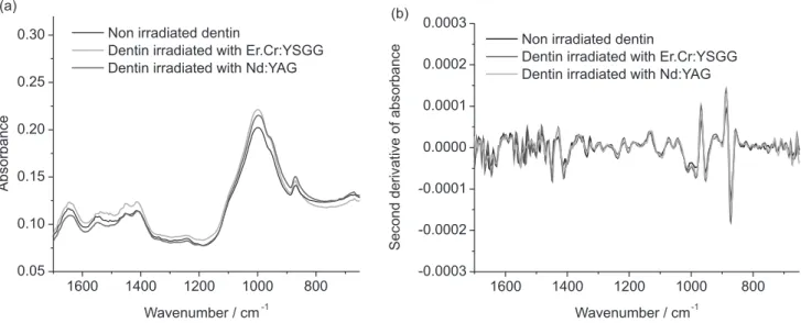
The infrared bands observed in enamel spectra are close to those reported on literature,22,25,26 and the absence of amide III and water bands is expected, since the enamel has only 1.5 wt.% of organic content (which corresponds to type-I collagen) and 97 wt.% of inorganic matrix, mainly composed by carbonated hydroxyapatite. In this way, due to the few amount of organic and water content of enamel, the infrared bands corresponding to collagen and water have lower intensities when compared to phosphate and carbonate bands, which are related to the inorganic content of enamel. The fingerprint region (1700-700 cm−1) mean infrared spectra of untreated dentin and dentin irradiated with Nd:YAG and Er,Cr:YSGG lasers are shown in Figure 3a. In these spectra, it is observed the presence of phosphate, as well as the infrared bands corresponding to water (3450-3300 cm−1), amide I, amide II and amide III. The presence of these bands are related to the chemical composition of dentin, composed by 20 wt.% of organic content (which corresponds to type-I collagen) and 70 wt.% of inorganic matrix (carbonated hydroxyapatite) and agrees with the literature.26 Figure 3b shows the second derivative of the same dentin spectra. As the same way than the observed for enamel, it was not evidenced the disappearance or changes in the position of the infrared bands after laser irradiation, which also indicates that both infrared lasers do not promote chemical damages on dentin structure when used at the conditions of the present study.
The results of the semi-quantitative analysis of organic and inorganic content of enamel and dentin after laser
1600 1400 1200 1000 800
-0.0015 -0.0010 -0.0005 0.0000 0.0005 0.0010
0.0015 Untreated enamel
Enamel irradiated with Er.Cr:YSGG Enamel irradiated with Nd:YAG
S
e
c
o
n
d
d
e
ri
v
a
ti
v
e
o
f
a
b
s
o
rb
a
n
c
e
1600 1400 1200 1000 800
0.0 0.2 0.4 0.6 0.8 1.0 1.2 1.4
A
b
s
o
rb
a
n
c
e
Wavenumber / cm-1 Wavenumber / cm-1
Untreated enamel
Enamel irradiated with Er.Cr:YSGG Enamel irradiated with Nd:YAG
(a) (b)
Figure 2. (a) Means of infrared spectra (1700-600 cm−1) obtained after the treatments proposed for enamel (spectra normalized by phosphate band); (b)
treatments are shown in Figures 4a and 4b, respectively. Considering these analysis for enamel, it was observed that both Nd:YAG and Er,Cr:YSGG laser irradiations did not significantly alter the proportion of collagen (amide I and amide II) in relation to phosphate band (ν
1 and ν3 vibrations); however, it was possible to observe a tendency to have a smaller proportion of collagen and the hydroxyapatite in the irradiated samples. Concerning the content of carbonate, the same tendency was observed, although it was not evidenced a statistically significant difference, as it can be noted in Figure 4a.
Both Er,Cr:YSGG and Nd:YAG lasers are widely used for irradiating dental hard tissues and, at the conditions used in the present study, they are indicated for preventing dental demineralization.21,31 The literature reports that
Er,Cr:YSGG laser, when used at the same parameters used in the present study (0.25 W, 2.8 J cm−2, 20 Hz), can induce averaged temperature rises up to 250 °C on enamel, when measured with a thermographic camera,11 and maximum temperature rises up to 400 °C when used at 8 J cm−2 and measured using an elliptical mirror and a HgCdZnTe detector.32 These temperature increases can induce chemical changes on enamel, such as loss of carbonate and water, which begins when temperature rises above 100 °C.13 Higher temperatures are necessary to promote the oxidation of phosphates and formation of pyrophosphates, as well as the formation of new crystalline phases due to laser irradiation.33 In this way, considering the energy density used in the present study, the observed tendency on reducing the carbonate content is expected. However, the low number
1600 1400 1200 1000 800
0.05 0.10 0.15 0.20 0.25 0.30
1600 1400 1200 1000 800
-0.0003 -0.0002 -0.0001 0.0000 0.0001 0.0002 0.0003
Non irradiated dentin
Dentin irradiated with Er.Cr:YSGG Dentin irradiated with Nd:YAG
Se
c
o
nd
de
ri
v
a
ti
v
e
of
ab
s
o
rb
an
c
e
Wavenumber / cm-1 (b)
A
b
s
o
rb
a
n
c
e
Wavenumber / cm-1 Non irradiated dentin
Dentin irradiated with Er.Cr:YSGG Dentin irradiated with Nd:YAG (a)
Figure 3. (a) Means of infrared spectra (1700-600 cm−1) obtained after the treatments proposed for dentin (spectra normalized by phosphate band); (b)
second derivative of the same spectra.
0.01 0.1 1
Amide I Amide II Amide III Carbonate
0.01 0.1
1 ControlEr.Cr:YSGG
Nd:YAG (b) Dentin
Carbonate Amide II
B B
a a
n n
d d
a a
r r
e e
a a
/ /
P P
h h
o o
s s
p p
h h
a a
t t
e e
b b
a a
n n
d d
a a
r r
e e
a a
Control Er.Cr:YSGG Nd:YAG
Amide I (a) Enamel
of samples used in the present study (n = 5) seems to be the reason for the non-significant statistical differences observed. The reduction of carbonate due to Er,Cr:YSGG laser irradiation on enamel, when used at the same energy density of this study, was previously reported on irradiated enamel powder by FTIR,22 or on human enamel evaluated by FT-Raman spectroscopy.30
At the same parameter used in the present study (0.6 W, 84.9 J cm−2, 10 Hz), Nd:YAG laser promotes temperature to rises up to 615 °C on enamel.21 In this way, it was also expected the reduction of carbonate content due to Nd:YAG laser irradiation, as it was previously demonstrated in a study using FT-Raman spectroscopy.30 However, in thepresent study, it was only demonstrated a tendency, which can be also explained due to the low number of samples used.
At the same way, it was noted that neither Nd:YAG nor Er,Cr:YSGG laser irradiation promoted statistically significant changes on the organic content of dentin, but the same tendency to decrease the organic content after laser irradiation was detected (Figure 4b). This effect is also attributed to the temperature rises on the surface due to laser irradiation, considering that the denaturation of the organic components can occur at temperatures up to 400 °C.33 However, the temperature rises due to Er,Cr:YSGG and Nd:YAG laser irradiations on the dentin surface, when used at the same parameters than used in the present study, are not known and must be further investigated in order to determine if the parameters chosen are reliable for a future clinical application.
Also concerning the effects on dentin, it was noted that laser irradiation did not change the carbonate content of dentin and, this time, no tendency was observed. Although
infrared laser irradiation promotes localized temperature rises on dentin surface, we must consider the low content of inorganic matrix of dentin when compared to enamel.19 In this way, the effects on the reduction of carbonate observed on dentin can be lower than those observed on enamel.
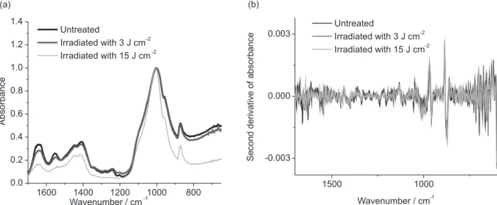
Figure 5a shows the means of infrared spectra obtained after Er,Cr:YSGG laser treatments proposed for bone and its second derivative, Figure 5b. The spectra are normalized by phosphate band. The infrared spectrum of bone is very similar to enamel and dentin spectra, since it is also composed by an organic matrix mainly formed by collagen type I and an inorganic matrix mainly formed by carbonated hydroxyapatite. The main difference between these tissues is related with the amount of organic component: in bone tissue it represents about 25 wt.%, while in dentin and enamel the organic matrix represents 20 and 1.5 wt.%, respectively.19 In this way, the main infrared bands observed in bone tissue are the same observed in dentin and enamel: water, amide I, amide II, amide III, carbonate and phosphate.
In spectra showed in Figure 5, it is possible to observe the maintenance of all the main infrared bands of bone tissue, which suggests that the Er,Cr:YSGG laser irradiation, in both conditions used in the present experiment, did not promote the total degradation of any component of bone. It is a positive effect for a future clinical application, considering that Er,Cr:YSGG laser can be used in surgery for cutting bone,6 since it is desirable that laser irradiation does not promote deleterious effects on bone, which can affect the healing of this tissue.
It is possible to observe changes in band profiles, mainly in the spectrum of the group irradiated with 15 J cm−2, which suggests that the Er,Cr:YSGG laser irradiation cause some
1600 1400 1200 1000 800 0.0
0.2 0.4 0.6 0.8 1.0 1.2 1.4
1500 1000
-0.003 0.000 0.003
Untreated
Irradiated with 3 J cm-2 Irradiated with 15 J cm-2
S
e
c
o
n
d
d
e
ri
v
a
tiv
e
o
f
a
b
s
o
rb
a
n
c
e
Wavenumber / cm-1 (a)
Untreated
Irradiated with 3 J cm-2 Irradiated with 15 J cm-2
A
b
s
o
rb
a
n
c
e
Wavenumber / cm-1
(b)
Figure 5. (a) Means of infrared spectra (1700-600 cm−1) obtained after the Er,Cr:YSGG laser irradiation of bone (spectra normalized by phosphate band);
chemical changes in bone samples. However, since none of the conditions used in this study were high enough to promote a total degradation of the organic components, it is possible to infer that the Er,Cr:YSGG laser irradiation did not promote an increase in surface temperature higher than 700-1500 °C. It is important to consider that these temperatures promote the total degradation of the organic material.34
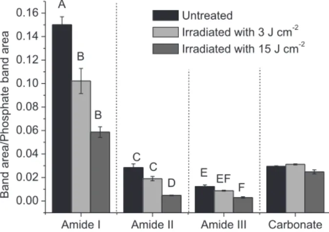
Figure 6 shows the average ratios of amide II/phosphate, amide II/phosphate, amide III/phosphate and carbonate/ phosphate obtained from untreated bone and bone treated with Er,Cr:YSGG laser at energy densities of 3 and 15 J cm−2. It was observed a statistically significant decrease of organic components between the untreated group and the group irradiated with 15 J cm−2 (considering the content of amide I, amide II and amide III). Significant differences between the untreated group and the group irradiated with 2.7 J cm−2 were only observed in the amide I analysis. Considering the analysis of carbonate band, no statistical difference was observed among the groups (p > 0.05).
The analysis of band ratios suggested that the temperature rise in bone during the laser irradiation was enough to promote the degradation of organic components, mainly considering the energy density of 15 J cm−2. The literature reports that the degradation of organic matrix starts at 175-200 °C;35 in this way, it is reasonable to infer that the temperature during irradiation reached this temperature range. However, in the group irradiated with 3 J cm−2, the organic degradation was only observed concerning the amide I band and, in the same way that it was observed in dentin (experiment 1), only a trend to decrease the content of amide II and amide III after laser irradiation was observed. It is
important to consider that the amide I band is overlapped with a vibration mode of water36 and, in this way it is difficult to know the exact contribution of each component for this band. Since the water evaporation starts nearly in 100 °C,37 it is possible to infer that the small changes on organic component observed in the group irradiated with 3 J cm−2 were due only to the water evaporation.
The Er,Cr:YSGG laser used in this study promotes thermal ablation of mineralized tissues. In this process, the laser energy is absorbed by the water molecules of the irradiated material; the temperature rise induces the water boiling and increases of internal pressure; a micro exposure happens, causing removal of material and the dissipation of heat.38 The minimal energy density able to remove material is called ablation threshold.39 The change in surface temperature during laser irradiation is reported to be the main responsible for the chemical modification of hard tissues.40
The energy densities used in this experiment represent two different situations: the energy density of 2.7 J cm−2 is lower than the Er,Cr:YSGG ablation threshold to bone tissue;41 in this way it is not able to cause the removal of material, but it is enough to promote an increase of the temperature surface taking into account the high absorption of the 2.78 µm radiation by water and hydroxyapatite.37 On the other side, the energy density of 15 J cm−2 is higher than the ablation threshold of Er,Cr:YSGG laser to bone tissue, so the ablation of bone is observed. In this situation, the material that had the greater increase in temperature is removed, and the remaining material is also heated, although at temperatures lower than the ablation threshold. In this way, the remaining tissue can be chemically altered by laser irradiation even after the ablation process.42
In the group irradiated with 15 J cm−2 the proportion between the amide I, amide II and amide III were significantly decreased in relation to the untreated bone, which suggests that the changes observed are related with the degradation of organic components in the remaining tissue. In fact, the proportions of amide I/phosphate, amide II/phosphate and amide III/phosphate in this group were more than 50% less when compared with the ratio values of the untreated samples, and this fact happens in organic components when temperature rises are at least 150 °C.43 In this way, the results suggested that the remaining bone tissue of the ablation process reached values nearly 200 °C.
No changes were observed in carbonate ratio analysis when comparing the irradiated samples with the untreated group. The literature reports that the reduction of carbonate content on bone happens in the range of 200 to 600 °C.34 In this way, the results suggest that the temperature rises promoted by Er,Cr:YSGG laser irradiation were not high enough to cause the degradation of the carbonate content. Amide I Amide II Amide III Carbonate
0.00 0.02 0.04 0.06 0.08 0.10 0.12 0.14 0.16
B
C C
D A
B
a
n
d
a
re
a
/P
h
o
s
p
h
a
te
b
a
n
d
a
re
a
Untreated
Irradiated with 3 J cm-2 Irradiated with 15 J cm-2 B
E EF F
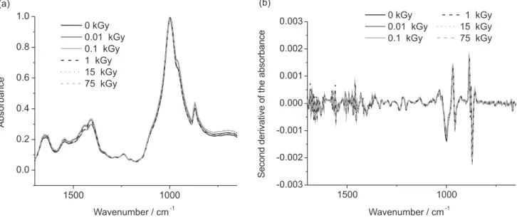
The mean of infrared spectra (1700-600 cm−1) obtained from bone tissue after gamma irradiation at different doses and their second derivative are shown in Figures 7a and 7b, respectively. It is possible to evidence the main absorption bands of bone and that gamma radiation, independent of the dose, did not promote the disappearance of infrared bands or even changed the position of bands. In this way, at the doses selected in the present study, it is possible to infer that gamma irradiation did not promote the degradation of organic or inorganic matrix of bone tissue.
The gamma radiation is widely used for sterilizing foods,44 biomaterials,45 as well as allograft soft46 and hard tissues47 aimed to transplantation and tissue engineering. Although other methods such as sterilization with high temperature or with chemical compounds have been reported,48 the sterilization using gamma irradiation has been demonstrated to be very effective for this purpose.49 However, the literature shows contradictory aspects concerning the effects of gamma irradiation on tissues, mainly related with the physical and biological properties of irradiated tissues.50 Considering the importance of bone allografts, it is important to know the compositional effects of this irradiation on bone tissue at different doses.
A previous study demonstrated that gamma radiation interacts with the bone at its molecular level in a dose-dependent manner, and can promote collagen degradation and the reduction of density of the intermolecular crosslinks.51 Concerning the mechanical properties, the literature shows reductions in ultimate strength and resistance to fatigue crack growth;52 on the other hand, it was reported that gamma irradiation does not alter the stiffness or strength of intact cancellous bone allograft.53
In this way, there are contradictory results on literature, mainly related with the substrate to be evaluated (only collagen fibers, different types and regions of bone tissue), as well as to the evaluation method and doses. For reducing the risk of disease transmission through bone allograft, the recommended dose of gamma radiation sterilization is 25-35 kGy.54
In the present work, distinct doses of gamma irradiation were evaluated on bone, in order to characterize the chemical changes promoted in a dose-dependent manner. Figure 8 shows the results of proportion of organic (amide I, amide II and amide III) related to phosphate (Figure 8a) and the proportion of carbonate related to phosphate content (Figure 8b). It is noted a statistically significant increase on the proportion of amide II as the gamma doses have increased. However, concerning the contents of amide I and amide III related to the phosphate band, it was not observed any statistical differences when compared to the control group (untreated bone). The same finding was observed when the ratio of carbonate related to phosphate was evaluated.
The analysis of amide I, amide II and amide III are related with the organic content of bone, mainly related with the type-I collagen. The amide I band results from the stretching vibration of the peptide carbonyl group55 and the ratio of its sub-bands (range of 1660 and 1690 cm−1) also corresponds to collagen cross-links that are abundant in mineralized tissues (pyridinoline and dehydrodihydroxylysinonorleucine).56 The proportion of these substances is also related to maturation of collagen present in bone. In this way, we can suggest that the maturation of collagen of bone is not altered by gamma irradiation at the doses used in the present study.
1500 1000 0.0
0.2 0.4
0.6 0.8 1.0
1500 1000
-0.003 -0.002 -0.001 0.000 0.001 0.002
0.003 0 kGy 1 kGy
0.01 kGy 15 kGy
0.1 kGy 75 kGy
S
e
c
o
n
d
d
e
ri
v
a
ti
v
e
o
f
th
e
a
b
s
o
rb
a
n
c
e
Wavenumber / cm-1 Wavenumber / cm-1
(b)
A
b
s
o
rb
a
n
c
e
0 kGy 0.01 kGy 0.1 kGy 1 kGy 15 kGy 75 kGy (a)
Figure 7. (a) Means of infrared spectra (1700-600 cm−1) obtained after treatments proposed for bone submitted to different doses of gamma irradiation
In the same way, the contents of amide II and amide III are also related to the organic content of bone, and refer to secondary structure of collagen. The amide II band correspond for the combination of N−H bending and C−N stretching vibrations, while the amide III band refers to the C−N stretching and N−H in-plane bending.55 In this study, it was observed a significant change on the amide II peak, which indicates that gamma radiation induced changes on helical structure of collagen, and these changes have a direct relation with the dose. These results confirm the literature findings,51 and suggest that the use of gamma radiation preferentially alters the collagen of bone.
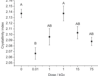
On the contrary to it was observed when using laser irradiation on bone, the results obtained in the present experiment indicate that gamma irradiation does not interfere in the inorganic matrix of bone, since the content of carbonate was not altered after gamma irradiation. For an additional analysis of the effects of gamma radiation on inorganic matrix of bone, it was also calculated the crystallinity index, and the results can be seen in Figure 9. It was detected that this index significantly decreased when the dose of 0.01 kGy was used; however, it was noted that other doses did not promote significant changes on the crystallinity index when compared to the non-irradiated samples.
The crystallinity index is useful for evaluating the order, organization and tension of the hydroxyapatite crystals.28 The literature shows that the crystal growth and crystallinity of hard tissues can be affected by factors such as the maturity degree of tissue or even due to the increase on the temperature.57 The present study evidenced that low doses of gamma radiation decrease the crystallinity of bone;
however, after 0.1 kGy, the crystallinity is not altered. In this way, we can infer that low doses of gamma radiation have a significant effect on the crystal characteristics of hydroxyapatite from the bone and this effect, together with the effect on collagen secondary structure, can be the main reason for the changes on the mechanical properties on sterilized bone showed on literature.
A literature study that evaluated the effects of gamma radiation on bone by Raman spectroscopy evidenced that changes in organic components occur in doses higher than 100 kGy, and no changes on inorganic components were observed in any dose used (from 10 kGy up to 1000 kGy).58 Using a similar methodology than we used in the present study, a previous ATR-FTIR analysis
Amide I Amide II Amide III
0.00 0.02 0.04 0.06 0.08 0.10 0.12 0.14
Carbonate 0.0
0.2 0.4 0.6 0.8 1.0 1.2 1.4 1.6 1.8
0 kGy 0.01 kGy 0.1 kGy 1 kGy 15 kGy 75 kGy
B B B A AB
B B
a a
n n
d d
a a
r r
e e
a a
/ /
P P
h h
o o
s s
p p
h h
a a
t t
e e
b b
a a
n n
d d
a a
r r
e e
a a
0 kGy 0.01 kGy
0.1 kGy 1 kGy
15 kGy 75 kGy
AB
(a) (b)
Figure 8. Means of amide I/phoshate, amide II/phosphate and amide III/phosphate ratios (a) and carbonate/phosphate ratios (b) obtained from bone samples irradiated with gamma radiation. Bars denote standard deviations. The absence of letters indicates that no statistic difference was observed, while distinct letters indicate statistically significant differences among the treatments according to Tukey’s test (p < 0.05).
0 0.01 1 1 15 75
2.05 2.06 2.07 2.08 2.09 2.10 2.11 2.12 2.13 2.14 2.15 2.16
AB AB
A
AB
B
C
ry
s
ta
lli
n
it
y
index
Dose / kGy A
in enamel showed no differences on the proportion of carbonate in relation to phosphate after gamma radiation at dose of 0.06 kGy.59 Our study differs from the Raman study since it was observed changes on amide II band after irradiation with 1 kGy, which can be explained by differences on methodology of analysis. Concerning the inorganic material (content of carbonate), the present work agrees with both mentioned studies. In this way, considering the few and divergent results observed in literature, more studies are necessary to evaluate the efficacy of each methodology for analyzing the effects of ionizing radiation on bone tissue.
Conclusions
According to the results observed in the present study, it is possible to conclude that infrared lasers can chemically alter the microstructure of enamel, dentin and bone; these changes are mainly related with the organic content of these tissues and are dependent on the laser wavelength, as well as the energy density used. Gamma irradiation can alter the chemical structure of bone, and these changes are mainly related to the amide II content. In this way, FTIR spectroscopy is a useful and confiable technique to evaluate the chemical changes promoted by non-ionizing and ionizing radiation on hard tissues.
Acknowledgements
The authors would like to thank to CAPES (PROCAD 8881.068505/2014-01), CNPq (473723/2007-7, INFO INCT 573.916/2008-0 and PQ 312397/2013-5) and FAPESP (2006/06746-0 and CEPID 05/51689-2) for giving financial support for this research.
References
1. Kjaer, A.; Knigge, U.; Scand. J. Gastroenterol. 2015, 6, 740.
2. Anderson, M.; Keyak, J.; J. Bone Jt. Surg., Am. Vol. 1992, 74, 747.
3. Featherstone, J. D. B.; Dent. Clin. North Am. 2000, 44, 955.
4. Kang, H. W.; Oh, J.; Welch, A. J.; Phys. Med. Biol. 2008, 53, 3381.
5. Zezell, D. M.; Ana, P. A.; Benetti, C.; Goulart, V. P.; Bachmann, L.; Tabchoury, C. P. M.; Cury, J. A.; Proc. SPIE
2010, 7549, 75490G.
6. Wang, X.; Zhang, C.; Matsumoto, K.; Laser Med. Sci. 2005,
20, 21.
7. Buyukhatipoglu, I.; Secilmis, A.; Eur. J. Dent. 2015, 9, 284.
8. Ana, P. A.; Tabchoury, C. P.; Cury, J. A.; Zezell, D. M.; Caries Res. 2012, 46, 441.
9. Archilla, J. R.; Moreira, M. S.; Miyagi, S. P.; Bombana, A. C.; Gutknecht, N.; Marques, M. M.; J. Biomed. Opt. 2012, 17, 118002.
10. Zezell, D. M.; Boari, H. G. D.; Ana, P. A.; Eduardo, C. P.; Powell, G. L.; Lasers Surg. Med. 2009, 41, 31.
11. Ana, P. A.; Blay, A.; Miyakawa, W.; Zezell, D. M.; Laser Phys. Letters 2007, 4, 827.
12. Morioka, T.; Tagomori, S.; Oho, T.; J. Clin.Laser Med. Surg.
1991, 9, 215.
13. Kuroda, S.; Fowler, B. O.; Calcif. Tissue Int. 1984, 36, 361. 14. Onisor, I.; Pecie, R.; Chaskelis, I.; Krejci, I.; Eur. J. Paediatr.
Dent. 2013, 14, 140.
15. Prasad, P. N.; Introduction to Biophotonics, 1st ed.; Wiley: New
York, USA, 2003.
16. Popp, J.; Ex-Vivo and In-Vivo Optical Molecular Pathology, 1st
ed.; Wiley: New York, USA, 2014.
17. Diem, M.; Griffiths, P. R.; Chalmers, J. M.; Vibrational Spectroscopy for Medical Diagnosis, 1st ed.; Wiley: New York,
USA, 2008.
18. Lima, C.; Goulart, V. P.; Correa, L.; Pereira, T. M.; Zezell, D. M.; Int. J. Mol. Sci. 2015, 16, 6621.
19. Dorozhkin, S. V.; Materials 2009, 2, 399.
20. Buijs, H. L.; Rochette, L.; Chateauneuf, F.; Proc. SPIE 2004, 5269, 132.
21. Boari, H. G. D.; Ana, P. A.; Eduardo, C. P.; Powell, G. L.; Zezell, D. M.; Laser Phys. 2009, 19, 463.
22. Rabelo, J. S.; Ana, P. A.; Benetti, C.; Valério, M. E. G.; Zezell, D. M.; Laser Phys. 2010, 20, 871.
23. Farlay, D.; Panczer, G.; Rey, C.; Delmas, P. D.; Boivin, G.;
J. Bone Miner. Metab. 2010, 28, 433.
24. Boskey, A.; Camacho, N. P.; Biomaterials 2007, 28, 246. 25. Corrêa-Afonso, A. M.; Bachmann, L.; Almeida, C. G.; Dibb,
R. G.; Borsatto, M. C.; Laser Med. Sci. 2014, 30, 1183. 26. Bachmann, L.; Diebolder, R.; Hibst, R.; Zezell, D. M.; Appl.
Spectrosc. Rev. 2003, 38, 1.
27. Benetti, C.; Santos, M. O.; Ana, P. A.; Bachmann, L.; Zezell, D. M.; Biomed Spectrosc. Imaging 2014, 3, 301.
28. Thompson, T. J. U.; Islam, M.; Bonniere, M.; J. Archaeol. Sci.
2013, 40416, 422.
29. Boskey, A.; Camacho, N. P.; Biomaterials 2007, 28, 2465. 30. Ana, P. A.; Kauffmann, C. M. F.; Bachmann, L.; Soares, L. E. S.;
Martin, A. A.; Gomes, A. S. L.; Zezell, D. M.; Laser Phys. 2014,
24, 35603.
31. Geraldo-Martins, V. R.; Lepri, C. P.; Palma-Dibb, R. G.; Laser Med. Sci. 2013, 28, 33.
32. Fried, D.; Featherstone, J. D. B.; Visuri, S. R.; Seka, W. D.; Walsh, J. T.; Proc. SPIE 1996, 2672, 73.
33. Bachmann, L.; Craievich, A. F.; Zezell, D. M.; Arch. Oral Biol.
34. Mkukuma, L. D.; Skakle, J. M. S.; Gibson, I. R.; Imrie, C. T.; Aspden, R. M.; Hukins, D. W. L.; Calcif. Tissue Int. 2004, 75, 321.
35. Bachmann, L.; Gomes, A. S. L.; Zezell, D. M.; Spectrochim. Acta, Part A 2005, 62, 1045.
36. Antunes, A.; Rossi, W.; Zezell, D. M.; Spectrochim. Acta, Part A
2006, 64, 1142.
37. Seka, W.; Featherstone, J. D. B.; Fried, D.; Visuri, S. R.; Walsh, J. T.; Proc. SPIE 1996, 2672, 144.
38. Beltrano, J. J.; Torrisi, L.; Campagna, E.; Rapisarda, E.; Finocchiaro, I.; Olivi, G.; Radiat. Eff. Defects Solids 2008, 163, 331.
39. Apel, C.; Meister, J.; Ioana, R. S.; Franzen, R.; Hering, P.; Gutknecht, N.; Laser Med. Sci. 2002, 17, 246.
40. Sasaki, K. M.; Aoki, A.; Ichinose, S.; Yoshino, T.; Yamada, S.; Ishikawa, I.; J. Periodontol. 2002, 73, 643.
41. Lin, S.; Liu, Q.; Peng, Q.; Lin, M.; Zhan, Z.; Zhang, X.; Sci. Res. Essays 2010, 5, 2128.
42. Benetti, C.; Santos, M. O.; Rabelo, J. S.; Ana, P. A.; Correa, P. R.; Zezell, D. M.; Proc. SPIE 2011, 7883, 78834P. 43. Bachmann, L.; Baffa, O.; Zezell, D. M.; Philos. Mag. 2007, 87,
1033.
44. Kim, G. C.; Rakovski, C.; Caporaso, F.; Prakash, A.; J. Food Sci. 2014, 79, 81.
45. Barron, D.; Birkinshaw, C.; Collins, M. N.; J. Mech. Behav. Biomed. Mater. 2015, 48, 46.
46. Hogg, P.; Rooney, P.; Leow-Dyke, S.; Brown, C.; Ingham, E.; Kearney, J. N.; Cell Tissue Banking 2014, 14, 365.
47. Arjmand, B.; Aghayan, H. R.; Larijani, B.; Sahebjam, M.; Ghaderi, F.; Goodarzi, P.; Acta Med. Iran. 2014, 52, 215.
48. Glowacki, J.; Cell Tissue Banking 2005, 6, 3.
49. Dziedzic-Goclawska, A.; Kaminiski, A.; Uhrynowska-Tyszkiewicz, I.; Stachowicz, W.; Cell Tissue Banking 2005, 6, 201.
50. Zhou, Z.; Qin, T.; Yang, J.; Shen, B.; Kang, P.; Peil, F.; Acta Orthop. Belg. 2011, 77, 670.
51. Cheung, D. T.; Perelman, N.; Tong, D.; Nimni, M. E.; J. Biomed. Mater. Res. 1990, 24, 581.
52. Mitchell, E. J.; Stawarz, A. M.; Kayacan, R.; Rimnac, C. M.;
J. Bone Jt. Surg., Am. Vol. 2004, 86, 2648.
53. Hernandez, C. J.; Ramsey, D. S.; Dux, S. J.; Chu, E. H.; Rimnac, C. M.; Clin. Orthop. Relat. Res. 2012, 470, 2488.
54. Dziedzic-Goclawska, A.; Kaminski, A.; Uhrynowska-Tyszkiewicz, I.; Stachowicz, W.; Cell Tissue Banking 2005, 6, 201.
55. Vidal, B. C.; Mello, M. L. S.; Micron 2011, 42, 283.
56. Paschalis, E. P.; Verdelis, K.; Doty, S. B.; Boskey, A. L.; Mendelsohn, R.; Yamauchi, M.; J.Bone Miner. Res. 2001, 16, 1821.
57. Liu, Q.; Pan, H.; Chen, Z.; Matinlinna, J. P.; BioMed Res. Int.
2015, 60, 1025.
58. Kubisz, L.; Połomska, M.; Spectrochim. Acta, Part A 2007, 66, 616.
59. Qing, P.; Huang, S.; Gao, S.; Qian, L.; Yu, H.; Sci. Rep. 2015,
23, 11568.
Submitted: June 26, 2015 Published online: September 22, 2015